[1,3]\fnmRizwan \surAhmad
1]\orgdivBiomedical Engineering, \orgnameOhio State University, \orgaddress \cityColumbus, \stateOH, \countryUSA 2]\orgdivDavis Heart and Lung Research Institute, \orgnameOhio State University Wexner Medical Center, \orgaddress \cityColumbus, \stateOH, \countryUSA 3]\orgdivElectrical and Computer Engineering, \orgnameOhio State University, \orgaddress \cityColumbus, \stateOH, \countryUSA 4]\orgdivKinesiology, \orgnameOhio State University, \orgaddress \cityColumbus,\stateOH, \countryUSA 5]\orgdivCardiovascular Medicine, \orgnameOhio State University Wexner Medical Center, \orgaddress \cityColumbus, \stateOH, \countryUSA
Accelerated Real-time Cine and Flow under In-magnet Staged Exercise
Abstract
Background: Cardiovascular magnetic resonance imaging (CMR) is a well-established imaging tool for diagnosing and managing cardiac conditions. The integration of exercise stress with CMR (ExCMR) can enhance its diagnostic capacity. Despite recent advances in CMR technology, ExCMR remains technically challenging due to motion artifacts and limited spatial and temporal resolution.
Methods: This study investigates the feasibility of biventricular functional and hemodynamic assessment using real-time (RT) ExCMR during a staged exercise protocol in 26 healthy volunteers. We introduce a coil reweighting technique to minimize motion artifacts. In addition, we identify and analyze heartbeats from the end-expiratory phase to enhance the repeatability of cardiac function quantification. To demonstrate clinical feasibility, qualitative results from five patients are also presented.
Results: Our findings indicate a consistent decrease in end-systolic volume (ESV) and stable end-diastolic volume (EDV) across exercise intensities, leading to increased stroke volume (SV) and ejection fraction (EF). Coil reweighting effectively reduces motion artifacts, improving image quality in both healthy volunteers and patients. The repeatability of cardiac function parameters, demonstrated by scan-rescan tests in nine volunteers, improves with the selection of end-expiratory beats.
Conclusions: The study demonstrates that RT ExCMR with in-magnet exercise is a feasible and effective method for dynamic cardiac function monitoring during exercise. The proposed coil reweighting technique and selection of end-expiratory beats significantly enhance image quality and repeatability.
keywords:
cardiac MRI, exercise stress, accelerated imaging1 Introduction
Cardiovascular magnetic resonance imaging (CMR) is an established tool with proven diagnostic and prognostic value. It is considered a gold standard for biventricular volume quantification and detecting myocardial scar [1]. When paired with pharmacological or exercise stress, CMR-based first-pass perfusion can accurately diagnose coronary artery disease [2, 3] by identifying myocardial ischemia [4]. More recently, cardiac stress imaging has emerged as an attractive option to investigate conditions other than ischemic heart disease. For instance, the myocardial contractile reserve’s response to stress is known to be a reliable prognostic indicator in conditions such as cardiomyopathies and heart failure [5, 6].
Stress CMR investigations commonly involve the use of vasodilators or inotropic agents [7], which fail to mimic the hemodynamic alterations or provide the prognostic data associated with physical exercise [8, 9]. Due to its ability to correlate symptoms (e.g., shortness of breath) and functional capacity with workload, CMR with exercise stress (ExCMR) is able to offer a more comprehensive and dynamic assessment of cardiovascular function. In pulmonary artery hypertension, for example, ExCMR has shown a superior ability to capture dynamic changes in the right heart than any other modality [10].
Several studies have demonstrated the use of CMR immediately after in-room treadmill exercise [11]. In multiple clinical trials, this strategy has demonstrated success in detecting myocardial ischemia [12]. However, the need to transfer the patient from the treadmill to the MRI scanner creates a time delay between peak exercise and imaging, and MRI-compatible treadmills are not readily available. In contrast, the commercially available ergometers that enable supine exercise on the MRI patient table are more desirable as they enable simultaneous in-magnet exercise and imaging.
With the recent advances in highly accelerated real-time (RT) imaging, ExCMR with in-magnet exercise has become an increasingly feasible and informative tool for assessing cardiac function under stress conditions. In 2009, Lurz et al. demonstrated the feasibility of accelerated RT ExCMR in 12 healthy subjects [13]. More recently, several other studies have shown the feasibility of RT ExCMR in small cohorts of healthy subjects or patients [14, 15] and the benefit of applying advanced processing techniques [16, 17]. Despite these advances, biventricular functional and hemodynamic assessment with ExCMR remains technically challenging [18]. In particular, motion artifacts, through-plane motion, and high acceleration rates can render RT ExCMR images difficult to interpret and analyze, potentially affecting the diagnostic accuracy and overall utility of this technique in clinical and research settings.
In this study, we demonstrate the feasibility of biventricular hemodynamic and functional assessment of the heart from highly accelerated RT cine and flow data collected during a staged exercise protocol from 26 healthy subjects. To improve image quality, we introduce a coil reweighting technique that effectively suppresses motion artifacts due to the torso movement. To suppress the impact of respiratory motion on cardiac function assessment, heartbeats from the end-expiratory phase are isolated and analyzed [19], resulting in improved repeatability of imaging parameters. To demonstrate the clinical applicability of RT ExCMR, qualitative results from five patients are also presented.
2 Methods
2.1 Data acquisition
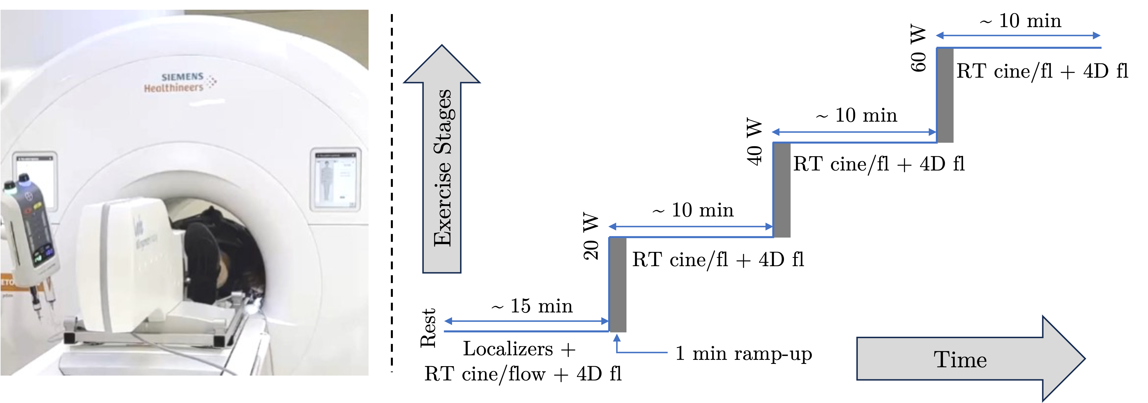
Twenty-eight healthy volunteers over the age of 18 were recruited to participate in an institutional review board-approved ExCMR study. Written informed consent was obtained from each subject. Data from two volunteers were excluded owing to their non-compliance with instructions. Twenty-six volunteers ( male, female; age = years; kg/m2; m2) were imaged on a 3T scanner (MAGNETOM Vida, Siemens Healthcare, Erlangen, Germany) fitted with an in-magnet supine ergometer (MR Ergometer Pedal, Lode, The Netherlands). The imaging protocol included a free-breathing RT short-axis cine stack (11 to 14 slices) covering the whole heart and at least two long-axis cine slices. Additionally, it comprised two sets of three closely spaced phase-contrast CMR (PC-CMR) slices to measure flow at the root of the aortic and pulmonic arteries, as well as 4D flow with whole-heart coverage. The data for cine were collected using balanced steady-state free-precession (bSSFP), while the data for PC-CMR and 4D flow were collected using gradient echo (GRE) sequences. For cine, the data were collected using a pseudo-random Cartesian sampling pattern, called GRO [20]. For flow, the data were collected using a different Cartesian sampling pattern, called CAVA [21]. The protocol used to collect data is shown in Figure 1, and the imaging parameters for RT cine and flow are summarized in Table 1.
| Parameter/Sequence | RT Cine | RT Flow |
|---|---|---|
| Sequence | bSSFP | GRE |
| Acquisition time (s/slice) | ||
| Acceleration rate | ||
| Repetition Time (ms) | ||
| Echo Time (ms) | ||
| Spatial Resolution (mm2) | ||
| Temporal Resolution (ms) | ||
| Flip Angle (deg) | ||
| Slice thickness (mm) | ||
| Sampling pattern | Cartesian [20] | Cartesian [21] |
| VENC (cm/s) | N/A | (rest), (stress) |
After data acquisition at rest, the volunteers were asked to pedal the ergometer at a rate of 60 cycles per minute by matching their cadence to a metronome played through the headphones. The initial workload was set to W and was increased in increments of W to the maximum of W. The same set of sequences was executed at each exercise stage after allowing a ramp-up time of one minute for the heart rate (HR) to stabilize. Each exercise stage lasted for 8 to 10 minutes. Due to exhaustion or leg fatigue, not all subjects were able to complete all three exercise stages. In total, we have , , , and volunteers at rest, W, W, and W, respectively. In one of the volunteers, an incidental finding during the resting stage of the protocol prompted termination of the experiment after the resting stage. For a subset of volunteers, we repeated RT cine and flow acquisitions in quick succession (within 10 minutes) at rest () and at the W exercise stage () to assess repeatability. This study is focused on the analysis of RT cine and flow images to track hemodynamic response to exercise. The data from the 4D flow acquisition are being processed and will be reported separately [22].
Upon completing the scan, volunteers were requested to assess their level of exertion during each stage of the exercise using the Borg rating of perceived exertion (BRPE) scale [23]. This scale categorizes exertion into four distinct levels: “no to extremely light exertion” (scores ), “light exertion” (scores ), “somewhat hard to hard” (scores ), and “very hard to maximal exertion” (scores ). It is important to note that the scale’s range from to , when multiplied by 10, corresponds closely with HR measurements, underscoring its effectiveness in quantifying exertion levels.
After completing the healthy subject study, five patient volunteers ( male, female; age = years; kg/m2; m2) were also imaged. In contrast to the healthy subjects, a W workload increment was employed when the W increase was considered excessively challenging by the patients. The maximum exercise intensity achieved by the five patients was W, W, W, W, and W.
2.2 Image reconstruction
The image reconstruction for RT cine and flow was performed inline with Gadgetron-based [24] implementation of SCoRe [25], which is a parameter-free compressive sensing method. Coil sensitivity maps were estimated from the temporally averaged k-space using ESPIRiT [26]. In the case of RT cine, three-dimensional undecimated wavelet transform (UWT) was applied to sparsify both the spatial and temporal dimensions. For RT flow, in addition to UWT, temporal principal components [27], inferred after stacking flow-encoded and flow-compensated images, were used as data-driven sparsifying transform. The reconstruction was performed inline on a dedicated GPU workstation equipped with NVIDIA GeForce RTX 3090. For a 6 s per slice acquisition, the reconstruction time was 10 seconds per slice for RT cine and 15 seconds per slice for RT flow.
Although subjects were instructed to firmly hold onto the handles connected to the table’s rails, periodic bulk motion was observed during paddling in almost all cases. For some subjects, this resulted in significant degradation of image quality due to the temporally varying sensitivity maps of the body coil array. We attempted to use dynamic sensitivity maps, estimated from a sliding window of a small number of frames, but this approach was unsuccessful. Specifically, a smaller window (less than 8 frames) did not yield a fully sampled region of sufficient size or quality for reliable estimation of coil sensitivity maps, while a larger window (greater than 8 frames) introduced artifacts similar to those from time-averaged sensitivity maps. To mitigate these artifacts, we employed our recently proposed coil reweighting method [28], designed to automatically suppress contributions from artifact-inducing coil elements. This preprocessing step is computationally efficient and can be integrated with any reconstruction method.
2.3 Cardiac function quantification
For both RT cine and flow imaging, multiple heartbeats were collected from each slice. The electrocardiogram (ECG) signal facilitated the segregation of frames into individual heartbeats. Arrhythmic heartbeats, defined as those deviating from the mean R-wave to R-wave interval by more than 15%, were excluded. Several studies have highlighted the impact of respiration-induced variation in cardiac output quantification, which is attributed to changes in intrathoracic pressure and through-plane motion [29]. This variation is expected to be more pronounced during exercise. To minimize the impact of respiratory motion on cardiac function assessment, heartbeats from the RT cine and flow captured during the end-expiratory phase were specifically isolated for analysis. The respiratory signal, critical for identifying this phase, was derived from RT images using a recently proposed principal component analysis-based method [19]. In cases involving RT images captured during exercise, the ECG signal proved unreliable. As illustrated in Supporting Movie S1, the respiratory signal from [19] was superimposed at the bottom of the exercise RT image series. A white cross marked the respiratory location for each frame. This signal served as a reference to manually identify systolic and diastolic frames from the end-expiratory phase. The steps of isolating the end-expiratory beat (for rest images) and superimposing the respiratory signal (for exercise images) were performed offline.
The DICOM images from the short-axis cine and flow acquisitions from the ascending aorta (AAo) and main pulmonary artery (MPA) were imported into suiteHEART (NeoSoft, Pewaukee, WI, USA) for hemodynamic and cardiac function quantification. For the RT data collected at rest, only one end-expiratory beat was imported. However, for RT data acquired during exercise stress, the entire cine and flow series, superimposed with the respiratory signal, were imported. Analyses were conducted on expiratory heartbeats, aided by the reference respiratory signal. For an initial cohort of volunteers imaged with an acquisition time of 3 s/slice, all effort was made to conduct the analyses on the end-expiratory beat where possible. The protocol was consequently modified to an acquisition time of 6 s/slice to accommodate slower breathing patterns at rest. Biventricular segmentation was initially performed using suiteHEART software and manually adjusted where necessary. The following parameters were derived for cardiac function analysis: left ventricular (LV) and right ventricular (RV) end-diastolic volume (EDV), end-systolic volume (ESV), stroke volume (SV), and ejection fraction (EF). From the flow images, peak velocity (Vmax), net forward flow (NFF), and SV were measured for both major arteries. To account for the exaggerated through-plane motion, three PC-CMR slices were acquired from AAo and MPA. From the three slices available, a slice closest to but above the aortic valve was used for AAo flow quantification, and a slice closest to the pulmonic valve but away from the bifurcation was used for MPA flow analysis. The HR at rest and during each exercise stage was extracted from both RT cine and flow images and converted to a percentage of the age-predicted maximal heart rate (APHR). Cardiac output (CO), computed from both cine and flow images, was calculated as the product of SV (mL/beat) and HR (beats/min). In repeat imaging at rest and during a 40 W workload, quantification was performed on a heartbeat from an arbitrary respiratory phase (AP) and on a heartbeat from end-expiration (EE).
3 Results
3.1 Results from healthy subjects
In Figure 2A, the cardiac function parameters extracted from the RT short-axis cine analysis are depicted, tracing the progression from rest to three stages of exercise for each volunteer. In summary, the EDV remained relatively consistent from rest through the exercise stages in both ventricles. In contrast, the ESV decreased as exercise intensity and HR increased, indicating more vigorous cardiac contractions during exercise stress. Consequently, the SV typically increased under stress. However, the pattern of increase in SV varied among volunteers, ranging from a rapid initial rise to plateauing in some, and a more gradual increase in others. One of the healthy volunteers had an incidental finding of congenital cardiovascular abnormality, resulting in dilated RV and a mismatch between aortic and pulmonic flow. The consistent elevation in CO was attributable to both the increased SV and HR. Both HR and APHR trends extracted from the cine images (not shown) were similar to the ones extracted from the flow images.
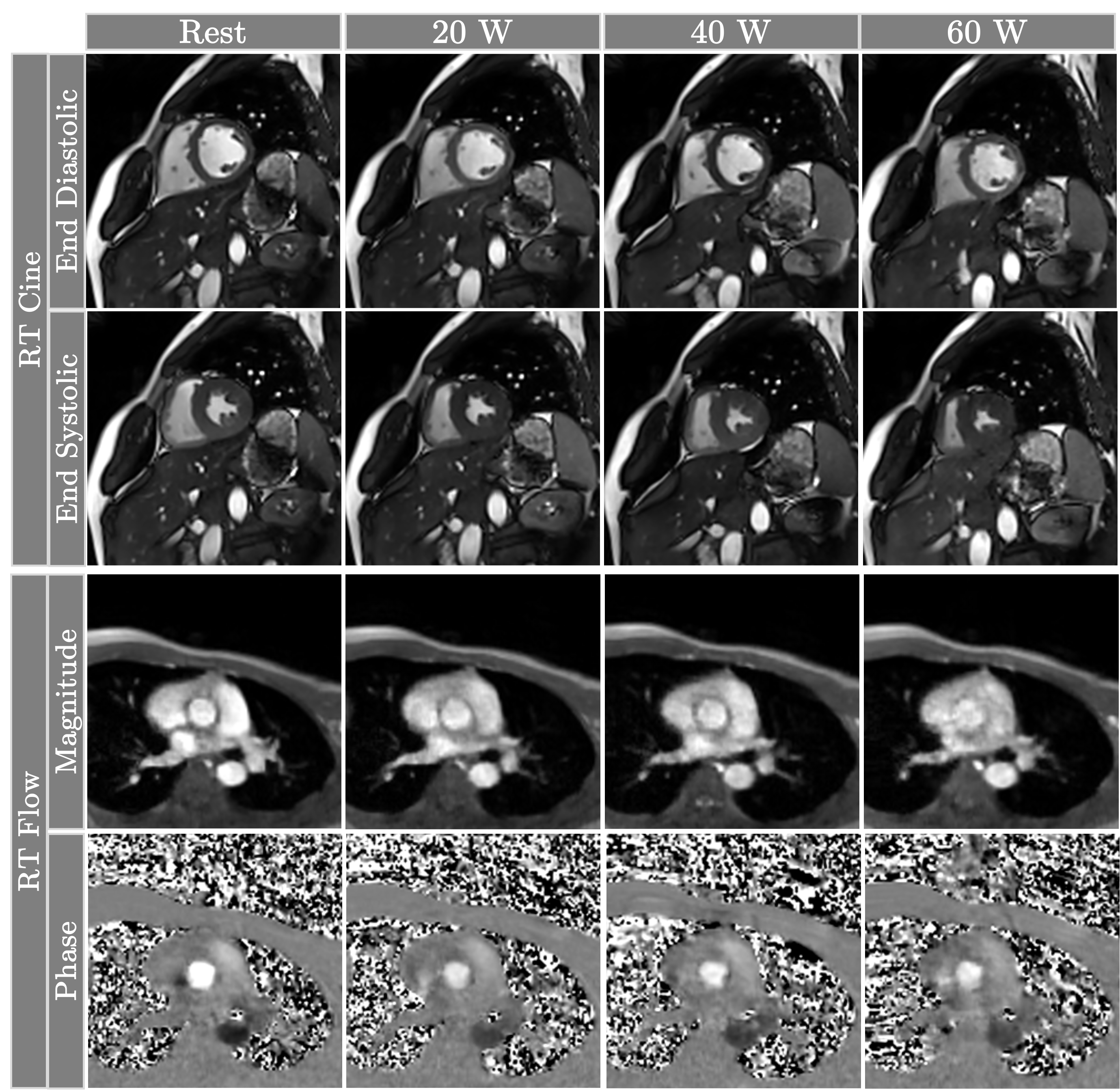
In Figure 2B, the aortic and pulmonic flow parameters, tracked from rest through three stages of exercise for each volunteer, are depicted. The NFF values demonstrate an overall increase with exercise, although this rise is less pronounced compared to the biventricular stroke volumes. However, CO exhibits a similar trend of increasing with exercise intensity. The peak velocities for both arteries also show an upward trend with exercise. Heart rates consistently rose with increasing stress, with each volunteer reaching up to 70% of their APHR on average. Representative cine and flow images at rest and various stages of exercise are shown in Figure 3.
The perceived exertion, measured using BRPE, ranged from 6 to 16, with an average value of 9.3. For the subjects who quit before reaching the 60 W workload, the most commonly expressed reasons were shortness of breath, leg fatigue, and overall discomfort. Some of the subjects also found it challenging to ramp up their cadence back to 60 cycles per minute if they fell below 40 cycles per minute. This is because, for a given workload, the ergometer automatically increases the resistance to account for the lower frequency. We addressed this issue by periodically reminding the volunteers to maintain a cadence of around 60 cycles per minute.
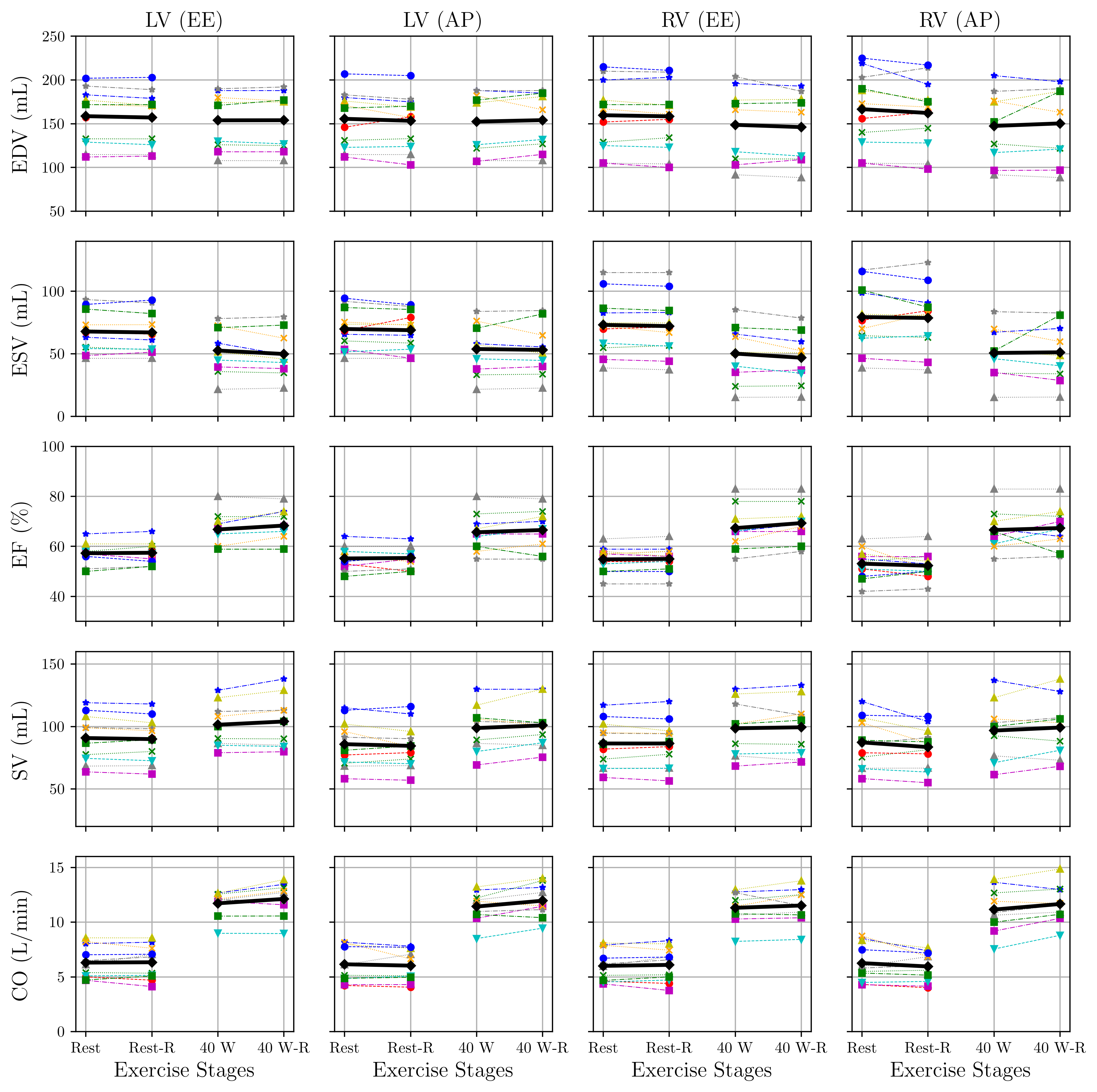
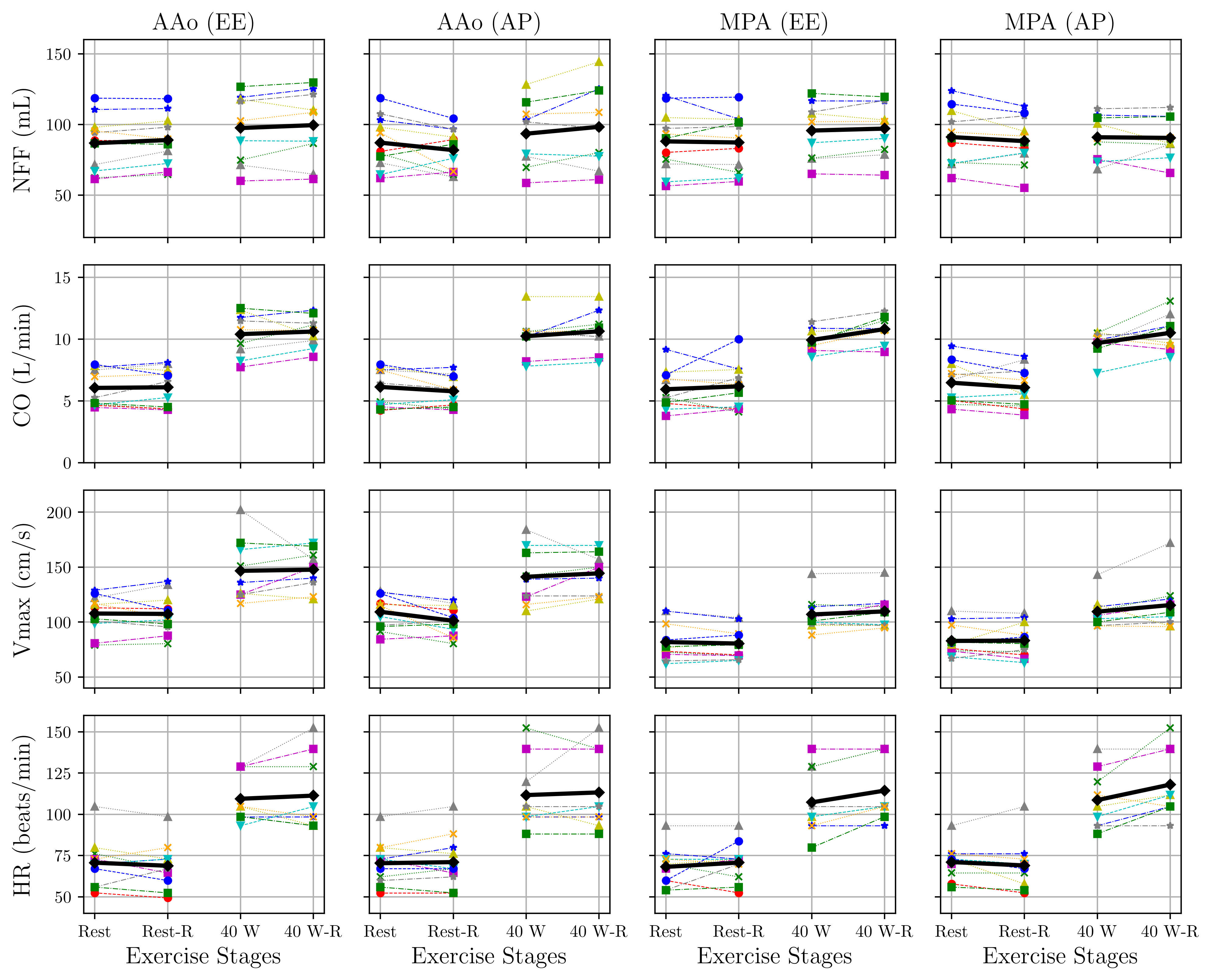
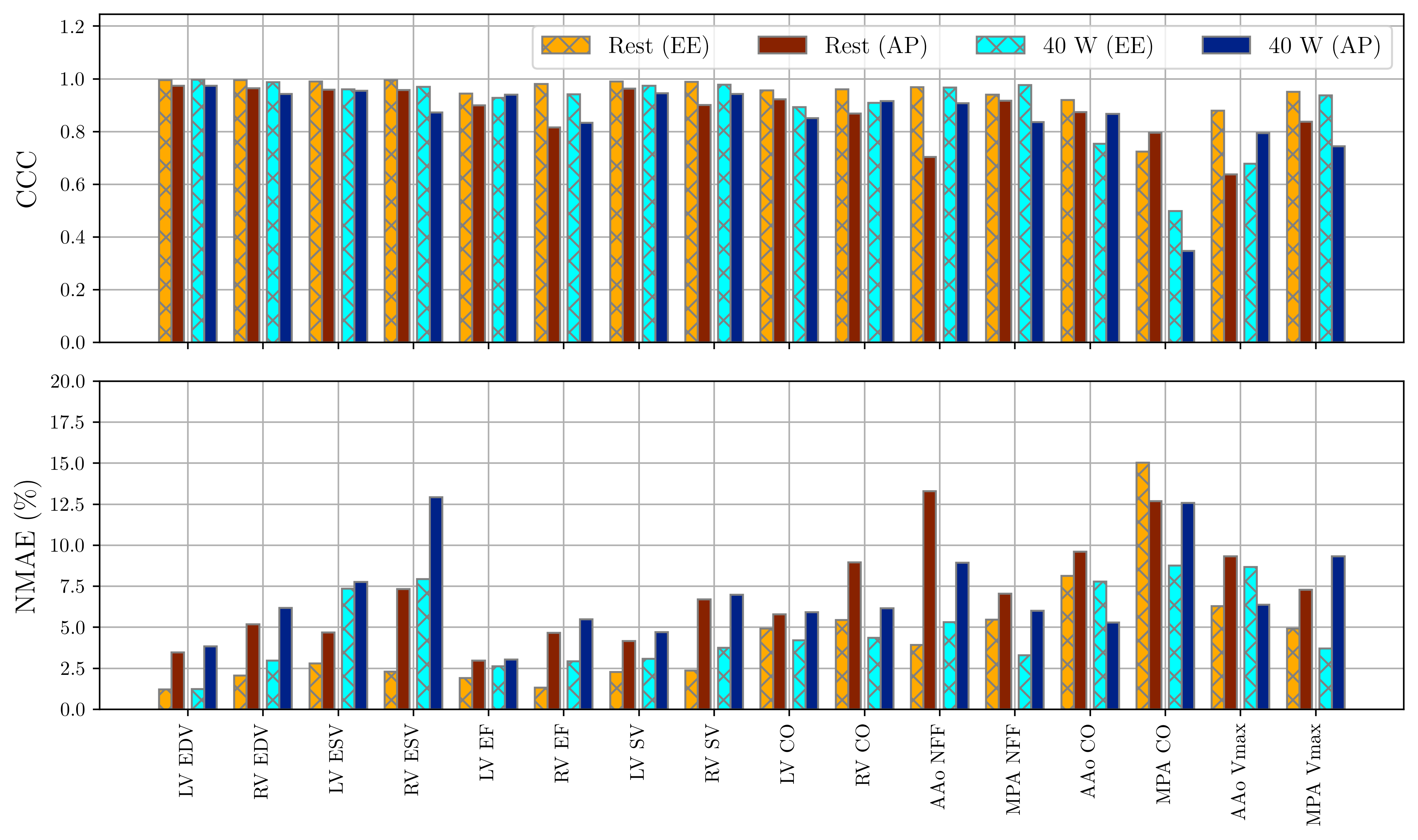
Figure 4 displays the repeat assessment of biventricular functional parameters from cine images at rest and a workload of W. Similarly, Figure 5 presents the repeat assessment of hemodynamic parameters from flow images at the same conditions. The repeatability comparison is conducted for both AP and EE beats. Overall, both AP and EE beats demonstrate a high level of repeatability. Quantitatively, the cardiac function parameters for AP and EE beats are compared in Figure 6. In terms of the concordance correlation coefficient (CCC) and normalized mean absolute error (NMAE), EE beats show a marginal but consistent improvement over AP beats. NMAE, inferred from the two repeats, and , is calculated as .
3.2 Results from patients
The RT cine and flow data from five patients were reconstructed and analyzed. From each subject, one mid-ventricular short-axis cine slice, one long-axis cine slice, one PC-CMR slide from AAo, and one PC-CMR slice from MPA were scored by an expert reader on a five-point Likert-type scale (5-excellent, 4-good, 3-adequate, 2-fair, 1-non-diagnostic) in terms of overall image quality. The perceived exertion, measured using BRPE, ranged from 6 to 19, with an average value of 9.3 All images, after coil reweighting, received a score of 3 or higher. Figure 7 shows representative images from one of the patients. The adequate quality of cine and flow images allowed us to perform cardiac function and hemodynamic quantification on all five patients. Due to the small sample size and lack of reference values, the cardiac function and hemodynamic parameters are not reported.
3.3 Artifact suppression
The artifact suppression method based on coil reweighting was applied to all RT cine data. In cases, where images had little to no artifact, coil reweighting had minimal impact on the image quality or quantification. However, in cases where image artifacts were severe, image interpretation or quantitative analysis was not feasible without coil reweighting. Figure 8 shows one such example, where images without coil reweighting exhibit severe artifacts, which are mostly mitigated after coil reweighting.
4 Discussion
ExCMR with in-magnet exercise allows dynamic monitoring of the quantitative cardiac function during exercise. Since breath-holding is generally not feasible during exercise, free-breathing RT imaging is often used to collect the data. Some of the technical challenges associated with ExCMR with in-magnet exercise include image quality degradation due to motion artifacts and limited spatial and temporal resolution. Recent studies have shown the promise of applying highly accelerated RT imaging techniques to enable ExCMR during exercise. In this work, we demonstrate the feasibility of ExCMR for measuring biventricular cardiac function and hemodynamics under staged exercise for both healthy subjects and patients.
Figure 2 summarizes the changes in cardiac function and hemodynamic parameters from rest through various stages of exercise. The values of ESV, both for LV and RV, consistently decrease due to more vigorous contractions under stress. The values of EDV remain relatively stable across different exercise intensities. As a result, both SV and EF gradually increase with exercise. Compared to SV, the increase in CO is more pronounced due to the increased HR. This biventricular increase is observed in both cine and flow images. Figure 3 shows an example of RT cine and flow images from a healthy subject. The cine images show a clear delineation of the blood-myocardium boundary, with some marginal blurring at 60 W. The end-systolic frames exhibit stronger contractility, especially at 60 W. The flow images are mostly free of motion artifacts, with the 60 W magnitude images showing marginal blurring.
Figure 4 and Figure 5 demonstrate the scan-rescan repeatability of measured parameters at rest and 40 W from eleven and nine subjects, respectively. With few exceptions, most parameter values are consistent across the scans. Figure 6 shows that selecting an EE beat, compared to an AP beat, results in a modest but consistent improvement in repeatability. With the sole exception of MPA CO, NMAE from all parameters from the EE beat is less than 10%. In contrast, the NMAE exceeds the 10% threshold four times for AP. A similar trend is observed in CCC. Relatively poor CCC and NMAE values for MPA CO can be attributed to HR variations between the two repeats and the small sample size.
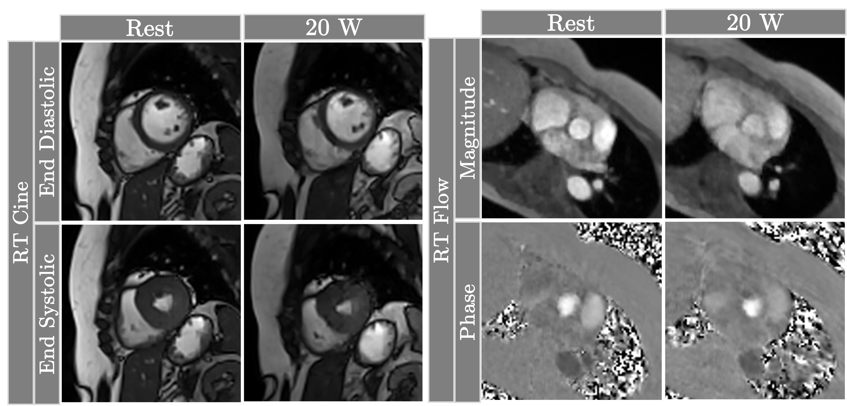
Figure 7 shows a representative frame from a patient who could only exercise at the workload of W. Both RT cine images show clear delineation of blood and myocardium during the systolic and diastolic phases. The flow images are also mostly free of artifacts, with the 20 W magnitude image showing slight blurring. The phase images, however, clearly capture the flow in the ascending and descending aorta. Note that a higher value of VENC was used at 20 W; therefore, the velocities are scaled differently in the 20 W image compared to the resting image. Finally, Figure 8 highlights the impact of coil reweighting on image quality. Without applying coil reweighting, some of the cine images are not interpretable and amenable to quantification. With coil reweighting, most of the artifacts are eliminated. Note, the coil reweighting comes at the cost of suppressing contribution from certain coil elements, which can lead to partial loss of signal in some areas of the image. However, we did not observe a case where the benefit of suppressing artifacts was outweighed by some loss of intensity.
5 Conclusion
In this preliminary study, we demonstrated the feasibility of ExCMR with in-magnet exercise for quantitative cardiac function evaluation in both healthy volunteers and patients under multi-stage exercise. The highly accelerated RT imaging was facilitated by parameter-free compressed sensing, overcoming the traditional challenges of limited resolution. Additional key innovative aspects of our technique are the incorporation of a coil reweighting technique and the selection of end-expiratory heartbeats, which resulted in significantly improved image quality and enhanced repeatability of cardiac function measurements. Data from both healthy subjects and patients underscored ExCMR’s ability to dynamically capture physiological changes in key cardiac parameters under stress. The advancement in ExCMR technology can be pivotal for the enhanced diagnosis and management of cardiovascular diseases, particularly for conditions where functional impairment is not elucidated on resting cardiovascular exams.
Acknowledgments and Funding This work was funded by NIH grants R01-EB029957, R01-HL151697, and R01- HL135489.
Ethics Declarations
-
•
Competing interests: The authors declare no competing interests.
-
•
Ethics approval and consent: For the human subject data, approval was granted by the Institutional Review Board (IRB) at The Ohio State University (2019H0076). Informed consent to participate in the study and to publish results was obtained from all individual participants.
-
•
Data and code availability: MRI data and the code to generate results are available upon request
-
•
Author contribution: P. Chandrasekaran assisted with data acquisition and processing as well as manuscript writing, C. Chen implemented data processing techniques, Y. Liu assisted with data acquisition and pulse sequence programming, S.M. Arshad assisted with experiment planning, C. Crabtree assisted with subject monitoring during exercise, M. Tong and Y. Han assisted with experiment design and results interpretation, and R. Ahmad supervised all aspects of the study.
References
- \bibcommenthead
- Lima and Desai [2004] Lima, J.A., Desai, M.Y.: Cardiovascular magnetic resonance imaging: current and emerging applications. Journal of the American College of Cardiology 44(6), 1164–1171 (2004)
- de Jong et al. [2012] Jong, M.C., Genders, T.S., Geuns, R.-J., Moelker, A., Hunink, M.M.: Diagnostic performance of stress myocardial perfusion imaging for coronary artery disease: a systematic review and meta-analysis. European Radiology 22, 1881–1895 (2012)
- Arai et al. [2023] Arai, A.E., Schulz-Menger, J., Shah, D.J., Han, Y., Bandettini, W.P., Abraham, A., Woodard, P.K., Selvanayagam, J.B., Hamilton-Craig, C., Tan, R.-S., et al.: Stress perfusion cardiac magnetic resonance vs SPECT imaging for detection of coronary artery disease. Journal of American College of Cardiology 82(19), 1828–1838 (2023)
- Sakuma et al. [2005] Sakuma, H., Suzawa, N., Ichikawa, Y., Makino, K., Hirano, T., Kitagawa, K., Takeda, K.: Diagnostic accuracy of stress first-pass contrast-enhanced myocardial perfusion MRI compared with stress myocardial perfusion scintigraphy. American Journal of Roentgenology 185(1), 95–102 (2005)
- Indorkar et al. [2019] Indorkar, R., Kwong, R.Y., Romano, S., White, B.E., Chia, R.C., Trybula, M., Evans, K., Shenoy, C., Farzaneh-Far, A.: Global coronary flow reserve measured during stress cardiac magnetic resonance imaging is an independent predictor of adverse cardiovascular events. JACC: Cardiovascular Imaging 12(8 Part 2), 1686–1695 (2019)
- Kobayashi et al. [2008] Kobayashi, M., Izawa, H., Cheng, X.W., Asano, H., Hirashiki, A., Unno, K., Ohshima, S., Yamada, T., Murase, Y., Kato, T.S., et al.: Dobutamine stress testing as a diagnostic tool for evaluation of myocardial contractile reserve in asymptomatic or mildly symptomatic patients with dilated cardiomyopathy. JACC: Cardiovascular Imaging 1(6), 718–726 (2008)
- Kramer et al. [2020] Kramer, C.M., Barkhausen, J., Bucciarelli-Ducci, C., Flamm, S.D., Kim, R.J., Nagel, E.: Standardized cardiovascular magnetic resonance imaging (CMR) protocols: 2020 update. Journal of Cardiovascular Magnetic Resonance 22(1), 1–18 (2020)
- Leischik et al. [2007] Leischik, R., Dworrak, B., Littwitz, H., Gülker, H.: Prognostic significance of exercise stress echocardiography in 3329 outpatients (5-year longitudinal study). International Journal of Cardiology 119(3), 297–305 (2007)
- Pagnanelli and Camposano [2017] Pagnanelli, R.A., Camposano, H.L.: Pharmacologic stress testing with myocardialperfusion imaging. Journal of Nuclear Medicine Technology 45(4), 249–252 (2017)
- Göransson et al. [2019] Göransson, C., Vejlstrup, N., Carlsen, J.: Exercise cardiovascular magnetic resonance imaging allows differentiation of low-risk pulmonary arterial hypertension. Journal of Heart and Lung Transplantation 38(6), 627–635 (2019)
- Foster et al. [2012] Foster, E.L., Arnold, J.W., Jekic, M., Bender, J.A., Balasubramanian, V., Thavendiranathan, P., Dickerson, J.A., Raman, S.V., Simonetti, O.P.: MR-compatible treadmill for exercise stress cardiac magnetic resonance imaging. Magnetic Resonance in Medicine 67(3), 880–889 (2012)
- Raman et al. [2016] Raman, S.V., Dickerson, J.A., Mazur, W., Wong, T.C., Schelbert, E.B., Min, J.K., Scandling, D., Bartone, C., Craft, J.T., Thavendiranathan, P., et al.: Diagnostic performance of treadmill exercise cardiac magnetic resonance: the prospective, multicenter exercise CMR’s accuracy for cardiovascular stress testing (EXACT) trial. Journal of the American Heart Association 5(8), 003811 (2016)
- Lurz et al. [2009] Lurz, P., Muthurangu, V., Schievano, S., Nordmeyer, J., Bonhoeffer, P., Taylor, A.M., Hansen, M.S.: Feasibility and reproducibility of biventricular volumetric assessment of cardiac function during exercise using real-time radial k-t SENSE magnetic resonance imaging. Journal of Magnetic Resonance Imaging 29(5), 1062–1070 (2009)
- Craven et al. [2021] Craven, T.P., Jex, N., Chew, P.G., Higgins, D.M., Bissell, M.M., Brown, L.A., Saunderson, C.E., Das, A., Chowdhary, A., Dall’Armellina, E., et al.: Exercise cardiovascular magnetic resonance: feasibility and development of biventricular function and great vessel flow assessment, during continuous exercise accelerated by Compressed SENSE: preliminary results in healthy volunteers. International Journal of Cardiovascular Imaging 37, 685–698 (2021)
- Edlund et al. [2022] Edlund, J., Haris, K., Ostenfeld, E., Carlsson, M., Heiberg, E., Johansson, S., Östenson, B., Jin, N., Aletras, A.H., Steding-Ehrenborg, K.: Validation and quantification of left ventricular function during exercise and free breathing from real-time cardiac magnetic resonance images. Scientific Reports 12(1), 5611 (2022)
- Li et al. [2021] Li, Y.Y., Zhang, P., Rashid, S., Cheng, Y.J., Li, W., Schapiro, W., Gliganic, K., Yamashita, A.-M., Grgas, M., Haag, E., et al.: Real-time exercise stress cardiac MRI with fourier-series reconstruction from golden-angle radial data. Magnetic Resonance Imaging 75, 89–99 (2021)
- Morales et al. [2022] Morales, M.A., Assana, S., Cai, X., Chow, K., Haji-Valizadeh, H., Sai, E., Tsao, C., Matos, J., Rodriguez, J., Berg, S., et al.: An inline deep learning based free-breathing ECG-free cine for exercise cardiovascular magnetic resonance. Journal of Cardiovascular Magnetic Resonance 24(1), 1–14 (2022)
- Craven et al. [2020] Craven, T.P., Tsao, C.W., La Gerche, A., Simonetti, O.P., Greenwood, J.P.: Exercise cardiovascular magnetic resonance: development, current utility and future applications. Journal of Cardiovascular Magnetic Resonance 22(1), 1–20 (2020)
- Chen et al. [2022] Chen, C., Chandrasekaran, P., Liu, Y., Simonetti, O.P., Tong, M., Ahmad, R.: Ensuring respiratory phase consistency to improve cardiac function quantification in real-time CMR. Magnetic Resonance in Medicine 87(3), 1595–1604 (2022)
- Joshi et al. [2022] Joshi, M., Pruitt, A., Chen, C., Liu, Y., Ahmad, R.: Technical report (v1.0)–pseudo-random cartesian sampling for dynamic MRI. arXiv preprint arXiv:2206.03630 (2022)
- Rich et al. [2020] Rich, A., Gregg, M., Jin, N., Liu, Y., Potter, L., Simonetti, O., Ahmad, R.: Cartesian sampling with variable density and adjustable temporal resolution (CAVA). Magnetic Resonance in Medicine 83(6), 2015–2025 (2020)
- Arshad et al. [2023] Arshad, S.M., Potter, L.C., Chen, C., Liu, Y., Chandrasekaran, P., Crabtree, C., Han, Y., Ahmad, R.: Motion-robust free-running cardiovascular MRI. arXiv preprint arXiv:2308.02088 (2023)
- Williams [2017] Williams, N.: The borg rating of perceived exertion (RPE) scale. Occupational Medicine 67(5), 404–405 (2017)
- Hansen and Sørensen [2013] Hansen, M.S., Sørensen, T.S.: Gadgetron: an open source framework for medical image reconstruction. Magnetic Resonance in Medicine 69(6), 1768–1776 (2013)
- Chen et al. [2019] Chen, C., Liu, Y., Schniter, P., Jin, N., Craft, J., Simonetti, O., Ahmad, R.: Sparsity adaptive reconstruction for highly accelerated cardiac MRI. Magnetic Resonance in Medicine 81(6), 3875–3887 (2019)
- Uecker et al. [2014] Uecker, M., Lai, P., Murphy, M.J., Virtue, P., Elad, M., Pauly, J.M., Vasanawala, S.S., Lustig, M.: ESPIRiT–An eigenvalue approach to autocalibrating parallel MRI: Where SENSE meets GRAPPA. Magnetic Resonance in Medicine 71(3), 990–1001 (2014)
- Petzschner et al. [2011] Petzschner, F.H., Ponce, I.P., Blaimer, M., Jakob, P.M., Breuer, F.A.: Fast MR parameter mapping using k-t principal component analysis. Magnetic Resonance in Medicine 66(3), 706–716 (2011)
- Chen et al. [2023] Chen, C., Liu, Y., Ding, Y., Tong, M., Han, Y., Ahmad, R.: Automatic coil selection to suppress motion artifacts in exercise real-time cine imaging. In: ISMRM ISMRT Annual Meeting Exhibition, Toronto, Canada, p. 1152 (2023)
- Claessen et al. [2014] Claessen, G., Claus, P., Delcroix, M., Bogaert, J., Gerche, A.L., Heidbuchel, H.: Interaction between respiration and right versus left ventricular volumes at rest and during exercise: a real-time cardiac magnetic resonance study. American Journal of Physiology-Heart and Circulatory Physiology 306(6), 816–824 (2014)