Hybrid Metamaterial Optical Tweezers for Dielectric Particles and Biomolecules Discrimination
Abstract
Although plasmonic nanotweezers can achieve manipulation on a scale smaller than the diffraction limit, direct trapping of sub-10 nm bioparticles in an aqueous environment without modifications remains challenging. Here, we demonstrate enzyme delivery-trapping and dynamic manipulation at the single-entity level by employing photothermal-assisted trapping via metamaterial optical tweezers at low trapping laser intensities. By analyzing the probability density function, we are able to distinguish dielectric particles from biomolecules. For enzyme-related data, the presence of two peaks in violin plots indicates conformational changes or interactions with the environment. We also observe that increasing the laser intensity increases the local temperature, resulting in enzyme aggregation. This study provides an alternative approach for identifying single enzymes and dielectric particles by combining plasmonic fields and photothermal heat, while paving the way for label-free characterization of biomolecules at the single-particle level.
keywords:
American Chemical Society, LaTeXLight-Matter Interactions for Quantum Technologies Unit, Okinawa Institute of Science and Technology Graduate University, Onna, Okinawa 904-0495, Japan \alsoaffiliationInstitute for Protein Research, Osaka University, Suita, Osaka 565-0871, Japan \abbreviationsIR,NMR,UV
Keywords: Metamaterial optical tweezers, enzyme trapping, thermoplasmonics, single-particle level, violin plots, aggregations
1 Introduction
In biomedical research and disease diagnosis, detecting single biomolecules in solution is essential. It allows for a better understanding of disease mechanisms, leading to early detection, and treatment responses. Several advanced techniques and methods are used to meet this goal 1. For example, fluorescence resonance energy transfer (FRET) can be used to detect conformational changes by measuring the transfer of energy between two fluorophores attached to interacting biomolecules 2. However, repeated excitation of fluorophores can lead to photobleaching, reducing the intensity of fluorescence signals over time which may affect the accuracy of measurements 3. Since optical tweezers 4 can measure forces and torques with high accuracy and temporal resolution, they could be considered as a preferred method for single-particle detection in many biophysical applications 5. Optical tweezers have been employed to investigate the movement of motor proteins such as kinesin 6 and myosin 7, as well as to measure the elasticity, stretching, and torsional properties of DNA 8, 5, 9 and RNA 8, 5. Despite the wide range of applications 10, 11, optical tweezers face certain challenges, particularly when it comes to tethering and labelling molecules. To overcome the limitations of conventional optical tweezers for detecting single molecules, plasmonic optical tweezers (POT) were developed 12, 13, 14, 15, 16. With the ability of metallic nanostructures to concentrate light in subwavelength volumes, POT techniques allow precise manipulation of nanoscale objects, such as biomolecules 17, 18, 15, nanoparticles 19, 20, 21, and quantum dots 22, 23. For example, double nanohole optical tweezers were used to investigate how protein p53 interacts with a single DNA-hairpin 18, to measure vibrational modes of proteins in the low-wavenumber regime 24, and to monitor the structure dynamics of single enzymes during catalytic cycles 25.
Among all nanostructure designs, metamaterials have emerged as the most promising owing to their exotic electromagnetic properties and functionalities that are not attainable from naturally occurring materials 26. In biosensing, metamaterials provide the large active area necessary for the assessment and quantification of interactions between the resonating metamolecules and biological entity 27, 28, 29, 30, 31. Exploiting this advantage, several metamaterials have been designed to detect viruses 28, bacteria 31 and other biomolecules 29. Metamaterials that support a Fano-like resonance are characterized by a narrow spectral window where scattering is suppressed and absorption is enhanced due to interference between super-radiant and subradiant plasmonic modes 32. The resonance of these nanostructures is extremely sensitive to their geometric characteristics and, in combination with subdiffraction volumes, can be employed for applications such as ultrasensitive protein recognition 29, 33 and efficient nanoparticle trapping using low trapping laser intensities 34, 16, 35.
Meanwhile, the energy losses of plasmonics and the associated heat generation at the nanoscale could benefit a broad range of applications, including photothermal-assisted plasmonic sensing 36. Specifically, the localized heat generated by metallic nanostructures can lead to changes in the refractive index of the surrounding medium that are detectable and can be correlated with analyte concentrations 37. For instance, dual-functional plasmonic biosensors have been demonstrated to facilitate the specific nucleic acid hybridization of SARS-CoV-2, while simultaneously detecting the nonamplified nucleic acid sequences of the virus 37. This finding suggests that plasmonics and near-field heating also have the potential to regulate the biocatalytic behaviors of the site-specific nucleases, leading to accurate biomolecule detection and discrimination 37.
In this work, we extend the utility of metamaterial optical tweezers and introduce a thermoplasmonic-assisted concept to optically trap and discriminate between dielectric nanoparticles and biomolecules in the nanoscale regime. We used 20 nm diameter polystyrene beads and urease that has a hydrodynamic radius of 7 nm suspended in polyethylene glycol(PEG)/heavy water solution. Urease is a nickel-containing enzyme that catalyzes the hydrolysis of urea to form ammonia and carbon dioxide in some bacteria, fungi, algae, and plants 38, 39. Healthy human organisms do not contain urease; however, Helicobacter pylori, a bacterial pathogen of the human stomach and a class I carcinogen 40, 41, secretes urease 42. PEG is a highly soluble polymer in water that has been used to modify biomolecules and to create more stable drug delivery systems for anticancer therapies 43. We observed that a single urease can be delivered to, and trapped in, the nano-aperture of the asymmetric, split-ring (ASR) metamaterial for trapping laser intensities lower than 0.5 mW/m2. Due to the large hydrodynamic motion and conformational changes of the molecule in the plasmonic hotspot, two peaks appeared in the probability density function for single and multiple trapping events of ureases. For dielectric particles, violin plots show a single peak both for single and multiple trapping events, indicating their consistent behavior during the trapping process. Furthermore, we investigated how the normalized root-mean-square of the first urease trapping event changes with trapping laser intensities and trap stiffness and we conclude that aggregations of molecules may form and be trapped for intensities greater than 1 mW/m2, i.e., at high temperatures. We also measured the average time until the first observed trapping event occurred for several molecular concentrations. Using the same range of trapping laser intensities, average trapping times for low molecule concentrations were measured as being similar to those for non-bioparticle trapping. According to our analysis, our metamaterial optical tweezer platform facilitates the identification of various particles in a simple and label-free way and this may open up new avenues for nanoscience and life science research.
2 Materials and Methods
A schematic of the experimental setup used to trap and manipulate a single enzyme molecule is shown in Figure 1. The optical tweezers consist of a 930 nm continuous-wave (CW) laser focused using a high numerical aperture (NA = 1.3) oil immersion objective lens (OLYMPUS UPlanFL N 100) to a spot size of 1 m. Detection of trapping events was conducted by collecting the transmitted laser light through a 50 objective lens (Nikon CF Plan), sending it to an avalanche photodiode (APD430A/M, Thorlabs), and recording it using a data acquisition card (DAQ) at a frequency of 100 kHz with LabVIEW software.
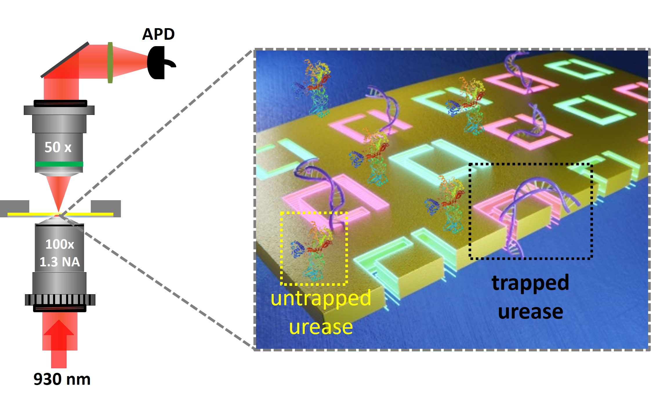
The unit cell of the metamaterial contains an asymmetrically split ring (ASR) consisting of two nano-apertures in a gold film (Figure 2(a)). The full array consists of 15 (-direction) 16 (-direction) units with a period of 400 nm and was fabricated using focused ion beam (FIB-FEI Helios G3UC) milling on a 50 nm thin gold film (PHASIS, Geneva, BioNano) at 30 kV and a 2 pA beam current. The metamaterial was attached to a microscope cover glass with adhesive microscope spacers of 10 m thickness, forming a microwell. Since the focus spot has a diameter of about 1 m means that 2 2 metamolecules of the metamaterial structures were illuminated. We used a solution containing heavy water, polyethylene glycol (PEG6000), and urease (U4002; Sigma-Aldrich). Several concentrations of urease suspended in the 0.4 %w/v PEG6000/D2O water solution were used and a microwell containing urease of various concentrations was mounted and fixed on top of a piezoelectric translation stage. The urease protein could then be trapped by plasmon-enhanced optical forces into the ASR nano-aperture of the metamaterial. In addition, a solution of polystyrene (PS) particles with a mean diameter of 20 nm (ThermoFisher Scientific, F8786) in 0.4 %w/v PEG6000/D2O water with a concentration of 0.0625% w/v was used as a control.
3 Results and Discussion
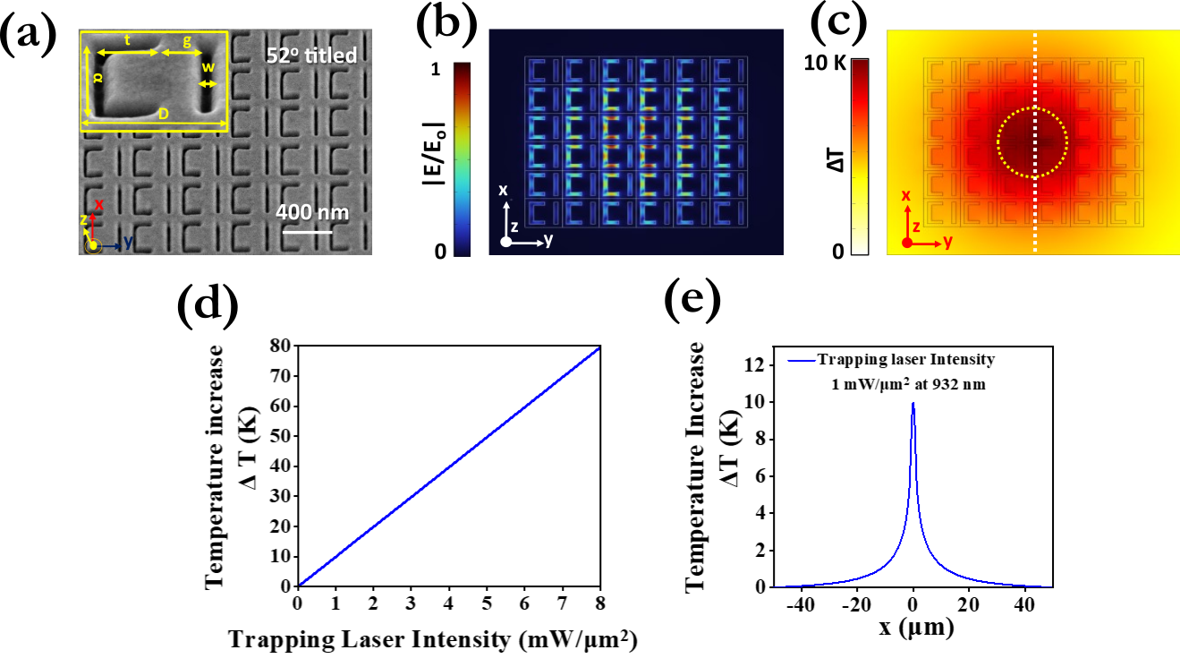
We performed finite-difference time-domain simulations using COMSOL Multiphysics software to calculate the electric field enhancement of the metamaterial (Figure 2(b)). The incident light was polarized along the y-axis. The mesh had a minimum size of 1.2 nm, maximum size of 44 nm, and resolution of narrow regions, i.e. the region of the titanium adhesion layer, at 0.7 nm. Using the shape of the metamaterial as determined by a scanning electron microscopy image (SEM), we determined the resonance wavelength in the absorption spectrum to be at 932 nm, just slightly above the 930 nm wavelength of the trapping laser 34. Enhanced light absorption results in a temperature increase and thermal gradient in the fluid. Figure 2(c) shows the increase in temperature due to laser irradiation of 1 mW/m2 on the metamaterial. We note that, at the center of the metamaterial, the strong heat source density leads to the temperature increase being around 10 K, while far from the center the heat source is weaker and the temperature of the structure does not change appreciably. The simulated temperature change versus trapping laser intensity (Itrap) is shown in Figure 2(d). Because the gold has high thermal conductivity compared with the surrounding medium, as expected from the heat diffusion equation 45, the temperature distribution in the metamaterial remains fairly uniform and increases linearly as a function of Itrap. Notably, at 8.0 mW/m2 the temperature increase in the metamaterial is predicted to exceed 80 K. This is not a negligible value and shows that the metamaterial is unable to dissipate heat from a focused laser beam at high trapping laser intensities. In addition, Figure 2(e) shows how the temperature increase changes as a function of distance from the center of the metamaterial. We observe that the temperature increase is reduced by 50% at 1.5 m from the center for a laser intensity of 1 mW/m2. Therefore, our metamaterial structure has the ability to deliver and trap sub-10 nm particles such as small protein molecules without the risk of photo-induced damage.
Since the resonance frequency of a metamaterial is sensitive to small changes in the local refractive index, the presence of biomolecules in the ASR nano-aperture can be tracked by changes in the transmitted signal. Figure 3(a) shows the time evolution of the transmitted signal as measured on the APD through the ASR metamolecules in the presence of urease solution. The abrupt jump in the APD signal at about 21 s and the change in the noise level right after the jump are signatures of biomolecular trapping by the metamaterial. In a control experiment, using a solution without urease molecules, no changes in the transmission signal were observed, confirming that the jump can be attributed to the trapping of urease molecules. Additionally, the cycle of trapping and then releasing the enzyme was repeated by blocking and unblocking the trapping laser beam.
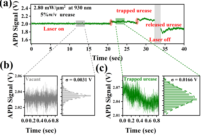
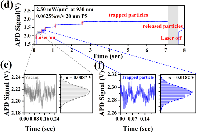
Two specific properties were extracted from the signal of each trapping event, the root-mean-square (RMS or ) deviation of the transmitted laser signal as measured on the APD, and the trap stiffness, k. The RMS of the transmitted signal was calculated by fitting a Gaussian distribution to its histogram. Figures 3(b) and (c) show the transmitted signal fluctuations with corresponding histograms for the two different states, i.e., vacant and trapped. The width of the Gaussian distribution for the vacant case displays a small RMS deviation, i.e, = 0.0031 V, while the RMS for the trapped event, = 0.0166 V, is significantly larger. For comparison, we show a sequence of trapping and releasing events of a 20 nm PS particle in an array of ASRs in Figure 3(d). As we illuminated 22 metamolecules, we excited eight plasmonic hotspots, which can trap up to eight nanoparticles 34. Therefore, we assume that each jump in the APD signal corresponded to a single trapping event. Figures 3(e) and (f) show fluctuations in the APD signal with corresponding histograms as well as the Gaussian fit for the trapped nanoparticle and the vacant positions. Similarly to the case for urease, we observe that the APD signal for trapped PS nanoparticles has a larger RMS deviation than that observed for no trapping event. Comparing Figure 3(c) with Figure 3(f), we notice that, in the case of protein trapping, the signal fluctuations are larger than for PS particle trapping. We assume that the hydrodynamical motion and conformational changes of the protein in the ASR may increase fluctuations in the transmitted intensity of the trapping laser 46, 47. The intensity of the transmitted signal at the trapping site (I) is proportional to the trapping potential energy (U) which itself is proportional to the polarizability 24 of the trapped particle; hence, the RMS deviation of the trapping laser signal may scale with the polarizability of the trapped biomolecule (i.e. the potential energy of a Rayleigh particle can be written in the dipole limit as where is the polarizability of the particle and E is the electric field) 46. Moreover, the biomolecule’s polarizability is proportional to its volume, V, and the volume scales with the mass. Since RMS () scales with the polarizability and the polarizability scales with the mass, we conclude that the M. Therefore, by calculating the RMS deviation of the transmitted signal for each trapping event, the total molecular weight of the trapped enzyme at the plasmonic hotspots of the metamaterial can be estimated.
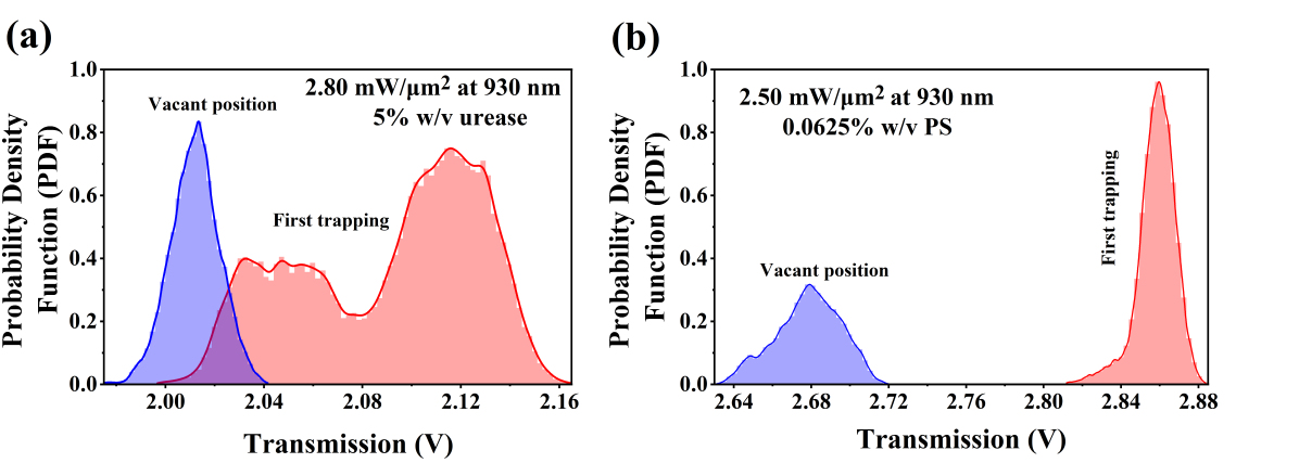
In addition, we calculated the probability density function (PDF) of the APD signal through the metamaterial both before (purple curve) and after (red curve) trapping of urease and polystyrene nanoparticles (Figures 4(a) and (b)). In this case, the PDF refers to the likelihood of finding the trapped particle in a specific arrangement or exhibiting a particular characteristic 48. We observe that trapping urease leads to a PDF with a wider amplitude distribution with two peaks compared to the vacant signal. This might indicate heterogeneity in enzyme activity, conformational states, or interactions with the environment 25. However, the PDF calculated from the PS nanoparticle trapping signal shows a relatively narrow peak compared to the vacant signal (Figure 4(b)). This observation reveals that the PS nanoparticles behave more uniformly or consistently when trapped. Urease and PS nanoparticles have different sizes, shapes, and optical properties. These characteristics can influence their trapping behavior and result in different PDF shapes. Therefore, the width and the shape of the PDF may be a key factor to distinguish the behavior of the particles in optical trapping applications.
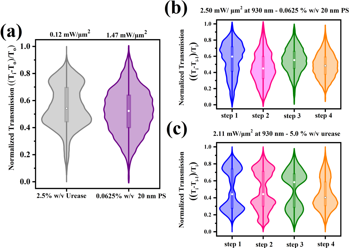
To further investigate the behavior of enzymes when trapped, we calculated the PDFs of the changes in the APD signal through the metamaterial for multiple trapping events for both types of particles. For this purpose, we defined and calculated the relative transmitted APD signal as , where is the transmitted signal for each trapping event, is the transmitted signal one trapping step/jump before and i is the number of trapping events ( trapping steps). This method minimizes effects caused by previously trapped particles, the metamaterial structure, and instrumentation noise. Note that by changing the trapping laser power and the trapping duration, we observed single or multiple trapping events. In Figure 5(a), we present violin plots for both types of particles at the lowest laser intensity for which we observed trapping. Violin plots are a data visualization tool that combines aspects of box plots and kernel density plots to represent the distribution of the experimental data 48. Using this method when studying enzymes, for example, can reveal the heterogeneity of enzyme properties or conformational states under several experimental conditions 48. Additionally, monitoring the median in a violin plot helps us to understand how the average behavior of the trapped particle is affected by various trapping conditions 48. In our case, the data set for the urease molecules has a median (white dot in Figure 5(a)) of the same magnitude as the data set obtained for PS nanoparticle trapping as a result of the normalization of the data. By comparing medians for nanoparticle and enzyme trapping experimental data, we can confirm that any effects due to the laser intensity differences are minimized. We also note that the violin plot for urease shows two peaks, while that for polystyrene displays only one.
Next, we generated violin plots for multiple trapping events for both polystyrene and urease particles. Note that we did not observe multiple urease trapping events for every laser trapping power used to illuminate the metamaterial, whereas we did for the PS particles. Hence, here we analyze data where up to four trapping events were observed for both types of particles at similar Itrap. In Figure 5(b) we present the violin plots of four PS nanoparticle trapping events. Due to polystyrene’s homogeneous nature, all violin plots show a peak, hence the data may have similar characteristics. Furthermore, we observe that the elongated kernel density distribution slightly decreases with increasing trap steps from one to four. The decrease in elongation of the PDF may suggest that the trapping strength increases as the number of trapping events increases. In the case of enzyme trapping at low (Figure 5(a)) and high (Figure 5(c)) trapping laser intensities, whether for a single trapping event or multiple events, two peaks appear in the violin plots with a roughly constant elongated distribution in contrast to the PS nanoparticle trapping. Proteins, such as enzymes, are held together by various weak interactions, including hydrogen bonds, van der Waals forces, and hydrophobic interactions. Hence, as the temperature rises, the thermal energy can overcome these weak interactions, leading to changes in protein conformation. Since urease is sensitive to changes in temperature 49, its structural conformation can be modulated by the local temperature increase on the metamaterial. Hence, we conclude that the presence of these two peaks for the enzyme-related data suggests conformational changes in the protein 50, 48.
We subsequently calculated the normalized root-mean-squared, i.e. NRMS, of the transmitted signal recorded on the APD for the first trapping event as a function of the incident Itrap for different urease concentrations and a PS nanoparticle concentration of 0.0625% w/v using a 5 s measuring window (Figure 6(a)). We notice that, for Itrap less than 1 mW/m2, the NRMS values of the APD signal for urease are roughly constant for all concentrations. As the Itrap increases, the NRMS of the APD signal increases, revealing an exponential-type behavior. By increasing the trapping laser intensity, more energy is transferred to the trapped molecule, resulting in greater hydrodynamic movement and large conformational fluctuations of the molecule in the harmonic trap, leading to an increase in the NRMS of the transmitted signals 47. On the other hand, we notice a constant NRMS of the APD signal for PS trapping as a function of trapping laser intensity. This arises due to the difference in properties between urease and polystyrene, i.e. the enzyme is not perfectly spherical and solid.
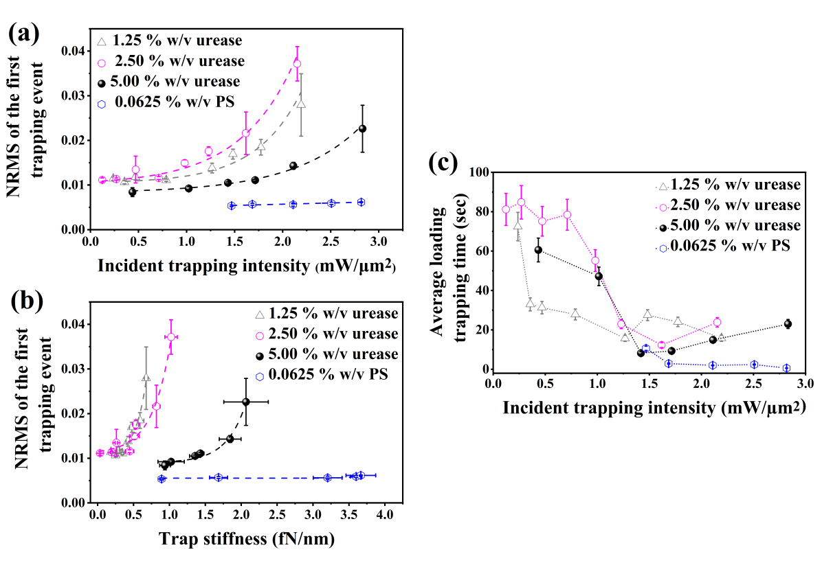
Additionally, we also calculated the trap stiffness ( = /) of the enzyme and the PS nanoparticle using the transient time method 51. For urease, we assumed a distance of 17 nm between the trapped molecule and ASR metamolecule wall 16 to modify the Faxén correction, . We used the viscosity of water at room temperature 52 and a hydrodynamic radius of 7 nm for urease 39. We noted a linear dependence of trap stiffness versus laser intensity (data not shown here). In Figure 6(b), we plot the NRMS of the APD transmitted signal as a function of trap stiffness and urease concentrations. The NRMS of the APD signal provides information on the fluctuation of the trapped particle inside the ASR metamolecule while the trap stiffness shows the strength of the optical forces exerted on the trapped particle. We observe that the NRMS of the recorded signal calculated for a 20 nm PS particle remains constant as the trap stiffness increases, i.e., as Itrap increases (Figures 6(a) and (b)). Note that the trap stiffness scales with the radius cubed, i.e., the volume (assuming a spherical particle), leading to a scaling with the mass of the particle assuming uniform density, (i.e, k M). As a result of the constant NRMS values for PS particles versus trap stiffness, we conclude that a single PS particle is trapped in the ASR during each experiment. For the low urease concentrations (magenta and grey lines in Figure 6(b)), and incident Itrap less than 0.5 mW/m2, we also observe the NRMS value of the transmitted signal for urease to be constant with trap stiffness when the concentration is low. We assume in this regime that single urease trapping was achieved. In addition to trap stiffness increasing as trapping laser intensity increases, the local temperature within the microwell also increases. With an increase in temperature, urease undergoes denaturation, where the three-dimensional structure is disrupted, causing aggregation 53, 49. Moreover, it has been reported that urease becomes thermally inactive at 53. Consequently, enzyme aggregates may form as the trapping laser intensities increase, leading to an increase in the molecular weight and its hydrodynamic radius, resulting in trap stiffness increases. In other words, we assume that for Itrap above 1 mW/m2, aggregations of urease were trapped. Note that the melting point of urease is around 54, which can be achieved at Itrap beyond 8.0 mW/m2 based on Figure 2(d), therefore we did not exceed this value in any of our experiments.
Finally, we investigated the average time taken to trap urease molecules as a function of the trapping laser intensity for different molecular concentrations (Figure 6(c)). The trapping time is defined as the time from when the trapping laser is turned on () to the transmission signal’s first observed discrete step, indicating the first trapping event. We calculated the experimental trapping time based on the average values of two runs. As noted in our previous works 55, 56, we assumed that convection fluid flow occurs as the Itrap increases, which causes the temperature increase. As a result, the probability of trapping a urease molecule in the fluid flow increases, thence delivering it to the illuminated area of the metamaterial, thus reducing the average trapping time. Specifically, the average time until the first trapping event observed was calculated to be 16 s, 23 s, 47 sec, and 11 s for concentrations of 1.25% w/v and 2.50%w/v for urease, and 5.00% w/v for PS particles, respectively, at a Itrap of around 1 mW/m2. When the enzyme concentration increases, we observed that the trapping time increased slightly as there are more molecules available to move and spread out. In other words, as more molecules are present in a given space, they may experience increased collisions and interactions with neighboring molecules. This crowding effect may delay the trapping process and result in a slight increase in the trapping time compared to that observed for a low urease concentration. However, this change in enzyme concentration did not strongly alter the rapid delivery of the molecule to the metamaterial surface but rather modified the absolute values of the enzyme trapping times. We also note that PS nanoparticle trapping required similar diffusion times as urease molecules at the lowest concentrations.
4 Conclusions
Through the manipulation of light and mass transfer at a liquid/metamaterial interface, we have demonstrated a versatile nanofluidic system on a chip that enables nano-object transportation, trapping, and identification. Using low trapping laser intensities, we successfully delivered and trapped single biomolecules using a metamaterial. We showed that the optical trapping signals from the same metamaterial but for different types of particles provide two parameters that allow us to discriminate their properties. By calculating the probability density function, we succeeded in distinguishing a dielectric from a biomolecule particle. Specifically, we used violin plots to study the differences; a single peak was observed for PS nanoparticle trapping, characteristic of a homogeneous particle, while two peaks appeared in the case of urease trapping due to the anisotropic nature of the molecule. The second parameter considered was a combination of the normalized-root-mean-square of the transmitted signal and the trap stiffness that confirmed the aggregation of the biomolecule and trapping of aggregates at Itrap larger than 1 mW/m2. Understanding the factors that contribute to protein aggregation and developing strategies to prevent or control it are critical for both biomedical research and in the pharmaceutical industry. This will be a subject of our future work. In summary, our noninvasive optical nanotweezers approach is expected to open new avenues in nanoscience and life science research by offering an unprecedented level of control of and discrimination between nano-objects without the limitations of strong bonding fluorescent groups, thereby making single biomolecule manipulation and characterization a reality.
The authors thank M. Ozer for technical assistance, N. Ishizu from the Engineering Section at Okinawa Institute of Science and Technology Graduate University (OIST), P. Puchenkov from the Scientific Computing and Data Analysis Section at OIST, T. Bouloumis for the device fabrication. SNC acknowledges initial discussions with F.A. Samatey and M.A. Price on protein structure and detection methods. This work was supported by funding from OIST Graduate University. DGK acknowledges support from the JSPS Grant-in-Aid for Scientific Research (C) Grant Number GD1675001 and the Sumitomo Foundation Grant for Basic Science Research Project.
References
- Farka et al. 2020 Farka, Z.; Mickert, M. J.; Pastucha, M.; Mikušová, Z.; Skládal, P.; Gorris, H. H. Advances in Optical Single-Molecule Detection: En-Route to Supersensitive Bioaffinity Assays. Angewandte Chemie International Edition 2020, 59, 10746–10773
- Rahul et al. 2008 Rahul, R.; Sungchul, H.; Taekjip, H. A practical guide to single-molecule FRET. Nature Methods 2008, 5, 507–516
- Leavesley and Rich 2016 Leavesley, S. J.; Rich, T. C. Overcoming limitations of FRET measurements. Cytometry Part A 2016, 89, 325–327
- Ashkin 1970 Ashkin, A. Acceleration and Trapping of Particles by Radiation Pressure. Phys. Rev. Lett. 1970, 24, 156–159
- Bustamante et al. 2021 Bustamante, C. J.; Chemla, Y. R.; Liu, S.; Wang, M. D. Optical tweezers in single-molecule biophysics. Nature Reviews Methods Primers 2021, 1:25, 1–25
- Svoboda et al. 1993 Svoboda, K.; Schmidt, C. F.; Schnapp, B. J.; Block, S. M. Direct observation of kinesin stepping by optical trapping interferometry. Nature 1993, 365, 721–727
- Cappello et al. 2007 Cappello, G.; Pierobon, P.; Symonds, C.; Busoni, L.; Christof, J.; Gebhardt, M.; Rief, M.; Prost, J. Myosin V stepping mechanism. Proceedings of the National Academy of Sciences 2007, 104, 15328–15333
- Zev et al. 2003 Zev, B.; Michael D., S.; Jeff, G.; B., S.; Smith, N. R.; Cozzarelli, C.; Bustamante Structural transitions and elasticity from torque measurements on DNA. Nature 2003, 424, 338–341
- Allemand et al. 2003 Allemand, J.-F.; Bensimon, D.; Croquette, V. Stretching DNA and RNA to probe their interactions with proteins. Current Opinion in Structural Biology 2003, 13, 266–274
- Yang et al. 2021 Yang, Y.; Ren, Y.; Chen, M.; Arita, Y.; Rosales-Guzmán, C. Optical trapping with structured light: a review. Advanced Photonics 2021, 3, 034001
- Zhu et al. 2023 Zhu, Z.; Zhang, Y.; Zhang, S.; Adam, A. J. L.; Min, C.; Urbach, H. P.; Yuan, X. Nonlinear optical trapping effect with reverse saturable absorption. Advanced Photonics 2023, 5, 046006
- Novotny et al. 1997 Novotny, L.; Bian, R. X.; Xie, X. S. Theory of Nanometric Optical Tweezers. Phys. Rev. Lett. 1997, 79, 645–648
- Crozier 0 Crozier, K. B. Plasmonic Nanotweezers: What’s Next? ACS Photonics 0, 0, null
- Reece et al. 2006 Reece, P. J.; Garcés-Chávez, V.; Dholakia, K. Near-field optical micromanipulation with cavity enhanced evanescent waves. Applied Physics Letters 2006, 88, 221116
- Gordon 2019 Gordon, R. Biosensing with nanoaperture optical tweezers. Optics & Laser Technology 2019, 109, 328–335
- Kotsifaki et al. 2020 Kotsifaki, D. G.; Truong, V. G.; Nic Chormaic, S. Fano-resonant, asymmetric, metamaterial-assisted tweezers for single nanoparticle trapping. Nano Letters 2020, 20, 3388–3395
- Pang and Gordon 2012 Pang, Y.; Gordon, R. Optical Trapping of a Single Protein. Nano Letters 2012, 12, 402–406
- Kotnala and Gordon 2014 Kotnala, A.; Gordon, R. Double nanohole optical tweezers visualize protein p53 suppressing unzipping of single DNA-hairpins. Biomed. Opt. Express 2014, 5, 1886–1894
- Wang et al. 2011 Wang, K.; Schonbrun, E.; Steinvurzel, P.; Crozier, K. B. Trapping and rotating nanoparticles using a plasmonic nano-tweezer with an integrated heat sink. Nature Communications 2011, 2, 469
- Wang and Crozier 2012 Wang, K.; Crozier, K. B. Plasmonic Trapping with a Gold Nanopillar. ChemPhysChem 2012, 13, 2639–2648
- Xu et al. 2018 Xu, Z.; Song, W.; Crozier, K. B. Direct Particle Tracking Observation and Brownian Dynamics Simulations of a Single Nanoparticle Optically Trapped by a Plasmonic Nanoaperture. ACS Photonics 2018, 5, 2850–2859
- Tsuboi et al. 2010 Tsuboi, Y.; Shoji, T.; Kitamura, N.; Takase, M.; Murakoshi, K.; Mizumoto, Y.; Ishihara, H. Optical Trapping of Quantum Dots Based on Gap-Mode-Excitation of Localized Surface Plasmon. The Journal of Physical Chemistry Letters 2010, 1, 2327–2333
- Jiang et al. 2021 Jiang, Q.; Roy, P.; Claude, J.-B.; Wenger, J. Single Photon Source from a Nanoantenna-Trapped Single Quantum Dot. Nano Letters 2021, 21, 7030–7036
- Wheaton et al. 2015 Wheaton, S.; Gelfand, R. M.; Gordon, R. Probing the Raman-active acoustic vibrations of nanoparticles with extraordinary spectral resolution. Nature Photonics 2015, 9, 68–72
- Ying et al. 2021 Ying, C.; Karakaci, E.; Bermudez-Urena, E.; Ianiro, A.; Foster, C.; Awasthi, S.; Guha, A.; Bryan, L.; List, J.; Balog, S.; Acuna, G. P.; Gordon, R.; Mayer, M. Watching single unmodified enzymes at work. 2021; https://arxiv.org/abs/2107.06407
- Luk’yanchuk et al. 2010 Luk’yanchuk, B.; Zheludev, N. I.; Maier, S. A.; Halas, N. J.; Nordlander, P.; Giessen, H.; Chong, T. C. The Fano resonance in plasmonic nanostructures and metamaterials. Nature Materials 2010, 9, 707–715
- Wang et al. 2021 Wang, Y.; Zhao, C.; Wang, J.; Luo, X.; Xie, L.; Zhan, S.; Kim, J.; Wang, X.; Liu, X.; Ying, Y. Wearable plasmonic-metasurface sensor for noninvasive and universal molecular fingerprint detection on biointerfaces. Science Advances 2021, 7, eabe4553
- Ahmadivand et al. 2018 Ahmadivand, A.; Gerislioglu, B.; Tomitaka, A.; Manickam, P.; Kaushik, A.; Bhansali, S.; Nair, M.; Pala, N. Extreme sensitive metasensor for targeted biomarkers identification using colloidal nanoparticles-integrated plasmonic unit cells. Biomedical Optics Express 2018, 9, 373–386
- Chihhui et al. 2012 Chihhui, W.; Khanikaev, A. B.; Adato, R.; Arju, N.; Ali, A. Y.; Altug, H.; Shvets, G. Fano-resonant asymmetric metamaterials for ultrasensitive spectroscopy and identification of molecular monolayers. Nature Materials 2012, 11, 69–75
- Wang et al. 2023 Wang, J.; Xu, Z.; Kotsifaki, D. G. Plasmonic and metamaterial biosensors: a game-changer for virus detection. Sens. Diagn. 2023, 2, 600–619
- Kotsifaki et al. 2023 Kotsifaki, D. G.; Singh, R. R.; Nic Chormaic, S.; Truong, V. G. Asymmetric split-ring plasmonic nanostructures for the optical sensing of Escherichia coli. Biomed. Opt. Express 2023, 14, 4875–4887
- Papasimakis and Zheludev 2009 Papasimakis, N.; Zheludev, N. I. Metamaterial-induced transparency: sharp fano resonances and slow light. Optics and Photonics News 2009, 20, 22–27
- Ahmadivand and Gerislioglu 2021 Ahmadivand, A.; Gerislioglu, B. Photonic and plasmonic metasensors. Laser & Photonics Reviews 2021, 16, 2100328
- Bouloumis et al. 2023 Bouloumis, T. D.; Kotsifaki, D. G.; Nic Chormaic, S. Enabling self-induced back-action trapping of gold nanoparticles in metamaterial plasmonic tweezers. Nano Letters 2023, 23, 4723–4731
- Kotsifaki et al. 2021 Kotsifaki, D. G.; Truong, V. G.; Nic Chormaic, S. Dynamic multiple nanoparticle trapping using metamaterial plasmonic tweezers. Applied Physics Letters 2021, 118, 021107
- Guillaume et al. 2020 Guillaume, B.; Frank, C.; Romain, Q. Applications and challenges of thermoplasmonics. Nature Materials 2020, 19, 946–958
- Qiu et al. 2020 Qiu, G.; Gai, Z.; Tao, Y.; Schmitt, J.; Kullak-Ublick, G. A.; Wang, J. Dual-Functional Plasmonic Photothermal Biosensors for Highly Accurate Severe Acute Respiratory Syndrome Coronavirus 2 Detection. ACS Nano 2020, 14, 5268–5277
- Blakeley and Zerner 1984 Blakeley, R. L.; Zerner, B. Jack bean urease: the first nickel enzyme. Journal of Molecular Catalysis 1984, 23, 263–292
- Follmer et al. 2004 Follmer, C.; Pereira, F. V.; da Silveira, N. P.; Carlini, C. R. Jack bean urease (EC 3.5.1.5) aggregation monitored by dynamic and static light scattering. Biophysical Chemistry 2004, 111, 79–87
- Amieva and El–Omar 2008 Amieva, M. R.; El–Omar, E. M. Host-bacterial interactions in Helicobacter pylori infection. Gastroenterology 2008, 134, 306–323
- Lynch et al. 1996 Lynch, D. A. F.; Mapstone, N. P.; Lewis, F.; Pentith, J.; Axon, A. T. R.; Dixon, M. F.; Quirke, P. Serum and gastric luminal epidermal growth factor in Helicobacter pylori—associated gastritis and peptic ulcer disease. Helicobacter 1996, 1, 219–226
- Weeks et al. 2000 Weeks, D. L.; Eskandari, S.; Scott, D. R.; Sachs, G. A H+-Gated Urea Channel: The link between Helicobacter pylori urease and gastric colonization. Science 2000, 287, 482–485
- Gupta et al. 2019 Gupta, V.; Bhavanasi, S.; Quadir, M.; Singh, K.; Ghosh, G.; Vasamreddy, K.; Ghosh, A.; Siahaan, T. J.; Banerjee, S.; Banerjee, S. K. Protein PEGylation for cancer therapy: bench to bedside. Journal of Cell Communication and Signaling 2019, 13, 319–330
- Balasubramanian and Ponnuraj 2010 Balasubramanian, A.; Ponnuraj, K. Crystal Structure of the First Plant Urease from Jack Bean: 83 Years of Journey from Its First Crystal to Molecular Structure. Journal of Molecular Biology 2010, 400, 274–283
- Baffou 2017 Baffou, G. Thermoplasmonics: Heating Metal Nanoparticles Using Light; Cambridge University Press, 2017
- Wheaton and Gordon 2015 Wheaton, S.; Gordon, R. Molecular weight characterization of single globular proteins using optical nanotweezers. Analyst 2015, 140, 4799–4803
- Yousefi et al. 2023 Yousefi, A.; Ying, C.; Parmenter, C. D. J.; Assadipapari, M.; Sanderson, G.; Zheng, Z.; Xu, L.; Zargarbashi, S.; Hickman, G. J.; Cousins, R. B.; Mellor, C. J.; Mayer, M.; Rahmani, M. Optical monitoring of in situ iron loading into single, native Ferritin proteins. Nano Letters 2023, 23, 3251–3258
- Madan et al. 2023 Madan, L. K.; Welsh, C. L.; Kornev, A. P.; Taylor, S. S. The “violin model”: Looking at community networks for dynamic allostery. The Journal of Chemical Physics 2023, 158, 081001
- Feder et al. 2021 Feder, M. J.; Akyel, A.; Morasko, V. J.; Gerlach, R.; Phillips, A. J. Temperature-dependent inactivation and catalysis rates of plant-based ureases for engineered biomineralization. Engineering Reports 2021, 3, e12299
- Monzon et al. 2017 Monzon, A. M.; Zea, D. J.; Fornasari, M. S.; Saldaño, T. E.; Fernandez-Alberti, S.; Tosatto, S. C. E.; Parisi, G. Conformational diversity analysis reveals three functional mechanisms in proteins. PLOS Computational Biology 2017, 13, 1–18
- Rohrbach 2005 Rohrbach, A. Stiffness of Optical Traps: Quantitative Agreement between Experiment and Electromagnetic Theory. Phys. Rev. Lett. 2005, 95, 168102
- Korson et al. 1969 Korson, L.; Drost-Hansen, W.; Millero, F. J. Viscosity of water at various temperatures. The Journal of Physical Chemistry 1969, 73, 34–39
- Grancic et al. 2012 Grancic, P.; Illeova, V.; Polakovic, M.; Sefcik, J. Thermally induced inactivation and aggregation of urease: Experiments and population balance modelling. Chemical Engineering Science 2012, 70, 14–21
- Fishbein 1965 Fishbein, W. N. Urease Catalysis III. Stoichiometry, Kinetics, and Inhibitory Properties of a Third Substrate: Dihydroxyurea. The Journal of Biological Chemistry 1965, 240, 2402–2406
- Bouloumis et al. 2020 Bouloumis, T. D.; Kotsifaki, D. G.; Han, X.; Nic Chormaic, S.; Truong, V. G. Fast and efficient nanoparticle trapping using plasmonic connected nanoring apertures. Nanotechnology 2020, 32, 025507
- Kotsifaki and Nic Chormaic 2022 Kotsifaki, D. G.; Nic Chormaic, S. The role of temperature-induced effects generated by plasmonic nanostructures on particle delivery and manipulation: a review. Nanophotonics 2022, 11, 2199–2218