revtex4-1Repair the float
Revealing fingerprints of valence excitons in x-ray absorption spectra with the Bethe-Salpeter equation
Abstract
The Bethe-Salpeter equation (BSE) is a powerful theoretical approach that is capable to accurately treat electron-hole interactions in materials in an excited state. We developed an ab initio framework based on the BSE to describe a pump-probe experiment, in which an x-ray pulse probes solid-state valence excitons by means of x-ray absorption spectroscopy. Our theoretical framework is of relevance for an accurate modeling of pump-probe experiments of photo-excited materials that utilize novel capabilities offered by x-ray science.
Excitons, bound electron-hole pairs, can be created in a material due to absorption of light and are responsible for energy-conversion processes. Excitonic effects can play an essential role for electronic excitations in solids and influence their dynamics. Despite the crucial role of excitons for functionality of optoelectronic devices, their properties are not completely understood. X-ray absorption spectroscopy (XAS) is an element-specific technique, in which an x-ray pulse resonantly excites an electron from a core orbital into unoccupied states of a material or a molecule [1, 2, 3]. Its ability to deliver information about electronic structure of matter made it a powerful method for studying materials in an excited state [4, 5, 6, 7]. Especially, it has been applied to investigate dynamics of excitons in functional materials [8, 9, 10, 11, 12, 13]. Accurate interpretation of features in XAS spectra and mapping them to properties of a photo-excited material relies on an accurate ab initio modelling of corresponding pump- and probe-induced processes. Several theoretical approaches were recently applied to describe XAS of excitons in materials using model Hamiltonians [14, 15] or using Time-Dependent Density Functional Theory [9, 12] as well as approaches applicable to molecular systems are being developed [16, 17, 18, 19]. In this work, we present an ab initio framework based on the Bethe-Salpeter equation (BSE) [20, 21, 22] to describe an experiment in which a pump pulse creates a valence exciton in a material and a probe x-ray pulse probes the exciton by inducing a transition of a core electron into unoccupied states below Fermi level. Our framework allows for advanced modelling of XAS spectra of photo-excited materials.
The Bethe-Salpeter equation (BSE) is a state-of-the-art ab initio many-body approach that can accurately treat excited-states in matter including excitonic effects. On the one hand, it has been proven to be a powerful tool for calculations of optical excitations in solids and for the description of valence excitons. On the other hand, this method substantially improves calculations of XAS in solid-state materials since excitonic effects can also play an important role for core-excited states. It is common to use the BSE for calculations of either optical absorption spectra, when an initial state is a ground state and final states are optically-excited states [23, 24, 25, 26], or x-ray absorption spectra, when an initial state is a ground state and final states are core-excited states [27, 28, 29]. In a recent study, the BSE was employed to describe an optical-pump–optical-probe experiment that involves valence-excited states [30]. In our study, we employ the BSE to describe initial states that are optically-excited states and to describe final states that are core-excited states.
We illustrate our results with calculations for 4H-SiC that is known to have a strong excitonic effect [31, 32, 33]. One advantage of this material is that its optical absorption spectra have been extensively investigated [34, 35, 36, 37, 38, 39, 40, 41, 42, 43] and its XAS has been measured [44, 45], which allows us to verify the accuracy of the BSE calculations. The other advantage is that it has two types of atoms in the unit cell and we can compare results for Si and C K-edges.
We derive XAS of a photo-excited material within the second-order time-dependent perturbation theory and second quantization formalism [46] in Supplementary material (SM) [47]. We show that the absorption cross section in the pump-probe experiment is proportional to
| (1) |
where this and all the following expressions are in atomic units. is the ground state a many-electron system with the energy , is its optically-excited state with eigenenergy , is its core-excited state with the eigenenergy after absorption of an x-ray photon. and are life-time broadenings of the states and . is the wave vector of the x-ray pulse. , , and , are the photon energies and polarizations of an x-ray probe pulse and an optical pump pulse, correspondingly. is the speed of light, is the momentum operator and , are field operators [48]. Within the BSE formalism, a many-body wave function are eigenfunctions of a two-particle excitonic Hamiltonian that are represented as [49, 50, 51]
| (2) |
where is a basis state, where an electron occupies a one-particle state and a hole occupies a one-particle state . We can also represent this state as , where () creates (annihilates) an electron from the one-particle state () of the many-body ground-state wave function . If many-body wave functions can be represented as a product state of the Fock space and one-particle wave functions form an orthonormal basis set, then the field operators can be represented as and [52]. Within a standard application of the BSE for solid-state materials, are Kohn-Sham orbitals, where is a band and a -point index. We now assume that the wave functions of both the initial state and the final state that enter Eq. (1) are the solutions of the BSE equation and derive the following expression for the absorption cross section in SM [47]
| (3) |
Here, are core states and are valence states, is a “weight” associated with a valence-excited state
| (4) |
where is the intensity of the pump pulse. The expression in Eq. (3) can be intuitively understood as the cross section resulting due to x-ray-induced transitions from states to states weighted by a probability amplitude of the optical excitation into the state .
Our assumption that the final state can be described by the BSE wave function in Eq. (2) implies that there is one exciton in the final state. This applies to experiments, in which an x-ray-pulse energy is below the x-ray absorption threshold (XAT) and an electron from a core shell fills the hole in valence bands as shown in Fig. 1. Such experiments carry information about holes of a photo-excited material. Since the hole is coupled to the electron, the XAS spectra are also affected by the presence of an electron in the conduction states. This effect is covered by our formalism.
We perform the BSE+G0W0 calculations for 4H-SiC implemented in exciting software package [28, 29] to calculate both valence-excited states [53, 54, 55, 56, 57] and core-excited states [58, 59, 60, 61]. exciting is an electronic-structure density-functional-theory (DFT) package that employs all-electron full-potential linearized augmented plane-wave method combined with local orbital approach (LAPW+lo) [62]. This method allows for the treatment of core orbitals in solid-state materials [63]. A core state has a -point index, since core states are represented as periodic Bloch functions within the LAPW+lo method. We use the generalized gradient approximation (GGA)[64] in the implementation of Perdew, Burke, and Ernzerhof [65] for exchange-correlation functional calculation in the DFT calculation. G0W0 approximation was applied on top of the DFT calculation to correct quasi-particle energies that enter the BSE equation [66, 67]. We performed calculations on a -point grid, the number of unoccupied bands considered in G0W0 calculations were 300. Valence-excited and core-excited BSE calculations were performed using 18 unoccupied bands, with Lorentzian broadening widths of 0.48 eV and 0.32 eV, respectively. With the two BSE calculations, we obtain the necessary components for the evaluation of XAS spectra with Eq. (3): the coefficients and , the energies and , and the momentum matrix elements between the Kohn-Sham orbitals.
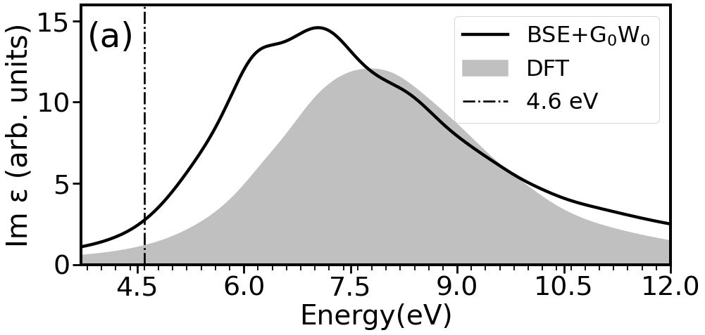
We first analyze the optical absorption spectrum of 4H-SiC calculated with the BSE. The absorption cross section averaged over polarizations is proportional to the imaginary part of the dielectric function that is shown in Fig. 2(a). It agrees with the experimental data reported in Refs. [42, 43], (see SM [47] for the data comparison). We also show calculated with the DFT for comparison in Fig. 2(a).

The significant difference between the DFT and the BSE+G0W0 optical spectra in the energy region 4–6 eV indicate that the excitonic effects are particularly strong for excited states with these energies. For this reason, we further consider the photon energy of a pump pulse to be eV.
The properties of a pump pulse determine the shape of created excitons. In Figs. 2(b)-(f), we analyze how the structure of excitons depends on the pump-pulse polarization . Figs. 2(b) - (d) show for different valence-excited states as a function of their energy . is proportional to the probability of a transition to a state. Figs. 2(e) - (f) show the averaged distribution of the hole density of created excitons . Details of the calculation of and the crystal structure of 4H-SiC are provided in SM [47].
Comparing Figs. 2(b) and (c), we see that a pump pulse with polarization along the and a pump pulse with a polarization along the lattice vectors excite same excitonic states with the same probability. We also obtain that the corresponding hole distributions shown in Fig. 2(e) are identical for these pump-pulse polarizations. They are predominantly of a Carbon -type character aligned along the both and directions slightly bent towards the vector. The excitons’ hole distributions for parallel to the lattice vector are predominantly of a Carbon -type character pointing along the direction.

We show the results of our calculations of Carbon K-edge and Si K-edge x-ray spectra of an optically-excited 4H-SiC probed by an x-ray pulse with an energy below XAT in Fig. 3. We also show the XAS from 4H-SiC in the ground state that is in a good agreement with experimental data [44, 45, 47]. We find that the presence of an exciton leads to an appearance of an additional peak in the spectra below the XAT. Such an additional peak in the XAS signal after a photo-excitation has been observed in experimental studies [8, 9, 10, 12]. It appears due to the creation of a coupled electron-hole pair. The hole of the pair provides an additional channel for an electron transition from a core to a state near the Fermi energy. The position of the peak at an energy lower than XAT can be intuitively understood in an one-particle picture. In such a picture, the energy of the peak would be given by the energy difference between a core orbital and a valence band, which is lower than the energy of transitions to conduction bands involved in a ground-state XAS (see Fig. 1).


If the energy of an x-ray pulse is above the XAT, then a final state in an optical-pump–x-ray-probe experiment is a doubly-excited excitonic state with one core-excited and one valence-excited exciton. Such transitions are beyond our formalism and that is why we do not observe any changes in the XAS spectra above the XAT. In an experiment, it would also be challenging to resolve the change of the XAS spectra above the threshold due to a strong background from a ground-state XAS signal. In our study, we focus on the signal below the XAT, which does not suffer from a background.
XAS spectra of 4H-SiC excited by a pump pulse with polarizations are shown in Figs. 3(a)-(c). K-edge XAS spectra are sensitive to a -type contribution to a wave function of unoccupied states, since they are governed by transitions from an orbital. In agreement with our observation that the hole is of a predominantly -type character pointing symmetrically in the and directions, we observe that intensive and identical peaks appear for the probe pulses polarized along the and along directions. A small peak appears in the case of as well, which is because the hole has also a non-zero projection along the direction (see also the two-dimensional (2D) cuts of the density in the SM [47]). The intensity of the peaks for a pump pulse polarized along the direction in Fig. 3(d)-(f) are the same as that in Fig. 3(a)-(c) in agreement with the fact that the averaged hole distributions are identical for the pump pulses polarized along and along .
XAS signals for 4H-SiC excited by an the optical pulse polarized along the direction are shown in Fig. 3(g)-(i). The strongest XAS signal in this case is for the probe pulse polarized along the direction. This indicates that the hole distribution is predominantly aligned along the vector, which agrees with our observation in Fig. 2(f). The hole distribution also has a small contribution of a -type character centred on Si atoms, which is visible on 2D density cuts in SM [47]. And this is why we observe the additional peak in the calculated Si K-edge XAS spectra in Fig. 3(j)-(l) as well, but find that Si K-edge XAS spectra are overall much less intensive than the Carbon K-edge XAS spectra. These results demonstrate that there is a direct correspondence between the shape of the excitons’ hole distribution and the dependence of the intensity of the corresponding feature in the XAS spectra on polarization of the x-ray probe pulse.
In conclusion, we developed an ab initio framework to describe optical-pump–x-ray-probe experiments in solid-state materials that treats excitonic effects at the BSE level, the state-of-the-art many-body approach. This way, it is capable to accurately describe modern experiments, where excited-state dynamics in functional materials or complexes are probed by means of x-ray pulses [69, 13]. Using the example of 4H-SiC, we demonstrated that a shape of an exciton can strongly depend on the excitation properties, which can eventually affect exciton transport. XAS is a powerful tool to reveal details about the shape of a positive charge of a photo-excited material with an element- and orbital-specificity.
Acknowledgement
We gratefully acknowledge the financial support of the Volkswagen Foundation. D P.-G. acknowledges support from the Cluster of Excellence ’CUI: Advanced Imaging of Matter’ of the Deutsche Forschungsgemeinschaft (DFG) - EXC 2056 - project ID 390715994.
References
- De Groot and Kotani [2008] F. De Groot and A. Kotani, Core level spectroscopy of solids (CRC press, 2008).
- Yano and Yachandra [2009] J. Yano and V. K. Yachandra, X-ray absorption spectroscopy, Photosynthesis research 102, 241 (2009).
- Baumgarten [2012] L. Baumgarten, X-ray absorption spectroscopy, Scattering Methods for Condensed Matter Research: Towards Novel Applications at Future Sources, Jülich (Germany) 33, F4.1 (2012).
- Ramasesha et al. [2016] K. Ramasesha, S. R. Leone, and D. M. Neumark, Real-time probing of electron dynamics using attosecond time-resolved spectroscopy, Annual Review of Physical Chemistry 67, 41 (2016).
- Santomauro et al. [2017] F. G. Santomauro, J. Grilj, L. Mewes, G. Nedelcu, S. Yakunin, T. Rossi, G. Capano, A. Al Haddad, J. Budarz, D. Kinschel, D. S. Ferreira, G. Rossi, M. Gutierrez Tovar, D. Grolimund, M. Samson, Valerieand Nachtegaal, G. Smolentsev, M. Kovalenko V, and M. Chergui, Localized holes and delocalized electrons in photoexcited inorganic perovskites: Watching each atomic actor by picosecond x-ray absorption spectroscopy, Structural Dynamics 4 (2017).
- Fracchia et al. [2018] M. Fracchia, P. Ghigna, A. Vertova, S. Rondinini, and A. Minguzzi, Time-resolved x-ray absorption spectroscopy in (photo) electrochemistry, Surfaces 1, 138 (2018).
- Huang et al. [2021] N. Huang, H. Deng, B. Liu, D. Wang, and Z. Zhao, Features and futures of x-ray free-electron lasers, The Innovation 2, 100097 (2021).
- Garratt et al. [2021] D. Garratt, L. Misiekis, D. Wood, E. Witting-Larsen, M. Matthews, O. Alexander, P. Ye, S. Jarosch, A. Bakulin, T. Penfold, and J. Marangos, Ultrafast exciton dynamics in poly(3-hexylthiophene) probed with time resolved x-ray absorption spectroscopy at the carbon k-edge, 2021 Conference on Lasers and Electro-Optics Europe and European Quantum Electronics Conference, 2021 Conference on Lasers and Electro-Optics Europe and European Quantum Electronics Conference , jsiii_2_4 (2021).
- Garratt et al. [2022] D. Garratt, L. Misiekis, D. Wood, E. W. Larsen, M. Matthews, O. Alexander, P. Ye, S. Jarosch, C. Ferchaud, C. Strüber, A. S. Johnson, A. A. Bakulin, and J. P. Penfold, T. J. Marangos, Direct observation of ultrafast exciton localization in an organic semiconductor with soft x-ray transient absorption spectroscopy, Nature Communications 13, 3414 (2022).
- Hillyard et al. [2009] P. Hillyard, S. Kuchibhatla, T. Glover, M. Hertlein, N. Huse, P. Nachimuthu, L. Saraf, S. Thevuthasan, and K. Gaffney, Atomic resolution mapping of the excited-state electronic structure of cu2o with time-resolved x-ray absorption spectroscopy, Physical Review B 80, 125210 (2009).
- Palmieri et al. [2020] T. Palmieri, E. Baldini, A. Steinhoff, A. Akrap, M. Kollár, E. Horváth, L. Forró, F. Jahnke, and M. Chergui, Mahan excitons in room-temperature methylammonium lead bromide perovskites, Nature communications 11, 850 (2020).
- Zürch et al. [2017] M. Zürch, H.-T. Chang, L. J. Borja, P. M. Kraus, S. K. Cushing, A. Gandman, C. J. Kaplan, M. H. Oh, J. S. Prell, D. Prendergast, C. D. Pemmaraju, D. M. Neumark, and S. R. Leone, Direct and simultaneous observation of ultrafast electron and hole dynamics in germanium, Nature Communications 8, 15734 (2017).
- Garratt et al. [2024] D. Garratt, M. Matthews, and J. Marangos, Toward ultrafast soft x-ray spectroscopy of organic photovoltaic devices, Structural Dynamics 11, 010901 (2024).
- Malakhov et al. [2023] M. Malakhov, G. Cistaro, F. Mart\́text{in}n, and A. Picón, Exciton migration in two-dimensional materials, arXiv e-prints , arXiv:2309.01190 (2023).
- Hansen et al. [2023] T. Hansen, T. Bezriadina, and D. Popova-Gorelova, Theoretical description of attosecond x-ray absorption spectroscopy of frenkel exciton dynamics, Molecules 28, 4502 (2023).
- Dutoi et al. [2013] A. D. Dutoi, K. Gokhberg, and L. S. Cederbaum, Time-resolved pump-probe spectroscopy to follow valence electronic motion in molecules: Theory, Phys. Rev. A 88, 013419 (2013).
- Skeidsvoll et al. [2020] A. S. Skeidsvoll, A. Balbi, and H. Koch, Time-dependent coupled-cluster theory for ultrafast transient-absorption spectroscopy, Phys. Rev. A 102, 023115 (2020).
- Khalili et al. [2020] K. Khalili, L. Inhester, C. Arnold, A. S. Gertsen, J. W. Andreasen, and R. Santra, Simulation of time-resolved x-ray absorption spectroscopy of ultrafast dynamics in particle-hole-excited 4‐(2-thienyl)-2,1,3-benzothiadiazole, Structural Dynamics 7, 044101 (2020).
- Rott et al. [2021] F. Rott, M. Reduzzi, T. Schnappinger, Y. Kobayashi, K. F. Chang, H. Timmers, D. M. Neumark, R. d. Vivie-Riedle, and S. R. Leone, Ultrafast strong-field dissociation of vinyl bromide: An attosecond transient absorption spectroscopy and non-adiabatic molecular dynamics study, Structural Dynamics 8, 034104 (2021).
- Salpeter and Bethe [1951] E. E. Salpeter and H. A. Bethe, A relativistic equation for bound-state problems, Physical Review 84, 1232 (1951).
- Strinati [1988] G. Strinati, Application of the green’s functions method to the study of the optical properties of semiconductors, La Rivista del Nuovo Cimento 11, 1 (1988).
- Onida et al. [2002] G. Onida, L. Reining, and A. Rubio, Electronic excitations: density-functional versus many-body green’s-function approaches, Reviews of modern physics 74, 601 (2002).
- Hirose et al. [2015] D. Hirose, Y. Noguchi, and O. Sugino, All-electron gw+ bethe-salpeter calculations on small molecules, Physical Review B 91, 205111 (2015).
- Gui et al. [2018] X. Gui, C. Holzer, and W. Klopper, Accuracy assessment of gw starting points for calculating molecular excitation energies using the bethe–salpeter formalism, Journal of Chemical Theory and Computation 14, 2127 (2018).
- Bruneval et al. [2015] F. Bruneval, S. M. Hamed, and J. B. Neaton, A systematic benchmark of the ab initio bethe-salpeter equation approach for low-lying optical excitations of small organic molecules, The Journal of Chemical Physics 142 (2015).
- Jacquemin et al. [2017] D. Jacquemin, I. Duchemin, and X. Blase, Is the bethe-salpeter formalism accurate for excitation energies? comparisons with td-dft, caspt2, and eom-ccsd, The Journal of Physical Chemistry Letters 8, 1524 (2017).
- Vorwerk et al. [2020] C. Vorwerk, F. Sottile, and C. Draxl, Excitation pathways in resonant inelastic x-ray scattering of solids, Physical Review Research 2, 042003 (2020).
- Gulans et al. [2014] A. Gulans, S. Kontur, C. Meisenbichler, D. Nabok, P. Pavone, S. Rigamonti, S. Sagmeister, U. Werner, and C. Draxl, Exciting: a full-potential all-electron package implementing density-functional theory and many-body perturbation theory, Journal of Physics: Condensed Matter 26, 363202 (2014).
- Draxl and Cocchi [2020] C. Draxl and C. Cocchi, exciting core-level spectroscopy, International Tables for Crystallography 1 (2020).
- Sangalli et al. [2023] D. Sangalli, M. D’Alessandro, and C. Attaccalite, Exciton-exciton transitions involving strongly bound excitons: An ab initio approach, Physical Review B 107, 205203 (2023).
- Bockstedte et al. [2010] M. Bockstedte, A. Marini, O. Pankratov, and A. Rubio, Many-body effects in the excitation spectrum of a defect in sic, Physical review letters 105, 026401 (2010).
- Klahold et al. [2018] W. M. Klahold, W. J. Choyke, and R. P. Devaty, High resolution optical spectroscopy of free exciton and electronic band structure near the fundamental gap in 4h sic, in Materials Science Forum, Vol. 924 (Trans Tech Publ, 2018) pp. 239–244.
- Klahold et al. [2020] W. Klahold, W. Choyke, and R. Devaty, Band structure properties, phonons, and exciton fine structure in 4 h-sic measured by wavelength-modulated absorption and low-temperature photoluminescence, Physical Review B 102, 205203 (2020).
- Zhang and Kioupakis [2023] X. Zhang and E. Kioupakis, Phonon-assisted optical absorption of sic polytypes from first principles, Physical Review B 107, 115207 (2023).
- Demmouche and Coutinho [2018] K. Demmouche and J. Coutinho, Electronic exchange-correlation, many-body effect issues on first-principles calculations of bulk sic polytypes, International Journal of Modern Physics B 32, 1850328 (2018).
- Peng-Shou et al. [2004] X. Peng-Shou, X. Chang-Kun, P. Hai-Bin, and X. Fa-Qiang, Theoretical study on the band structure and optical properties of 4h-sic, Chinese Physics 13, 2126 (2004).
- de Oliveira et al. [2003] A. de Oliveira, J. Freitas Jr, W. Moore, A. Silva, I. Pepe, J. Almeida, J. Osório-Guillén, R. Ahuja, C. Persson, K. Järrendahl, O. Lindquist, N. Edwards, and Q. Wahab, Spectroscopy studies of 4h-sic, Materials Research 6, 43 (2003).
- Adolph et al. [1997] B. Adolph, K. Tenelsen, V. Gavrilenko, and F. Bechstedt, Optical and loss spectra of sic polytypes from ab initio calculations, Physical Review B 55, 1422 (1997).
- Ahuja et al. [2002] R. Ahuja, A. Ferreira da Silva, C. Persson, J. Osorio-Guillen, I. Pepe, K. Järrendahl, O. Lindquist, N. Edwards, Q. Wahab, and B. Johansson, Optical properties of 4h–sic, Journal of applied physics 91, 2099 (2002).
- Ching et al. [2006] W. Ching, Y.-N. Xu, P. Rulis, and L. Ouyang, The electronic structure and spectroscopic properties of 3c, 2h, 4h, 6h, 15r and 21r polymorphs of sic, Materials Science and Engineering: A 422, 147 (2006).
- Gao [2015] S.-P. Gao, Band gaps and dielectric functions of cubic and hexagonal diamond polytypes calculated by many-body perturbation theory, physica status solidi (b) 252, 235 (2015).
- Lindquist et al. [2001] O. Lindquist, K. Järrendahl, S. Peters, J. Zettler, C. Cobet, N. Esser, D. Aspnes, A. Henry, and N. Edwards, Ordinary and extraordinary dielectric functions of 4h–and 6h–sic from 3.5 to 9.0 ev, Applied Physics Letters 78, 2715 (2001).
- Harris [1995] G. L. Harris, Properties of silicon carbide, EMIS datareviews series (INSPEC, Institution of Electrical Engineers, 1995).
- Tallarida et al. [2006] M. Tallarida, D. Schmeißer, F. Zheng, and F. Himpsel, X-ray absorption and photoemission spectroscopy of 3c-and 4h-sic, Surface science 600, 3879 (2006).
- Lüning et al. [1999] J. Lüning, S. Eisebitt, J.-E. Rubensson, C. Ellmers, and W. Eberhardt, Electronic structure of silicon carbide polytypes studied by soft x-ray spectroscopy, Physical Review B 59, 10573 (1999).
- Santra [2008] R. Santra, Concepts in x-ray physics, Journal of Physics B: Atomic, Molecular and Optical Physics 42, 023001 (2008).
- [47] See Supplemental Material at [URL will be inserted by publisher] for details of derivation of presented equations, definition of an average hole distribution of an exciton and its two-dimensional-plane cuts, and theoretical data that can be directly compared to experimental data showing optical and x-ray absorption spectra .
- Fetter and Walecka [2012] A. L. Fetter and J. D. Walecka, Quantum theory of many-particle systems (Courier Corporation, 2012).
- Quintela et al. [2022] M. Quintela, J. Henriques, and N. Peres, Theoretical methods for excitonic physics in two-dimensional materials, physica status solidi (b) 259 (2022).
- Karni et al. [2021] O. Karni, E. Barré, V. Pareek, J. D. Georgaras, M. K. Man, C. Sahoo, D. R. Bacon, X. Zhu, H. B. Ribeiro, A. L. O’Beirne, J. Hu, A. Al-Mahboob, M. M. M. Abdelrasoul, N. S. Chan, A. Karmakar, A. J. Winchester, B. Kim, K. Watanabe, T. Taniguchi, K. Barmak, J. Madéo, F. H. d. Jornada, T. F. Heinz, and K. M. Dani, Moiré-localized interlayer exciton wavefunctions captured by imaging its electron and hole constituents, arXiv preprint arXiv:2108.01933 (2021).
- Man et al. [2021] M. K. Man, J. Madéo, C. Sahoo, K. Xie, M. Campbell, V. Pareek, A. Karmakar, E. L. Wong, A. Al-Mahboob, N. S. Chan, D. R. Bacon, X. Zhu, M. M. M. Abdelrasoul, X. Li, T. F. Heinz, F. H. da Jornada, T. C. Cao, and K. M. Dani, Experimental measurement of the intrinsic excitonic wave function, Science advances 7, eabg0192 (2021).
- Cistaro et al. [2022] G. Cistaro, M. Malakhov, J. J. Esteve-Paredes, A. J. Ur\́text{in}a-Álvarez, R. E. Silva, F. Martin, J. J. Palacios, and A. Picon, Theoretical approach for electron dynamics and ultrafast spectroscopy (edus), Journal of Chemical Theory and Computation 19, 333 (2022).
- Sagmeister and Ambrosch-Draxl [2009] S. Sagmeister and C. Ambrosch-Draxl, Time-dependent density functional theory versus bethe–salpeter equation: an all-electron study, Physical Chemistry Chemical Physics 11, 4451 (2009).
- Puschnig and Ambrosch-Draxl [2002] P. Puschnig and C. Ambrosch-Draxl, Optical absorption spectra of semiconductors and insulators including electron-hole correlations: An ab initio study within the lapw method, Physical Review B 66, 165105 (2002).
- Leng et al. [2016] X. Leng, F. Jin, M. Wei, and Y. Ma, Gw method and bethe–salpeter equation for calculating electronic excitations, Wiley Interdisciplinary Reviews: Computational Molecular Science 6, 532 (2016).
- Vorwerk et al. [2019] C. Vorwerk, B. Aurich, C. Cocchi, and C. Draxl, Bethe-salpeter equation for absorption and scattering spectroscopy: implementation in the exciting code, Electronic Structure 1, 037001 (2019).
- Henneke et al. [2020] F. Henneke, L. Lin, C. Vorwerk, C. Draxl, R. Klein, and C. Yang, Fast optical absorption spectra calculations for periodic solid state systems, Communications in Applied Mathematics and Computational Science 15, 89 (2020).
- Vorwerk et al. [2022] C. Vorwerk, F. Sottile, and C. Draxl, All-electron many-body approach to resonant inelastic x-ray scattering, Physical Chemistry Chemical Physics 24, 17439 (2022).
- Urquiza et al. [2023] M. L. Urquiza, M. Gatti, and F. Sottile, Pseudopotential bethe-salpeter calculations for shallow-core x-ray absorption near-edge structures: Excitonic effects in - al 2 o 3, Physical Review B 107, 205148 (2023).
- Olovsson et al. [2009] W. Olovsson, I. Tanaka, T. Mizoguchi, P. Puschnig, and C. Ambrosch-Draxl, All-electron bethe-salpeter calculations for shallow-core x-ray absorption near-edge structures, Physical Review B 79, 041102 (2009).
- de Groot et al. [2021] F. M. de Groot, H. Elnaggar, F. Frati, R.-p. Wang, M. U. Delgado-Jaime, M. van Veenendaal, J. Fernandez-Rodriguez, M. W. Haverkort, R. J. Green, G. van der Laan, Y. Kvashnin, A. Hariki, H. Ikeno, H. Ramanantoanina, C. Daul, B. Delley, M. Odelius, M. Lundberg, O. Kuhn, S. I. Bokarev, E. Shirley, J. Vinson, K. Gilmore, M. Stener, G. Fronzoni, P. Decleva, P. Kruger, M. Retegan, Y. Joly, C. Vorwerk, C. Draxl, J. Rehr, and A. Tanaka, 2p x-ray absorption spectroscopy of 3d transition metal systems, Journal of Electron Spectroscopy and Related Phenomena 249, 147061 (2021).
- Kutepov [2021] A. L. Kutepov, Atomic forces from dirac–kohn–sham equations: implementation in flexible (apw+ lo/lapw)+ lo basis set, Journal of Physics: Condensed Matter 33, 235503 (2021).
- Unzog et al. [2022] M. Unzog, A. Tal, and G. Kresse, X-ray absorption using the projector augmented-wave method and the bethe-salpeter equation, Physical Review B 106, 155133 (2022).
- Stampfl et al. [2001] C. Stampfl, W. Mannstadt, R. Asahi, and A. J. Freeman, Electronic structure and physical properties of early transition metal mononitrides: Density-functional theory lda, gga, and screened-exchange lda flapw calculations, Physical Review B 63, 155106 (2001).
- Yuan et al. [2018] J.-H. Yuan, Q. Chen, L. R. Fonseca, M. Xu, K.-H. Xue, and X.-S. Miao, Gga-1/2 self-energy correction for accurate band structure calculations: the case of resistive switching oxides, Journal of Physics Communications 2, 105005 (2018).
- Nabok et al. [2016] D. Nabok, A. Gulans, and C. Draxl, Accurate all-electron g 0 w 0 quasiparticle energies employing the full-potential augmented plane-wave method, Physical Review B 94, 035118 (2016).
- Salas-Illanes et al. [2022] N. Salas-Illanes, D. Nabok, and C. Draxl, Electronic structure of representative band-gap materials by all-electron quasiparticle self-consistent g w calculations, Physical Review B 106, 045143 (2022).
- Momma and Izumi [2008] K. Momma and F. Izumi, Vesta: a three-dimensional visualization system for electronic and structural analysis, Journal of Applied crystallography 41, 653 (2008).
- Wernet [2019] P. Wernet, Chemical interactions and dynamics with femtosecond x-ray spectroscopy and the role of x-ray free-electron lasers, Philosophical Transactions of the Royal Society A 377, 20170464 (2019).