nnMamba: 3D Biomedical Image Segmentation, Classification and Landmark Detection with State Space Model
Abstract
In the field of biomedical image analysis, the quest for architectures capable of effectively capturing long-range dependencies is paramount, especially when dealing with 3D image segmentation, classification, and landmark detection. Traditional Convolutional Neural Networks (CNNs) struggle with locality respective field, and Transformers have a heavy computational load when applied to high-dimensional medical images. In this paper, we introduce nnMamba, a novel architecture that integrates the strengths of CNNs and the advanced long-range modeling capabilities of State Space Sequence Models (SSMs). nnMamba adds the SSMs to the convolutional residual-block to extract local features and model complex dependencies. For diffirent tasks, we build different blocks to learn the features. Extensive experiments demonstrate nnMamba’s superiority over state-of-the-art methods in a suite of challenging tasks, including 3D image segmentation, classification, and landmark detection. nnMamba emerges as a robust solution, offering both the local representation ability of CNNs and the efficient global context processing of SSMs, setting a new standard for long-range dependency modeling in medical image analysis. Code is available at https://github.com/lhaof/nnMamba
1 Introduction
Biomedical image analysis plays a pivotal role in healthcare, encompassing key tasks such as segmentation, classification, and landmark detection, which have been extensively studied in the literature [1, 2]. Image segmentation, in particular, is crucial for converting unstructured biomedical images into structured and informative representations, thereby propelling scientific discovery and clinical applications forward [3, 4]. Semantic segmentation is indispensable across various AI-enabled clinical applications, from diagnostic assistance and therapeutic planning to intra-operative support and monitoring tumor evolution [5]. Similarly, image based classification [6, 7, 8] and landmark detection tasks [9, 10] are vital for a range of downstream biomedical applications. For instance, Alzheimer’s disease (AD), a prevalent neurological disorder, can be better managed through classification models that help in understanding its progression [11]. Automated landmark detection plays a critical role in assisting radiologists by providing key reference points, thereby reducing workload and the potential for diagnostic errors [9, 12].
In the field, CNNs [13] have been a dominant force, particularly with FCN-based methods [14, 15, 16, 17] that are adept at hierarchical feature extraction. Transformers, with their origins in NLP and subsequent adaptation for visual tasks via architectures like ViT [18] and SwinTransformer [19], excel in global information aggregation. Their integration into CNN frameworks has led to hybrid models such as TransUNet [20], UNETR[21], and nnFormer [22], which significantly enhance the modeling of long-range dependencies.
However, the computational intensity of Transformers, particularly due to the self-attention mechanism’s scaling with input size, poses challenges in high-dimensional biomedical image analysis. State Space Sequence Models (SSMs), and specifically Structured State Space (S4) models [23], have emerged as efficient alternatives, delivering cutting-edge results in long-sequence data analysis [24, 23]. The Mamba model [25] refines the S4 approach by introducing an input-adaptive mechanism, outperforming Transformers in dense data scenarios. Recent works have begun exploring Mamba’s application in medical image segmentation [26, 27, 28] and visual recognition [29]. Despite these developments, a comprehensive evaluation of Mamba’s effectiveness in 3D medical segmentation, classification, and landmark detection tasks remains an open area of investigation.
Inspired by the promising results of SSMs in vision tasks and the capabilities of Mamba to handle long sequences efficiently, we explore the integration of Mamba blocks within CNNs to enhance long-range dependency modeling. Thus we proposed the nnMamba, a various of structure for both segmentation, classification, and landmark detection.
-
•
We present nnMamba, a framework adept at handling the intrinsic complexities of three core tasks in 3D medical imaging, namely image segmentation, classification, and landmark detection. The architecture of nnMamba is meticulously crafted to address the unique demands of each task while maintaining a cohesive structural foundation.
-
•
Through extensive experimentation, we demonstrate the superior performance of the nnMamba framework over existing methodologies. Our results indicate that nnMamba achieves the state-of-the-art effectiveness across the board, setting a new benchmark for future research and applications in the field of 3D medical image analysis.
2 Methodology
2.1 State Space Sequence Models
State Space Sequence Models (SSMs) efficiently transform one-dimensional input sequences to outputs through a system of linear Ordinary Differential Equations (ODEs):
| (1) |
| (2) |
with the latent state and parameters , . Despite their linear time complexity and parallelizability, SSMs often face memory and vanishing gradient issues. Structured State Space (S4) models mitigate these drawbacks by structuring the state matrix using the High-Order Polynomial Projection Operator (HIPPO), which improves long-range dependency modeling. Mamba, an extension of SSMs, introduces an input-dependent selection mechanism for adaptive parameter tuning and employs a hardware-optimized algorithm for efficient computation. This innovation leads to performance gains in processing long sequences, achieving state-of-the-art results with enhanced computational efficiency.
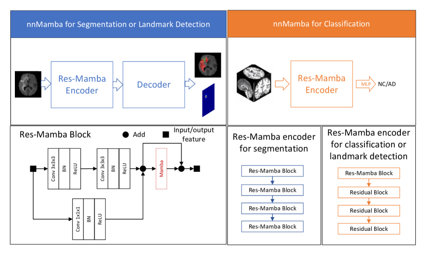
2.2 nnMamba for segmentation and landmark detection
In this study, we concentrate on enhancing the representational capacity of the encoder in neural network architectures through the incorporation of the Mamba layer, which is designed to capture long-range dependencies within the input data. Our methodology is applied to two distinct tasks: segmentation and landmark detection. We employ an encoder-decoder framework, utilizing a residual-based encoder coupled with a convolutional decoder. Segmentation tasks, characterized by their requirement for dense prediction, naturally demand intricate feature interactions. To address this, we introduce Res-Mamba blocks, which integrate Mamba layers subsequent to each residual block layer, thereby enhancing the network’s ability to facilitate effective feature communication at multiple scales.
For landmark detection, the objective is to generate accurate heatmaps that pinpoint anatomical landmarks. We posit that the initial stages of feature extraction are crucial for this task. Consequently, we incorporate a singular Mamba layer at the first block of the network. This strategic placement is intended to bolster the network’s proficiency in modeling long-range relationships early on in the processing pipeline, which is pivotal for the precise localization inherent to landmark detection. Our approach leverages the strengths of the Mamba layer to enrich feature representation, thereby improving performance across both segmentation and landmark detection tasks, as evidenced by our experimental results.
2.3 nnMamba for classification
In addressing the classification task, our architecture primarily employs a residual encoder. The underlying hypothesis is that when feature maps are reduced in size, the capacity to encapsulate long-range dependencies via convolutional operations is enhanced. Building on this rationale, we judiciously integrate the Mamba layer at an early stage, immediately following the initial feature extraction phase. This strategic insertion of the Mamba layer is designed to leverage the smaller spatial dimensions of the feature map, thereby enabling a more effective capture of long-range interactions. Such interactions are pivotal for classification, where the global understanding of the input data is crucial for accurate label assignment. The configuration of the Mamba layer at this juncture in the network ensures that the subsequent layers operate on features that already possess a global contextual awareness, potentially reducing the need for further complex operations to achieve high-level reasoning. Our experimental results confirm the efficacy of this approach, demonstrating enhanced classification performance attributed to the improved representational capabilities introduced by the Mamba layer.
3 Experiments
3.1 Implementation and Metric
All the models are trained with NVIDIA V100 GPU with 24GB memory. The framework is implemented in PyTorch 2.0.1[30] and CUDA 11.6. We train the models with the Adam optimizer at a learning rate of 0.002, batch size at 2, training epoch at 100, and weight decay at 0.001. For the segmentation task, we use the dice score and HD95 for evaluation. For the landmark detection task, we take the MRE as the metric for evaluation following [10]. For the classification task, we use the AUC, accuracy, recall ,specificity, precision, and F1-score by following [11].
3.2 Segmentation Evaluation
We make the evaluation of the segmentation on the BraTS 2023 GIL track [7]. We use the official training set, and split them to the training, validation, and testing with the factor of 7:1:2. We use the nnUNet framework to evaluate our code. In Table 1, we present the comparative evaluation of our proposed nnMamba network against other state-of-the-art methods on the BraTS 2023 GIL track. Performance metrics include the Dice similarity coefficient (Dice) and the 95th percentile Hausdorff Distance (HD95). Our findings demonstrate that nnMamba achieves superior performance in terms of the average Dice score, with a value of 90.01, outperforming all other methods including ViT, DIT, UNETR, and nnUNet. Specifically, nnMamba shows notable improvements in the ET category with a Dice score of 85.65. Additionally, nnMamba’s average HD95 score is competitive at 7.43. Notably, it outperforms the pioneering ViT model by a margin of 0.60 in HD95. These results underscore nnMamba’s efficacy in segmenting brain tumors with high precision, substantiating its potential as a robust tool for medical image analysis.
| BraTS 2023 GIL | Dice | HD95 | ||||||
|---|---|---|---|---|---|---|---|---|
| Methods | WT | TC | ET | Average | WT | TC | ET | Average |
| ViT[18] | 92.97 | 88.88 | 84.30 | 88.72 | 5.20 | 6.06 | 12.83 | 8.03 |
| DIT[31] | 93.49 | 90.22 | 84.38 | 89.36 | 4.21 | 5.27 | 13.64 | 7.71 |
| UNETR[21] | 93.33 | 89.89 | 85.19 | 89.47 | 4.76 | 7.27 | 12.78 | 8.27 |
| nnUNet[5] | 93.31 | 90.24 | 85.18 | 89.58 | 4.49 | 4.95 | 11.91 | 7.12 |
| nnMamba | 93.70 | 90.67 | 85.65 | 90.01 | 3.88 | 6.82 | 11.59 | 7.43 |
3.3 Classification Evaluation
We evaluate the classification task on the ADNI dataset[32, 33] with the AD and NC classification task. Table 2 showcases the performance of our nnMamba network compared to the 3D-ResNet baseline on the challenging ANDI (Alzheimer’s Disease Neuroimaging Initiative) classification task. The performance metrics evaluated include Accuracy, Sensitivity/Recall, Specificity, Precision, F1 score, and the Area Under the ROC Curve (AUC). The nnMamba model demonstrates a commendable improvement in Accuracy, achieving a score of 80.07, which is a relative increase from the 79.39 accuracy of the 3D-ResNet model. Remarkably, nnMamba exhibits a significant enhancement in Sensitivity/Recall, achieving an impressive 89.78 in distinguishing between Normal Control (NC) and Alzheimer’s Disease (AD) cases. This is a substantial improvement over the 77.37 sensitivity of the 3D-ResNet model, indicating the robustness of nnMamba in identifying AD cases. Specificity is lower for nnMamba (71.70) compared to 3D-ResNet (81.13), which may indicate a trade-off between identifying true positives and reducing false positives. Despite a slight decrease in Precision, nnMamba achieved a higher F1 score of 80.66, compared to 77.66 of the baseline model, suggesting a better balance between Precision and Recall. Most notably, nnMamba’s AUC of 90.04 is higher than that of 3D-ResNet, which stands at 87.90, underscoring its superior diagnostic ability in distinguishing between the two classes.
| NC VS AD | Accuracy | Sensitivity/Recall | Specificity | Precision | F1 | AUC |
|---|---|---|---|---|---|---|
| 3D-ResNet[34] | 79.39 | 77.37 | 81.13 | 77.94 | 77.66 | 87.90 |
| nnMamba | 80.07 | 89.78 | 71.70 | 73.21 | 80.66 | 90.04 |
3.4 Landmark Detection Evaluation
For the landmark detection task, we evaluate our method on the our private fetal Brain landmark detection dataset. In Table 3, we report the performance comparison between the proposed nnMamba framework and the ResUNet baseline for anatomical landmark detection. Performance is quantified using mean error values in millimeters for landmarks. nnMamba outperforms ResUNet on HDV 1, ADV 1, and ADV 2 with mean errors of 2.23 mm, 2.05 mm, and 1.84 mm, respectively, compared to ResUNet’s 2.32 mm, 2.45 mm, and 2.05 mm in the same order. Despite nnMamba’s slightly increased mean errors in the CBD category, it shows a lower overall average mean error (2.21 mm versus ResUNet’s 2.23 mm), indicating a marginal yet consistent improvement in general landmark detection accuracy. These results underscore nnMamba’s potential as a reliable model for detecting specific anatomical landmarks, especially for HDV and ADV, corroborating its utility in medical imaging applications.
| Landmark Detection | CBD 1 | CBD 2 | HDV 1 | HDV 2 | ADV 1 | ADV 2 | Average |
|---|---|---|---|---|---|---|---|
| ResUNet[34] | 2.23 | 2.15 | 2.32 | 2.16 | 2.45 | 2.05 | 2.23 |
| nnMamba | 2.45 | 2.59 | 2.23 | 2.10 | 2.05 | 1.84 | 2.21 |
4 Conclusion
In summary, our work has introduced nnMamba, a novel framework that effectively fuses the granular feature extraction capabilities of CNNs with the expansive long-range dependency modeling afforded by State Space Sequence Models (SSMs). By innovatively integrating SSMs within the convolutional residual blocks, nnMamba has been tailored to excel in the domain of 3D medical image analysis, tackling image segmentation, classification, and landmark detection with unprecedented efficacy.
Our extensive experimental analysis confirms that nnMamba sets a new benchmark, surpassing existing state-of-the-art methodologies in handling complex image analysis tasks. The architecture’s capacity to seamlessly blend attention to local details with an understanding of global context makes it an essential tool in the field. The implications of nnMamba’s performance are substantial, offering a robust and versatile framework that promises to elevate the standard of medical image analysis and support the advancement of medical diagnostic and therapeutic applications.
It should be noted that current version only provides primary results. We are still working on this project by evaluating more backbones and datasets.
Acknowledgments and Disclosure of Funding
This work was supported in part by the National Natural Science Foundation of China (NO. 62102267), and in part by the Natural Science Foundation of Guangdong Province of China (2023A1515011464).
References
- [1] Dinggang Shen, Guorong Wu, and Heung-Il Suk. Deep learning in medical image analysis. Annual review of biomedical engineering, 19:221–248, 2017.
- [2] Xiang He, Sibei Yang, Guanbin Li, Haofeng Li, Huiyou Chang, and Yizhou Yu. Non-local context encoder: Robust biomedical image segmentation against adversarial attacks. In Proceedings of the AAAI Conference on Artificial Intelligence, volume 33, pages 8417–8424, 2019.
- [3] Junjia Huang, Haofeng Li, Guanbin Li, and Xiang Wan. Attentive symmetric autoencoder for brain mri segmentation. In International Conference on Medical Image Computing and Computer-Assisted Intervention, pages 203–213. Springer, 2022.
- [4] Zihang Xu, Haifan Gong, Xiang Wan, and Haofeng Li. Asc: Appearance and structure consistency for unsupervised domain adaptation in fetal brain mri segmentation. In International Conference on Medical Image Computing and Computer-Assisted Intervention, pages 325–335. Springer, 2023.
- [5] Fabian Isensee, Paul F Jaeger, Simon AA Kohl, Jens Petersen, and Klaus H Maier-Hein. nnu-net: a self-configuring method for deep learning-based biomedical image segmentation. Nature methods, 18(2):203–211, 2021.
- [6] Hee E Kim, Alejandro Cosa-Linan, Nandhini Santhanam, Mahboubeh Jannesari, Mate E Maros, and Thomas Ganslandt. Transfer learning for medical image classification: a literature review. BMC medical imaging, 22(1):69, 2022.
- [7] Bjoern H Menze, Andras Jakab, Stefan Bauer, Jayashree Kalpathy-Cramer, Keyvan Farahani, Justin Kirby, Yuliya Burren, Nicole Porz, Johannes Slotboom, Roland Wiest, et al. The multimodal brain tumor image segmentation benchmark (brats). IEEE transactions on medical imaging, 34(10):1993–2024, 2014.
- [8] Haifan Gong, Hui Cheng, Yifan Xie, Shuangyi Tan, Guanqi Chen, Fei Chen, and Guanbin Li. Less is more: Adaptive curriculum learning for thyroid nodule diagnosis. In International Conference on Medical Image Computing and Computer-Assisted Intervention, pages 248–257. Springer, 2022.
- [9] Junshen Xu, Molin Zhang, Esra Abaci Turk, Larry Zhang, P Ellen Grant, Kui Ying, Polina Golland, and Elfar Adalsteinsson. Fetal pose estimation in volumetric MRI using a 3d convolution neural network. In Medical Image Computing and Computer Assisted Intervention–MICCAI 2019: 22nd International Conference, Shenzhen, China, October 13–17, 2019, Proceedings, Part IV 22, pages 403–410. Springer, 2019.
- [10] Xiang Li, Songcen Lv, Minglei Li, Jiusi Zhang, Yuchen Jiang, Yong Qin, Hao Luo, and Shen Yin. Sdmt: Spatial dependence multi-task transformer network for 3d knee MRI segmentation and landmark localization. IEEE Transactions on Medical Imaging, 2023.
- [11] Luoyao Kang, Haifan Gong, Xiang Wan, and Haofeng Li. Visual-attribute prompt learning for progressive mild cognitive impairment prediction. In International Conference on Medical Image Computing and Computer-Assisted Intervention, pages 547–557. Springer, 2023.
- [12] Netanell Avisdris, Leo Joskowicz, Brian Dromey, Anna L David, Donald M Peebles, Danail Stoyanov, Dafna Ben Bashat, and Sophia Bano. Biometrynet: Landmark-based fetal biometry estimation from standard ultrasound planes. In International Conference on Medical Image Computing and Computer-Assisted Intervention, pages 279–289. Springer, 2022.
- [13] Yann LeCun, Yoshua Bengio, et al. Convolutional networks for images, speech, and time series. The handbook of brain theory and neural networks, 3361(10):1995, 1995.
- [14] Jonathan Long, Evan Shelhamer, and Trevor Darrell. Fully convolutional networks for semantic segmentation. In Proceedings of the IEEE conference on computer vision and pattern recognition, pages 3431–3440, 2015.
- [15] Guanbin Li and Yizhou Yu. Visual saliency based on multiscale deep features. In Proceedings of the IEEE conference on computer vision and pattern recognition, pages 5455–5463, 2015.
- [16] Olaf Ronneberger, Philipp Fischer, and Thomas Brox. U-net: Convolutional networks for biomedical image segmentation. In Medical Image Computing and Computer-Assisted Intervention–MICCAI 2015: 18th International Conference, Munich, Germany, October 5-9, 2015, Proceedings, Part III 18, pages 234–241. Springer, 2015.
- [17] Liang-Chieh Chen, Yukun Zhu, George Papandreou, Florian Schroff, and Hartwig Adam. Encoder-decoder with atrous separable convolution for semantic image segmentation. In Proceedings of the European conference on computer vision (ECCV), pages 801–818, 2018.
- [18] Alexey Dosovitskiy, Lucas Beyer, Alexander Kolesnikov, Dirk Weissenborn, Xiaohua Zhai, Thomas Unterthiner, Mostafa Dehghani, Matthias Minderer, Georg Heigold, Sylvain Gelly, Jakob Uszkoreit, and Neil Houlsby. An image is worth 16x16 words: Transformers for image recognition at scale. In International Conference on Learning Representations, 2021.
- [19] Ze Liu, Yutong Lin, Yue Cao, Han Hu, Yixuan Wei, Zheng Zhang, Stephen Lin, and Baining Guo. Swin transformer: Hierarchical vision transformer using shifted windows. In Proceedings of the IEEE/CVF international conference on computer vision, pages 10012–10022, 2021.
- [20] Jieneng Chen, Yongyi Lu, Qihang Yu, Xiangde Luo, Ehsan Adeli, Yan Wang, Le Lu, Alan L Yuille, and Yuyin Zhou. Transunet: Transformers make strong encoders for medical image segmentation. arXiv preprint arXiv:2102.04306, 2021.
- [21] Ali Hatamizadeh, Yucheng Tang, Vishwesh Nath, Dong Yang, Andriy Myronenko, Bennett Landman, Holger R Roth, and Daguang Xu. Unetr: Transformers for 3d medical image segmentation. In Proceedings of the IEEE/CVF winter conference on applications of computer vision, pages 574–584, 2022.
- [22] Hong-Yu Zhou, Jiansen Guo, Yinghao Zhang, Xiaoguang Han, Lequan Yu, Liansheng Wang, and Yizhou Yu. nnformer: Volumetric medical image segmentation via a 3d transformer. IEEE Transactions on Image Processing, 2023.
- [23] Albert Gu, Karan Goel, and Christopher Ré. Efficiently modeling long sequences with structured state spaces. arXiv preprint arXiv:2111.00396, 2021.
- [24] Albert Gu, Isys Johnson, Karan Goel, Khaled Saab, Tri Dao, Atri Rudra, and Christopher Ré. Combining recurrent, convolutional, and continuous-time models with linear state space layers. Advances in neural information processing systems, 34:572–585, 2021.
- [25] Albert Gu and Tri Dao. Mamba: Linear-time sequence modeling with selective state spaces. arXiv preprint arXiv:2312.00752, 2023.
- [26] Jun Ma, Feifei Li, and Bo Wang. U-mamba: Enhancing long-range dependency for biomedical image segmentation. arXiv preprint arXiv:2401.04722, 2024.
- [27] Zhaohu Xing, Tian Ye, Yijun Yang, Guang Liu, and Lei Zhu. Segmamba: Long-range sequential modeling mamba for 3d medical image segmentation. arXiv preprint arXiv:2401.13560, 2024.
- [28] Yijun Yang, Zhaohu Xing, and Lei Zhu. Vivim: a video vision mamba for medical video object segmentation. arXiv preprint arXiv:2401.14168, 2024.
- [29] Lianghui Zhu, Bencheng Liao, Qian Zhang, Xinlong Wang, Wenyu Liu, and Xinggang Wang. Vision mamba: Efficient visual representation learning with bidirectional state space model. arXiv preprint arXiv:2401.09417, 2024.
- [30] Adam Paszke, Sam Gross, Francisco Massa, Adam Lerer, James Bradbury, Gregory Chanan, Trevor Killeen, Zeming Lin, Natalia Gimelshein, Luca Antiga, et al. Pytorch: An imperative style, high-performance deep learning library. Advances in neural information processing systems, 32, 2019.
- [31] William Peebles and Saining Xie. Scalable diffusion models with transformers. In Proceedings of the IEEE/CVF International Conference on Computer Vision, pages 4195–4205, 2023.
- [32] Clifford R Jack Jr, Matt A Bernstein, Nick C Fox, Paul Thompson, Gene Alexander, Danielle Harvey, Bret Borowski, Paula J Britson, Jennifer L. Whitwell, Chadwick Ward, et al. The Alzheimer’s disease neuroimaging initiative (ADNI): MRI methods. Journal of Magnetic Resonance Imaging, 27(4):685–691, 2008.
- [33] Chunfeng Lian, Mingxia Liu, Jun Zhang, and Dinggang Shen. Hierarchical fully convolutional network for joint atrophy localization and alzheimer’s disease diagnosis using structural MRI. IEEE Transactions on Pattern Analysis and Machine Intelligence, 42(4):880–893, 2020.
- [34] Kaiming He, Xiangyu Zhang, Shaoqing Ren, and Jian Sun. Deep residual learning for image recognition. In Proceedings of the IEEE conference on computer vision and pattern recognition, pages 770–778, 2016.