AI-Enhanced Virtual Reality in Medicine: A Comprehensive Survey
Abstract
With the rapid advance of computer graphics and artificial intelligence technologies, the ways we interact with the world have undergone a transformative shift. Virtual Reality (VR) technology, aided by artificial intelligence (AI), has emerged as a dominant interaction media in multiple application areas, thanks to its advantage of providing users with immersive experiences. Among those applications, medicine is considered one of the most promising areas. In this paper, we present a comprehensive examination of the burgeoning field of AI-enhanced VR applications in medical care and services. By introducing a systematic taxonomy, we meticulously classify the pertinent techniques and applications into three well-defined categories based on different phases of medical diagnosis and treatment: Visualization Enhancement, VR-related Medical Data Processing, and VR-assisted Intervention. This categorization enables a structured exploration of the diverse roles that AI-powered VR plays in the medical domain, providing a framework for a more comprehensive understanding and evaluation of these technologies. To our best knowledge, this is the first systematic survey of AI-powered VR systems in medical settings, laying a foundation for future research in this interdisciplinary domain.
1 Introduction
The intersection of artificial intelligence (AI) and virtual reality (VR) is an emerging frontier in the realm of medical technologies. Recent advances in these fields have given rise to innovative applications that promise to reshape various aspects of medical care. The integration of AI and VR brings forth a unique confluence of data-driven analytics and immersive experiences, making it a critical area of studies and applications in contemporary healthcare. AI’s contribution to VR in medicine ranges from enhancing diagnostic precision to offering new paradigms in patient care. These technologies not only are redefining existing medical procedures but also are paving the way for novel treatment methods. The purpose of this survey is to delve into the technical nuances, practice workflows, and diverse application scenarios of AI-powered VR in medical settings, examining their impacts on the efficiency, accuracy, and effectiveness of healthcare services.
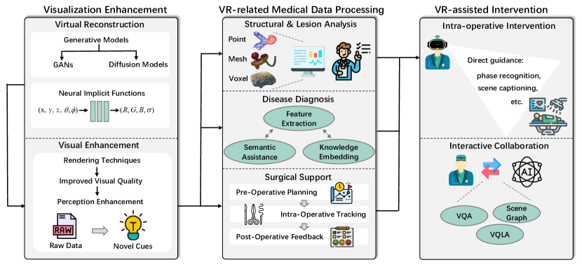
To systematically understand and analyze the role of AI in medical VR, this paper introduces a taxonomy categorizing the applications into three primary categories: Visualization Enhancement, VR-related Medical Data Processing, and VR-assisted Intervention (see Figure 1). This classification allows for a comprehensive overview of the current state of the art of AI in VR applications tailored to medical needs, highlighting each category’s unique contributions to healthcare. By categorizing these applications based on their functions and utilities in the spectrum of patient care, we aim to provide an extensive examination that facilitates easier comprehension and identification of existing research gaps.
Visualization Enhancement focuses on the augmentation of a user’s visual and sensory perception within a virtual space. This enhancement is crucial in complex medical procedures where understanding intricate anatomical structures and spatial relationships is imperative. We delve into the subcategories of virtual object reconstruction and visual enhancement, both pivotal in improving clinical outcomes and professional training. VR-related Medical Data Processing addresses the advanced capabilities of VR systems to analyze and interpret complex medical data. This category discusses how VR, combined with AI, facilitates in-depth structural and lesion analysis, enhances disease diagnosis, and supports the entire stages of surgical procedures. It underscores the transformation from traditional 2D data interpretation to a more dynamic, 3D analytical approach. VR-assisted Intervention demonstrates the practical application of AI-driven VR in real-time and interactive medical scenarios. This category covers the utilization of VR for direct procedural guidance, intra-operative intervention, and fostering interactive collaborations among medical professionals. It exemplifies the potential of VR to not only assist but also augment medical practice in live environments.
To our best knowledge, this is the first systematic survey of AI-powered VR technologies in medical settings, laying a foundation for future research in this interdisciplinary domain. The scope of this survey extends beyond mere categorization of current techniques and applications. It also explores the potential future trajectories of AI in VR for medicine, contemplating how ongoing advancements might unfold. The discussion encompasses not only technological aspects but also considers ethical, legal, and practical implications of deploying such advanced systems in sensitive medical environments.
0\float@count-1\float@count\e@alloc@chardef\forest@temp@box\float@count=
AI-powered VR in Medical Services
2 Taxonomy
The objective of the taxonomy is to group AI-powered VR applications in medical services with similar goals into the same category, facilitating in-depth investigation in subsequent studies. We classify the techniques and applications into three distinct categories based on diagnosis and treatment procedures: Visualization Enhancement, VR-related Medical Data Processing, and VR-assisted Intervention. A visual presentation of the taxonomy is shown in Figure 2.
2.1 Visualization Enhancement
Rapid advance of AI technologies offers VR platforms the capability of enhancing medical professionals’ visual perception during treatment and surgical stages. The techniques of visualization enhancement can be divided into the following two subcategories.
Virtual Reconstruction enables medical professionals to visualize medical data in a more intuitive manner, enhancing their understanding and interpretation of complex anatomical structures. By reconstructing virtual objects (e.g., organs or anatomical models) in a VR environment, professionals can gain a clearer and more comprehensive view of the patient’s condition, aiding treatment planning and decision-making.
Visual Enhancement focuses on improving the visual perception of professionals during the training or surgical phase. By immersing the professionals in augmented or virtual surgical scenes, such techniques create an immersive visual perception that enables them to perform treatment procedures with greater precision, resulting in improved treatment outcomes.
2.2 VR-related Medical Data Processing
Enhanced vision in a virtual environment offers an additional analytical capability for processing medical data. By leveraging the immersive and interactive aspect of virtual reality, professionals gain access to a wealth of additional visual information that surpasses the limitations of traditional 2D data usage in different treatment phases, as follows.
Structural and Lesion Analysis. In VR medical contexts, the utilized data formats such as point cloud, mesh, and voxel facilitate AI-powered systems to conduct more comprehensive analysis of anatomical structures and lesion situations, thereby providing additional cognitive information for accurate diagnosis.
Disease Diagnosis. Comprehensive analysis of VR-based medical data serves as an underlying foundation for disease diagnosis through multiple methods, including semantic segmentation, feature extraction, and knowledge embedding.
Surgical Support. Building upon data analysis and diagnosis, surgical support in VR enhances the precision and effectiveness of surgical procedures. Current progress has covered multiple aspects including pre-operative planning, intra-operative recognition and tracking, and post-operative feedback.
2.3 VR-assisted Intervention
VR-assisted intervention harnesses the power of AI-enhanced visualization and analysis technologies, showcasing its immense potential in augmenting professionals’ capabilities during the inspection and surgical phases. This topic explores how VR technologies directly provide guidance in intra-operative interventions and deliver context-aware feedback in an interactive and collaborative manner.
Intra-operative Intervention offers professionals direct guidance and assistance by integrating AI-powered functionalities into VR platforms such as object segmentation, phase recognition, etc. This encompasses the integration of diverse input data formats and platforms.
Interactive Collaboration provides interactive feedback to professionals by leveraging advancements in human-machine collaboration. Techniques such as Vision Question Answering (VQA) and Visual Question Localized-Answering (VQLA) have played a key role in this progress.
3 Visualization Enhancement
Enhanced visualization capability is one of the key advantages of VR technologies, which also shows a great potential in the enhancement of medical services. In this section, we cover two key aspects in the application of visualization enhancement: virtual reconstruction and visual enhancement.
3.1 Virtual Reconstruction
VR technologies offer the capability to provide users with an immersive experience with infinite viewpoints, enabling them to visualize an object freely. This feature shows great potential in medical services, which enables professionals to visualize complex medical data in a more intuitive manner. Traditional medical data acquisition methods like X-ray and ultrasound mostly provide only flattened 2D images which limit the effectiveness of diagnosis. In contrast, AI-powered reconstruction methods provide solutions for reconstructing complete objects with high quality, which can be visualized with VR devices to provide professionals with a better perception of the target objects. Compared with traditional 2D monitoring methods, 3D reconstruction offers notable advantages since it allows users to observe a surgical site from any viewpoint, which dramatically benefits downstream medical applications such as surgeon-centered augmented reality Nicolau et al. (2011) and virtual reality Chong et al. (2022). Multiple learning-based reconstruction methodologies have gained prominence, including neural implicit functions and generative models as two mainstream approaches.
Generative Models. In practical medical inspection scenarios, due to the limitations of safe radiation exposure, visual occlusion, or anisotropic spatial resolution, the reconstruction of high-quality 3D models in medical scenarios has been challenging. Thanks to the capability to synthesize image details with higher fidelity, generative adversarial networks (GANs) Goodfellow et al. (2020) have been widely adopted to reconstruct high-quality medical images Kaplan and Zhu (2019). In Lei et al. (2019), a cycle-consistent generative adversarial network (Cycle GAN) model was proposed to estimate diagnostic quality PET images using low count data. Recently, due to improved sample quality and higher log-likelihood scores compared to GANs, diffusion probabilistic models Ho et al. (2020) have emerged as a compelling alternative. Such methods have been employed in 3D teeth reconstruction Park et al. (2023) and cardiac volume reconstruction He et al. (2023). Likewise, to tackle the problem of inadequate natural medical data for temporal dynamics analysis and disease progression monitoring, diffusion models have been exploited to generate 4D volumetric data. In Kim and Ye (2022), a diffusion deformable model (DDM) was proposed to generate intermediate temporal volumes between source and target volumes.
| Representation | Size | Visual Quality | Computing Resource | Editability | Visualization |
| Point Cloud Yang et al. (2020) | Large | Low | Low | Easy |
![[Uncaptioned image]](/html/2402.03093/assets/Figures/representations/point_cloud.png)
|
| Mesh Wu et al. (2022a) | Medium | Medium | Medium | Medium |
![[Uncaptioned image]](/html/2402.03093/assets/Figures/representations/mesh2.png)
|
| Voxel Lei et al. (2023) | Medium | Low | Low | Easy |
![[Uncaptioned image]](/html/2402.03093/assets/Figures/representations/voxel2.png)
|
| Implicit Surfaces Zha et al. (2023) | Medium | Medium | Medium | Hard |
![[Uncaptioned image]](/html/2402.03093/assets/Figures/representations/implicit2.png)
|
| NeRF Chen et al. (2023) | Medium | Very high | Very high | Hard |
![[Uncaptioned image]](/html/2402.03093/assets/Figures/representations/nerf2.png)
|
Neural Implicit Functions. In virtual reality, typical methodologies for 3D shape representations encompass point-based Yang et al. (2020), voxel-based Lei et al. (2023), and mesh-based Wu et al. (2022a) techniques. These approaches explicitly utilize individual points, vertices, or faces to delineate 3D structures, which commonly result in substantial data size. Moreover, they necessitate high-quality raw data for precise reconstruction. A notable limitation of these traditional methods, particularly in medical context, is their inability to produce watertight surfaces, leading to significant drawbacks. In contrast, recent advances in neural implicit representation methods have substantially enhanced reconstruction performance. Neural implicit functions represent a scene by learning a mapping from 3D input coordinates to a shape or surface representation using neural networks, typically through a multi-layer perceptron (MLP) architecture. These functions capture the scene’s characteristics in terms of local densities and associated colors, allowing detailed and flexible modeling of complex shapes and surfaces. In Chen et al. (2023), an implicit function network was utilized to yield super-resolution medical images at arbitrary scales and free viewpoints in a continuous domain. For deformable tissues that pose a high requirement for watertight reconstruction, EndoSurf Zha et al. (2023) was proposed, which effectively learns three neural fields to transform 3D points, predict signed distance functions (SDFs), and estimate colors.
3.2 Visual Enhancement
Multiple learning-based methods have been developed to enhance visual perception during the training or surgical phase, improving the skills of surgeons and treatment effect.
Rendering. Recent advances in rendering techniques have significantly enhanced rendering performance, leading to improved visual quality of medical data perception. These developments contributed to a more immersive and realistic experience for medical professionals. In Semantic Virtual Shadows (SVS) Sommersperger et al. (2023), instrument-specific shadows are artificially generated, enabling naturally non-existent but important perceptual cues that are present in microscopic surgeries. On the other hand, in order to deal with the problem of significant topology changes, occlusion, and spare viewpoints, neural rendering has been recently developed to address the limitations of traditional 3D rendering techniques. In Wang et al. (2022), dynamic surgical scenes are reconstructed from single-view stereo endoscope videos based on D-NeRF, even when non-rigid deformation and occlusion exist.
Perception Enhancement. VR devices enable professionals to visualize medical data in a more flexible manner, offering additional cognitive cues during surgical or training procedures. With the aid of AI-powered VR techniques, professionals are able to acquire further information for treatment in a virtual vision. In Palumbo et al. (2022), a deep learning-based workflow was implemented to support emergency treatment where automatic segmentation of skill, face skin, and ventricles is achieved. In this work, 3D U-Net Çiçek et al. (2016) architecture is exploited for segmentation. Then, the segmentation results are superimposed on the MR representation in 3D point clouds. In clinical practice, a deep learning-based method Tamersoy et al. (2023) was proposed to accurately estimate a patient’s height and weight by formulating the problem as an end-to-end single-value regression problem given only depth images as input. To provide complementary information for image-guided therapies, multi-modal image alignment is essential. In Chen et al. (2022), a discriminator-free translation network to facilitate the training of the registration network and a patch-wise contrastive loss to encourage the translation network to preserve object shapes are incorporated to tackle the challenges caused by complicated and unknown spatial correspondences between different modalities.
4 VR Medical Data Analytics
Enhanced vision in virtual space offers a multitude of visual cues for both human and computer perception, thereby enhancing the analytical capability of medical data. By exploiting the immersive and interactive nature of VR, medical professionals can access a wealth of additional visual information that goes beyond traditional 2D displays.
4.1 Structural and Lesion Analysis
In medical context, the application of VR predominantly utilizes three principal data formats: voxel-based, point-based, and mesh-based. These formats facilitate more sophisticated and nuanced visualization of anatomical structures and disease lesions, which is pivotal for a comprehensive analysis in clinical practice. We provide comparisons and examples of principle data formats in Table 1.
Point-based Format. A point cloud employs a large group of individual data points in space to represent 3D objects. Each point contains spatial coordinates and additional attributes (e.g., color in RGB, transparency). This unique characteristic allows straightforward and direct applications of downstream tasks, including object detection and segmentation. However, the use of point clouds also comes with the drawback of high computational resource consumption due to their large data sizes. In Liu et al. (2023); Cui et al. (2021), operations are directly conducted on point clouds, without requiring further manipulations of the input point cloud data. IntrA Yang et al. (2020) and 3DTeethSeg Ben-Hamadou et al. (2023) are two of the most mainstream point cloud datasets in medical scenarios, and a series of methods in both medical scenarios Liu et al. (2023); Cui et al. (2021) and general 3D vision Wu et al. (2019); Li et al. (2018) were implemented on such datasets, showing the transfer-ability of 3D segmentation methods from the general domain to the medical domain. There are also some works Zhang et al. (2023) that combine different 3D formats to jointly promote the understanding and analysis of anatomical structures. In Zhang et al. (2023), a point-voxel fusion framework was presented for accurately segmenting the liver into anatomical segments, addressing the challenge of no intensity contrast between adjacent segments by incorporating vessel structure prior.
Mesh-based Format. A polygon mesh consists of vertices, edges, and faces that define the shape of a polyhedral object. The 3D mesh format is a collection of meshes that represent the spatial surface, color, and texture of the object. Compared with point clouds, the mesh is suitable for representing complex geometry by a combination of smooth surfaces with a smaller data size. However, the complicated composition of the mesh introduces higher processing costs. Mesh-based methods Wu et al. (2022a); Zhang et al. (2021); Lian et al. (2020) take triangle meshes as input and produce corresponding labels for each mesh. In Lian et al. (2020), cascaded graph-constrained learning modules were introduced for extracting multi-scale local contextual features for automated tooth labeling on raw dental meshes. In Zhang et al. (2021), a two-stream graph convolutional network was proposed to learn multi-view geometric information from different geometric attributes. In Wu et al. (2022a), a two-stage framework was designed for concurrent segmentation of meshes and regression of anatomical landmarks.
Voxel-based Format. Compared with pixels in 2D images, voxels are the most analogous representation in 3D space with a smaller domain gap. Therefore, many 2D methods for medical data processing, by taking into account the additional spatial dimension, can be adapted to encode 3D features and capture the relationships in three-dimensional space. In Lei et al. (2023); Huang et al. (2022); Dong et al. (2022), voxel-based methods operate on volumetric data represented as a grid of 3D pixels, known as voxels. CiT-Net Lei et al. (2023) was designed as a hybrid architecture of 3D convolutional neural networks (CNNs) hand in hand with 3D Vision Transformers for volumetric medical image segmentation. Furthermore, 2D imaging Salari et al. (2023), projections Wu et al. (2022b), and depth maps Dima et al. (2023) are frequently employed to facilitate the comprehension and interpretation of voxel-based data. In Dima et al. (2023), 2D labels were mapped to the 3D space using depth information to bridge the gap between 3D supervision and 2D supervision for 3D arterial segmentation. In Salari et al. (2023), real-time 2D imaging was leveraged to assist high-precision 3D voxel-based visual landmark detection.
4.2 Disease Diagnosis
Recently, based on enhanced visual data, various methods for disease diagnosis have been developed, which can be grouped into three lines according to their design philosophies.
Extractor-focused Methods. This line of methods Wang et al. (2024); Wu et al. (2023b); Shao et al. (2022); Jang et al. (2022) focuses on designing efficient and effective local feature extractors. Early work was usually based on using MLPs Liu et al. (2018), 3D CNNs Çiçek et al. (2016), and GNNs Zhang et al. (2021). More recently, inspired by the success of Transformers in the general vision domain, Transformer-based backbones Wang et al. (2024); Wu et al. (2023b); Shao et al. (2022); Jang et al. (2022) have gradually unified feature extraction for various 3D medical enhanced visual data. For example, D-former Wu et al. (2023b) introduced a dilated attention to effectively reduce computation for long-dependent feature extraction process of Transformer.
Semantics-enhanced Methods. Semantics-based methods Pan et al. (2023); Zhao et al. (2023); Xia et al. (2019) are concerned with integrating various intrinsic information sources to enhance the comprehension of the underlying semantics associated with features and target objects. This can involve leveraging multi-view consistency Pan et al. (2023), multi-task consistency Zhao et al. (2023), and multi-modality consistency Xia et al. (2019) to improve the accuracy of diagnostic algorithms. In Pan et al. (2023), a multi-view relation-aware consistency and virtual feature compensation (MRC-VFC) framework was proposed for long-tailed medical image classification.
Knowledge-embedded Methods. Knowledge-embedded methods Rygiel et al. (2023); Wu et al. (2023a) aim to incorporate external knowledge and priors to enable networks to conduct diagnosis in a more informative and effective manner. Based on geometric priors, a geometry-based architecture was proposed to utilize implicit geometry embedding for directly estimating hemodynamic features Rygiel et al. (2023). Meta labels Wu et al. (2023a), representing specific attribute information of medical images, serve as valuable cues for networks to enhance the identification and classification of pathological findings. Such incorporation of domain-specific knowledge not only enhances the interpretability of AI-driven diagnostics but also increases the reliability of systems in clinical settings.
4.3 Surgical Support
An important application scenario of AI-powered VR is in surgical support, where it can offer surgeons a more precise planning, real-time tracking, and reliable feedback throughout the surgical procedures.
Pre-operative Planning. Pre-operative planning is a crucial phase in surgery, involving detailed patient-specific analysis and strategic procedure mapping to ensure optimal surgical outcomes and minimize risks. VR-enhanced planning in surgery primarily serves two key purposes: (1) facilitating the arrangement of surgical procedures Bukas et al. (2021), and (2) enabling the simulation of surgical outcomes Friedrich et al. (2023). In Bukas et al. (2021), a personalized automatic high-level framework was presented for planning osteoplasty procedures. In Friedrich et al. (2023), a novel approach was proposed for implant generation based on a combination of 3D point cloud diffusion models and voxelization networks.
Real-time Object Recognition and Tracking. Real-time object recognition and tracking play a crucial role in surgical procedures, ensuring immediate and accurate identification of surgical instruments and anatomical structures. Based on the categories of the targets, existing works can be broadly classified into two types: lesion-centric and device-centric. Lesion-centric works Szeskin et al. (2023); Cai et al. (2021) involve accurate measurement and monitoring of pathological changes during surgical procedures. In Szeskin et al. (2023), a fully automatic end-to-end pipeline was presented for liver lesion change analysis in consecutive (prior and current) abdominal CECT scans of oncology patients. Highlighting unusual lesion labels and lesion change patterns helps radiologists identify possibly overlooked or faintly visible lesions. In contrast, device-centric approaches Gsaxner et al. (2021) focus on real-time localization and control of surgical instruments. In Gsaxner et al. (2021), 6-DOF tracking was novelly achieved by purely utilizing on-board stereo cameras of a HoloLens2 to track the same retro-reflective marker spheres used by current optical navigation systems.
Post-operative Feedback. Post-operative feedback is essential in evaluating the success of surgical procedures, guiding patient recovery, and informing future improvements in surgical practice and patient care, where motion analysis and outcome assessment are two key technical aspects. Motion analysis Nwoye et al. (2022); Twinanda et al. (2016) refers to precisely detecting fine-grained actions of surgeons, with a particular focus on their hand movement. Rendezvous (RDV) Nwoye et al. (2022) was proposed to recognize surgical action triplets (i.e., instrument, verb, target) in endoscopic videos by leveraging attention at the spatial and semantic levels, respectively. POV-Surgery dataset Wang et al. (2023) is a large-scale, synthetic, egocentric dataset focusing on pose estimation of hands with different surgical gloves and orthopedic surgical instruments. Outcome assessment Jamzad et al. (2023) involves systematic evaluation of the results and impact of a treatment or intervention, measuring its effectiveness, safety, and patient satisfaction.
5 VR-assisted Intervention
Based on combination of AI-powered visualization and analysis technologies, VR-assisted intervention technologies have significantly enhanced professionals’ capabilities during the inspection and surgical phases. This section explores two key applications of VR-assisted intervention: Intra-operative Intervention and Interactive Collaboration.
5.1 Intra-operative Intervention
Intra-operative intervention focuses on leveraging VR technologies to enhance professionals’ abilities during surgery. VR platforms attached with AI algorithms offer additional guidance/assistance during the complex intervention phases. Leveraging the advantages of augmented vision, a virtual assistance platform based on commodity optical see-through head-mounted displays (OST-HMDs) was implemented Doughty et al. (2021), which provides users with action-and-workflow-driven assistance with near-real-time performance on the Microsoft HoloLens2 OST-HMD. To achieve real-time performance, CNN for spatial feature extraction and RNN for temporal relationship learning were introduced to form the core of the system’s analytical capabilities. A similar attempt Palumbo et al. (2022) was also implemented on the same platform (Microsoft HoloLens2) in the emergency treatment scenario, where 3D U-Net Çiçek et al. (2016) architecture is exploited for skill, face skin, and ventricle segmentation. These works provide valuable insights into developing real-time surgical guidance systems based on VR platforms. Furthermore, regarding surgical scene understanding and reasoning, which serve as a backbone of VR-assisted intervention, phase recognition Ding et al. (2022) and scene captioning Beddiar et al. (2023) techniques play a crucial role, aiding surgeons to obtain direct responses and comprehensive understanding of the current state of surgery. This task encompasses real-time processing and integration of diverse data streams, including visual inputs from the surgical field, patient vitals, and historical medical data.
5.2 Interactive Collaboration
Recent advances in human-machine collaboration have significantly reduced human effort in field operations, thereby fostering widespread implementation of AI-aided interactive collaboration. At the forefront of these advances is Interactive Query. In Seenivasan et al. (2023), a Vision Question Answering (VQA) system was proposed specifically for medical scenarios, providing a robust and reliable surgical visual question answering solution that can respond to questions by inferring from context-enriched surgical scenes. Beyond that, in Bai et al. (2023), Visual Question Localized-Answering (VQLA) was further developed that offers more guidance by highlighting the specific areas in the images related to the question and answer. This allows a better understanding of complex medical diagnoses and surgical scenes. Notably, Scene Graph Holm et al. (2023); Özsoy et al. (2023) is a more holistic, semantically meaningful, and human-readable way to represent surgical videos while encoding all anatomical structures, tools, and their interactions.
Datasets play a critical role in this domain. For instance, in Sharghi et al. (2020), a dataset was created to capture different robot-assisted interventions, focusing on the phase recognition of the OR scene. The MVOR dataset Srivastav et al. (2018) contains synchronized multi-view frames from real surgeries with human pose annotations. This dataset facilitates research on human motion analysis and understanding during surgical procedures. Additionally, the 4D-OR dataset Özsoy et al. (2022) is the first publicly available 4D surgical semantic scene graphs dataset, contributing to advancements in surgical planning and decision-making.
6 Challenges and Future Prospects
6.1 Challenges
Data and Integration Limitations. A primary challenge in AI-driven VR for medical applications lies in the quality and availability of data. High-quality, diverse datasets are essential for training effective AI models. Yet, such data currently are still scarce and fragmented. Additionally, integrating the advanced AI-VR technologies with existing healthcare systems and workflows is a complex task, requiring both technical compatibility and operational adjustments.
Ethical and Legal Issues. Ethical issues, particularly concerning with patient privacy, data security, and informed consent, are crucial. AI-VR systems must comply with stringent healthcare regulations to ensure patient confidentiality and safety. Furthermore, the issue of liability in the cases of AI-VR related errors remains unresolved, complicating the legal landscape for healthcare providers, technology developers, and governments.
Technological and User Acceptance. Achieving realism and accuracy in VR simulations is vital for effective medical training and treatment, yet it remains technically challenging. The ‘black box’ nature of some AI systems can impede user trust and acceptance, as medical professionals often need to understand the decision-making processes behind AI recommendations. Additionally, designing user-friendly interfaces for a diverse range of users and overcoming technophobia among healthcare professionals and patients are critical for widespread adoption of AI-driven VR systems.
6.2 Future Prospects
Immersive Medicine through Technological Advancements. Future advancements in AI algorithms and VR technologies promise more accurate, efficient, and immersive medical simulations. The integration of Natural Language Processing (NLP) approaches within VR environments will facilitate a much enhanced understanding of patient speech and text records. Besides, advanced interactive sonification technologies Matinfar et al. (2023) can also lead to more comprehensive and detailed diagnostics, thereby enhancing the accuracy and effectiveness of diagnoses and treatments.
Tailored Therapeutic Interventions with AI-Driven Analytics. Utilizing AI algorithms to analyze patient interactions within VR scenarios will allow for customization of therapeutic approaches and options, especially in mental healthcare settings. Furthermore, the integration of biofeedback into VR, wherein AI adjusts experiences based on real-time physiological data, represents a significant stride in personalizing patient care and treatment.
AI-Enhanced Real-Time Clinical Analytics. AI-powered VR has the potential to revolutionize tele-medicine and remote medical training, particularly in under-served or under-developed areas. Implementing real-time analytical capabilities within VR, AI algorithms will provide immediate diagnostic support, enhancing the decision-making processes in clinical and non-clinical settings. Besides, by providing high-quality medical services remotely, these technologies can bridge the gaps in healthcare accessibility, quality, and equality.
References
- Bai et al. [2023] Long Bai, Mobarakol Islam, et al. Surgical-VQLA: Transformer with gated vision-language embedding for visual question localized-answering in robotic surgery. In ICRA, 2023.
- Beddiar et al. [2023] Djamila-Romaissa Beddiar, Mourad Oussalah, et al. Automatic captioning for medical imaging (MIC): A rapid review of literature. Artificial Intelligence Review, 2023.
- Ben-Hamadou et al. [2023] Achraf Ben-Hamadou, Oussama Smaoui, et al. 3DTeethSeg’22: 3D teeth scan segmentation and labeling challenge. arXiv, 2023.
- Bukas et al. [2021] Christina Bukas, Bailiang Jian, et al. Patient-specific virtual spine straightening and vertebra inpainting: an automatic framework for osteoplasty planning. In MICCAI, 2021.
- Cai et al. [2021] Jinzheng Cai, Youbao Tang, et al. Deep lesion tracker: Monitoring lesions in 4D longitudinal imaging studies. In CVPR, 2021.
- Chen et al. [2022] Zekang Chen, Jia Wei, et al. Unsupervised multi-modal medical image registration via discriminator-free image-to-image translation. In IJCAI, 2022.
- Chen et al. [2023] Zixuan Chen, Lingxiao Yang, et al. CuNeRF: Cube-based neural radiance field for zero-shot medical image arbitrary-scale super resolution. In ICCV, 2023.
- Chong et al. [2022] Nannan Chong, Yazhong Si, et al. Virtual reality application for laparoscope in clinical surgery based on siamese network and census transformation. In MICCAI, 2022.
- Çiçek et al. [2016] Özgün Çiçek, Ahmed Abdulkadir, et al. 3D U-Net: Learning dense volumetric segmentation from sparse annotation. In MICCAI, 2016.
- Cui et al. [2021] Zhiming Cui, Changjian Li, et al. TSegNet: An efficient and accurate tooth segmentation network on 3D dental model. MIA, 2021.
- Dima et al. [2023] Alina F Dima, Veronika A Zimmer, et al. 3D arterial segmentation via single 2D projections and depth supervision in contrast-enhanced CT images. In MICCAI, 2023.
- Ding et al. [2022] Xinpeng Ding, Xiaomeng Li, et al. Exploring segment-level semantics for online phase recognition from surgical videos. TMI, 2022.
- Dong et al. [2022] Zhangfu Dong, Yuting He, et al. MNet: Rethinking 2D/3D networks for anisotropic medical image segmentation. In IJCAI, 2022.
- Doughty et al. [2021] Mitchell Doughty, Karan Singh, et al. SurgeonAssist-Net: Towards context-aware head-mounted display-based augmented reality for surgical guidance. In MICCAI, 2021.
- Friedrich et al. [2023] Paul Friedrich, Julia Wolleb, et al. Point cloud diffusion models for automatic implant generation. In MICCAI, 2023.
- Goodfellow et al. [2020] Ian Goodfellow, Jean Pouget-Abadie, et al. Generative adversarial networks. Commun. ACM, 2020.
- Gsaxner et al. [2021] Christina Gsaxner, Jianning Li, et al. Inside-out instrument tracking for surgical navigation in augmented reality. In VRST, 2021.
- He et al. [2023] Xiaoxiao He, Chaowei Tan, et al. DMCVR: Morphology-guided diffusion model for 3D cardiac volume reconstruction. In MICCAI, 2023.
- Ho et al. [2020] Jonathan Ho, Ajay Jain, et al. Denoising diffusion probabilistic models. NeurIPS, 2020.
- Holm et al. [2023] Felix Holm, Ghazal Ghazaei, et al. Dynamic scene graph representation for surgical video. In ICCV, 2023.
- Huang et al. [2022] Huimin Huang, Shiao Xie, et al. ScaleFormer: Revisiting the Transformer-based backbones from a scale-wise perspective for medical image segmentation. In IJCAI, 2022.
- Jamzad et al. [2023] Amoon Jamzad, Fahimeh Fooladgar, et al. Bridging ex-vivo training and intra-operative deployment for surgical margin assessment with evidential graph Transformer. In MICCAI, 2023.
- Jang et al. [2022] Jinseong Jang, Dosik Hwang, et al. M3T: Three-dimensional medical image classifier using multi-plane and multi-slice Transformer. In CVPR, 2022.
- Kaplan and Zhu [2019] Sydney Kaplan and Yang-Ming Zhu. Full-dose PET image estimation from low-dose PET image using deep learning: A pilot study. Dig. imaging, 2019.
- Kim and Ye [2022] Boah Kim and Jong Chul Ye. Diffusion deformable model for 4D temporal medical image generation. In MICCAI, 2022.
- Lei et al. [2019] Yang Lei, Xue Dong, et al. Whole-body PET estimation from low count statistics using cycle-consistent generative adversarial networks. Physics in Medicine & Biology, 2019.
- Lei et al. [2023] Tao Lei, Rui Sun, et al. CiT-Net: Convolutional neural networks hand in hand with vision Transformers for medical image segmentation. In IJCAI, 2023.
- Li et al. [2018] Yangyan Li, Rui Bu, et al. PointCNN: Convolution on X-transformed points. NeurIPS, 2018.
- Lian et al. [2020] Chunfeng Lian, Li Wang, et al. Deep multi-scale mesh feature learning for automated labeling of raw dental surfaces from 3D intraoral scanners. TMI, 2020.
- Liu et al. [2018] Bingbin Liu, Michelle Guo, et al. 3D point cloud-based visual prediction of ICU mobility care activities. In MLHC, 2018.
- Liu et al. [2023] Yifan Liu, Wuyang Li, et al. GRAB-Net: Graph-based boundary-aware network for medical point cloud segmentation. TMI, 2023.
- Matinfar et al. [2023] Sasan Matinfar, Mehrdad Salehi, et al. From tissue to sound: Model-based sonification of medical imaging. In MICCAI, 2023.
- Nicolau et al. [2011] Stéphane Nicolau, Luc Soler, et al. Augmented reality in laparoscopic surgical oncology. Surgical Oncology, 2011.
- Nwoye et al. [2022] Chinedu Innocent Nwoye, Tong Yu, et al. Rendezvous: Attention mechanisms for the recognition of surgical action triplets in endoscopic videos. MIA, 2022.
- Özsoy et al. [2022] Ege Özsoy, Evin Pınar Örnek, et al. 4D-OR: Semantic scene graphs for or domain modeling. In MICCAI, 2022.
- Özsoy et al. [2023] Ege Özsoy, Tobias Czempiel, et al. Holistic OR domain modeling: A semantic scene graph approach. International Journal of Computer Assisted Radiology and Surgery, 2023.
- Palumbo et al. [2022] Maria Chiara Palumbo, Simone Saitta, et al. Mixed reality and deep learning for external ventricular drainage placement: A fast and automatic workflow for emergency treatments. In MICCAI, 2022.
- Pan et al. [2023] Li Pan, Yupei Zhang, et al. Combat long-tails in medical classification with relation-aware consistency and virtual features compensation. In MICCAI, 2023.
- Panigrahy et al. [2020] Chinmaya Panigrahy, Ayan Seal, et al. MRI and SPECT image fusion using a weighted parameter adaptive dual channel PCNN. IEEE SPL, 2020.
- Park et al. [2023] Sihwa Park, Seongjun Kim, et al. 3D teeth reconstruction from panoramic radiographs using neural implicit functions. In MICCAI, 2023.
- Rygiel et al. [2023] Patryk Rygiel, Paweł Płuszka, et al. CenterlinePointNet++: A new point cloud based architecture for coronary artery pressure drop and vFFR estimation. In MICCAI, 2023.
- Salari et al. [2023] Soorena Salari, Amirhossein Rasoulian, et al. Towards multi-modal anatomical landmark detection for ultrasound-guided brain tumor resection with contrastive learning. In MICCAI, 2023.
- Seenivasan et al. [2023] Lalithkumar Seenivasan, Mobarakol Islam, et al. SurgicalGPT: End-to-end language-vision GPT for visual question answering in surgery. arXiv, 2023.
- Shao et al. [2022] Di Shao, Xuequan Lu, et al. 3D intracranial aneurysm classification and segmentation via unsupervised dual-branch learning. JBHI, 2022.
- Sharghi et al. [2020] Aidean Sharghi, Helene Haugerud, et al. Automatic operating room surgical activity recognition for robot-assisted surgery. In MICCAI, 2020.
- Sommersperger et al. [2023] Michael Sommersperger, Shervin Dehghani, et al. Semantic virtual shadows (SVS) for improved perception in 4D OCT guided surgery. In MICCAI, 2023.
- Srivastav et al. [2018] Vinkle Srivastav, Thibaut Issenhuth, et al. MVOR: A multi-view RGB-D operating room dataset for 2D and 3D human pose estimation. arXiv, 2018.
- Szeskin et al. [2023] Adi Szeskin, Shalom Rochman, et al. Liver lesion changes analysis in longitudinal CECT scans by simultaneous deep learning voxel classification with SimU-Net. MIA, 2023.
- Tamersoy et al. [2023] Birgi Tamersoy, Felix Alexandru Pîrvan, et al. Accurate and robust patient height and weight estimation in clinical imaging using a depth camera. In MICCAI, 2023.
- Twinanda et al. [2016] Andru P Twinanda, Sherif Shehata, et al. EndoNet: A deep architecture for recognition tasks on laparoscopic videos. TMI, 2016.
- Wang et al. [2022] Yuehao Wang, Yonghao Long, et al. Neural rendering for stereo 3D reconstruction of deformable tissues in robotic surgery. In MICCAI, 2022.
- Wang et al. [2023] Rui Wang, Sophokles Ktistakis, et al. POV-Surgery: A dataset for egocentric hand and tool pose estimation during surgical activities. In MICCAI, 2023.
- Wang et al. [2024] Yifeng Wang, Ke Chen, et al. MedTransformer: Accurate AD diagnosis for 3D MRI images through 2D Vision Transformers. arXiv, 2024.
- Wu et al. [2019] Wenxuan Wu, Zhongang Qi, et al. PointConv: Deep convolutional networks on 3D point clouds. In CVPR, 2019.
- Wu et al. [2022a] Tai-Hsien Wu, Chunfeng Lian, et al. Two-stage mesh deep learning for automated tooth segmentation and landmark localization on 3D intraoral scans. TMI, 2022.
- Wu et al. [2022b] Yixuan Wu, Bo Zheng, et al. Self-learning and one-shot learning based single-slice annotation for 3D medical image segmentation. In MICCAI, 2022.
- Wu et al. [2023a] Yixuan Wu, Jintai Chen, et al. GCL: Gradient-guided contrastive learning for medical image segmentation with multi-perspective meta labels. In ACM Multimedia, 2023.
- Wu et al. [2023b] Yixuan Wu, Kuanlun Liao, et al. D-former: A U-shaped dilated transformer for 3D medical image segmentation. Neural Computing and Applications, 2023.
- Xia et al. [2019] Kai-jian Xia, Hong-sheng Yin, et al. A novel improved deep convolutional neural network model for medical image fusion. Cluster Computing, 2019.
- Yang et al. [2020] Xi Yang, Ding Xia, et al. IntrA: 3D intracranial aneurysm dataset for deep learning. In CVPR, 2020.
- Zha et al. [2023] Ruyi Zha, Xuelian Cheng, et al. EndoSurf: Neural surface reconstruction of deformable tissues with stereo endoscope videos. In MICCAI, 2023.
- Zhang et al. [2021] Lingming Zhang, Yue Zhao, et al. TSGCNet: Discriminative geometric feature learning with two-stream graph convolutional network for 3D dental model segmentation. In CVPR, 2021.
- Zhang et al. [2023] Xukun Zhang, Yang Liu, et al. Anatomical-aware point-voxel network for Couinaud segmentation in liver CT. In MICCAI, 2023.
- Zhao et al. [2023] Yan Zhao, Xiuying Wang, et al. Multi-task deep learning for medical image computing and analysis: A review. Computers in Biology and Medicine, 2023.