Fluidization and anomalous density fluctuations in epithelial tissues
with pulsating activity
Abstract
Cells not only can have self-motility by crawling, but also are capable of other types of active motions like periodic contraction or pulsation. In this work, by using the Voronoi cell model, we show how this non-motility activity affects the structure, dynamic and density fluctuations of cellular monolayers. We found that random cell pulsation can fluidize solid epithelial tissues, resulting in a hyperuniform fluid state, while pulsation synchronization inhibits the fluidity, causes a reversed solidification. We prove this solidification is BKT-type transition, characterized by strong density/dynamic heterogeneity arising from the creation of topological defects in the pulsating phase space. The magnitude and length scale of density heterogeneity diverge as the pulsating period increases, resulting in an opposite giant density fluctuation. Our findings accord with recent experimental observations and can be quantitatively explained by a proposed fluctuating hydrodynamic theory.
Introduction
Collective motions of cells in epithelial tissues play important roles in many vital life processes, like embryonic development, wound healing and cancer invasion Friedl & Gilmour (2009); Hakim & Silberzan (2017); Alert & Trepat (2020), among which self-motility of cells was reported to induce solid-liquid (fluidization) transition Park et al. (2015); Bi et al. (2015, 2016); Mongera et al. (2018), active turbulence or flocking Lin et al. (2018); Giavazzi et al. (2018); Cai et al. (2022) etc. Petrolli et al. (2019); Peyret et al. (2019); Yu et al. (2021). On the other hand, non-motility activities, e.g., active cell deformation or periodic pulsation Tjhung & Berthier (2017); Zhang & Fodor (2023); Manacorda & Fodor (2023); Manning (2023), also widely exist in cell tissues. For example, pulsating contractions induced by actin and myosin dynamics Banerjee et al. (2015); Lin et al. (2017); Hino et al. (2020); Krajnc et al. (2021); Boocock et al. (2021, 2023); Pérez-Verdugo et al. (2023); Pérez-Verdugo & Banerjee (2023); Dierkes et al. (2014); Curran et al. (2017); Krajnc (2020); Devany et al. (2021); Yamamoto et al. (2022); Duclut et al. (2022), which are closely related to contraction waves, morphogenesis and pattern formations in epithelial tissues Solon et al. (2009); Martin et al. (2009); Armon et al. (2018); Staddon et al. (2022); Thiagarajan et al. (2022). Meanwhile, pulsating activity can also be caused by the exchange of fluid between cells gap junctions Zehnder et al. (2015a, b, 2016), or induced by active nuclear movements Bocanegra-Moreno et al. (2023). During these active pulsating processes, synchronization has been observed Zehnder et al. (2015a); Boocock et al. (2023). How pulsating activities and their synchronization affect the fluidity of epithelial tissue still remains poorly understood. Moreover, epithelial tissue shows giant density fluctuations driven by collective cell migration or cell pulsation Zehnder et al. (2015a); Giavazzi et al. (2017), while in other cellular systems, hyperuniform states were reported Jiao et al. (2014); Li et al. (2018); Zheng et al. (2020); Liu et al. (2023). Thus, it is also interesting to reveal the connection between non-motility activities and anomalous density fluctuations in epithelial tissues.
In this work, we focus on the effect of pulsating activity on the structure, dynamics and density fluctuations of epithelial monolayers. Based on the Voronoi cell model, we find that random cell pulsation fluidizes epithelial tissue, generating a hyperuniform fluid state Torquato (2018); Lei et al. (2019); Lei & Ni (2019). When cell pulsations are synchronised, the fluid state is reversely solidified. This synchronization-induced solidification is a Berezinskii-Kosterlitz-Thouless (BKT) type transition. At the transition point, density and dynamic heterogeneity emerge due to the creation of topological defects in the pulsating phase space. Both the magnitude and length scale of the density heterogeneity diverge as the pulsating period increases to infinity, which results in an opposite giant density fluctuation Shi & Ma (2013). We generalize the underlying mechanism based on a fluctuating hydrodynamic theory and prove that the above phenomena is insensitive to the form of pulsation. Our findings provide a simple explanation for recently observed giant density fluctuations and associated topological defects in MDCK cell monolayer Zehnder et al. (2015a); Boocock et al. (2021, 2023).
Model
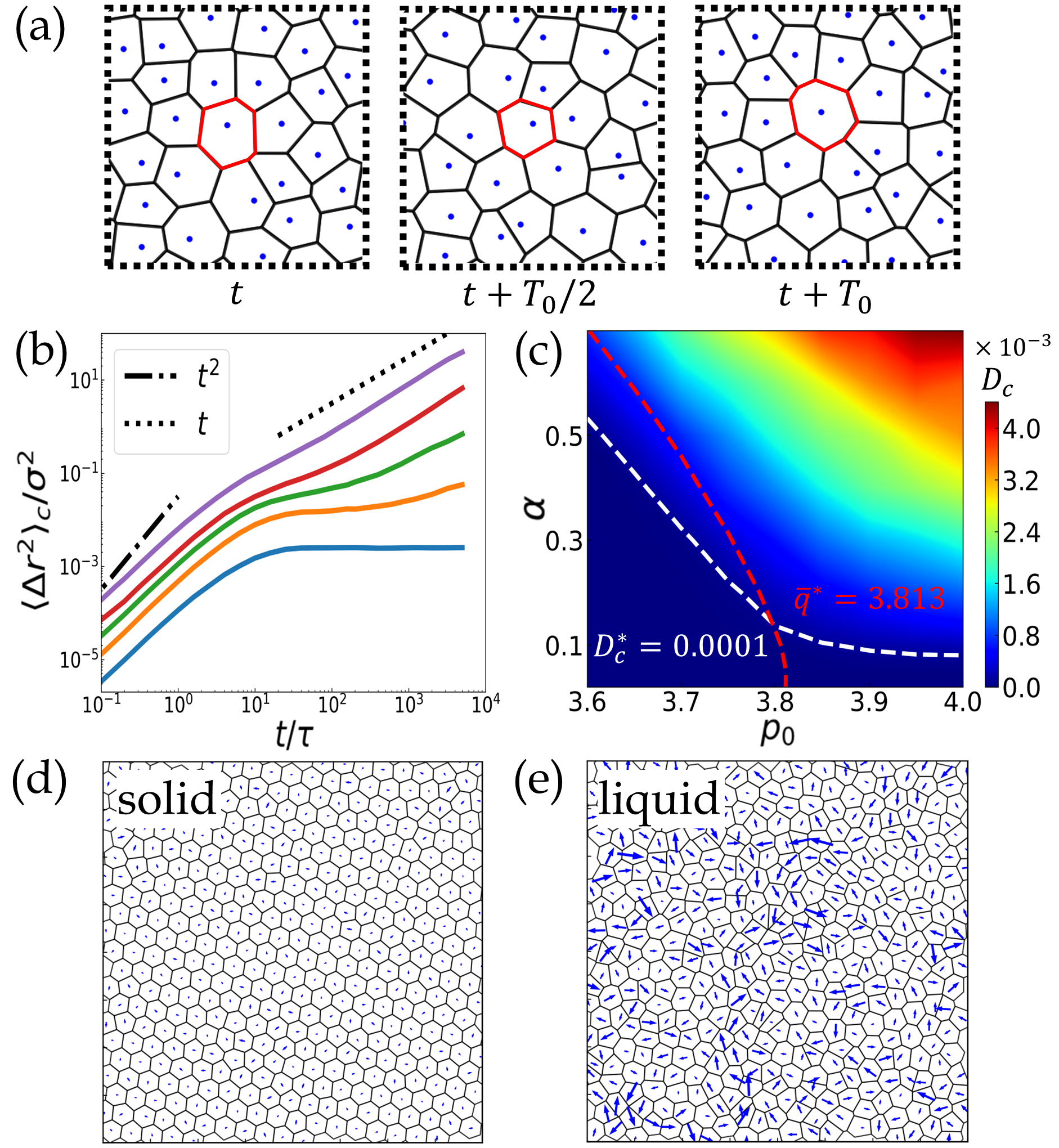
We consider a 2D confluent cell monolayer composed of pulsating cells whose area and shape can change with time (Fig. 1a). Cells are modelled as polygons determined by the Voronoi tessellation of the space Bi et al. (2016). We assume the pulsation is strong enough that the effect of thermal noise can be neglected. The center of cell thus obeys the overdamped dynamics, i.e., with is the mobility coefficient and the inter-cellular force acting on particle . The system energy can be written as Fletcher et al. (2014); Bi et al. (2016); Alt et al. (2017); Li et al. (2019a)
| (1) |
Here, is the averaged cell area and the area modulus of cell. and are the dimensionless area and perimeter of cell , while the corresponding and are their targeted values. is the dimensionless perimeter modulus. To model cell pulsation, we assume each do stochastic oscillation as where is the pulsating strength and is the pulsating phase. with the so-called target shape factor Bi et al. (2016). To account for collective cell pulsation, we assume obeys the simple stochastic Kuramoto-like dynamics Acebrón et al. (2005); Sarkar & Gupte (2021) inspired from biological models Staddon et al. (2022); Zhang & Fodor (2023); Manacorda & Fodor (2023); Boocock et al. (2023); Pérez-Verdugo et al. (2023), i.e.,
| (2) |
where frequency and the corresponding period are supposed to be associated with certain intrinsic cell cycles Zehnder et al. (2015a). The second term represents the coupling between adjacent cells with the coupling strength. The last term is the noise term with noise strength and standard Gaussian white noise . In our simulation, the unit length, unit energy and unit time are defined as , , , respectively. We fixed and if no state otherwise. Detail simulation information can be found in the Supplemental Material Sup (????).
Cell-pulsation induced fluidization
We first study the case of zero pulsation coupling (). To accurately characterize the dynamical states of cellular monolayer, we calculate the cage relative mean square displacement of cells, , which excludes the influence of the long-wavelength fluctuations in 2D Lin et al. (2018); Sup (????). As shown in Fig. 1b, with increasing , the plateau of gradually disappears and exhibits diffusion scaling. This indicates a pulsation-induced fluidization transition analogous to the one induced by motility Bi et al. (2016). Similar behaviors have been reported in the actively-deforming spherical particle system Tjhung & Berthier (2017). Microscopically, the fluidization transition is because random pulsation induces unbalanced intercellular forces, which results in frequent T1 transition (see Movie. S1 in Sup (????)). In Fig. 1c, we plot the diffusion coefficient of the system in dimension of and , where the solid and liquid phases are tentatively divided by a threshold Bi et al. (2016). We confirm that the solid phase in our model has the crystal order (see Movie. S2 and Fig. S1 in Sup (????)). From Fig. 1c, one can see that systems with large are more easily fluidized as long as . This accords with the energy barrier scenario of T1 transition Bi et al. (2015). Nevertheless, by accurately calculating the average shape factor , we find the line of zero-energy-barrier, i.e., Bi et al. (2015, 2016), does not correspond to the solid-liquid phase boundary determined by , especially for small . Similar discrepancy has also been observed experimentally Devany et al. (2021).
Synchronization-induced solidification
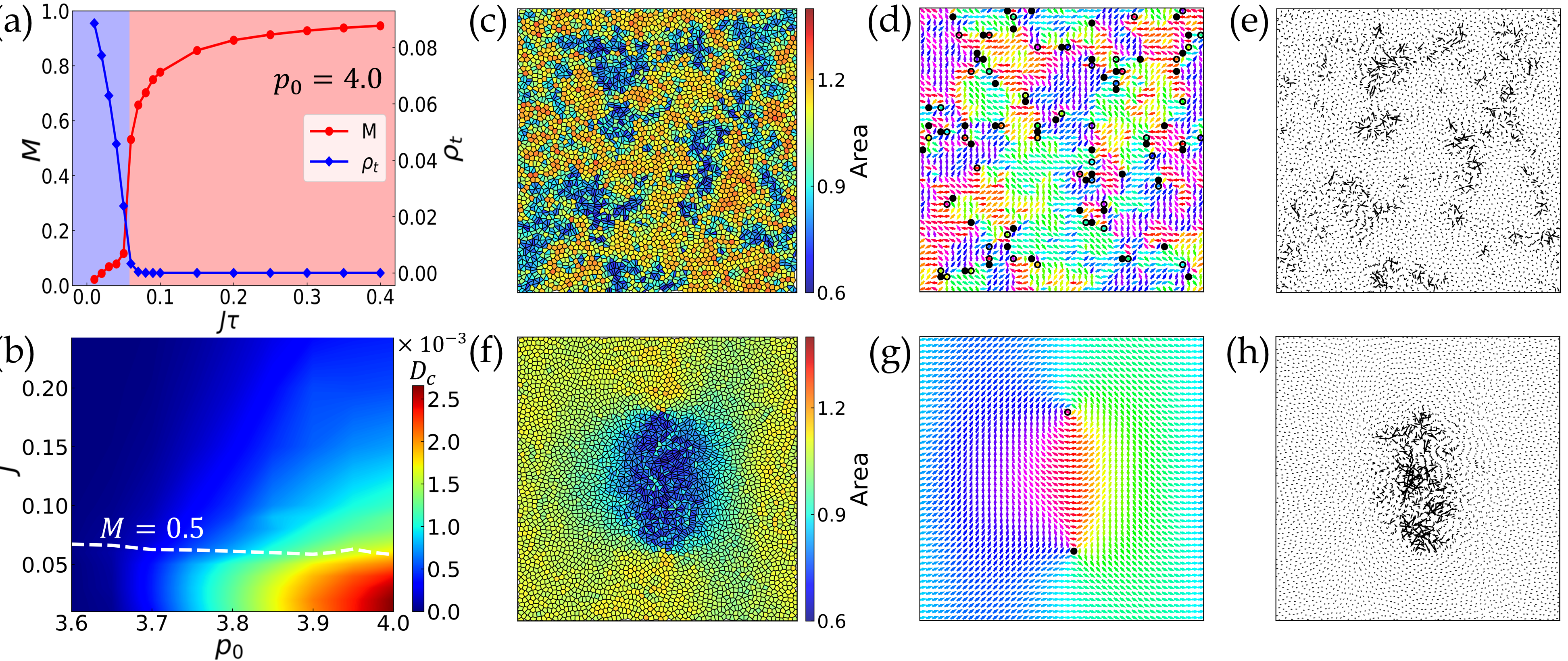
We further consider the intercellular coupling in the system. we find that increasing leads to synchronization transition, which can be characterized by the phase polarity . We plot as a function of and observe a transition happening at critical in Fig. 2a, which is almost invariant to the change of and (see Fig. S2 in Sup (????)). This is because the dynamic equation Eq. (2) for pulsating phase is formally decoupled from the energy function Eq. (1). Similar to the order-disorder transition in 2D XY model, 2D synchronization transition described by Kuramoto model belongs to BKT universality. In Fig. 2a, we plot the topological defect density as a function of and confirm that (de-)synchronization transition is indeed accompanied by the creation and annihilation of () topological defects in the pulsating phase space. In Fig. 2b, we further plot in dimension of and at fixed . We find that as increases above , drops quickly, suggesting a synchronization-induced solidification. This collective phenomenon is insensitive to the choice of and Sup (????). The reason behind this solidification is that compared with the unsynchronized state, the cell sizes in the synchronized state are more uniformly distributed Sup (????). Thus cells experience more balanced intercellular force during the collective pulsation, resulting in suppressed cell motions. This synchronization pulsation can capture the recently observed collective motions of the MDCK monolayer (see Movie. S3 in Sup (????) and Thiagarajan et al. (2022)).
In Fig. 2c, we give snapshots of the monolayer near the synchronization transition point. One can see the coexistence of high density contracted regions and low density expanded regions that periodically switch their states (see Movie. S4 in Sup (????)). This density heterogeneity is strongest at , as proven by the height of the first peak in structure factor (Fig. 3c). In Fig. 2e, we also show that cell motility in the expanded regions is negligible like a solid, while in the contracted regions it is much higher akin to the liquid state. This dynamic heterogeneity is a result of heterogeneous distribution of local shape factor which is large (small) in the contracted (expanded) regions. A larger (smaller) shape factor corresponding to a more anisotropic (isotropic) cell shape and stronger (weaker) imbalanced intercellular forces which favour liquid (solid) phase Bi et al. (2015, 2016). We notice that similar density heterogeneities and local synchronized pulsation patterns were observed in MDCK monolayers Zehnder et al. (2015a); Hino et al. (2020); Boocock et al. (2021). Furthermore, we plot the pulsating phase field of the cell monolayer near the critical point in Fig. 2d. We identify topological defects and find they are mostly located on the boundaries between contracted and expanded regions. Surprisingly, these topological defects were also recently identified experimentally Boocock et al. (2023). To further disclose the relationship between the topological defects and density/dynamic heterogeneities, we create a single pair of topological defects in the phase field and observe its evolution (Fig. 2f-h). One can see that the topological defects create a synchronized cell droplet in between, which contracts (fluidizes) or expands (solidifies) alternatively. It also creates an local pulsating wave around the defect pair (see Movie. S5 in Sup (????)). Thus, the synchronization-induced solidification of the monolayers is essentially a BKT-type transition, resulting from the annihilation of topological defects in the phase field.
Anomalous density fluctuations
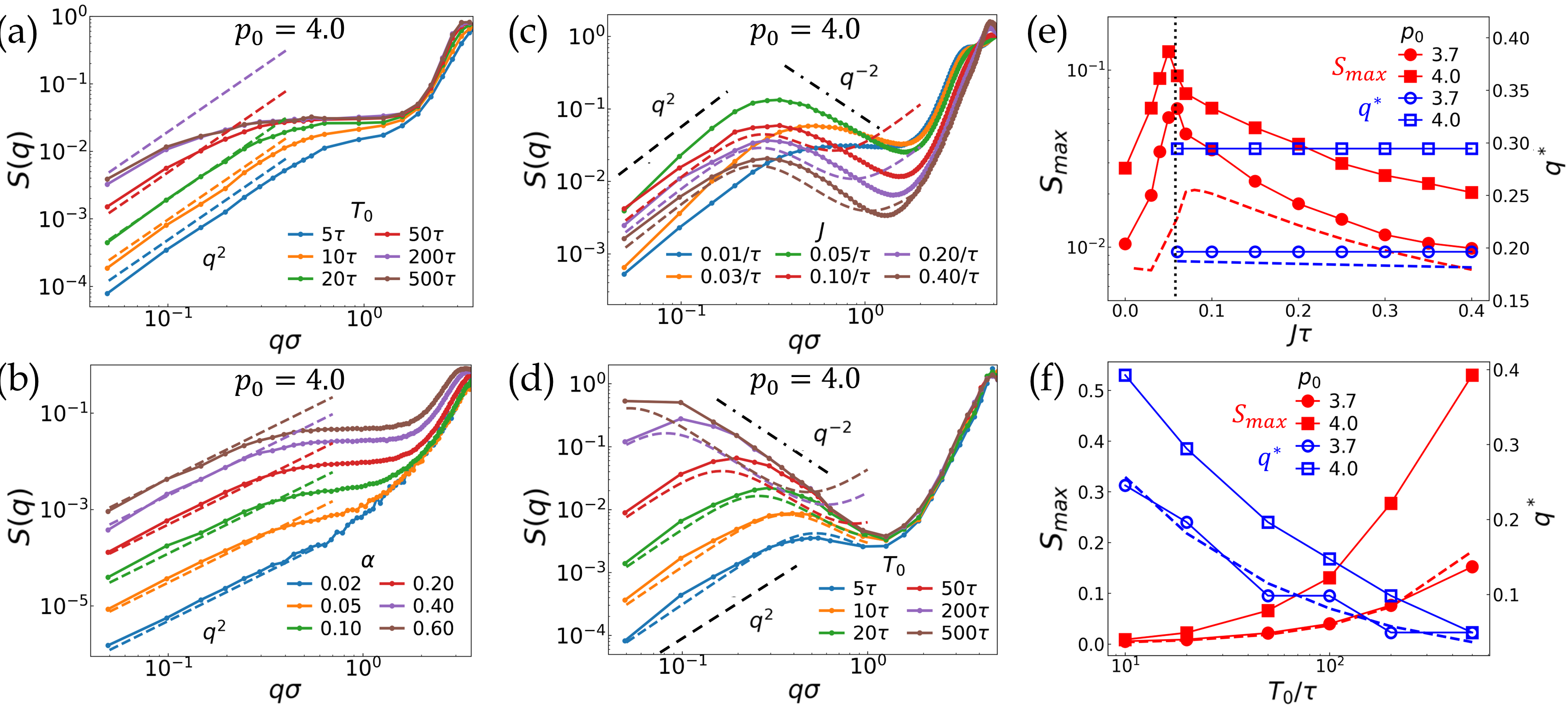
At last, we focus on the density fluctuations of the system. In Fig. 3a, we plot static structure factor for random pulsation systems under different in the liquid state ( and ). For systems with small , we observe , an hallmark of hyperuniform fluid state with vanishing density fluctuations at infinite wave length. Different from previously reported static hyperuniform states in cellular systems Jiao et al. (2014); Li et al. (2018); Zheng et al. (2020), the hyperuniform fluid state here is dynamically robust to perturbation Lei et al. (2019); Lei & Ni (2019). With increasing , we find that the scaling remains unchanged, but the strength of hyperuniformity gradually decreases. Increasing has similar effect (Fig. 3b). Thus the hyperuniformity is the strongest when cells undergo fast and weak pulsation in the liquid state.
With increasing from zero to , we find that a peak in gradually develops around without modifying scaling at small regime (Fig. 3c). This indicates the coexistence of local large density fluctuations with global hyperuniformity, a scenario similar to circle swimmer systems Lei et al. (2019). Interestingly, with further increasing , the height of first peak in denoted as gradually decreases. This is best shown in Fig. 3e, where we plot as a function of for two different . We find exhibits diverging behavior when , which is more pronounced for large . In Fig. 3d, we further show for various in synchronized states with . With increasing , increases and the location of the peak shifts to the small . This indicates that both the magnitude and length scale of the density heterogeneity are controlled by , which diverge when , resulting in giant density fluctuations ( scaling). We find this mechanism is quite robust,independent whether the system is in solid or liquid phase (see Fig. S4 in Sup (????)).
To understand the above anomalous density fluctuations, we construct a hydrodynamic theory based on cell density field and pulsating phase field Sup (????),
| (3) | |||||
| (4) |
where is the local pressure Sup (????). The second term in Eq. (3) is the center-of-mass conserved noise terms Hexner & Levine (2017); Lei & Ni (2019); Lei et al. (2019) caused by the active reciprocal interaction, i.e., cell pulsation. The strength of the noise is Sup (????). Eq. (4) is obtained by coarse-graining Eq. (2). and are two uncorrelated standard Gaussian white noise. From Eq. (3-4), we can obtain Sup (????)
| (5) |
The coefficients are , , , and . In the case of random pulsation ( and ), exhibits a hyperuniform scaling, as a result of the emergent long-range hydrodynamic correlation Lei & Ni (2019). In Fig. 3a-b, we plot the theoretical prediction as the dashed line. We find the theory matches the simulation data well under different and with only one adjustable parameter . In the case of synchronized state (), the theory predicts the emerging of the first peak in at finite and giant fluctuation scaling as . In Fig. 3e-f, we plot the predicted height and location of the first peak for the system as a function of and , respectively. One can see that theoretical lines quantitatively match the simulation data without additional fitting parameter for the synchronized solid phase (). Especially, the theory predicts a diffusion scaling relationship between the length scale of density heterogeneity and pulsating period, i.e., . For the liquid phase, due to large cell deformation, a correction is needed in to obtain the good fitting (Fig. 3c-d). The above fluctuating hydrodynamic theory indicates that the active reciprocal interaction and the competition of time scales between pulsation and stress relaxation are two key factors determining anomalous density fluctuations in the system.
Generalization to other pulsation form
Our above modelling of cell pulsation is based on the stochastic oscillation of . Biologically, cell pulsation might also come from time-dependent junction tension Dierkes et al. (2014); Curran et al. (2017); Krajnc (2020); Krajnc et al. (2021); Devany et al. (2021); Yamamoto et al. (2022). Thus, we extend our studies to the case where do stochastic oscillation as while . Here represents the pulsating strength. We find similar hyperuniform fluid states at large and synchronization-induced dynamical slowdown at large pulsating period (see Fig. S5-S7 in Sup (????)). The hydrodynamic theory can also be extended to this pulsating scenario and obtain similar predictions Sup (????). All these results demonstrate the robustness of pulsating activity in determining the fluidization and anomalous density fluctuations in epithelial tissue.
Conclusion and discussion
In this work, we show that random cell pulsation acting as reciprocal active force, melts solid epithelial tissues into hyperuniform fluids, while pulsating synchronization induces a reverse solidification. We prove that this solidification is BKT-type and the creation of topological defects in the pulsating phase space induces strong dynamical and structural heterogeneities at the transition point. Our findings can explain recent observed giant density fluctuations and topological defects in the MDCK monolayer Zehnder et al. (2015a); Hino et al. (2020); Boocock et al. (2021, 2023). In the future, it is tempting to study the fluctuating physics of systems with complex couplings between pulsating phase and the motion of cells Zhang & Fodor (2023); Boocock et al. (2023), or pulsating cellular systems with nematic order Saw et al. (2017); Zhang & Yeomans (2023); Rozman et al. (2023). Such exploration would help us understand how squeezing and contraction forces sculpt cell tissues Heisenberg & Bellaïche (2013); Dance (2021); Bailles et al. (2022) and guide the design of soft robot swarms Li et al. (2019b).
Acknowledgements.
Acknowledgments:
The authors thank Ran Ni, Xia-qing Shi, Yan-Wei Li for careful reading the manuscript and helpful discussion, and are grateful to the High-Performance Computing Center of Collaborative Innovation Center of Advanced Microstructures, the High-Performance Computing Center (HPCC) of Nanjing University for the numerical calculations. This work is supported by the National Key Research and Development Program of China (No. 2022YFA1405000) and the National Natural Science Foundation of China (No. 12275127, 11974175, 12174184, and 12247102).
References
- Friedl & Gilmour 2009 Friedl, P. & Gilmour, D. Collective cell migration in morphogenesis, regeneration and cancer. Nature reviews Molecular cell biology 10, 445–457 (2009).
- Hakim & Silberzan 2017 Hakim, V. & Silberzan, P. Collective cell migration: a physics perspective. Reports on Progress in Physics 80, 076601 (2017).
- Alert & Trepat 2020 Alert, R. & Trepat, X. Physical models of collective cell migration. Annual Review of Condensed Matter Physics 11, 77–101 (2020).
- Park et al. 2015 Park, J.-A. et al. Unjamming and cell shape in the asthmatic airway epithelium. Nature materials 14, 1040–1048 (2015).
- Bi et al. 2015 Bi, D., Lopez, J., Schwarz, J. M. & Manning, M. L. A density-independent rigidity transition in biological tissues. Nature Physics 11, 1074–1079 (2015).
- Bi et al. 2016 Bi, D., Yang, X., Marchetti, M. C. & Manning, M. L. Motility-driven glass and jamming transitions in biological tissues. Physical Review X 6, 021011 (2016).
- Mongera et al. 2018 Mongera, A. et al. A fluid-to-solid jamming transition underlies vertebrate body axis elongation. Nature 561, 401–405 (2018).
- Lin et al. 2018 Lin, S.-Z., Ye, S., Xu, G.-K., Li, B. & Feng, X.-Q. Dynamic migration modes of collective cells. Biophysical journal 115, 1826–1835 (2018).
- Giavazzi et al. 2018 Giavazzi, F. et al. Flocking transitions in confluent tissues. Soft matter 14, 3471–3477 (2018).
- Cai et al. 2022 Cai, L.-b., Ji, W., Luo, J., Lei, Q.-l. & Ma, Y.-q. Numerical study of dynamic zigzag patterns in migrating epithelial tissue. Science China Physics, Mechanics & Astronomy 65, 217011 (2022).
- Petrolli et al. 2019 Petrolli, V. et al. Confinement-induced transition between wavelike collective cell migration modes. Physical review letters 122, 168101 (2019).
- Peyret et al. 2019 Peyret, G. et al. Sustained oscillations of epithelial cell sheets. Biophysical journal 117, 464–478 (2019).
- Yu et al. 2021 Yu, J. et al. Spatiotemporal oscillation in confined epithelial motion upon fluid-to-solid transition. ACS nano 15, 7618–7627 (2021).
- Tjhung & Berthier 2017 Tjhung, E. & Berthier, L. Discontinuous fluidization transition in time-correlated assemblies of actively deforming particles. Physical Review E 96, 050601 (2017).
- Zhang & Fodor 2023 Zhang, Y. & Fodor, É. Pulsating Active Matter. Physical Review Letters 131, 238302 (2023).
- Manacorda & Fodor 2023 Manacorda, A. & Fodor, É. Pulsating with discrete symmetry. arXiv preprint arXiv:2310.14370 (2023).
- Manning 2023 Manning, M. L. Essay: Collections of deformable particles present exciting challenges for soft matter and biological physics. Physical Review Letters 130, 130002 (2023).
- Banerjee et al. 2015 Banerjee, S., Utuje, K. J. & Marchetti, M. C. Propagating stress waves during epithelial expansion. Physical review letters 114, 228101 (2015).
- Lin et al. 2017 Lin, S.-Z., Li, B., Lan, G. & Feng, X.-Q. Activation and synchronization of the oscillatory morphodynamics in multicellular monolayer. Proceedings of the National Academy of Sciences 114, 8157–8162 (2017).
- Hino et al. 2020 Hino, N. et al. ERK-mediated mechanochemical waves direct collective cell polarization. Developmental cell 53, 646–660 (2020).
- Krajnc et al. 2021 Krajnc, M., Stern, T. & Zankoc, C. Active instability and nonlinear dynamics of cell-cell junctions. Physical Review Letters 127, 198103 (2021).
- Boocock et al. 2021 Boocock, D., Hino, N., Ruzickova, N., Hirashima, T. & Hannezo, E. Theory of mechanochemical patterning and optimal migration in cell monolayers. Nature physics 17, 267–274 (2021).
- Boocock et al. 2023 Boocock, D., Hirashima, T. & Hannezo, E. Interplay between mechanochemical patterning and glassy dynamics in cellular monolayers. bioRxiv 2023–03 (2023).
- Pérez-Verdugo et al. 2023 Pérez-Verdugo, F., Banks, S. & Banerjee, S. Excitable dynamics driven by mechanical feedback in biological tissues. arXiv preprint arXiv:2310.04950 (2023).
- Pérez-Verdugo & Banerjee 2023 Pérez-Verdugo, F. & Banerjee, S. Tension Remodeling Regulates Topological Transitions in Epithelial Tissues. PRX Life 1, 023006 (2023).
- Dierkes et al. 2014 Dierkes, K., Sumi, A., Solon, J. & Salbreux, G. Spontaneous oscillations of elastic contractile materials with turnover. Physical review letters 113, 148102 (2014).
- Curran et al. 2017 Curran, S. et al. Myosin II controls junction fluctuations to guide epithelial tissue ordering. Developmental cell 43, 480–492 (2017).
- Krajnc 2020 Krajnc, M. Solid–fluid transition and cell sorting in epithelia with junctional tension fluctuations. Soft Matter 16, 3209–3215 (2020).
- Devany et al. 2021 Devany, J., Sussman, D. M., Yamamoto, T., Manning, M. L. & Gardel, M. L. Cell cycle–dependent active stress drives epithelia remodeling. Proceedings of the National Academy of Sciences 118, e1917853118 (2021).
- Yamamoto et al. 2022 Yamamoto, T., Sussman, D. M., Shibata, T. & Manning, M. L. Non-monotonic fluidization generated by fluctuating edge tensions in confluent tissues. Soft Matter 18, 2168–2175 (2022).
- Duclut et al. 2022 Duclut, C., Paijmans, J., Inamdar, M. M., Modes, C. D. & Jülicher, F. Active T1 transitions in cellular networks. The European Physical Journal E 45, 29 (2022).
- Solon et al. 2009 Solon, J., Kaya-Copur, A., Colombelli, J. & Brunner, D. Pulsed forces timed by a ratchet-like mechanism drive directed tissue movement during dorsal closure. Cell 137, 1331–1342 (2009).
- Martin et al. 2009 Martin, A. C., Kaschube, M. & Wieschaus, E. F. Pulsed contractions of an actin–myosin network drive apical constriction. Nature 457, 495–499 (2009).
- Armon et al. 2018 Armon, S., Bull, M. S., Aranda-Diaz, A. & Prakash, M. Ultrafast epithelial contractions provide insights into contraction speed limits and tissue integrity. Proceedings of the National Academy of Sciences 115, E10333–E10341 (2018).
- Staddon et al. 2022 Staddon, M. F., Munro, E. M. & Banerjee, S. Pulsatile contractions and pattern formation in excitable actomyosin cortex. PLoS Computational Biology 18, e1009981 (2022).
- Thiagarajan et al. 2022 Thiagarajan, R., Bhat, A., Salbreux, G., Inamdar, M. M. & Riveline, D. Pulsations and flows in tissues as two collective dynamics with simple cellular rules. Iscience 25 (2022).
- Zehnder et al. 2015a Zehnder, S. M., Suaris, M., Bellaire, M. M. & Angelini, T. E. Cell volume fluctuations in MDCK monolayers. Biophysical journal 108, 247–250 (2015a).
- Zehnder et al. 2015b Zehnder, S. M. et al. Multicellular density fluctuations in epithelial monolayers. Physical Review E 92, 032729 (2015b).
- Zehnder et al. 2016 Zehnder, S. M., Zegers, F. M. & Angelini, T. E. A Langevin model of physical forces in cell volume fluctuations. Journal of biomechanics 49, 1286–1289 (2016).
- Bocanegra-Moreno et al. 2023 Bocanegra-Moreno, L., Singh, A., Hannezo, E., Zagorski, M. & Kicheva, A. Cell cycle dynamics control fluidity of the developing mouse neuroepithelium. Nature Physics 1–9 (2023).
- Giavazzi et al. 2017 Giavazzi, F. et al. Giant fluctuations and structural effects in a flocking epithelium. Journal of Physics D: Applied Physics 50, 384003 (2017).
- Jiao et al. 2014 Jiao, Y. et al. Avian photoreceptor patterns represent a disordered hyperuniform solution to a multiscale packing problem. Physical Review E 89, 022721 (2014).
- Li et al. 2018 Li, X., Das, A. & Bi, D. Biological tissue-inspired tunable photonic fluid. Proceedings of the National Academy of Sciences 115, 6650–6655 (2018).
- Zheng et al. 2020 Zheng, Y., Li, Y.-W. & Ciamarra, M. P. Hyperuniformity and density fluctuations at a rigidity transition in a model of biological tissues. Soft Matter 16, 5942–5950 (2020).
- Liu et al. 2023 Liu, Y., Chen, D., Jiao, Y. & Tian, J. Universal Hyperuniform Organization of Cellular Structures in Leaf Vein Networks. arXiv preprint arXiv:2311.09551 (2023).
- Torquato 2018 Torquato, S. Hyperuniform states of matter. Physics Reports 745, 1–95 (2018).
- Lei et al. 2019 Lei, Q.-L., Ciamarra, M. P. & Ni, R. Nonequilibrium strongly hyperuniform fluids of circle active particles with large local density fluctuations. Science advances 5, eaau7423 (2019).
- Lei & Ni 2019 Lei, Q.-L. & Ni, R. Hydrodynamics of random-organizing hyperuniform fluids. Proceedings of the National Academy of Sciences 116, 22983–22989 (2019).
- Shi & Ma 2013 Shi, X.-q. & Ma, Y.-q. Topological structure dynamics revealing collective evolution in active nematics. Nature communications 4, 3013 (2013).
- Fletcher et al. 2014 Fletcher, A. G., Osterfield, M., Baker, R. E. & Shvartsman, S. Y. Vertex models of epithelial morphogenesis. Biophysical journal 106, 2291–2304 (2014).
- Alt et al. 2017 Alt, S., Ganguly, P. & Salbreux, G. Vertex models: from cell mechanics to tissue morphogenesis. Philosophical Transactions of the Royal Society B: Biological Sciences 372, 20150520 (2017).
- Li et al. 2019a Li, X., Das, A. & Bi, D. Mechanical heterogeneity in tissues promotes rigidity and controls cellular invasion. Physical review letters 123, 058101 (2019a).
- Acebrón et al. 2005 Acebrón, J. A., Bonilla, L. L., Vicente, C. J. P., Ritort, F. & Spigler, R. The Kuramoto model: A simple paradigm for synchronization phenomena. Reviews of modern physics 77, 137 (2005).
- Sarkar & Gupte 2021 Sarkar, M. & Gupte, N. Phase synchronization in the two-dimensional kuramoto model: Vortices and duality. Physical Review E 103, 032204 (2021).
- Sup ???? See Supplemental Material at [URL] for methods, supplementary datas, movies and complete hydrodynamic theory, which includes Refs. 64, 65, 66, 67, 68, 36, 69 (????).
- Hexner & Levine 2017 Hexner, D. & Levine, D. Noise, diffusion, and hyperuniformity. Physical review letters 118, 020601 (2017).
- Saw et al. 2017 Saw, T. B. et al. Topological defects in epithelia govern cell death and extrusion. Nature 544, 212–216 (2017).
- Zhang & Yeomans 2023 Zhang, G. & Yeomans, J. M. Active forces in confluent cell monolayers. Physical Review Letters 130, 038202 (2023).
- Rozman et al. 2023 Rozman, J., Yeomans, J. M. & Sknepnek, R. Shape-tension coupling produces nematic order in an epithelium vertex model. Physical Review Letters 131, 228301 (2023).
- Heisenberg & Bellaïche 2013 Heisenberg, C.-P. & Bellaïche, Y. Forces in tissue morphogenesis and patterning. Cell 153, 948–962 (2013).
- Dance 2021 Dance, A. The secret forces that squeeze and pull life into shape. Nature 589, 186–189 (2021).
- Bailles et al. 2022 Bailles, A., Gehrels, E. W. & Lecuit, T. Mechanochemical principles of spatial and temporal patterns in cells and tissues. Annual review of cell and developmental biology 38, 321–347 (2022).
- Li et al. 2019b Li, S. et al. Particle robotics based on statistical mechanics of loosely coupled components. Nature 567, 361–365 (2019b).
- Fabri & Pion 2009 Fabri, A. & Pion, S. CGAL: The computational geometry algorithms library. In Proceedings of the 17th ACM SIGSPATIAL international conference on advances in geographic information systems, 538–539 (2009).
- Li & Ciamarra 2018 Li, Y.-W. & Ciamarra, M. P. Role of cell deformability in the two-dimensional melting of biological tissues. Physical Review Materials 2, 045602 (2018).
- Pasupalak et al. 2020 Pasupalak, A., Yan-Wei, L., Ni, R. & Ciamarra, M. P. Hexatic phase in a model of active biological tissues. Soft matter 16, 3914–3920 (2020).
- Killeen et al. 2022 Killeen, A., Bertrand, T. & Lee, C. F. Polar fluctuations lead to extensile nematic behavior in confluent tissues. Physical Review Letters 128, 078001 (2022).
- Rouzaire & Levis 2021 Rouzaire, Y. & Levis, D. Defect superdiffusion and unbinding in a 2D XY model of self-driven rotors. Physical Review Letters 127, 088004 (2021).
- Shi & Chaté 2024 Shi, X.-q. & Chaté, H. Effect of Persistent Noise on the XY Model and Two-Dimensional Crystals. arXiv preprint arXiv:2401.11175 (2024).