DoseGNN: Improving the Performance of Deep Learning Models in Adaptive Dose-Volume Histogram Prediction through Graph Neural Networks.
Abstract
Dose-Volume Histogram (DVH) prediction is fundamental in radiation therapy that facilitate treatment planning, dose evaluation, plan comparison and etc. It helps to increase the ability to deliver precise and effective radiation treatments while managing potential toxicities to healthy tissues as needed to reduce the risk of complications. This paper extends recently disclosed research findings Dong et al. (2023b) presented on AAPM (AAPM 65th Annual Meeting Exhibition) and includes necessary technique details. The objective is to design efficient deep learning models for DVH prediction on general radiotherapy platform equipped with high performance CBCT system, where input CT images and target dose images to predict may have different origins, spacing and sizes. Deep learning models widely-adopted in DVH prediction task are evaluated on the novel radiotherapy platform, and graph neural networks (GNNs) are shown to be the ideal architecture to construct a plug-and-play framework to improve predictive performance of base deep learning models in the adaptive setting.
1 Introduction
Intensity-modulated radiation therapy (IMRT) enhances the precision of high-radiation-dose regions conforming to the planning target volume (PTV) while minimizing radiation exposure to each organ at risk (OAR) Tham et al. (2009). Dose-Volume Histogram (DVH) prediction aids in IMRT from different perspectives, including dose evaluation, plan comparison, adaptive replanning, and etc Palta (2003). In the context of radiotherapy, 3D dose prediction Dong & Xing (2020); Shiraishi & Moore (2016) estimates the three-dimensional distribution of radiation doses within a patient’s anatomy to ensure that prescribed dose is delivered accurately to the target while minimizing the dose to surrounding healthy tissues. Once the 3D dose distribution is calculated, a (DVH) is generated, providing a comprehensive view of the dose distribution in relation to the volumes of interest.
The CBCT (Cone Beam Computed Tomography) system has been widely used to provide a three-dimensional imaging representation of the patient’s anatomy in guiding the delivery of radiation dose. Various deep learning models Cagni et al. (2017); Sumida et al. (2020) are developed to predict what is likely achievable for patients’ DVHs based on these CBCT/CT images. Typically, these models are constructed upon recent advancements in the field of computer vision, such as 3D U-net Çiçek et al. (2016) and Vision Transformer Han et al. (2022).
In many radiotherapy platform equipped with high performance CBCT system Dong et al. (2023a), some training plans contain mismatched input CT images and target dose images. That is, the 3D CT images and 3D dose image to predict can have different origins, sizes, and spacings. One intuitive heuristic is to generate a 3D CT image by greedily searching a point/pixel in the original 3D CT image for each point/pixel in the 3D dose image based on the geometry distance, then the image-processing deep learning models can be used for the prediction task. However, the CT images after the greedy conversion are distorted and will fail to capture the patient’s anatomy accurately. In contrast, another heuristic is to directly apply image-processing deep learning models on the origin CT images, and then take the average of learnt representations of k-nearest points/pixels in CT images to each point/pixel in the dose image for predicting it’s corresponding dose value. However, this heuristic is over-smoothed and fail to capture the geometry variance.
To address aforementioned challenges, we introduce DoseGNN, a plug-and-play architecture based on graph neural networks (GNNs). GNNs Kipf & Welling (2016); Hamilton et al. (2017); Xu et al. (2019); Velickovic et al. (2018); Dong et al. (2022b); You et al. (2018); Scarselli et al. (2008); Duvenaud et al. (2015); Gilmer et al. (2017); Zhang et al. (2018); Dong et al. (2022a) have revolutionized the field of representation learning over graphs and is widely used in clinical applications including drug synergy prediction (Hopkins, 2008; Dong et al., 2023c; Podolsky & Greene, 2011), Alzheimer’s disease (AD) detection (Song et al., 2019; Qin et al., 2022; Li et al., 2023) and cancer subtype classification (Lu & Han, 2003). Specifically, we formulate the 3D dose prediction problem as a semi-supervised graph regression problem, where the graph consists of points/pixels in the input CT images and target dose images. CT nodes (i.e. nodes associated with points/pixels in the CT images) and dose nodes (i.e. nodes associated with points/pixels in the dose image) are connected if their geometric distance is smaller than a given threshold. Then, the image-processing deep learning models are used as base model to extract features of CT nodes, and a GNN is used to predict dose values of dose nodes.
DoseGNN is evaluated on 20 plans created by certified medical dosimetrists, with 15 used for training and the remaining 5 used for performance testing. Four deep learning models are used as the image-processing base models: Vision Transformer (ViT) Han et al. (2022), 3D U-net CNN Çiçek et al. (2016), vanilla multilayer perceptron MLP Cybenko (1989) and support vector machine SVM Suthaharan & Suthaharan (2016). Experimental results indicate that the porposed plug-and-play framework DoseGNN significantly and consistantly improves the predictive performance in adaptive 3D dose prediction.
2 Methodology
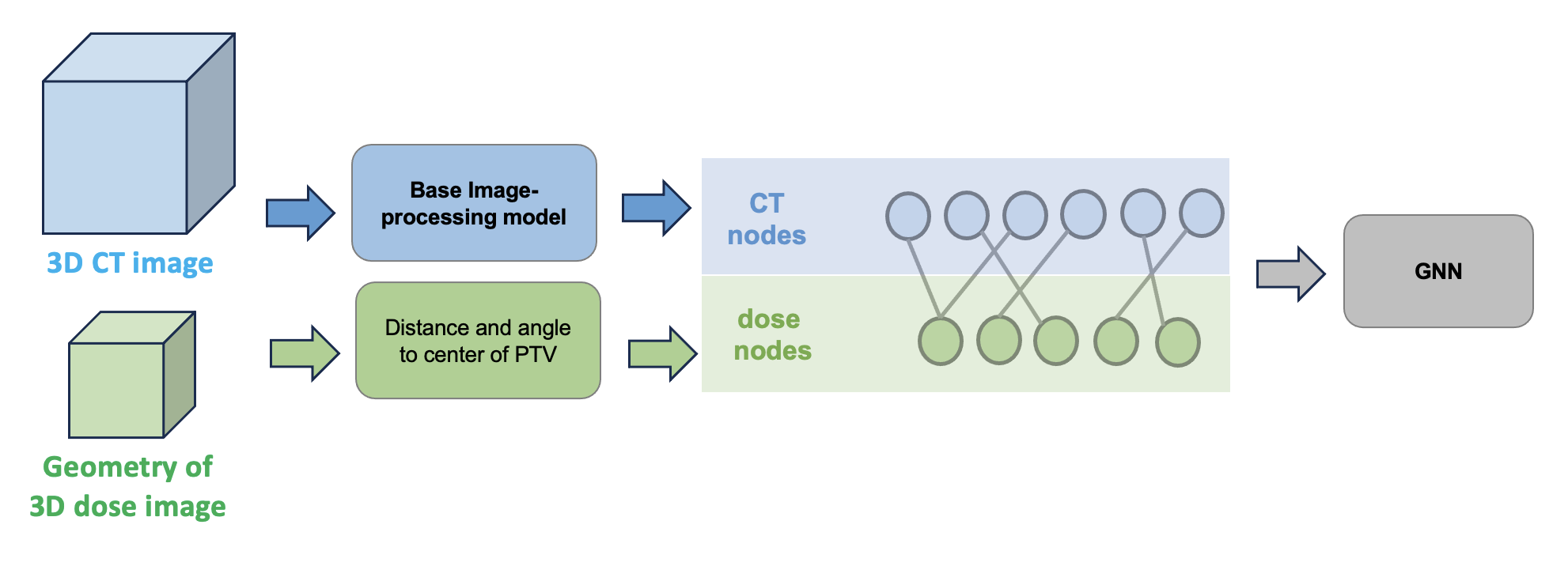
In this section, we introduce the proposed DoseGNN framework. Figure 1 illustrates the architecture. DoseGNN is a plug-and-play framework that can be combined with various base image-processing deep learning models.
The core idea is to formulate a bipartite graph to modulate the geometry relation of pixels in the input CT images and the target dose images. Specifically,
-
•
For each node in the bipartite graph associated to a point/pixel in the CT image, the base image-processing model is used to extract a vector representation of un-distorted geometric information, which accurately captures patient’s anatomy. Thus, the learnt vectorial representation is then used as the node embeddings of CT node.
-
•
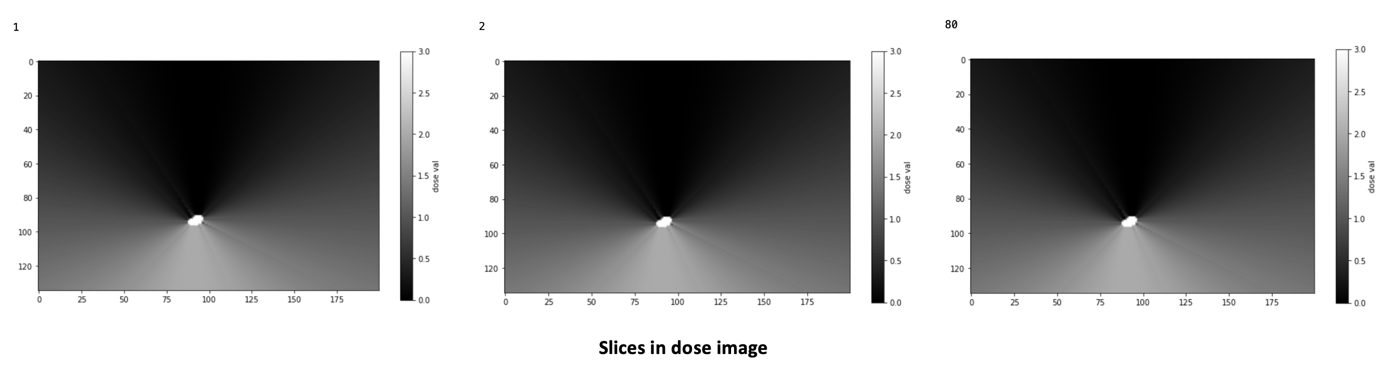
For each node in the bipartite graph associated to a point/pixel in the dose image, it takes two features: distance and angle to the center of the PTV. As Figure 2 illustrates, these two features well captures the shape of the dose image distribution. Then, DoseGNN takes the positional encoding framework to generate node embeddings of dose nodes, whose dimension is set to be the same as the embeddings of CT nodes.
-
•
In the bipartite graph, a CT node is connected with a dose node if and only if the geometry distance of these two pixels are bounded by a given threshold.
Following this pipeline, the 3D dose prediction problem is converted as a semi-supervised graph regression problem. Consequently, a GNN is used to predict the dose value of each dose node in the bipartite graph.
3 Experiments
In this section, we evaluate the effectiveness of proposed DoseGNN framework based on various based image-processing deep learning models. In total, 20 treatment plans created by certified medical dosimetrists in the experiments, with 15 used for training models and the remaining 5 used for performance testing.
3.1 Predictive performance
We use root mean square error (RMSE) of predicted dose and true dose as the evaluation metric in the experiment to evaluate how close the deep learning model can approximate the true dose information.
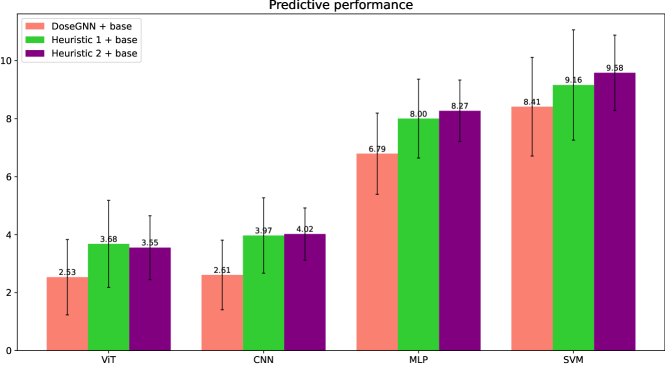
Two heuristics are used as baseline method to deal with CT images and dose images of different origins, sizes, and spacings.
-
•
Heuristic 1 firstly generates a distorted 3D CT image by greedily searching the nearst point/pixel in the original 3D CT image to each point/pixel in the 3D dose image in terms of the geometry distance, then the image-processing deep learning models is used as a predictor.
-
•
Heuristic 2 directly applies image-processing deep learning models on the origin CT images, and then take the average of learnt representations of k-nearest points/pixels in CT images to each point/pixel in the dose image as the final embedding, which is feed to a MLP to predict the corresponding dose value.
Figure 3 presents the results. We find that DoseGNN significantly and consistently improves the baseline heuristics. In addition, DoseGNN with a based of ViT achieves the state-of-the-art performance.
3.2 Visualization of CDVH
In the experiment, we visualize the CDVH (cumulative dose volume histogram) of predicted 3D dose from different method. In the experiment, DoseGNN indicates the DoseGNN with a base model of ViT, which beats DoseGNN with other base models. For other base models like ViT and MLP, we visualize the best predictive results the base model can achieve with different heuristics.
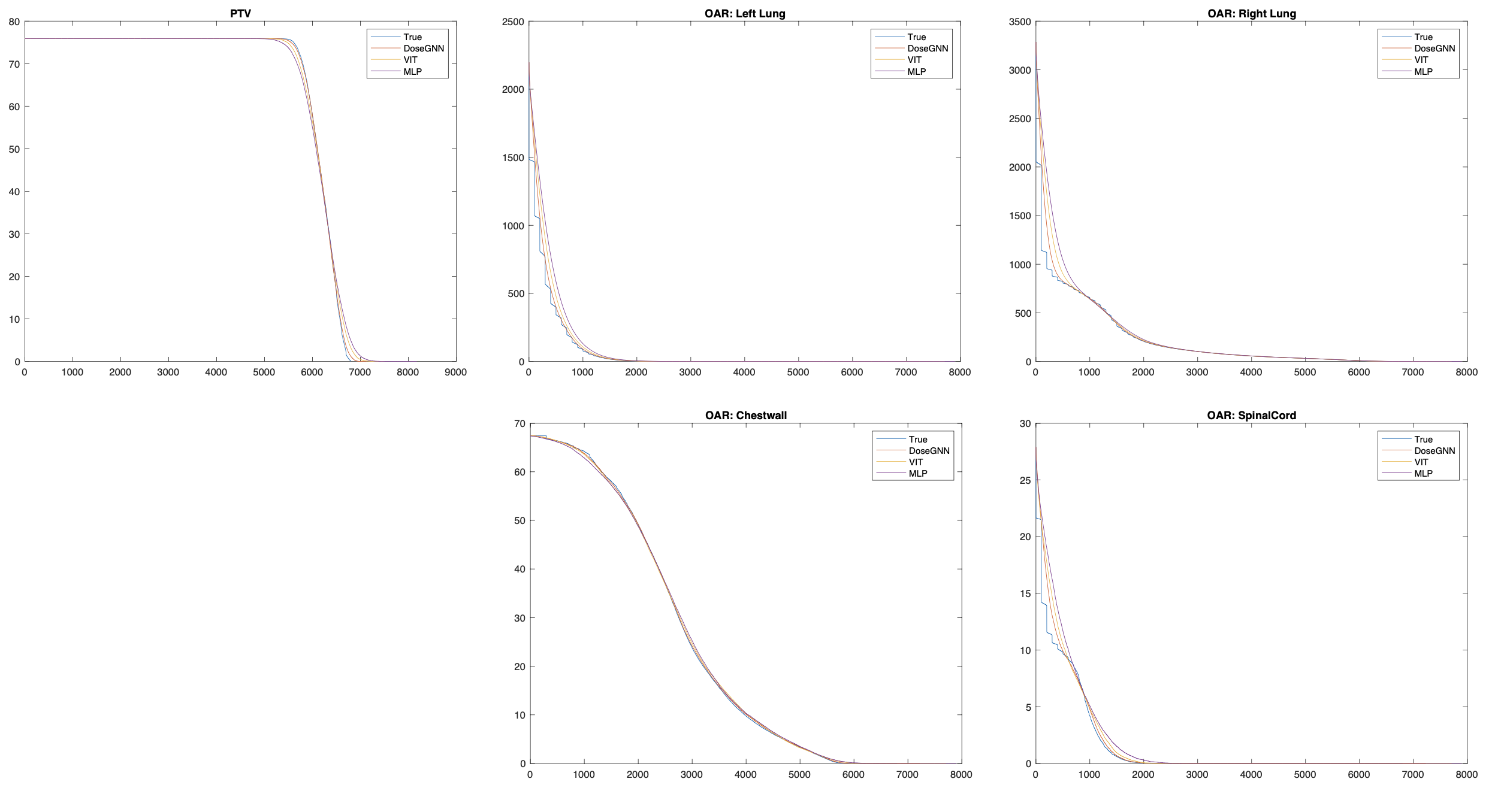
In Figure 4 demonstrates that DoseGNN has the best ability to capture the shape of the CDVH curve of true dose values.
4 Conclusion
In this paper, we propose a plug-and-play framework, DoseGNN, to enhance the predictive performance of base image-processing deep learning models in adaptive 3D dose prediction tasks. Experimental results demonstrate that DoseGNN significantly and consistently improve base deep learning models, and can be applied in radiotherapy plans with different number of OARs and CT slices.
References
- Cagni et al. (2017) Elisabetta Cagni, Andrea Botti, Renato Micera, Maria Galeandro, Roberto Sghedoni, Matteo Orlandi, Cinzia Iotti, Luca Cozzi, and Mauro Iori. Knowledge-based treatment planning: An inter-technique and inter-system feasibility study for prostate cancer. Physica Medica, 36:38–45, 2017.
- Çiçek et al. (2016) Özgün Çiçek, Ahmed Abdulkadir, Soeren S Lienkamp, Thomas Brox, and Olaf Ronneberger. 3d u-net: learning dense volumetric segmentation from sparse annotation. In Medical Image Computing and Computer-Assisted Intervention–MICCAI 2016: 19th International Conference, Athens, Greece, October 17-21, 2016, Proceedings, Part II 19, pp. 424–432. Springer, 2016.
- Cybenko (1989) George Cybenko. Approximation by superpositions of a sigmoidal function. Mathematics of control, signals and systems, 2(4):303–314, 1989.
- Dong & Xing (2020) Peng Dong and Lei Xing. Deep dosenet: a deep neural network for accurate dosimetric transformation between different spatial resolutions and/or different dose calculation algorithms for precision radiation therapy. Physics in Medicine & Biology, 65(3):035010, 2020.
- Dong et al. (2023a) Z Dong, Y Hao, E Laugeman, GD Hugo, P Samson, Y Chen, and T Zhao. Performance of adaptive deep learning models for dose predictions on high-quality cone-beam computed tomography images. International Journal of Radiation Oncology, Biology, Physics, 117(2):e661, 2023a.
- Dong et al. (2022a) Zehao Dong, Weidong Cao, Muhan Zhang, Dacheng Tao, Yixin Chen, and Xuan Zhang. Cktgnn: Circuit graph neural network for electronic design automation. In The Eleventh International Conference on Learning Representations, 2022a.
- Dong et al. (2022b) Zehao Dong, Muhan Zhang, Fuhai Li, and Yixin Chen. Pace: A parallelizable computation encoder for directed acyclic graphs. arXiv preprint arXiv:2203.10304, 2022b.
- Dong et al. (2023b) Zehao Dong, Yixin Chen, and Tianyu Zhao. Dosegnn: An adaptive deep learning model for dose-volume histogram prediction. In AAPM 65th Annual Meeting & Exhibition. AAPM, 2023b.
- Dong et al. (2023c) Zehao Dong, Heming Zhang, Yixin Chen, Philip RO Payne, and Fuhai Li. Interpreting the mechanism of synergism for drug combinations using attention-based hierarchical graph pooling. Cancers, 15(17):4210, 2023c.
- Duvenaud et al. (2015) David Duvenaud, Dougal Maclaurin, Jorge Aguilera-Iparraguirre, Rafael Gómez-Bombarelli, Timothy Hirzel, Alán Aspuru-Guzik, and Ryan P Adams. Convolutional networks on graphs for learning molecular fingerprints. Advances in Neural Information Processing Systems, 2015:2224–2232, 2015.
- Gilmer et al. (2017) Justin Gilmer, Samuel S Schoenholz, Patrick F Riley, Oriol Vinyals, and George E Dahl. Neural message passing for quantum chemistry. In International Conference on Machine Learning, pp. 1263–1272. PMLR, 2017.
- Hamilton et al. (2017) Will Hamilton, Zhitao Ying, and Jure Leskovec. Inductive representation learning on large graphs. In I. Guyon, U. V. Luxburg, S. Bengio, H. Wallach, R. Fergus, S. Vishwanathan, and R. Garnett (eds.), Advances in Neural Information Processing Systems, volume 30. Curran Associates, Inc., 2017. URL https://proceedings.neurips.cc/paper/2017/file/5dd9db5e033da9c6fb5ba83c7a7ebea9-Paper.pdf.
- Han et al. (2022) Kai Han, Yunhe Wang, Hanting Chen, Xinghao Chen, Jianyuan Guo, Zhenhua Liu, Yehui Tang, An Xiao, Chunjing Xu, Yixing Xu, et al. A survey on vision transformer. IEEE transactions on pattern analysis and machine intelligence, 45(1):87–110, 2022.
- Hopkins (2008) Andrew L Hopkins. Network pharmacology: the next paradigm in drug discovery. Nature chemical biology, 4(11):682–690, 2008.
- Kipf & Welling (2016) Thomas N Kipf and Max Welling. Semi-supervised classification with graph convolutional networks. arXiv preprint arXiv:1609.02907, 2016.
- Li et al. (2023) F. Li, Z. Dong, Q. Zhao, P. Payne, M. Province, C. Cruchaga, M. Zhang, T. Zhao, and Y. Chen. Highly accurate disease diagnosis and highly reproducible biomarker identification with pathformer. 2023. doi: 10.21203/rs.3.rs-3576068/v1.
- Lu & Han (2003) Ying Lu and Jiawei Han. Cancer classification using gene expression data. Information Systems, 28(4):243–268, 2003.
- Palta (2003) Jatinder R Palta. Intensity-modulated radiation therapy—the state of the art. Medical physics, 30(12):3265, 2003.
- Podolsky & Greene (2011) Scott H Podolsky and Jeremy A Greene. Combination drugs—hype, harm, and hope. New England Journal of Medicine, 365(6):488–491, 2011.
- Qin et al. (2022) Zhiwei Qin, Zhao Liu, and Ping Zhu. Aiding alzheimer’s disease diagnosis using graph convolutional networks based on rs-fmri data. In 2022 15th International Congress on Image and Signal Processing, BioMedical Engineering and Informatics (CISP-BMEI), pp. 1–7. IEEE, 2022.
- Scarselli et al. (2008) Franco Scarselli, Marco Gori, Ah Chung Tsoi, Markus Hagenbuchner, and Gabriele Monfardini. The graph neural network model. IEEE transactions on neural networks, 20(1):61–80, 2008.
- Shiraishi & Moore (2016) Satomi Shiraishi and Kevin L Moore. Knowledge-based prediction of three-dimensional dose distributions for external beam radiotherapy. Medical physics, 43(1):378–387, 2016.
- Song et al. (2019) Tzu-An Song, Samadrita Roy Chowdhury, Fan Yang, Heidi Jacobs, Georges El Fakhri, Quanzheng Li, Keith Johnson, and Joyita Dutta. Graph convolutional neural networks for alzheimer’s disease classification. In 2019 IEEE 16th international symposium on biomedical imaging (ISBI 2019), pp. 414–417. IEEE, 2019.
- Sumida et al. (2020) Iori Sumida, Taiki Magome, Indra J Das, Hajime Yamaguchi, Hisao Kizaki, Keiko Aboshi, Hiroko Yamaguchi, Yuji Seo, Fumiaki Isohashi, and Kazuhiko Ogawa. A convolution neural network for higher resolution dose prediction in prostate volumetric modulated arc therapy. Physica Medica, 72:88–95, 2020.
- Suthaharan & Suthaharan (2016) Shan Suthaharan and Shan Suthaharan. Support vector machine. Machine learning models and algorithms for big data classification: thinking with examples for effective learning, pp. 207–235, 2016.
- Tham et al. (2009) Ivan Weng-Keong Tham, Siew Wan Hee, Richard Ming-Chert Yeo, Patemah Bte Salleh, James Lee, Terence Wee-Kiat Tan, Kam Weng Fong, Eu Tiong Chua, and Joseph Tien-Seng Wee. Treatment of nasopharyngeal carcinoma using intensity-modulated radiotherapy—the national cancer centre singapore experience. International Journal of Radiation Oncology* Biology* Physics, 75(5):1481–1486, 2009.
- Velickovic et al. (2018) Petar Velickovic, Guillem Cucurull, A. Casanova, Adriana Romero, P. Lio’, and Yoshua Bengio. Graph attention networks. ArXiv, abs/1710.10903, 2018.
- Xu et al. (2019) Keyulu Xu, Weihua Hu, Jure Leskovec, and Stefanie Jegelka. How powerful are graph neural networks? In International Conference on Learning Representations, 2019. URL https://openreview.net/forum?id=ryGs6iA5Km.
- You et al. (2018) Jiaxuan You, Rex Ying, Xiang Ren, William Hamilton, and Jure Leskovec. Graphrnn: Generating realistic graphs with deep auto-regressive models. In International Conference on Machine Learning, pp. 5708–5717. PMLR, 2018.
- Zhang et al. (2018) Muhan Zhang, Zhicheng Cui, Marion Neumann, and Yixin Chen. An end-to-end deep learning architecture for graph classification. In Proceedings of the AAAI Conference on Artificial Intelligence, volume 32, 2018.