Towards reliable synthesis of superconducting infinite-layer nickelate thin films by topochemical reduction
Abstract
Infinite-layer nickelates provide a new route beyond copper oxides to address outstanding questions in the field of unconventional superconductivity. However, their synthesis poses considerable challenges, largely hindering experimental research on this new class of oxide superconductors. That synthesis is achieved in a two-step process that yields the most thermodynamically stable perovskite phase first, then the infinite-layer phase by topotactic reduction, the quality of the starting phase playing a crucial role. Here, we report on reliable synthesis of superconducting infinite-layer nickelate films after successive topochemical reductions of a parent perovskite phase with nearly optimal stoichiometry. Careful analysis of the transport properties of the incompletely reduced films reveals an improvement of the strange metal behaviour of their normal state resistivity over subsequent topochemical reductions, offering insight into the reduction process.
I Introduction
The first observation of superconductivity at relatively high temperature in single-crystal thin films of infinite-layer NdNiO2 upon hole doping was a significant breakthrough.Li et al. (2019) Thereafter, superconductivity has also been observed in other families of hole-doped infinite-layer (IL) nickelate thin films, such as (Pr, Sr)NiO2,Osada et al. (2020a, b); Wang et al. (2022) (La, Sr)NiO2,Osada et al. (2021, 2023) (La, Ca)NiO2,Zeng et al. (2022) and (Nd, Eu)NiO2;Wei et al. (2023) in reduced Ruddlesden-Popper Nd6Ni5O12 thin films without chemical doping;Pan et al. (2021) and in bilayer Ruddlesden–Popper La3Ni2O7 bulk single-crystals under high pressure.Sun et al. (2023) Thus, this new class of oxide superconductors provides a new route beyond copper oxides to address outstanding questions in the field of unconventional superconductivity,Zhou et al. (2021) such as the mechanism that causes the electrons to form pairs, unanswered despite decades of intense research activity.Scalapino (2012); Tsuei and Kirtley (2000); Keimer et al. (2015); Proust and Taillefer (2019); Stewart (2017); O’Mahonya et al. (2022)
Despite an unremarkable critical temperature of around 10 K, that initial observation generated intense interest.Pickett (2021); Norman (2020); Botana et al. (2022); Mitchell (2021) Part of the reason for that is the discovery of superconducting (SC) nickelates was driven by the decades-long search of cuprate-like physics in other strongly correlated metallic oxides. Within this context, Ni-based compounds with Ni ions arranged in corner-sharing NiO4 square units and ultralow chemical valence (Ni1+ and 3d9 configuration) were suggested to exhibit superconductivity due to their electronic and structural similarities with the Cu2+ ions in cuprates.Anisimov et al. (59) However, besides these similarities, very different behaviour between LaNiO2 and CaCuO2 was pointed out early on.Lee and Pickett (2004) And, whether there is a universal mechanism that explains superconductivity in both cuprates and nickelates remains an open question.S.Botana and Norman (2020); Nomura and Arita (2022); Gu and Wen (2022); Kitatani et al. (2023); Hepting et al. (2021); Lechermann (2020); Rossi et al. (2021); Goodge et al. (2021); Carrasco-Alvarez et al. (2022)
Further progress in the field crucially depends on the synthesis of high quality SC nickelate samples, which can provide reliable experimental data. However, only a few groups worldwide have developed the appropriate expertise to date,Li et al. (2019); Wang et al. (2022); Zeng et al. (2022); Wei et al. (2023) since the synthesis of these films poses serious challenges.
Epitaxial growth of complex oxides thin films, such as nickelates, is achieved under vacuum at high substrate temperatures to increase surface mobility of adatoms and improve crystallinity of the grown films. At these elevated temperatures, the process yields the most thermodynamically stable phase with octahedral NiO6 coordination. Thus, the synthesis of the reduced structure with square-planar coordination around Ni1+ ions, arranged in infinite layers, should proceed via the subsequent removal of relatively mobile oxygen anions at low temperatures, by means of kinetically controlled reactions that enable the preparation of metastable phases.Hayward and Rosseinsky (2007)
This transformation of the starting complex oxide phases into oxygen-deficient metastable phases can be triggered by different methods,Ranmohotti et al. (2011); Meng et al. (2023); Kageyama et al. (2018) and is topotactic since it does not involve diffusive rearrangement of the host cations, although lattice parameters and bond lengths change, giving rise to drastic changes in electronic structures. Thus far, in the case of hole-doped nickelates, the topotactic transition from the perovskite phase into the highly metastable SC IL phase has been accomplished ex-situ by low temperature annealing within a sealed glass ampoule using CaH2 as the reducing agent,Li et al. (2019); Osada et al. (2020a, 2021) and in-situ using an oxygen getter metal layer.Wei et al. (2023) Under the former topochemical approach, the resulting phase can be tuned by the choice of the metal hydride and by the reaction conditions, in particular, temperature and time.Hayward et al. (1999); Hayward and Rosseinsky (2003); Kawai et al. (2010) On the one hand, the temperature of the topochemical reaction has to be high enough to bring about the reduction as the activity of metal hydrides in solid state reduction declines at lower temperature. On the other hand, the perovskite framework is more stable at low temperature since an increase in temperature can result in non-topotactic reactions, if the cations in the resulting metastable phase become mobile, leading to degradation of the sample crystallinity. Furthermore, the crystalline quality of the starting perovskite phase greatly affects the reaction.Lee et al. (2020) Indeed, decreasing the lattice mismatch (tensile strain) between the parent perovskite phase and the substrate enhances crystallinity of the subsequent reduced phase, even though the increase in the in-plane lattice parameter upon reduction leads to higher compressive strain for the reduced phase in that case.Lee et al. (2023)
Here, we report the successful synthesis of SC strontium-doped praseodymium nickelate thin films. Complementary to previous approaches, we study the cation stoichiometry of the starting perovskite films by X-ray photoelectron spectroscopy. Furthermore, we carry out a comprehensive study on transport properties of intermediate reduced films, and find an enhancement of the strange metal behaviour of the normal state resistivity of incompletely reduced films over subsequent topochemical reductions. Moreover, the removal of apical oxygen anions from the perovskite phase is confirmed through the structural analysis of the IL phase using four dimensional scanning transmission electron microscopy (4D-STEM).
II Results and discussion
As a first step, we optimize the growth conditions of the Pr0.8Sr0.2NiO3 parent perovskite films (hereafter, PSNO3) with thicknesses between 10 and 13 unit cells (u.c.) by pulsed laser deposition (PLD) on TiO2-terminated (001)-oriented SrTiO3 (STO) substrates. Bulk PrNiO3 crystallizes in an orthorhombic structure (space group #62 Pbnm, GdFeO3-type) with lattice constants at room temperature of , , (pseudocubic constant ).Garcia-Muñoz et al. (1992) Its crystalline structure is not modified by doping with Sr2+ although this brings about a contraction of the unit cell, despite the larger effective size of Sr2+ compared to Pr3+.Garcia-Muñoz et al. (1995) Thus, assuming a Poisson ratio of , a value common to other oxide perovskites,Ledbetter et al. (1990) the expected out-of-plane lattice parameter for epitaxially grown PSNO3 films on STO substrates, inducing 2.23% of tensile strain, is . This estimate leads to a value of 2 for the reflexion in the x-ray diffraction pattern (using Copper K- radiation). Interestingly, that value is in agreement with the threshold experimentally found in the (Nd, Sr)NiO3 system, where if the (002) pseudocubic perovskite peak position is below , the subsequently reduced film never exhibits superconductivity.Lee et al. (2020).
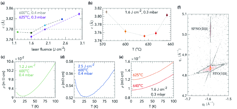
We use complementary information from X-ray Diffraction (XRD) and resistivity as function of temperature, , to elucidate the optimal growth conditions. Laser fluence has a major impact on the film quality, as expected.Ohnishi et al. (2005, 2008) Initially, we have explored laser fluences ranging from 1.2 to 3 at temperatures around 600 ∘C in a strongly oxidizing environment with 0.3-0.4 mbar of O2, required to stabilize a Ni3.2+ oxidation state.
As illustrated in Fig.1(a), the out-of-plane lattice parameter of the PSNO3 films shows a minimum for a laser fluence of 1.6 for different conditions of oxygen pressure and substrate temperature. The cell expansion observed as the fluence departs from that value is indicative of cation vacancies in the films. Oxygen vacancies can be ruled out as the cause of the increase in lattice parameter, since the higher the oxygen pressure is, the more the unit cell expands. A more detailed exploration of the out-of-plane lattice parameter as a function of the substrate temperature at a laser fluence of 1.6 in 0.3 mbar of O2 is shown in Fig.1(b). Under these conditions, lattice constants consistent with a low density of defects are found for substrate temperatures in the range from 600∘C to 640∘C. We confirm the superior quality of the films grown at 1.6 by electrical transport measurements, as depicted in Fig.1(c, d, e). Indeed, at fluences of 1.2 or 2.5 , we observed a resistivity upturn at around 25 K, Fig.1(c, d, respectively), that may be attributed to localization induced by structural disorder, while a metallic state is observed down to 2 K in films grown under 1.6 , 0.3 mbar, and 625∘C or 640∘C, Fig.1(e). Moreover, reciprocal space maps (RSM) around the asymmetric reflection of PSNO3 films grown under the latter conditions demonstrates that the films are fully strained to the STO substrate, as depicted in Fig.1(f). See also Fig. S1 for supplementary information on the optimal growth conditions.
We probed cation stoichiometry of the PSNO3 films by means of X-ray photoelectron spectroscopy (XPS). Elemental composition of the films and assignments of the peaks in a XPS survey spectrum are plotted in Fig. S2 (Supplementary Information). Quantitative XPS for the determination of the [Pr]/[Ni] ratio was derived from the area under the core-level and satellite peaks, Pr 3d and Ni 2p, and relative sensitivity factors derived from photoionization cross-sections by Scofield.Scofield (1973). The precise details regarding the quantification of cation stoichiometry of PSNO3 films are given in Supplementary Information, section 2 and Fig. S3.
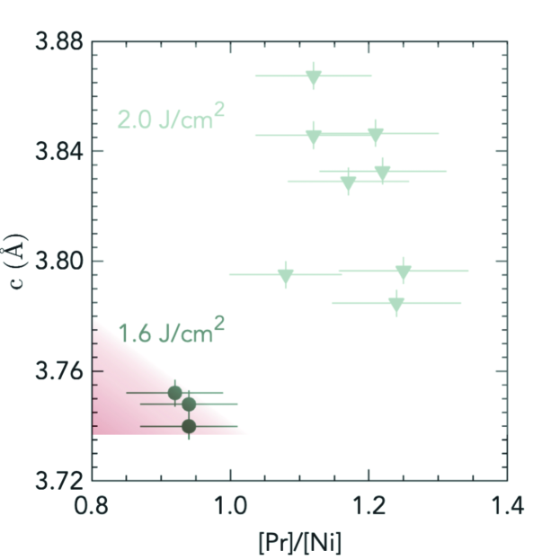
Fig.2 shows -axis lattice parameter as a function of [Pr]/[Ni] ratio of PSNO3 films grown under optimal growth conditions of substrate temperature and oxygen pressure and a laser fluence of 1.6 , or at a laser fluence of 2 . Given the complex satellite structure of the XPS spectra, one may expect accuracy for the XPS quantification.Brundle and Crist (2020) Within these limits of accuracy, nearly stoichiometric films are obtained for an optimal laser fluence of 1.6 , while films grown at a laser fluence of 2 Jcm-2 present a dramatic increase in the [Pr]/[Ni] ratio, with strong deviations from the expected film stoichiometry. Fig. S4 (Supplementary Information) shows in detail the dependence of [Pr]/[Ni] ratio on oxygen pressure and substrate temperature at 2 . We also assessed the [Pr]/[Sr] ratio from Pr 3 and Sr 3, and found values close to , indicating no strong deviations relative to the nominal hole-doping levels across different growth conditions, as depicted in Fig. S5 (Supplementary Information).
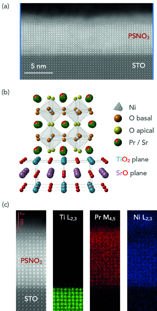
To provide further structural characterization of the grown films and visualize possible defects of the atomic lattice, cross-sectional scanning transmission electron microscopy (STEM) imaging was carried out. Fig.3(a) depicts a high-angle annular dark-field STEM (HAADF-STEM) image of an optimized PSNO3 film over a wide area of the film with very few vertical Ruddlesden–Popper faults (the structural model is illustrated in Fig.3(b)). Electron energy-loss spectroscopy (EELS) map analysis indicate a uniform distribution of Ni and Pr across the film and an abrupt interface with the STO substrate, Fig.3(c)).
Having accomplished the growth of PSNO3 films with close to optimal stoichiometry and minimal defect densities, we now focus on the study of reduced samples Pr0.8Sr0.2NiO2 (hereafter, PSNO2). The as-grown 5 5 mm2 films were cut into two pieces with lateral dimensions of 2.5 5 mm2 or four pieces of 2.5 2.5 mm2, approximately, before carrying out the hydride reduction. A same piece from the as-grown sample is repeatedly reduced. Between annealings, the sealed, evacuated glass tube that contains the reducing agent (CaH2, physically separated from the sample) is unsealed to measure the sample, and, hence, a new tube is evacuated and sealed to carry out the next step.
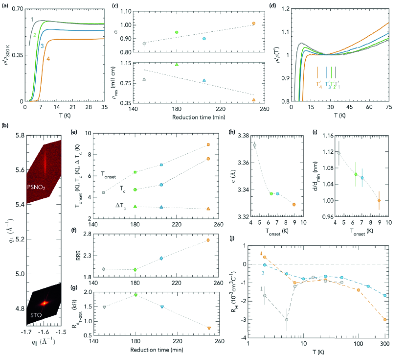
Fig.4(a) shows temperature dependent resistivity normalised to its room temperature value (to remove the influence of geometric factors) of a SC PSNO2 film over successive reductions carried out at 260∘C for periods of 150 min (1), 30 min (2), 25 min (3), and 45 min (4), (see Fig. S6, Supplementary Information for the extended range 0 K - 300 K). Although superconductivity arises after the first reduction, the zero-resistance state is achieved as a result of further heating periods. The absolute value of resistivity is for the fully reduced sample (Fig. S7, supplementary information), matching the previously published value for SC PSNO2 films on STO.Osada et al. (2020b) No diffraction reflections of the parent perovskite phase are detected in the XRD patterns of the reduced samples, and positions of the observed reflections are consistent with the (001) and (002) diffractions of the tetragonal IL phase (Fig. S8, Supplementary Information). In addition, the RSM in Fig.4(b) reveals that after the fourth step the reduced phase remains fully strained to the substrate. This is noteworthy because perovskite PSNO3 thin films on STO substrates undergo a change from tensile strain to compressive strain as they turn into the IL tetragonal PSNO2 phase. Indeed, upon deintercalation of apical oxygen atoms, the in-plane (out-of-plane) lattice parameter of the IL PSNO2 phase expands (drastically shrinks) relative to that of the parent perovskite PSNO3 phase,Hayward et al. (1999); Hayward and Rosseinsky (2003) and STO substrates induce a compressive strain () on that reduced IL phase.
Resistivity of the SC fully reduced film decreases linearly in from 300 K down to around 60 K, and deviates from linearity below that temperature, likely because of scattering due to disorder, as we discuss below. -linear normal-state resistivity has been observed in other families of unconventional superconductors, in particular in the high and low (superconductivity supressed with a magnetic field) normal-state of cuprates,L.Taillefer (2010); Legros et al. (2019) and has also been recently found in optimally doped (Nd, Sr)NiO2 films with improved crystallinity and reduced disorder synthesised on LSAT substrates.Lee et al. (2023)
We carried out least-square fitting of the experimental data to in the range from 300 K to 60 K to determine the power law dependence of resistivity (Fig. S9, Supplementary Information), and found that linearity within this region increases over subsequent reductions, i.e. : fitted exponents go from for the first incomplete reduction to for the fully reduced film, as depicted in Fig.4(c, top panel). We also observed that the value of , usually taken as a signature of the disorder contribution to the resistivity, tends to lower values throughout the reduction, Fig.4(c, bottom panel). Moreover, the gradient of linear resistivity in the high-T range upon complete reduction is , within the same order of magnitude than empirically found across different materials,Bruin et al. (2013) including highly crystalline (Nd,Sr)NiO2 films.Lee et al. (2023)
Proximity to a quantum critical point, Planckian dissipation and existence of a charge gap (Mott insulation) that remains upon doping have been proposed as underlying principles of linear-in temperature resistivity observed in some metals,Phillips et al. (2022) but there is no widely accepted explanation. To further elucidate the resistive behaviour of nickelates superconductors, additional experimental exploration of different manifestations of anomalous behaviour in magnetotransport properties of the normal state, such as -linear magnetoresistance at high field , over a broad doping range, is needed. Nonetheless, the fact that -linear resistivity is observed in superconductors that are not doped Mott insulators is significant, and supports the results recently found in the IL phase on LSAT substrates.Lee et al. (2023)
Upon further cooling, we find a resistivity upturn that holds between a temperature , at which reaches its local minimum, and the onset of the SC transition, Fig.4(d). There is evidence of this behaviour for previously reported SC nickelate thin films on STO substrates.Li et al. (2019); Osada et al. (2021) This feature appears to provide additional support for a decrease of disorder in our films throughout the reduction since the temperature lowers along the reduction process from 34.3 K after the first step to 26.7 K after the third, and reaches 19.8 K after the fourth, narrowing the temperature range over which the resistivity increases. Also, the upturn gradually smooths out and the resistivity curve flattens, nearly vanishing for the fully reduced sample, whose resistivity exhibits a very weak minimum.
Fig.4(e) illustrates the evolution of SC parameters of the reduced sample over successive annealings. Incremental reduction of the film results in a gradual increase of the onset of superconductivity, , and of the critical temperature , defined as the temperature at which resistance reaches half the normal state value at 20 K. The highest , which corresponds to a critical temperature of , and the lowest resistivity (Fig. S7, Supplementary Information) are found after the whole reduction process. The width of the resistive transition (90%–10% of normal state at 20 K) in the absence of an applied magnetic field, , is around 3 K, and slightly decreases throughout the process. No transition widths are given in literature for SC nickelates, and they are difficult to discern from the plots, but widths of the order of 2 K-3 K are usually found in high quality samples of doped cuprates.Saadaoui et al. (2015)
The observed increase in over incremental reduction processes can be attributed to a decrease of disorder, supporting the discussion above. Furthermore, intrinsic inhomogeneity inherent to doping, such as inhomogeneous charge density due to random distribution of dopants and disorder owing to difference in the ionic radius of Pr3+ and Sr2+, can reduce the attainable as it has been reported in cuprates.Eisaki et al. (2004); Fujita et al. (2005) Although, on the other hand, nanoscale electronic disorder, studied using scanning tunnelling microscopy and spectroscopy, has been found to coexist with high SC transition temperatures in cuprates.Pan et al. (2001); McElroy et al. (2005) In our results, being the dopant distribution constant along the reduction process, the increase in suggests an overall decrease of structural defects as the reduction progresses. However, scanning tunnelling microscopy experiments are needed on nickelates to reveal the effect of dopant disorder on their local superconducting properties.
The Residual Resistivity Ratio (RRR), which is a measure of the scattering rate of charge carriers by impurities or defects in the films, and is also thought to be indicative of residual apical oxygen atoms in the NiO6 octahedra in the reduced phase, is depicted in Fig.4(f). As a reference, the RRR in the as-grown sample was . In the reduced films, the RRR increases through the overall reduction process, suggesting a progressive decreasing of structural disorder over the orthorhombic to tetragonal transition along with a gradual removal of apical oxygen anions. RR ratios of around 2.8 are achieved, approximately equal to those attained elsewhere.Osada et al. (2020a, b) The significant improvement of the SC transition after the second step does not result in a subsequent increase in the RRR. This may indicate the formation of a restricted SC region in the sample, surrounded by regions that do not superconduct.
As shown in Fig.4(g), the normal state sheet resistance measured at 20 K lowers down to ( in units, where is Planck’s constant and , elementary charge, taken as an approximative value for the Mott-Ioffe-Regel limit in two dimensions). The increase of the absolute value after the second reduction may be due to slight uncertainties in the contact sizes in the Van der Pauw measurement and not to a degradation of the film, since the RRR value keeps the same as for the first reduction, and when normalised at 300 K, , shows a subsequent improvement (Fig.4(a) and Fig. S6,Supplementary Information).
The removal of oxygen anions in the reduced phase can also be tracked by the shrinkage of the -axis lattice parameter. Fig.4(h) shows the -axis lattice parameter as function of . We find a striking 12% decrease in the lattice parameter along from the parent perovskite phase to the complete topotactic oxygen deintercalation, and although a precise lattice parameter determination is hindered by the limited number of accessible Bragg reflections, our values are in agreement with those in literature for the SC IL PSNO2 phase.Osada et al. (2020a, b) This contraction along leads to a decrease in the thickness of the film (estimated from the Scherrer equation), as depicted in Fig.4(i), in agreement with STEM measurements as discussed below.
Temperature dependence of the Hall coefficient is depicted in Fig.4(j). for the fully reduced SC IL phase exhibits the zero-crossing previously reported in the SC doping range of IL nickelates:Li et al. (2019, 2020); Osada et al. (2021); Lee et al. (2023) from negative at high temperatures to positive in the low-temperature region. This behaviour of has been interpreted in accordance with a simplified two-band modelLi et al. (2020); Lee et al. (2023) although electrical transport of SC nickelates is yet to be understood. Significantly different behaviour of doped holes in IL nickelates compared to cuprates is supported by experimental evidence via spectroscopy measurementsGoodge et al. (2021) that are consistent with calculations suggesting a multiband character for nickelates,Werner and Hoshino (2020) different from the single-band cuprates. However, a single Ni band description as in the one-band Hubbard model with additional Fermi pockets has also been proved to be valid for nickelates in the superconducting doping range.Kitatani et al. (2020) In contrast, is negative for the incomplete reduced films over the entire temperature range, a behaviour also found in underdoped nickelates.Lee et al. (2023)
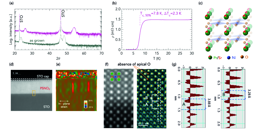
The structural analysis of the IL phase was completed by STEM on an optimized IL PSNO2 film whose XRD pattern and temperature dependent resistivity are shown in Fig.5(a, b), showing a critical temperature of . Fig.5(c) depicts the schematic of the crystal structure of the IL phase, where the infinite NiO2 planes are separated only by Pr/Sr atoms, once the apical oxygen anions of the starting perovskite phase have been removed. The cross-sectional STEM image of the IL film in Fig.5(d) shows a high-quality infinite-layer structure. And, the geometrical phase analysis (GPA) algorithm applied to that STEM image in Fig.5(e) reveals only a few possible Ruddlesden–Popper defects. Furthermore, 4D-STEM divergence of center of mass (dCOM) image, approximating to a projected charge density image in Fig.5(f) confirms the removal of apical oxygen anions from the perovskite phase so that Pr/Sr planes alternate with NiO2 planes in the film. We also observed an abrupt oxygen-oxygen distance variation at the interface of the reduced film (Fig.5(g)), changing from in the STO substrate (left panel, A) to in the IL PSNO2 film (right panel, B), which confirms the topotactic transformation at the interface. Moreover, the out-of-plane lattice parameter estimated from the GPA analysis depicted in Fig. S10 corresponds to a decrease of 15% related to the STO lattice parameter (), closely matching the values determined for the fully reduced sample by X-ray diffraction in Fig.4(i).
We finally mention that attempts to obtain SC films from uncut samples were always unproductive, as illustrated in Fig. S11 (Supplementary Information). Immediately following unsealing of the ampoule, the uncut film shows XRD patterns or temperature dependence of resistivity typical of a reduced phase, but it readily reoxidizes in less than 24 hours even when stored in a glovebox under nitrogen atmosphere or under vacuum. This swift reoxidation prevents us from measuring different properties after a reduction step. Remarkably, once that sample is cut and the reduction is carried out on one of the resulting pieces, we did not observe reoxidation after successive annealings, as shown in Fig. S12 (Supplementary Information). Previous works report perovskite films are cut in half before being reduced to the infinite-layer phase,Li et al. (2019); Lee et al. (2020, 2023) regardless of the size of the substrate,Fowlie et al. (2022) but a deeper understanding of the topotactic process is needed to explain this empirical observation. It is worth noting that reoxidation observed on the uncut reduced samples is not avoided by a STO capping layer, while no reoxidation on capped SC films that have been previously cut was detected within the course of several weeks after reduction (Fig. S13, Supplementary Information).
III Conclusions
In summary, we have accomplished the synthesis of SC infinite-layer IL praseodymium nickelate thin films. Our results highlight the importance of the combined optimization of both steps of the process: perovskite growth, with control over cation stoichiometry, and topochemical reduction. Starting from nearly stoichiometric perovskite films with minimal defect densities, the linear-in-temperature resistivity of intermediate reduced films can be used as a proxy to assess the performance of subsequent reduction processes. These results contribute towards the goal of yielding high quality superconducting nickelate samples, still scarce to date, to enable further experimental progress in this field. Furthermore, better understanding of the topochemical aspects of the reduction process is a critical issue for exploring this new class of superconductors and could push forward experimental research on the field.
IV Experimental methods
Pulsed Laser Deposition and CaH2 reduction
We grew perovskite nickelate thin films in a PLD system which utilizes a KrF excimer laser (248 nm) focused onto the target. The laser pulse rate was fixed at 4 Hz. During the growth, oxygen was supplied in the PLD chamber yielding a background pressure up to 0.5 mbar. The laser fluence was varied between 1.2 and 2.5 . The substrate temperature was set in the range 570 ∘C - 675 ∘C. We optimized these parameters to produce high-quality epitaxial films as discussed in the text. The films were cooled down to room temperature at a rate of 5 at the growth pressure. Single crystals of (001)SrTiO3 (STO) were used as substrates. Prior to the growth, they were etched in buffered HF and annealed at 1000 ∘C for 3 h to obtain stepped surfaces. We sintered the PLD target from a mixture of Pr2O3 (99.99%), Nickel(II) oxide (99.99%) and SrCO3 with controlled cation stoichiometry ([Ni]/([Pr]+[Sr])=1.1; [Pr]/[Sr]=4) by a solid state reaction. These mixtures were ground in an agate mortar and, after initial decarbonation at 1200 ∘C for 12 h, pressed into pellets, and heated in a box furnace at 1300 ∘C for 24 h. To get high-density PLD targets, the powders were reground and repressed, and then fired at 1300 ∘C for further 24 h.
Reduction of the perovskite phase into the IL phase was carried out in evacuated glass tubes. The tubes were filled with 0.1 g of CaH2 powder in an N2-filled glovebox. Samples were wrapped in aluminum foil and inserted into the glass tubes. The tubes were evacuated by means of a rotary pump ( mbar) and sealed. Heating and cooling rates in the oven were 5 .
X-ray Diffraction
XRD /2 scans of the films were performed in a four-circle diffractometer (Cu source, Ge(220) 2-bounce incident beam monochromator). The lattice parameters are calculated from Gaussian fits of the (001) and (002) XRD peaks (indices with respect to the pseudocubic unit cell) and extrapolated against to to reduce systematic errors. The thickness of the reduced films was estimated from the width of the (002) XRD reflection through the Scherrer equation with a constant fitted for our samples. Scans around asymmetrical reflections of the films were transformed to Reciprocal Space Maps (RSMs).
X-ray Photoelectron Spectroscopy
XPS measurements (in situ and ex situ) were performed using a Mg K source (1253.6 eV, 20 mA, 15 kV). Survey spectra were acquired with a pass energy of 60 eV and detailed spectra with a pass energy of 20 eV in the energy analyzer. All spectra were measured in normal emission. XPS data were processed with the CasaXPS software.
Electrical transport
Transport measurements were performed in a Physical Property Measurement System (PPMS, Quantum Design). Four-point resistivity measurements were performed in a Van der Pauw geometry by means of wire-bonded Au wires. Temperature-dependent Hall coefficients were calculated from linear fits of antisymmetrized field sweeps up to 9 T.
Transmission Electron Microscopy
The cross-sectional lamellae for Transmission Electron Microscopy were prepared using a Focused Ion Beam (FIB) technique at Centre de Nanosciences et de Nanotechnologies (C2N), University Paris-Saclay, France. Prior to FIB lamellae preparation, around 20-30 nm of amorphous carbon was deposited on top of the samples for protection. The High-angle annular dark-field (HAADF) imaging and 4D-STEM was carried out in a NION UltraSTEM 200 C3/C5-corrected scanning transmission electron microscope (STEM). The experiments were done at 200 keV with a probe current of 12 pA and convergence semi-angles of 23 mrad. A MerlinEM (Quantum Detectors Ltd) in a 4 × 1 configuration (1024 × 256) had been installed on a Gatan ENFINA spectrometer mounted on the microscope.Tencé et al. (2020) For 4D-STEM, the electron energy loss spectroscopy (EELS) spectrometer was set into non-energy dispersive trajectories and 6-bit detector mode that gave a diffraction pattern with a good signal to noise ratio without compromising much on the scanning speed was used. The geometrical phase analysis (GPA)Hytch et al. (1998) had been done choosing the STO substrate with as a reference parameter. The lattice parameters of the PSNO2 were estimated by averaging the GPA maps over square areas of 50 (in-plane) 50 (out-of-plane) nm giving a strain accuracy determination better than 1%, that is, better than for the lattice parameters. Such an approach has been previously employed to accurately determine the -axis variation in an apical oxygen ordered nickelate thin-film on an STO substrate.Raji et al. (2023) The EELS spectra were obtained using the full 4 1 configuration and the 4D-STEM by selecting only one of the chips (256 256 pixels). The element maps were done by integrating the core EELS edge-signal of the respective elements and mapping them in the spectrum image.
Acknowledgments
The authors thank Jin-Hong Lee for his help at the early stage of this project and Richard Lebourgeois for his support with target preparation. A.G.L. acknowledges financial support through a research grant from the Next Generation EU plan 2021, European Union. D. Z. acknowledges finantial support from École Doctorale 564 Physique en Ile de France (EDPIF). This work received funding from the ERC Advanced Grant No. 833973 (Fresco).
Additional Information
Supporting Information is available for this manuscript.
Author contributions
A.G.L. designed and performed the experiments (growth, XRD, XPS, topochemical reductions, electrical transport), processed and analysed the data, produced the graphics and wrote the manuscript. A.R. and A.G. designed and performed electron microscopy experiments, processed and analysed the data. D.Z. and F.G. performed complementary experiments. L.D. and C.G. designed and implemented the experimental setup for sealing the glass ampoules used in the reduction process. L.I. initiated the optimization (growth, XRD, topochemical reduction, electrical transport) of nickelate samples at the beginning of the project and assisted in reduction experiments. M.B. proposed the project, provided equipement and acquired funding. A.G.L., A.R., D.Z., A.G., L.I. and M.B. discussed the data. All co-authors reviewed the manuscript, and approved its final form.
Competing interests
The authors declare no competing interests.
Data availability
The data that support the findings of this study are available from the corresponding author upon reasonable request.
Keywords
nickelates, superconductivity, thin films, topochemical, reduction
References
- Li et al. (2019) D. F. Li, K. Lee, B. Y. Wang, M. Osada, S. Crossley, H. R. Lee, Y. Cui, Y. Hikita, and H. Y. Hwang, Nature 572 (2019), 10.1038/s41586-019-1496-5.
- Osada et al. (2020a) M. Osada, B. Y. Wang, B. H. Goodge, K. Lee, H. Yoon, K. Sakuma, D. F. Li, M. Miura, L. F. Kourkoutis, and H. Y. Hwang, Nano Letters 20 (2020a), 10.1021/acs.nanolett.0c01392.
- Osada et al. (2020b) M. Osada, B. Y. Wang, K. Lee, D. F. Li, and H. Y. Hwang, Physical Review Materials 4 (2020b), 10.1103/PhysRevMaterials.4.121801.
- Wang et al. (2022) N. N. Wang, M. W. Yang, Z. Y. K. Y. C. H. Zhang, Q. H. Z. Z. H. Zhu, Y. Uwatoko, L. Gu, X. L. Dong, J. P. Sun, K. J. Jin, and J. G. Cheng, Nature Communications 13 (2022), 10.1038/s41467-022-32065-x.
- Osada et al. (2021) M. Osada, B. Y. Wang, B. H. Goodge, S. P. Harvey, K. H. Lee, D. F. Li, L. F. Kourkoutis, and H. Y. Hwang, Advanced Materials 33 (2021), 10.1002/adma.202104083.
- Osada et al. (2023) M. Osada, K. Fujiwara, T. Nojima, and A. Tsukazaki, Physical Review Materials 7 (2023), 10.1103/PhysRevMaterials.7.L051801.
- Zeng et al. (2022) S. Zeng, C. Li, L. E. Chow, Y. Cao, Z. Zhang, C. S. Tang, X. Yin, Z. S. Lim, J. Hu, P. Yang, and A. Ariando, Science Advances 8 (2022), 10.1126/sciadv.abl9927.
- Wei et al. (2023) W. Z. Wei, D. Vu, Z. Zhang, F. J. Walker, and C. H. Ahn, Science Advances 9 (2023), 10.1126/sciadv.adh3327.
- Pan et al. (2021) G. A. Pan, D. F. Segedin, H. LaBollita, Q. Song, E. M. Nica, B. H. Goodge, A. T. Pierce, S. Doyle, S. Novakov, D. C. Carrizales, A. T. NDiaye, P. Shafer, H. Paik, J. T. Heron, J. A. Mason, A. Yacoby, L. F. Kourkoutis, O. Erten, C. M. Brooks, A. S. Botana, and J. A. Mundy, Nature Materials 21 (2021), 10.1038/s41563-021-01142-9.
- Sun et al. (2023) H. Sun, M. Huo, X. Hu, J. Li, Z. Liu, Y. Han, L. Tang, Z. Mao, P. Yang, B. Wang, J. Cheng, D. X. Yao, G. M. Zhang, and M. Wang, Nature (2023), 10.1038/s41586-023-06408-7.
- Zhou et al. (2021) X. J. Zhou, W. S. Lee, M. Imada, N. Trivedi, P. Phillips, H. Y. Kee, P. Torma, and M. Eremets, Nature Reviews Physics 3 (2021), Zhou.
- Scalapino (2012) D. J. Scalapino, Reviews of Modern Physics 84 (2012), 10.1103/RevModPhys.84.1383.
- Tsuei and Kirtley (2000) C. C. Tsuei and J. R. Kirtley, Reviews of Modern Physics 72, 969 (2000).
- Keimer et al. (2015) B. Keimer, S. A. Kivelson, M. R. Norman, S. Uchida, and J. Zaanen, Nature 518 (2015), 10.1038/nature14165.
- Proust and Taillefer (2019) C. Proust and L. Taillefer, Annual Review of Condensed Matter Physics 10, 409 (2019).
- Stewart (2017) G. R. Stewart, Advances in Physics 66, 75 (2017).
- O’Mahonya et al. (2022) S. M. O’Mahonya, W. Ren, W. Chen, Y. X. Chong, X. Liu, H. Eisakie, S. Uchidaf, M. H. Hamidianc, and J. C. S. Davis, Proceedings of the National Academy of Sciences of the United States of America 119 (2022), 10.1073/pnas.2207449119.
- Pickett (2021) W. E. Pickett, Nature Reviews Physics 3 (2021), 10.1038/s42254-020-00257-3.
- Norman (2020) M. R. Norman, Physics 13 (2020), https://physics.aps.org/articles/v13/85.
- Botana et al. (2022) A. S. Botana, K. Lee, M. R. Norman, V. Pardo, and W. E. Pickett, Frontiers in Physics 9 (2022), 10.3389/fphy.2021.813532.
- Mitchell (2021) J. F. Mitchell, Frontiers in Physics 9 (2021), 10.3389/fphy.2021.813483.
- Anisimov et al. (59) V. I. Anisimov, D. Bukhvalov, and T. M. Rice, Physical Review B 12 (59), 10.1103/PhysRevB.59.7901.
- Lee and Pickett (2004) K. W. Lee and W. E. Pickett, Physical Review B 70 (2004), 10.1103/PhysRevB.70.165109.
- S.Botana and Norman (2020) A. S.Botana and M. R. Norman, Physical Review X 10 (2020), 10.1103/PhysRevX.10.011024.
- Nomura and Arita (2022) Y. Nomura and R. Arita, Reports on Progress in Physics 85 (2022), 10.1088/1361-6633/ac5a60.
- Gu and Wen (2022) Q. Q. Gu and H. H. Wen, The Innovation 3 (2022), 10.1016/j.xinn.2021.100202.
- Kitatani et al. (2023) M. Kitatani, Y. Nomura, M. Hirayama, and R. Arita, APL Materials 11 (2023), 10.1063/5.0097618.
- Hepting et al. (2021) M. Hepting, M. P. M. Dean, and W. S. Lee, Frontiers in Physics 9 (2021), 10.3389/fphy.2021.808683.
- Lechermann (2020) F. Lechermann, Physical Review B 101, 5 (2020).
- Rossi et al. (2021) M. Rossi, H. Lu, A. Nag, D. Li, M. Osada, K. Lee, B. Y. Wang, S. Agrestini, M. Garcia-Fernandez, Y. Chuang, Z. X. Shen, H. Y. Hwang, B. Moritz, K. J. Zhou, T. P. Devereaux, and W. S. Lee, Physical Review B 104 (2021), 10.1103/PhysRevB.104.L220505.
- Goodge et al. (2021) B. H. Goodge, D. Li, K. Lee, M. Osada, B. Y. Wang, G. A. Sawatzky, H. Y. Hwang, and L. F. Kourkoutis, Proceedings of the National Academy of Sciences of the United States of America 118 (2021), 10.1073/pnas.2007683118.
- Carrasco-Alvarez et al. (2022) A. A. Carrasco-Alvarez, L. Iglesias, S. Petit, W. Prellier, M. Bibes, and J. Varignon, arXiv:2211.04870 (2022), 10.48550/arXiv.2211.04870.
- Hayward and Rosseinsky (2007) M. A. Hayward and M. J. Rosseinsky, Nature 450, 960 (2007).
- Ranmohotti et al. (2011) K. G. S. Ranmohotti, E. Josepha, J. Choi, J. Zhang, and J. B. Wiley, Advanced Materials 23 (2011), 10.1002/adma.201002274.
- Meng et al. (2023) Z. Meng, H. Yan, P. Qin, X. Zhou, X. Wang, H. Chen, L. Liu, and Z. Liu, Advanced Functional Materials 24 (2023), 10.1002/adfm.202305225.
- Kageyama et al. (2018) H. Kageyama, K. Hayashi, K. Maeda, J. P. Attfield, Z. Hiroi, J. M. Rondinelli, and K. R. Poeppelmeier, Nature Communications 9 (2018), 10.1038/s41467-018-02838-4.
- Hayward et al. (1999) M. A. Hayward, M. A. Green, M. J. Rosseinsky, and J. Sloan, Journal of the American Chemical Society 121, 8843 (1999).
- Hayward and Rosseinsky (2003) M. A. Hayward and M. J. Rosseinsky, Solid State Sciences 5, 839 (2003).
- Kawai et al. (2010) M. Kawai, K. Matsumoto, N. Ichikawa, M. Mizumaki, O. Sakata, N. Kawamura, S. Kimura, and Y. Shimakawa, Crystal Growth & Design 10, 2044 (2010).
- Lee et al. (2020) K. Lee, B. H. Goodge, D. Li, M. Osada, B. Y. Wang, Y. Cui, L. F. Kourkoutis, and H. Y. Hwang, APL Materials 8 (2020), 10.1063/5.0005103.
- Lee et al. (2023) K. Lee, B. Y. Wang, M. Osada, B. H. Goodge, T. C. Wang, Y. Lee, S. Harvey, W. J. Kim, Y. Yu, C. Murthy, S. Raghu, L. F. Kourkoutis, and H. Y. Hwang, Nature 619 (2023), 10.1038/s41586-023-06129-x.
- Garcia-Muñoz et al. (1992) J. L. Garcia-Muñoz, J. Rodriguez-Carvajal, P. Lacorre, and J.B.Torrance, Physical Review B 46 (1992), 10.1103/PhysRevB.46.4414.
- Garcia-Muñoz et al. (1995) J. L. Garcia-Muñoz, M. Suaaidi, M. J. Martinez-Lope, and J. A. Alonso, Physical Review B 52 (1995), 10.1103/PhysRevB.52.13563.
- Ledbetter et al. (1990) H. Ledbetter, M. Lei, and S. Kim, Phase Transitions 23 (1990), 10.1080/01411599008241819.
- Ohnishi et al. (2005) T. Ohnishi, M. Lippmaa, T. Yamamoto, S. Meguro, and H. oinuma, Applied Physics Letters 87 (2005), 10.1063/1.2146069.
- Ohnishi et al. (2008) T. Ohnishi, K. Shibuya, T. Yamamoto, and M. Lippmaa, Journal of Applied Physics 103 (2008), 10.1063/1.2921972.
- Scofield (1973) J. H. Scofield, Journal of Electron Spectroscopy and Related Phenomena 8, 129 (1973).
- Brundle and Crist (2020) C. R. Brundle and B. V. Crist, Journal Vacuum Science Technology A 38 (2020), 10.1116/1.5143897.
- L.Taillefer (2010) L.Taillefer, Annual Review of Condensed Matter Physics 1, 51 (2010).
- Legros et al. (2019) A. Legros, S. Benhabib, W. Tabis, F. Laliberteé, M. Dion, M. Lizaire, B. Vignolle, D. Vignolles, H. Raffy, Z. Z. Li, P. Auban-Senzier, N. Doiron-Leyraud, P. Fournier, D. Colson, L. Taillefer, and C. Proust, Nature Physics 15 (2019), 10.1038/s41567-018-0334-2.
- Bruin et al. (2013) J. A. N. Bruin, H. Sakai, R. S. Perry, and A. P. Mackenzie, Science 6121, 804 (2013).
- Phillips et al. (2022) P. W. Phillips, N. E. Hussey, and P. Abbamonte, Science 377 (2022), 10.1126/science.abh4273.
- Saadaoui et al. (2015) H. Saadaoui, Z. Salman, H. Luetkens, T. Prokscha, A. Suter, W. MacFarlane, Y. Jiang, K. Jin, R. Greene, E. Morenzoni, and R. Kiefl, Nature Communications 6 (2015), 10.1038/ncomms7041.
- Eisaki et al. (2004) H. Eisaki, N. Kaneko, D. L. Feng, A. Damascelli, P. K. Mang, K. M. Shen, Z. X. Shen, and M. Greven, Physical Review B 69 (2004), 10.1103/PhysRevB.69.064512.
- Fujita et al. (2005) K. Fujita, T. Noda, K. M. Kojima, H. Eisaki, and S. Uchida, Physical Review Letters 95 (2005), 10.1103/PhysRevLett.95.097006.
- Pan et al. (2001) S. H. Pan, J. P. O’Neal, R. L. Badzey, C. Chamon, H. Ding, J. R. Engelbrecht, Z. Wang, H. Eisaki, S. Uchida, A. K. Guptak, K. W. Ngk, E. W. Hudson, K. M. Lang, and J. C. Davis, Nature 413, 282 (2001).
- McElroy et al. (2005) K. McElroy, J. Lee, J. A. Slezak, D. Lee, H. Eisaki, S. Uchida, and J. C. Davis, Science 309 (2005), 10.1126/science.1113095.
- Li et al. (2020) D. Li, B. Y. Wang, K. Lee, S. P. Harvey, M. Osada, B. H. Goodge, L. F. Kourkoutis, and H. Y. Hwang, Physical Review Letters 125 (2020), 10.1103/PhysRevLett.125.027001.
- Werner and Hoshino (2020) P. Werner and S. Hoshino, Physical Review B 101 (2020), 10.1103/PhysRevB.101.041104.
- Kitatani et al. (2020) M. Kitatani, L. Si, O. Janson, R. Arita, Z. Zhong, and K. Held, npj Quantum Materials 5 (2020), 10.1038/s41535-020-00260-y.
- Fowlie et al. (2022) J. Fowlie, M. Hadjimichael, M. M. Martins, D. Li, M. Osada, B. Y. Wang, K. Lee, Y. Lee, Z. Salman, T. Prokscha, J. M. Triscone, H. Y. Hwang, and A. Suter, Nature Physics 18 (2022), 10.1038/s41567-022-01684-y.
- Tencé et al. (2020) M. Tencé, J. D. Blazit, X. Li, M. Krajnak, R. S. L. C. E. Nebot del Busto, M. Kociak, O. Stephan, and A. Gloter, Microscopy and Microanalysis 26, 1940 (2020).
- Hytch et al. (1998) M. J. Hytch, E. Snoeck, and R. Kilaas, Ultramicroscopy 74, 131 (1998).
- Raji et al. (2023) A. Raji, G. Krieger, N. Viart, D. Preziosi, J. P. Rueff, and A. Gloter, Small (2023), 10.1002/smll.202304872.