:
\theoremsep
\jmlrvolume
\firstpageno1
\jmlryear2023
\jmlrworkshopSymmetry and Geometry in Neural Representations
Full-dimensional Characterisation of Time-Warped\titlebreakSpike-Time Stimulus-Response Distribution Geometries
Abstract
Characterising the representation of sensory stimuli in the brain is a fundamental scientific endeavor, which can illuminate principles of information coding. Most characterizations reduce the dimensionality of neural data by converting spike trains to firing rates or binned spike counts, applying explicitly named methods of “dimensionality reduction”, or collapsing trial-to-trial variability. Characterisation of the full-dimensional geometry of timing-based representations may provide unexpected insights into how complex high-dimensional information is encoded. Recent research shows that the distribution of representations elicited over trials of a single stimulus can be geometrically characterized without the application of dimensionality reduction, maintaining the temporal spiking information of individual neurons in a cell assembly and illuminating rich geometric structure. We extend these results, showing that precise spike time patterns for larger cell assemblies are time-warped (i.e. stretched or compressed) on each trial. Moreover, by geometrically characterizing distributions of large spike time patterns, our analysis supports the hypothesis that the degree to which a spike time pattern is time-warped depends on the cortical area’s background activity level on a single trial. Finally, we suggest that the proliferation of large electrophysiology datasets and the increasing concentration of “neural geometrists”, creates ideal conditions for characterization of full-dimensional spike time representations, in complement to dimensionality reduction approaches.
keywords:
Neural coding, cell assemblies, time-warp, spike timing, latent factors, neural representational geometry, high-dimensional representations, cortex, stimulus-responses.1 Introduction
Trial-to-trial variability implies that characterizing the neural representation of a single stimulus, requires characterization of the underlying distribution from which single trial representations are drawn. Whilst dimensionality reduction techniques have helped demystify aspects of neural processing (Gallego et al., 2017; Humphries, 2020) we have become accustomed to low-dimensional perspectives. Characterisation of the full-dimensional geometry of timing-based representations may provide unexpected insights into how high-dimensional sensory and motor information is encoded. This could facilitate improved decoding and would constrain how cortical architecture parses information on a millisecond timescale.
Recent research shows that the distribution of representations elicited over trials of a single stimulus can be geometrically characterized without the application of dimensionality reduction (Isbister et al., 2021), maintaining the temporal spiking information of individual neurons in a cell assembly and illuminating rich geometric structure. By studying responses to single whisker deflections in the barrel cortex, this work investigated the simplest correspondence between a piece of information and its cortical representation. Interestingly, single whisker deflections elicit only a single spike in the majority of responding neurons (Reyes-Puerta et al., 2015), supporting the view that temporally atomic stimuli and units of information are represented by short multi-neuron patterns of single spikes (Fig. LABEL:fig:introA). In the visual cortex, such patterns even support visual discrimination (Resulaj et al., 2018).
fig:intro
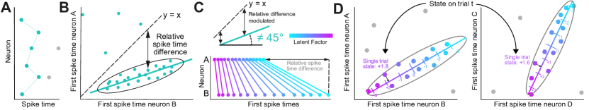
Specifically, Isbister et al. (2021) studied how single spike patterns of two (and later four) cortical neurons co-represent a stimulus by plotting the first spike times of two neurons over repeated trials, and characterising the observed geometry (Fig. LABEL:fig:introB). The study observed isolated regions (“clusters”) of high response probability, in which the spike times of the two neurons were positively correlated (i.e. the two neurons spiked earlier or later together on each trial). After controlling for stimulus-response adaptation, which can increase spike time latencies over repeated trials, the clusters remained positively correlated. This showed that the spike times of the two neurons, depended on a shared hidden variable, which varied from trial-to-trial. Interestingly, the correlation angle was often different from , showing that the spike times of the two neurons were modulated differently by the hidden variable, such that the the timing difference between the spikes of the two neurons was also modulated (Fig. LABEL:fig:introC). Finally, the study found that the variable modulating spike times was correlated between pairs of clusters (i.e. 4 neurons) and hypothesised that the cortical area’s background activity level might correspond with the variable modulating spike times.
To test this hypothesis, we explore whether a single dimensional hidden variable modulates the spike times of larger cell assemblies. In doing so, we geometrically characterize distributions of large spike time patterns for single stimuli. This demonstrates that multi-neuron single spike patterns, which could be the building blocks of neural representation, provide a tractable opportunity to characterize full-dimensional neural representations. Moreover, trial-to-trial variability in spike time patterns and dimensionality-reduced representations might be governed by the same low-dimensional mechanisms, and could explain why millisecond-precise spike time patterns are challenging to detect (Russo and Durstewitz, 2017; Stella et al., 2019). Whilst biases towards explaining task variables promote dimensionality reduction approaches (see Discussion), we suggest that the increasing concentration of “neural geometrists” and electrophysiology datasets, creates ideal conditions for complementary characterization of full-dimensional spike time representations.
2 Results
Data analysed here and in Isbister et al. (2021) was reused from Reyes-Puerta et al. (2015). Stimuli were 2ms single whisker deflections with each stimulus condition defined by the identity of the stimulated whisker and the inter-trial-interval, with 80-200 trials made per condition. Spike sorting was applied to recordings from eight 16 electrode shanks spanning layers 2/3, 4 and 5, and separated across three to four cortical barrels in anaesthetized rats.
Isbister et al. (2021) developed a clustering pipeline that detected isolated regions of high-response probability (“clusters”) in first spike response distributions for neuron pairs. The algorithm was validated to prevent detection of spurious clusters. If the spike times of cluster samples were modulated by stimulus-response adaptation, the adaptative trends were removed. Fig. LABEL:fig:resultsA visualizes the clusters extracted for a single condition after control for adaptation. All 12 clusters are positively correlated (many with correlation angles different from ), with 8 neurons participating in at least one cluster. Supplementary Video 1 shows figures for all experimental conditions.
Here we test whether a shared single dimensional hidden variable is responsible for observed spike time co-variability across larger cell assemblies, as this would support the dependence of trial-to-trial variability on the background activity level. Firstly in support of this, the single trial states (i.e. the global background activity level) predicted by each cluster are correlated between clusters (Fig. LABEL:fig:resultsB). Secondly, using the spike times from clusters we can calculate the correlation matrix between participating neurons, including for neuron pairs for which a cluster was not originally detected (Fig. LABEL:fig:resultsC, left). A large proportion of neuron pairs show significant correlations (; Fig. LABEL:fig:resultsC, right). Moreover, almost all of the neuron pairs are positively correlated; even neuron pairs which were not shown to be significantly correlated (Fig. LABEL:fig:resultsC), supporting low-dimensional modulation. These findings were general over conditions (Fig. LABEL:fig:resultsD). Importantly, for the majority of conditions, only the 1st and 2nd eigenvalues of factor analysis were significant based on the Kaiser criterion (), with the 1st eigenvalue strongly explaining variance (Fig. LABEL:fig:resultsE).
fig:results
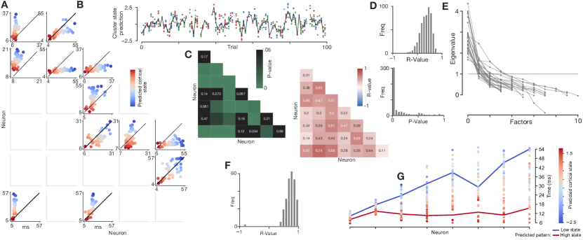
These results motivate a single dimensional “global cortical state” estimate: the mean of single cluster cortical state estimates (Fig. LABEL:fig:resultsB). This estimate explained a large amount of variance in cluster first spike times (Fig. LABEL:fig:resultsF); future work will employ a leave one out analysis. We next plot the spike time of each neuron coloured by the single trial global cortical state estimate, illustrating heterogeneous modulation of single neuron spike times by the global state (Fig. LABEL:fig:resultsG). The visualisation shows the average response for the 10 highest state trials vs the 10 lowest state trials, highlighting how the large multi-neuron single spike patterns is dramatically modulated by the shared latent factor.
These results suggest that the response distribution is highly structured and can be characterized as a high-dimensional high-response probability cluster surrounded by currently unexplained noise (or poorly sampled regions). The cluster is positively correlated due to a single dimensional latent factor oriented away from the unitary line. The cluster can be approximated by a Gaussian. For each single trial, the response distribution has missing values for different neurons due to low response reliability. As a first approximation, the response distribution was characterized by finding clusters in two-dimensional slices of the distribution, and analyzing the hidden variables modulating clusters in different slices.
3 Discussion
To our knowledge, such a precise geometrical characterisation has never previously been made. Low-dimensional modulation of larger multi-neuron single spike patterns strengthens the hypothesis of Isbister et al. (2021) that modulation reflects low-dimensional latent factors observed in dimensionality-reduced representations (Kao et al., 2015; Pandarinath et al., 2018). The single trial global cortical state estimate can thus be compared with trial-to-trial variability in reduced-dimensions. The metric may also correlate with the spiking activity of other neurons (not just cluster neurons), as well as secondary and tertiary spikes. As spike times are modulated positively, the biophysical nature of such low-dimensional modulation is likely to correspond with fluctuations in “shared excitability”, which also modulate spike counts (Lin et al., 2015). Spatially, this may correspond with travelling waves, indirectly measurable through the LFP (Davis et al., 2020).
The characterized representations may aid in understanding neural coding, such as theoretical encoding capacity or how neurons integrate temporal patterns. Relative timing differences between spikes must be considered, which are interpretable downstream. Isbister et al. (2021) previously suggested that low-dimensional modulation over multiple cortical layers and columns makes it possible that downstream neurons are also modulated by the same fluctuations introducing the possibility that downstream neurons can decode patterns conditioned on cortical state. Estimates of state-dependent coding capacity were made by Isbister et al. (2021), and will likely be improved using the estimated global cortical state.
The precise characterisation of multi-neuron first spike response distributions comes nearly 100 years after the discovery of the action potential (Adrian and Zotterman, 1926), which were described as “scarcely more complex than a succession of dots in Morse Code” (Adrian, 1932). In addition to the challenges of multi-electrode recording, a number of factors perhaps explain why such a characterization was long overlooked. Rather than studying the fundamental correspondence between a simple stimulus and its stochastic neural representation, researchers and funders are often biased towards demonstrating encoding of features or task relevant information. As such, duration limited experimental sessions often favor larger numbers of conditions over high trial counts per condition. Analyses are then applied which work robustly with low trial counts, in exchange for temporal granularity. In a self-proliferating cycle, statistical techniques and experiments are then developed in the same vein. Task variables are often low-dimensional, due to the complexity of training animals, and the need to create controlled and interpretable paradigms. This leads to low-dimensional modulation of population activity by task variables, which again proliferates dimensionality-reduction. In sensory modalities such as vision, the brain must rapidly create high-dimensional representations, however.
Openly available, large biophysically-detailed models are becoming gradually more abundant (Billeh et al., 2020; Isbister et al., 2023). A recent cortical model precisely captures layer-wise population response to whisker deflections, and selective propagation of activity to downstream areas (Isbister et al., 2023). Such a model provides the opportunity to generate unlimited samples from stimulus-response distributions, whilst controlling nonstationarities. As the model is refined, it is likely that the response distributions will better reflect those characterized in vivo, and the model can be used to study the propagation of activity through cortical architecture at full spatiotemporal scale.
Funding
This study was supported by funding to the Blue Brain Project, a research center of the École polytechnique fédérale de Lausanne (EPFL), from the Swiss government’s ETH Board of the Swiss Federal Institutes of Technology.
Supplementary Video 1
Supplementary Video 1 is available at: t.ly/iOhhX
References
- Adrian (1932) Edgar D Adrian. The mechanism of nervous action. electrical studies of the neurone. page 12, 1932.
- Adrian and Zotterman (1926) Edgar D Adrian and Yngve Zotterman. The impulses produced by sensory nerve-endings: Part ii. the response of a single end-organ. The Journal of physiology, 61(2):151, 1926.
- Billeh et al. (2020) Yazan N Billeh, Binghuang Cai, Sergey L Gratiy, Kael Dai, Ramakrishnan Iyer, Nathan W Gouwens, Reza Abbasi-Asl, Xiaoxuan Jia, Joshua H Siegle, Shawn R Olsen, et al. Systematic integration of structural and functional data into multi-scale models of mouse primary visual cortex. Neuron, 106(3):388–403, 2020.
- Davis et al. (2020) Zachary W Davis, Lyle Muller, Julio Martinez-Trujillo, Terrence Sejnowski, and John H Reynolds. Spontaneous travelling cortical waves gate perception in behaving primates. Nature, 587(7834):432–436, 2020.
- Gallego et al. (2017) Juan A Gallego, Matthew G Perich, Lee E Miller, and Sara A Solla. Neural manifolds for the control of movement. Neuron, 94(5):978–984, 2017.
- Humphries (2020) Mark D Humphries. Strong and weak principles of neural dimension reduction. arXiv preprint arXiv:2011.08088, 2020.
- Isbister et al. (2021) James B Isbister, Vicente Reyes-Puerta, Jyh-Jang Sun, Illia Horenko, and Heiko J Luhmann. Clustering and control for adaptation uncovers time-warped spike time patterns in cortical networks in vivo. Scientific Reports, 11(1):15066, 2021.
- Isbister et al. (2023) James B Isbister, András Ecker, Christoph Pokorny, Sirio Bolaños-Puchet, Daniela Egas Santander, Alexis Arnaudon, Omar Awile, Natali Barros-Zulaica, Jorge Blanco Alonso, Elvis Boci, et al. Modeling and simulation of neocortical micro- and mesocircuitry. part ii: Physiology and experimentation. bioRxiv, 2023.
- Kao et al. (2015) Jonathan C Kao, Paul Nuyujukian, Stephen I Ryu, Mark M Churchland, John P Cunningham, and Krishna V Shenoy. Single-trial dynamics of motor cortex and their applications to brain-machine interfaces. Nature communications, 6(1):7759, 2015.
- Lin et al. (2015) I-Chun Lin, Michael Okun, Matteo Carandini, and Kenneth D Harris. The nature of shared cortical variability. Neuron, 87(3):644–656, 2015.
- Pandarinath et al. (2018) Chethan Pandarinath, Daniel J O’Shea, Jasmine Collins, Rafal Jozefowicz, Sergey D Stavisky, Jonathan C Kao, Eric M Trautmann, Matthew T Kaufman, Stephen I Ryu, Leigh R Hochberg, et al. Inferring single-trial neural population dynamics using sequential auto-encoders. Nature methods, 15(10):805–815, 2018.
- Resulaj et al. (2018) Arbora Resulaj, Sarah Ruediger, Shawn R Olsen, and Massimo Scanziani. First spikes in visual cortex enable perceptual discrimination. Elife, 7:e34044, 2018.
- Reyes-Puerta et al. (2015) Vicente Reyes-Puerta, Jyh-Jang Sun, Suam Kim, Werner Kilb, and Heiko J Luhmann. Laminar and columnar structure of sensory-evoked multineuronal spike sequences in adult rat barrel cortex in vivo. Cerebral Cortex, 25(8):2001–2021, 2015.
- Russo and Durstewitz (2017) Eleonora Russo and Daniel Durstewitz. Cell assemblies at multiple time scales with arbitrary lag constellations. Elife, 6:e19428, 2017.
- Stella et al. (2019) Alessandra Stella, Pietro Quaglio, Emiliano Torre, and Sonja Grün. 3d-spade: Significance evaluation of spatio-temporal patterns of various temporal extents. Biosystems, 185:104022, 2019.