Understanding Hepatitis B Virus Infection through Hepatocyte Proliferation and Capsid Recycling
1 Abstract
Proliferation of uninfected as well as infected hepatocytes and recycling of DNA-containing capsids are two major mechanisms playing significant roles in the clearance of hepatitis B virus (HBV) infection. In this study, the temporal dynamics of this infection is investigated through two in silico bio-mathematical models considering both proliferation of hepatocytes and recycling of capsids. Both models are formulated on the basis of a key finding in existing literature: Mitosis of infected yields in two uninfected progenies. In the first model, we examine regular proliferation (occur continuously), while the second model deals with the irregular proliferation (happen when the total number of liver cells decreases to less than 70% of its initial volume). The models are calibrated with the experimental data obtained from an adult chimpanzee. Results of this study suggest that when both hepatocytes proliferate with equal rate, proliferation aids the individual in a rapid recovery from the acute infection whereas in case of chronic infection, the severity of the infection increase if the proliferation occur frequently. On the other hand, if the infected cells proliferate at a slower rate than uninfected cells, the proliferation of uninfected hepatocytes contributes to increase the infection, but the proliferation of infected hepatocytes acts to reduce the infection from the long-term perspective. Furthermore, it is also observed that the differences between the outcomes of regular and irregular proliferations are substantial and noteworthy.
Keywords: Hepatitis B, capsids, recycling, chronic, cellular proliferation, irregular proliferation.
2 Introduction
Hepatitis B virus (HBV) infection stands as an extensively widespread infection with more than 300 million chronically infected individuals across the world. This viral infection leads to approximately 1 million fatalities annually despite the fact that it is preventable and treatable [1]. HBV is a partially double-stranded, enveloped DNA virus. This viruses mainly target hepatocytes, the primary cells of the liver. Even with the availability of antiviral drugs (interferons (IFN)-alpha-2a, pegylated (PEG)-IFNalpha-2a (immune system modulators), and some nucleoside analogues, such as lamivudine, adefovir, entecavir, telbivudine, and tenofovir) and effective vaccine, HBV remains a significant public health concern. However, the long-term use of these medications can lead to the development of drug resistance, diminishing their effectiveness over time [2]. The amalgamation of in vitro experiments and in vivo explorations in ducks, woodchucks, mice, and chimpanzees has greatly advanced our knowledge of HBV infection and its interplay with the immune system. Extensive efforts have been dedicated over the past three decades to gain a comprehensive understanding of HBV[3, 4, 5, 6, 7, 8, 9, 10, 11, 12, 13, 14, 15, 16, 17, 18]. Especially, regarding both acute and chronic HBV infection in humans, our knowledge remains still restricted due to the paucity of available data. Even so, there exists some data [19, 20], and using this data with mathematical models, possible mechanisms underlying HBV infection are investigated [21, 22].
Cellular proliferation or mitosis cell division stands as one of the pivotal biological process influenced by this viral infection as HBV promotes cell proliferation through HBx-induced microRNA-21 in hepatocellular carcinoma (HCC) [23]. In the literature, a limited number of studies are dedicated in investigating the proliferation of hepatocytes. For example, in the year 2010, Hews et al. [11] introduced a HBV infection dynamics model considering the logistic growth of uninfected hepatocytes and standard incidence function. However, it’s worth noting that their model incorporated numerous assumptions while also omitting several crucial pieces of information that could have been included in their model. Taking into account the supposition that infected hepatocytes also undergo proliferation at a rate comparable to or lower than that of healthy hepatocytes during the infection, Hews et al. [24] modified their previously proposed model [11] and obtained some new outcomes. Dahari et al. [25] suggested that variations in proliferation between uninfected and infected hepatocytes could offer a rich explanation for the diverse patterns that are observed in the decline of viral loads. Some studies have also addressed the concept of proliferation of infected hepatocytes (POIH) with the assumption that POIH only can produce the infected hepatocytes [26, 27]. Based on the data collected regarding cellular proliferation, it is noted that three different scenarios may occur when an infected hepatocyte undergoes proliferation (mitosis).
-
1.
Two uninfected daughter cells.
-
2.
One uninfected and one infected daughter cell.
-
3.
Two infected daughter cells.
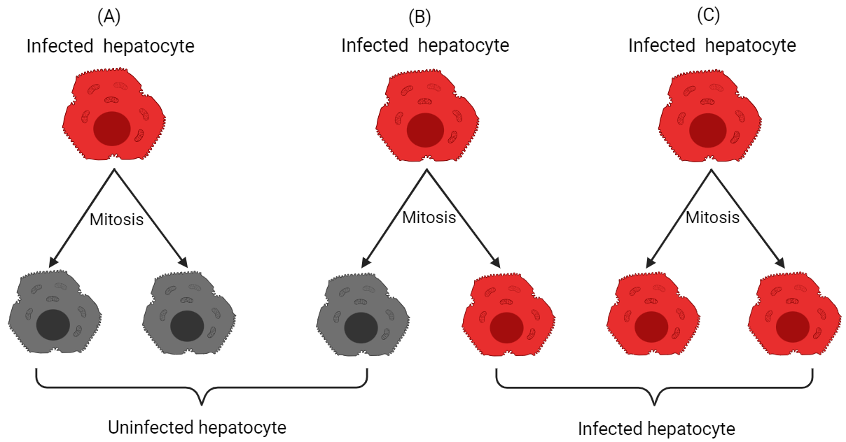
The fate of viral DNA after cell mitosis is a matter of controversy. The roles of proliferation in the clearance of both acute and chronic HBV infections continue to be subjects of ongoing debate [28, 29, 30]. During mitosis cell division, when the nuclear membrane is reformed, there is a possibility of cccDNA loss if the cccDNAs fail to reintegrate into the nucleus. Also, if the count of cccDNA within infected hepatocytes is extremely low, the progeny will be devoid of cccDNA, leading to the resolution of acute HBV infection [31]. Thus, the involvement of this additional mechanism is crucial in achieving the clearance of acute infection. Recently, Tu et al. [16] corroborate that the mitosis cell division of hepatitis B virus-infected cells leads to the generation of two uninfected daughter cells. This research team also suggests that mitosis of infected hepatocytes offers a potent avenue for addressing HBV persistence. Murray et al. [32] also highlighted the significance of cccDNA loss during cellular proliferation in the context of non-destructive clearance of HBV infection. Using experimental data from six HBV-infected patients, Goyal et al. [31] further affirmed that the resolution of acute HBV infection is probably contingent on the cellular proliferation of an infected cell leading to the generation of two uninfected daughter cells. To the best of our knowledge, no relevant article has been found in the existing literature that addresses the impacts of POIH on chronic infection. In this study, we mainly focus on the proliferation of uninfected as well as infected hepatocytes along the important mechanism: recycling of HBV DNA-containing capsids which is recently introduced by Sutradhar and Dalal [2023]. Based on the conclusion of the previous works [31, 16], we proceed with the fact that POIH produces two healthy progeny. In this study, the following things will be discussed:
-
•
When both uninfected and infected hepatocytes proliferate with equal rate.
-
–
Effects of proliferation rates on the acute infection.
-
–
Effects of proliferation rates on the chronic infection.
-
–
-
•
When both uninfected and infected hepatocytes proliferate with different rates.
-
–
Effects of proliferation rates on the acute infection.
-
–
Effects of proliferation rates on the chronic infection.
-
–
-
•
Proliferation is not a continuous process: regular proliferation vs irregular proliferation.
Considering all the aforementioned factors, we put forth a mathematical model that incorporates both the proliferation of infected hepatocytes (produce two uninfected daughter cells) and recycling of capsids. Due to high non-linearity, the proposed model is solved through numerical methods, and the outcomes for various scenarios are comprehensively elucidated.
3 Model formulation and analysis
In order to analyze the HBV infection focusing on the role of proliferation of susceptible and infected hepatocytes to the HBV infection, a new dynamics model is proposed in this study. The total number of liver cells of a healthy adult is denoted by which contains both hepatocytes and nonparenchymal cells (cells in the liver that cannot be infected). It is estimated that for an adult the total number of liver cells is and out of them only are virus-targeted liver cells [33, 31, 34]. This type of hepatocytes (virus-targeted cells) are grouped into two classes in this model: susceptible hepatocytes and infected hepatocytes . On the other hand, as model compartments, we take into account two components of the virus life cycle: (i) rcDNA containing capsids , and (ii) complete virions . In addition, it is assumed that the susceptible and virus infected hepatocytes proliferate following the logistic growth law with proliferation rate and , respectively. It is considered that the infection occurs at a constant rate . The parameter represents the natural death rate of susceptible hepatocytes. Accordingly, the susceptible hepatocytes adhere to the following dynamical equation:
| (1) |
where denotes the fraction of nonparenchymal cells. The term represents the fact that upon proliferation, an uninfected cell always produces uninfected cells. The second term of RHS of equation (1) i.e. indicates that one infected hepatocyte proliferates and results in two uninfected hepatocytes. We denote the per capita death rate of infected hepatocytes with the parameter . The corresponding governing equation of infected compartment is given by
| (2) |
The model also incorporates the recycling of capsids. In equation (3), the parameter represents the recycling rate or capsids to capsids production via recycling whereas indicates the volume fraction of newly produced capsids which are responsible for new virus production. During the infection, it is considered that the rcDNA-containing capsids are generated from infected hepatocytes with rate . Mathematically, the dynamical change of capsid compartment is represented in the following way:
| (3) |
where the production rate of virus from capsid is described by and performs as decay rate of viruses. Here, equation (4) describes the reaction equation for virions compartment.
| (4) |
Clearly, the functions are polynomial functions of & . The initial conditions are always non-negative. Each is continuous and satisfies the well-known Lipschitz condition for uniqueness of solution. Hence, the initial value problems (1), (2), (3), (4) has unique solution in any interval . With non-negative initial conditions, the solutions of the proposed model will remain non-negative. In order to show the boundedness of the solution with respect to non-negative initial conditions, we assume that
proliferation rate of uninfected hepatocytes () is greater than the proliferation rate of infected hepatocytes () i.e. .
Consider a new variable then
Using the above assumption, we have
| (5) |
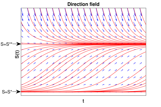
The equilibrium points of inequation (5) are and In order to show boundedness of inequation (5), the following cases are considered:
-
•
Case 1: Let be the initial condition satisfying the relation which implies . Upon simplification, we see that
(6) Combining, inequation (5) and (6), one can observe that
(7) -
–
Subcase-1: If , then the solution converges to .
-
–
Subcase-2: If , then the solution converges to .
-
–
-
•
Case-2: When , then
which indicates that . Therefore, the solution decreases and converges to the equilibrium point .
Hence, in all cases is bounded which implies that both are bounded. The associated direction field of the inequation (5) is given by Figure 2. Subsequently, when , by employing the same approach outlined in the article of Sutradhar and Dalal [35], one can easily establish the boundedness of both and .
Based on the proliferation rate of healthy and unhealthy hepatocytes, two different scenarios are analyzed here. In the first case, it is assumed that the both type of liver cells proliferate with the same rate i.e. . In the second, we consider that as compared to healthy hepatocytes, infected hepatocytes proliferate at a lower rate. In both cases, we consider non-negative initial conditions. Since in both cases, the covalently closed circular DNA (cccDNA) pool contained in the infected hepatocytes is disappeared after mitosis, this type of model is sometime refereed to as the “cccDNA loss model” [16].
4 Parameter estimation and initial condition
The parameter values are taken from the existing literature and shown in Table (1). The quantity denotes the fraction of the total liver cells that are hepatocytes and primary target of the viruses. Based on the prior studies [36, 37], it is considered that due to the fact that 60% of liver cells are hepatocytes. The value of proliferation rate of infected hepatocytes is constrained to be between 0.001 and 0.35/day [31]. The value of proliferation rate of uninfected hepatocytes lies between 0.001 and 3.4/day [27].
| Parameters | Descriptions | Values | Units | Sources |
| Proliferation rate of uninfected hepatocytes | [31] | |||
| Proliferation rate of infected hepatocytes | [27, 38] | |||
| Infection rate | [31] | |||
| Total number of liver cells | [24, 39] | |||
| Fraction of liver cells that cannot be infected | 0.4 | unitless | [31] | |
| Natural death rate of uninfected hepatocytes | [40] | |||
| Production rate of capsid | 150 | [6] | ||
| Production rate of virus from capsid | 0.87 | [6] | ||
| Death rate of infected hepatocytes and capsids | 0.053 | [6] | ||
| hepatocyte & capsid | ||||
| Death rate of virus | 3.8 | [6] | ||
| Volume fraction of HBV capsids | unitless | – | ||
| Capsid to capsid production rate or | 0.6931 | [41] | ||
| recycling rate |
5 Model calibration
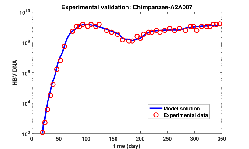
Once the proposed model is validated with experimental data, it becomes a powerful tool for analyzing the infection dynamics under various conditions. In order to validate our proposed model, the experimental data of HBV DNA from a male chimpanzee are utilized. This chimpanzee was labeled as A2A007 and was inoculated with GE of HBV. The age and the body weight of this chimpanzee were 5.1 years and 17.7 kg, respectively. These data have been previously published in the work of Asabe et al. [19]. In the study carried out by this group, the experiment was done on a cohort of nine healthy adult chimpanzees. Ethical guidelines governing the use and care of the chimpanzees were meticulously followed during the study.
Analyzing the experimental data of Chimpanzee A2A007, significant variations in HBV DNA levels were noted precisely at time intervals and days. To delineate and characterize these three sudden changes, we need to divide the time course into three distinct sub-intervals, namely , , and . The model parameters are estimated in each sub-interval minimizing the sum square error (SSE) objective function. The SSE function is defined as
| (8) |
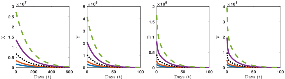
where model solution and experimental data are denoted by and , respectively. This optimization process is carried out through the utilization of the ’fminsearchbnd’ built-in MATLAB function. The upper and lower bound of the parameters are collected from the existing literature. The estimated values of each parameter in each time interval are shown in Table 2. The solution of our model and the experimental data are compared in Figure 3, and it is observed that the model solution exhibits a robust agreement with the experimental data.
| Estimated Parameters | Time interval | ||
|---|---|---|---|
| 20-82 | 82-185 | 185-346 | |
| 0.100558 | 0.150707 | 3.374189 | |
| 0.010721 | 0.062152 | 0.540862 | |
| 7.96E-11 | 5.83E-11 | 3.2E-10 | |
| 108.2001 | 40.97998 | 10.00146 | |
| 2.758616 | 2.107031 | 1.845693 | |
| 1.766558 | 0.887594 | 1.283164 | |
| 0.764174 | 0.702477 | 0.640821 | |
| 0.027786 | 0.198765 | 0.120172 | |
| 0.06998 | 0.050187 | 0.049513 | |
| 0.147755 | 0.347936 | 0.241115 | |
6 Optimal criteria for maintaining smoothly the essential functions of liver
In literature, many authors have proposed different types of HBV infection dynamics model without considering the lower bound of uninfected hepatocytes and upper bound of infected hepatocytes for survival of the patients. During liver transplantation, resections that leave 20–30% of the original liver volume are considered safe and allow the donor liver to regenerate [42, 43]. According to the experimental study of Kishi et al. [44] on more than 300 right lobe hepatectomy patients, it is come to know that the liver remnant volumes greater than 20% are sufficient to resect the liver safely. So, it is very important to maintain a certain level of uninfected hepatocytes throughout the infection period. As per the discussion, it is assumed that a minimum 20% of the total hepatocytes (excluding the population of non-parenchymal cells) or 52% with respect to entire liver size is required to maintain the critical liver functions during the infection. Any number less than 20% is lethal for the sufferer. Thus, for further study we pay close attention to this optimum value of uninfected hepatocytes.
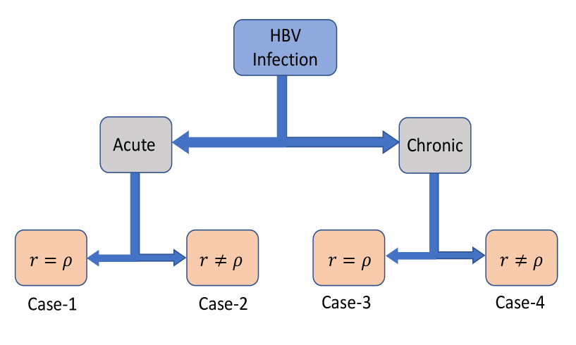
7 How does the proliferation rate influence the infection?
Both healthy and unhealthy hepatocytes have the ability to undergo proliferation in order to compensate the deficiency of the liver cells. In this study, we mainly focus to explore the roles of proliferation of liver cells during HBV infection. The understanding of how proliferation affects chronic infections is relatively scarce compared to the extensive knowledge available for acute infections. However, this experiment seeks to investigate the influence of proliferation on both acute as well as chronic infection. In order to achieve this purpose, we examine the impacts of proliferation from two distinct perspectives. In the first case, it is assumed that both types of hepatocytes proliferate with same rate (), and secondly that the rate differs (). In this context, four distinct cases are arrived as follows:
-
Case-1:
Same proliferation rates of uninfected and infected hepatocytes in acute infection.
-
Case-2:
Different proliferation rates of uninfected and infected hepatocytes in acute infection.
-
Case-3:
Same proliferation rates of uninfected and infected hepatocytes in chronic infection.
-
Case-4:
Different proliferation rates of uninfected and infected hepatocytes in chronic infection.
7.1 Effects of same proliferation rates on acute and chronic infection
In this case, it is considered that both types of hepatocytes proliferate with same rate . Accordingly, equations (1) and (2) reduce to the following forms:
| (9) |
7.1.1 Steady states
The steady states of the system (equations (3),(4) and (9)) are obtained by solving the system of equations given by
| (10) | |||
| (11) | |||
| (12) | |||
| (13) |
- (i)
- (ii)
- (iii)
-
(iv)
Endemic steady-state: The system has two endemic steady-states. The individual expressions of these two equilibria are not given here due to the complexity.
The basic reproduction number of the model is calculated using next-generation approach [45] and given by
7.1.2 Stability of equilibrium points
-
•
Liver failure equilibrium point
Acute liver failure (ALF) also known as fulminant hepatic failure, is the loss of liver function that happens quickly, usually in a matter of days or weeks, and in people who do not already have liver disease. Although ALF is quite uncommon. Most frequently, the main cause of ALF are the use of drugs or the hepatitis virus infection. In literature, it is seen that chronic liver failure (CLF), which develops itself more slowly, is more common than acute liver failure. In order to study the stability of this steady-state, we first calculate the Jacobian matrix at , and it is given by
| (14) |
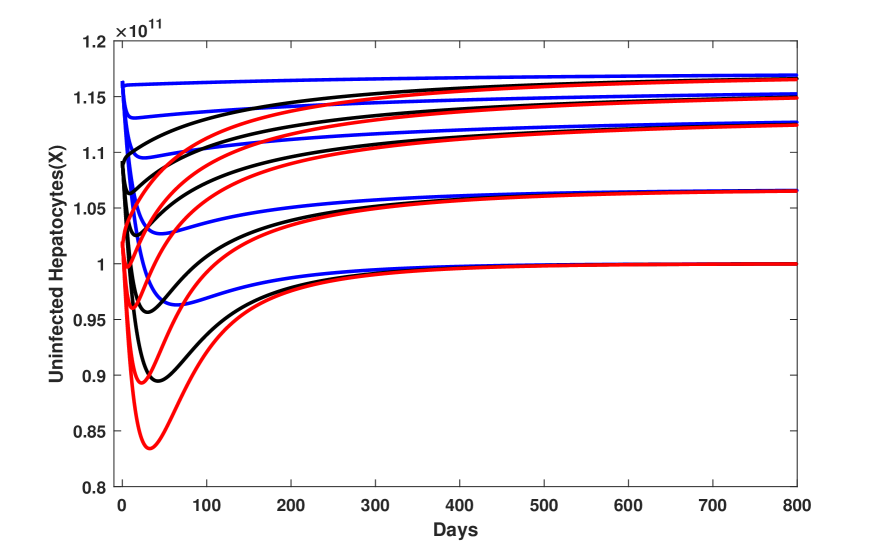
The corresponding eigenvalues are . All eigenvalues except are negative. The eigenvalue will be negative if . Hence, liver failure equilibrium will be locally asymptotically stable if . In Figure 5, the trajectories of both uninfected and infected hepatocytes, capsids and viruses are presented considering five different initial conditions. In this case, it is seen that when the parameters and follow the inequality , the solution of the system converges to the liver failure equilibrium point.
Remark 1.
In biological terminology, the inequality provides the following crucial information: if the proliferation rate and the death rate of uninfected hepatocytes meet this inequality, the health condition of the patient will deteriorate day by day, and the liver will subsequently become damage permanently.
Remark 2.
In general, for all humans, the death or degradation rate of uninfected hepatocytes is almost constant. Humans have no control over it. Proliferation rates differ between individuals. In this circumstance, the only option to prevent liver failure is to keep the proliferation rate above a certain threshold value.
-
•
Disease-free steady-state
The Jacobian matrix at is given by
| (15) |
Clearly, is an eigenvalue of the matrix (15), and it will be negative if . Other three eigenvalues are the eigenvalues of the matrix
| (16) |
Let the characteristic equation of this matrix (16) be
where
So, using Routh Hurwitz criteria [45], we have the disease-free equilibrium is locally stable when the following conditions are satisfied:
-
i.
-
ii.
-
iii.
.
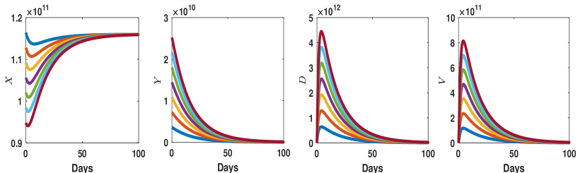
In addition, the system is numerically solved under eight different initial conditions, and in each instance, the system stabilizes to the disease-free steady-state as shown in Figure 6.
Remark 3.
The stability criteria ( for liver failure, for disease-free steady-states) indicate that the proliferation of hepatocytes has a significant influence on the dynamics of infection.
The stability of the other two equilibrium points can not be investigated analytically due to the high complexity of the model.
7.1.3 Effects of proliferation rate
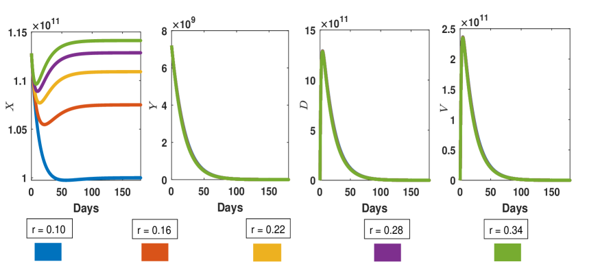
The proliferation of infected hepatocytes can vary between acute and chronic infections. One of the prime objective of this study is to determine how hepatocyte proliferation contributes to HBV infection. To study the effects of proliferation rate, we set five different values of proliferation rate within realistic range, as mentioned in Table 1. In addition, the possible relationships between initial situation of the patients and proliferation are also explored for both acute and chronic infection.
Acute infection
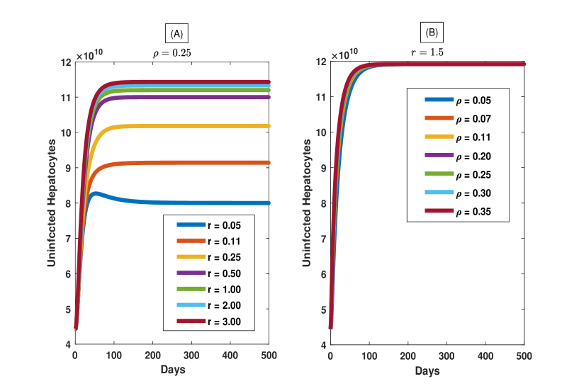
Figure 7 illustrates how the infection is influenced by the simultaneous proliferation of infected and uninfected hepatocytes. In this case, the parameter values are chosen in such a way that the value of remains below unity. When proliferation rate equal to 0.1 (signifying negligible proliferation), maximum liver cell becomes infected within few months. For this value of proliferation rate, the number of uninfected cells falls below the minimum required level for the liver to function smoothly, as mentioned in Section 6. The number of uninfected hepatocytes increases at a fixed time as the proliferation rate rises. Therefore, examining the profile of uninfected hepatocyte, it can be concluded that the proliferation reduces infection in acute case.
Chronic infection
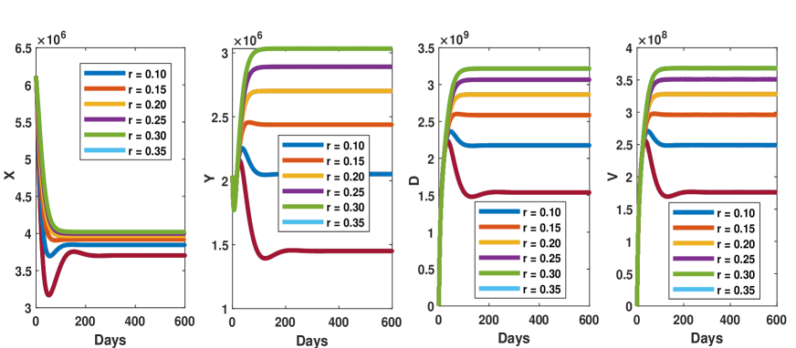
In the case of chronic infection, we have examined the impacts of the proliferation rate and explored the relationship between proliferation and the initial phage of the sufferers. The proposed model is solved for different values of proliferation rate and the solutions are plotted in Figure 8. As observed in acute infection, the growth of proliferation rate leads to negligible change in the concentration of infected hepatocytes, capsids and virus compartments but in this case the change is substantial and considerable. Infected hepatocytes exhibit a more pronounced change in their profile compared to uninfected counterparts. Since the stability levels of infected hepatocytes, capsids, and viruses rise significantly with the increase of proliferation as depicted in the Figure 8, it is conspicuous that proliferation serves as a positive factor in the persistence of the infection.
In order to elucidate the association between initial condition of the patients and the proliferation, five different initial conditions are considered, and the simulation results are presented in Figure 9. For all initial states, proliferation seems to play equal roles. The long-term behavior of the infection is solely governed by mitosis. We don’t observe any notable role of the initial condition of the patients in the chronicity of the infection. The conclusions, we draw are grounded on the results of a finite number of simulations, taking into account the smooth functional criteria of the liver outlined in the Section 6. To better understand the roles of patients’ initial conditions globally, further research is imperative.
7.2 Effects of different proliferation rates on the infection
In this section, it is considered that the rate at which infected hepatocytes proliferate is lower than the rate of proliferation observed in healthy hepatocytes, i.e., [24]. As a result, the reaction equations corresponding to uninfected and infected hepatocytes transform into the following equations:
| (17) |
In Section 3, we have already discussed that this system of equations (equations (17), (3) and (4)) possesses a distinct, bounded, and non-negative solution initiating from a non-negative initial condition. Our main cynosure of this section lies on understanding the infection dynamics influenced by the different proliferation rates of uninfected and infected cells. We rigorously explore how different proliferation rates affect the dynamics of infection in the present of standard incidence function and capsid recycling. In the following section, a thorough study is carried out on the proliferation of both types of hepatocytes.
7.2.1 Acute infection
In this case, we have conducted two experiments:
-
•
Experiment-1: Proliferation rate of uninfected hepatocytes varies within a reasonable range and proliferation rate of infected hepatocytes is kept fixed at (average value, according to Table 1).
-
•
Experiment-2: Proliferation rate of infected hepatocytes varies within a reasonable range and proliferation rate of uninfected hepatocytes is kept fixed at (average value, according to Table 1).
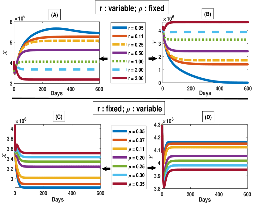
Figures 10(A) and 10(B) display the simulation results for Experiment-1 and Experiment-2, respectively. Since the severity of the infection is generally determined by the number of uninfected hepatocytes, we only focus on the profile of uninfected cells. In the Experiment-1, it is observed that when uninfected hepatocytes undergo rapid proliferation, there is a significance increase in the concentration of healthy hepatocytes. Consequently, the disease gradually diminishes over time.
On the other hand, in case of Experiment-2, the trend is clear. If only infected hepatocytes are allowed to proliferate, the number of uninfected hepatocytes convergences swiftly to the infection-free steady-state, resulting in a speedy recovery from the infection.
Remark 4.
The changes in the profile of healthy cells due to variations in are negligible compared to the changes observed in the case of variations in . However, the rapid proliferation of both cells reduce the severity of the infection.
7.2.2 Chronic infection
The understanding of liver cell proliferation during chronic infection is still incomplete and inadequate. It also remains unclear whether the roles of proliferation in chronic infections resemble those observed in acute infections or not. In order to explore the biologically plausible dynamics in chronic infection, two more experiments are carried out similarly to the case discussed above.
-
•
Experiment-3 ( : free variable, : fixed) : The influence of proliferation of uninfected hepatocytes are discussed in the context of long-term infection. In Figures 11(A) and 11(B), the dynamic temporal behavior of uninfected and infected hepatocytes are demonstrated. Compare to the acute infection, chronic infections manifest an opposite trend on both compartments. In this circumstance, the infection becomes more severe due to rapid proliferation. Based on the Figures 11(A) and 11(B) and rigorous analysis of the proposed model, the probable reason behind this opposite trend in the profiles of liver cells can be clarified by considering the fact that the rapid proliferation of uninfected hepatocytes results quick increase in the population of healthy hepatocytes. Consequently, this leads to the frequent interaction between viruses and healthy cells which intensifies infection.
-
•
Experiment-4: (: fixed, : free variable) In this case, we study how the proliferation of infected hepatocytes shapes various outcomes. The correlation between infection and the proliferation of infected hepatocytes during the infection is highlighted based on numerical results. The variations in the outcomes of uninfected and infected liver cells are visualized in Figures 11(C) and 11(D). The increase in the rate of infected cells proliferation leads to an increase in the number of uninfected cells and a decrease in the number of infected cells. Therefore, the persistence of chronic infection is closely associated with the cellular proliferation of infected cells resulting in two uninfected cells.
For our easily understanding, all the results obtained up-to this section are put together in Table 3. With the increase of proliferation rates, if the number of hepatocytes increases, we enter “Increase” in the corresponding cell of the Table 3. In case of decrease in hepatocytes, we put “Decrease”.
| (increase) | ||||
|---|---|---|---|---|
| increase, fixed | increase, fixed | |||
| Acute | Uninfected hepatocytes | Increase | Increase | Increase |
| Infected hepatocytes | Decrease | Increase | Decrease | |
| Chronic | Uninfected hepatocytes | Increase | Decrease | Increase |
| Infected hepatocytes | Increase | Increase | Decrease | |
8 Modified model: Proliferation is not a continuous biological process
In the proposed model (equations (1), (2), (3) and (4)), it was assumed that proliferation always occurs i.e. proliferation is a continuous biological process. In the literature it is seen that during liver regeneration after partial resection or injury, the remaining hepatocytes rapidly multiply to restore liver function. In response to liver damage or diseases like hepatitis or cirrhosis, compensatory hepatocyte proliferation occurs to replace damaged cells. According to a study of Miyaoka and Miyajima [46], cellular proliferation is stimulated solely when the total number liver cell population decreases to less than 70% of its initial volume. In order to include this phenomenon in the proposed model, the standard proliferation terms and are replaced by
and
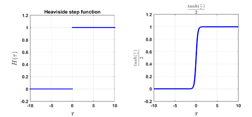
respectively, where denotes Heaviside step function defined by
The associated modified governing equations for uninfected and infected classes are given by the following equations:
| (18) |
Since, is a discontinuous function at , its applicability in continuous systems is restricted as it is unable to capture accurately an uninterrupted process. So, we approximate by a smooth function as
where is very small in magnitude. The function provides a smooth, monotonic description of proliferation. The graphical representation of these two functions are shown in Figure 12. The capsids and virus classes remain unchanged; no modifications are made in this context.
8.1 Regular proliferation vs irregular proliferation
For the convenience of our discussion, the following things are defined as:
-
•
Regular or standard proliferation: Both the terms and are defined as regular or standard proliferation due to the genesis of these terms.
-
•
Irregular proliferation: The expressions and used in the equation (18) are termed as irregular proliferation because it occurs only when the number of liver cell falls below 70% of the total number of liver cells.
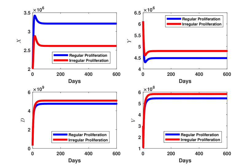
We also define the system of equations ((1), (2), (3) and (4)) as model-p (primary model) and system of equations ((18), (3) and (4)) as model-m (modified model). Both models (model-p and model-s) are solved numerically due to the presence of high non-linearity, and the corresponding solutions are demonstrated in Figure 13. As a result, it is observed that despite the consideration of same parameters values and a same fixed initial condition, there are substantial variations in the solutions. It is also noticed that in case of irregular proliferation, the infection becomes more severe. Although much information are unexplored in the context of irregular proliferation, these findings hold the promise of enriching our prior knowledge. However, a more comprehensive and detailed study is imperative in this area.
9 Conclusions
In this paper, the effects of proliferation of uninfected and infected hepatocytes on hepatitis B viral infection are studied by considering all possible cases. The recycling of capsids is also incorporated in this model. Capsids recycling is one of the distinctive features in the proposed model. We prove that the solution of the system starting from non-negative initial condition remains non-negative and bounded. The stability of the equilibrium points are established based on the value of basic reproduction number and proliferation rate. This study also addresses the consequences of both regular and irregular proliferation for the first time.
The results of our study reveal the following key findings.
-
1.
When both uninfected and infected hepatocytes proliferate with equal rate in acute infection, the number of uninfected hepatocytes increases as the proliferation rate rises. At the same time, the number of infected hepatocytes decreases to zero. This suggests that during acute infection, proliferation aids the individual in a rapid recovery from the infection.
In case of chronic infection, the severity of the infection increase if the proliferation occur frequently.
-
2.
When uninfected and infected hepatocytes proliferate with different rates , proliferation of uninfected hepatocytes cure the disease quickly where we do not find any significant contribution of proliferation of infected hepatocytes in acute case.
On the other hand, the experiments on chronic infection indicate that the infection is highly influenced by proliferation of both types of cells. The proliferation of uninfected increase the infection whereas proliferation of infected hepatocytes decrease the infection.
-
3.
The difference between the solutions of regular and irregular proliferation is substantial and noteworthy. In order to determine which solution better approximates reality, additional research and investigation are needed.
The analysis of our study represents some testable biological findings. This research provides greater insights and holds more significance. It is expected that this model will be used in a variety of purposes such as, in the development of any novel therapeutics strategies, future studies, clinical trials.
Availability of data and materials
Data sharing is not applicable to this article.
Competing interests
The authors declare that they have no competing interests.
Authors’ contributions
Both authors contribute equally.
Acknowledgments
First author would like to acknowledge the financial support obtained from CSIR (New Delhi) under the CSIR-SRF Fellowship scheme (File No: 09/731(0171)/2019-EMR-I). The first author also thanks the research facilities received from the Department of Mathematics, Indian Institute of Technology Guwahati, India.
References
- [1] Hepatitis b. https://www.hepb.org/what-is-hepatitis-b/what-is-hepb/. 2023.
- [2] Fabien Zoulim. Hepatitis b virus resistance to antiviral drugs: where are we going? Liver International, 31:111–116, 2011.
- [3] Martin A Nowak, Sebastian Bonhoeffer, Andrew M Hill, Richard Boehme, Howard C Thomas, and Hugh McDade. Viral dynamics in hepatitis b virus infection. Proceedings of the National Academy of Sciences, 93(9):4398–4402, 1996.
- [4] Sharon Lewin, Tomos Walters, and Stephen Locarnini. Hepatitis b treatment: rational combination chemotherapy based on viral kinetic and animal model studies. Antiviral Research, 55(3):381–396, 2002.
- [5] Dominik Wodarz. Hepatitis c virus dynamics and pathology: the role of ctl and antibody responses. Journal of General Virology, 84(7):1743–1750, 2003.
- [6] John M Murray, Robert H Purcell, and Stefan F Wieland. The half-life of hepatitis b virions. Hepatology, 44(5):1117–1121, 2006.
- [7] Luca G Guidotti and Francis V Chisari. Immunobiology and pathogenesis of viral hepatitis. Annu. Rev. Pathol. Mech. Dis., 1:23–61, 2006.
- [8] Stanca M Ciupe, Ruy M Ribeiro, Patrick W Nelson, and Alan S Perelson. Modeling the mechanisms of acute hepatitis b virus infection. Journal of Theoretical Biology, 247(1):23–35, 2007.
- [9] Lequan Min, Yongmei Su, and Yang Kuang. Mathematical analysis of a basic virus infection model with application to hbv infection. The Rocky Mountain Journal of Mathematics, 38(5):1573–1585, 2008.
- [10] Steffen Eikenberry, Sarah Hews, John D Nagy, and Yang Kuang. The dynamics of a delay model of hbv infection with logistic hepatocyte growth. Mathematical Biosciences and Engineering, 6:1–17, 2009.
- [11] Sarah Hews, Steffen Eikenberry, John D Nagy, and Yang Kuang. Rich dynamics of a hepatitis b viral infection model with logistic hepatocyte growth. Journal of Mathematical Biology, 60(4):573–590, 2010.
- [12] Jun Nakabayashi and Akira Sasaki. A mathematical model of the intracellular replication and within host evolution of hepatitis type b virus: Understanding the long time course of chronic hepatitis. Journal of Theoretical Biology, 269(1):318–329, 2011.
- [13] Xiao Chen, Lequan Min, Yu Zheng, Yang Kuang, and Yongan Ye. Dynamics of acute hepatitis b virus infection in chimpanzees. Mathematics and Computers in Simulation, 96:157–170, 2014.
- [14] F Fatehi Chenar, YN Kyrychko, and KB Blyuss. Mathematical model of immune response to hepatitis b. Journal of Theoretical Biology, 447:98–110, 2018.
- [15] Sanhong Liu and Ran Zhang. On an age-structured hepatitis b virus infection model with hbv dna-containing capsids. Bulletin of the Malaysian Mathematical Sciences Society, 44(3):1345–1370, 2021.
- [16] Thomas Tu, Benno Zehnder, Jochen M Wettengel, Henrik Zhang, Sally Coulter, Vikki Ho, Mark W Douglas, Ulrike Protzer, Jacob George, and Stephan Urban. Mitosis of hepatitis b virus-infected cells in vitro results in uninfected daughter cells. JHEP Reports, 4(9):100514, 2022.
- [17] Georgia-Myrto Prifti, Dimitrios Moianos, Erofili Giannakopoulou, Vasiliki Pardali, John E Tavis, and Grigoris Zoidis. Recent advances in hepatitis b treatment. Pharmaceuticals, 14(5):417, 2021.
- [18] Chunkyu Ko, Anindita Chakraborty, Wen-Min Chou, Julia Hasreiter, Jochen M Wettengel, Daniela Stadler, Romina Bester, Theresa Asen, Ke Zhang, Karin Wisskirchen, et al. Hepatitis b virus genome recycling and de novo secondary infection events maintain stable cccdna levels. Journal of hepatology, 69(6):1231–1241, 2018.
- [19] Shinichi Asabe, Stefan F Wieland, Pratip K Chattopadhyay, Mario Roederer, Ronald E Engle, Robert H Purcell, and Francis V Chisari. The size of the viral inoculum contributes to the outcome of hepatitis b virus infection. Journal of virology, 83(19):9652–9662, 2009.
- [20] Kosaku Kitagawa, Kwang Su Kim, Masashi Iwamoto, Sanae Hayashi, Hyeongki Park, Takara Nishiyama, Naotoshi Nakamura, Yasuhisa Fujita, Shinji Nakaoka, Kazuyuki Aihara, Alan Perelson, Lena Allweiss, Maura Dandri, Koichi Watashi, Yasuhito Tanaka, and Shingo Iwami. Multiscale modeling of hbv infection integrating intra- and intercellular viral propagation for analyzing extracellular viral markers, 2023.
- [21] Rupchand Sutradhar and D. C. Dalal. Intracellular dynamics of hepatitis b virus infection: A mathematical model and global sensitivity analysis of its parameters. arXiv preprint arXiv:2307.02835, 2023.
- [22] Rupchand Sutradhar and D. C. Dalal. Fractional-order models of hepatitis b virus infection with recycling effects of capsids. Mathematical Methods in the Applied Sciences, 46(14):15599–15625.
- [23] Preeti Damania, Bijoya Sen, Sadaf Bashir Dar, Satendra Kumar, Anupama Kumari, Ekta Gupta, Shiv Kumar Sarin, and Senthil Kumar Venugopal. Hepatitis b virus induces cell proliferation via hbx-induced microrna-21 in hepatocellular carcinoma by targeting programmed cell death protein4 (pdcd4) and phosphatase and tensin homologue (pten). PloS one, 9(3):e91745, 2014.
- [24] Sarah Hews, Steffen Eikenberry, John D Nagy, Tin Phan, and Yang Kuang. Global dynamics and implications of an hbv model with proliferating infected hepatocytes. Applied Sciences, 11(17):8176, 2021.
- [25] Harel Dahari, Emi Shudo, Ruy M Ribeiro, and Alan S Perelson. Modeling complex decay profiles of hepatitis b virus during antiviral therapy. Hepatology, 49(1):32–38, 2009.
- [26] Andrea Carracedo Rodriguez, Matthias Chung, and Stanca M Ciupe. Understanding the complex patterns observed during hepatitis b virus therapy. Viruses, 9(5):117, 2017.
- [27] Timothy C Reluga, Harel Dahari, and Alan S Perelson. Analysis of hepatitis c virus infection models with hepatocyte homeostasis. SIAM journal on applied mathematics, 69(4):999–1023, 2009.
- [28] Christoph Seeger and William S Mason. Hbv replication, pathobiology and therapy: Unanswered questions, 2016.
- [29] Lena Allweiss, Tassilo Volz, Katja Giersch, Janine Kah, Giuseppina Raffa, Joerg Petersen, Ansgar W Lohse, Concetta Beninati, Teresa Pollicino, Stephan Urban, et al. Proliferation of primary human hepatocytes and prevention of hepatitis b virus reinfection efficiently deplete nuclear cccdna in vivo. Gut, 67(3):542–552, 2018.
- [30] Marc Lutgehetmann, Tassilo Volz, Anne Köpke, Tim Broja, Eike Tigges, Ansgar W Lohse, Eberhard Fuchs, John M Murray, Joerg Petersen, and Maura Dandri. In vivo proliferation of hepadnavirus-infected hepatocytes induces loss of covalently closed circular dna in mice. Hepatology, 52(1):16–24, 2010.
- [31] Ashish Goyal, Ruy M Ribeiro, and Alan S Perelson. The role of infected cell proliferation in the clearance of acute hbv infection in humans. Viruses, 9(11):350, 2017.
- [32] John M Murray and Ashish Goyal. In silico single cell dynamics of hepatitis b virus infection and clearance. Journal of Theoretical Biology, 366:91–102, 2015.
- [33] Stanca M Ciupe, Ruy M Ribeiro, Patrick W Nelson, Geoffrey Dusheiko, and Alan S Perelson. The role of cells refractory to productive infection in acute hepatitis b viral dynamics. Proceedings of the National Academy of Sciences, 104(12):5050–5055, 2007.
- [34] Zbigniew Kmiec. Cooperation of liver cells in health and disease: with 18 tables. 2001.
- [35] Rupchand Sutradhar and D. C. Dalal. Re-cycling of dna-containing capsids enhances hepatitis b.
- [36] Erwin Kuntz and Hans-Dieter Kuntz. Systemic diseases and the liver. Hepatology Textbook and Atlas: History· Morphology Biochemistry· Diagnostics Clinic· Therapy, pages 837–850, 2008.
- [37] S Sherlock and J Dooley. Chapter 3: Biopsy of the liver. Sherlock S, Dooley J. Diseases of the liver and biliary system. 11th ed. Oxford, UK: Blackwell Science Ltd, pages 37–44, 2002.
- [38] Harel Dahari, Jennifer E Layden-Almer, Eric Kallwitz, Ruy M Ribeiro, Scott J Cotler, Thomas J Layden, and Alan S Perelson. A mathematical model of hepatitis c virus dynamics in patients with high baseline viral loads or advanced liver disease. Gastroenterology, 136(4):1402–1409, 2009.
- [39] Ogechi Ogoke, Janet Oluwole, and Natesh Parashurama. Bioengineering considerations in liver regenerative medicine. Journal of biological engineering, 11:1–16, 2017.
- [40] Harel Dahari, Arthur Lo, Ruy M Ribeiro, and Alan S Perelson. Modeling hepatitis c virus dynamics: Liver regeneration and critical drug efficacy. Journal of Theoretical Biology, 247(2):371–381, 2007.
- [41] John M Murray and Ashish Goyal. In silico single cell dynamics of hepatitis b virus infection and clearance. Journal of Theoretical Biology, 366:91–102, 2015.
- [42] Salvatore Gruttadauria, Vishal Parikh, Duilio Pagano, Fabio Tuzzolino, Davide Cintorino, Roberto Miraglia, Marco Spada, Giovanbattista Vizzini, Angelo Luca, and Bruno Gridelli. Early regeneration of the remnant liver volume after right hepatectomy for living donation: a multiple regression analysis. Liver Transplantation, 18(8):907–913, 2012.
- [43] Alfredo Guglielmi, Andrea Ruzzenente, Simone Conci, Alessandro Valdegamberi, and Calogero Iacono. How much remnant is enough in liver resection? Digestive surgery, 29(1):6–17, 2012.
- [44] Yoji Kishi, Eddie K Abdalla, Yun Shin Chun, Daria Zorzi, David C Madoff, Michael J Wallace, Steven A Curley, and Jean-Nicolas Vauthey. Three hundred and one consecutive extended right hepatectomies: evaluation of outcome based on systematic liver volumetry. Annals of surgery, 250(4):540–548, 2009.
- [45] Maia Martcheva. An Introduction to Mathematical Epidemiology, volume 61. Springer, 2015.
- [46] Yuichiro Miyaoka and Atsushi Miyajima. To divide or not to divide: revisiting liver regeneration. Cell division, 8:1–12, 2013.