RudolfV: A Foundation Model
by Pathologists for Pathologists
2 Machine Learning Group, Technische Universität Berlin, Germany
3 BIFOLD – Berlin Institute for the Foundations of Learning and Data, Germany
4 Department of Artificial Intelligence, Korea University, Republic of Korea
5 Max-Planck Institute for Informatics, Germany
6 German Cancer Research Center (DKFZ) & German Cancer Consortium (DKTK), Berlin & Munich Partner Sites
7 Institute of Pathology, Ludwig-Maximilians-Universität München, Germany
8 Institute of Pathology, Charité – Universitätsmedizin Berlin, Germany
∗† Equal Contribution, ‡ Work done during internship at Aignostics )
Abstract
Histopathology plays a central role in clinical medicine and biomedical research. While artificial intelligence shows promising results on many pathological tasks, generalization and dealing with rare diseases, where training data is scarce, remains a challenge. Distilling knowledge from unlabeled data into a foundation model before learning from, potentially limited, labeled data provides a viable path to address these challenges. In this work, we extend the state of the art of foundation models for digital pathology whole slide images by semi-automated data curation and incorporating pathologist domain knowledge.
Specifically, we combine computational and pathologist domain knowledge (1) to curate a diverse dataset of 103k slides corresponding to 750 million image patches covering data from different fixation, staining, and scanning protocols as well as data from different indications and labs across the EU and US, (2) for grouping semantically similar slides and tissue patches, and (3) to augment the input images during training. We evaluate the resulting model on a set of public and internal benchmarks and show that although our foundation model is trained with an order of magnitude less slides, it performs on par or better than competing models. We expect that scaling our approach to more data and larger models will further increase its performance and capacity to deal with increasingly complex real world tasks in diagnostics and biomedical research.
1 Introduction
Pathology plays a central role in clinical medicine for tissue-based diagnostics and in biomedical research as a basis for understanding cellular and molecular mechanisms of disease. While molecular and omics based data complement histological assessments, the microscopic evaluation of morphological changes has persisted as a key component of pathology. Consequently most digital pathology work focuses on whole slide image analysis.
Despite artificial intelligence (AI) having led to promising proof-of-concepts and applications (e.g., [1, 2, 3, 4, 5]), generalization and robustness are still a challenge when translating applications into clinical setups. Moreover, limited numbers of samples available for training pose a particular challenge in medicine, because the distribution of disease frequencies is highly skewed with only relatively few frequent and a large number of rare diseases. Therefore, generating enough labeled data to sufficiently cover observed variations regarding morphology, staining, scanner type, lab, and biology is (prohibitively) expensive.

Thus, while learning from labeled data will remain the standard for most AI use cases, strategies will be required to address the above outlined challenges. Here, foundation models [6] may contribute to a solution. This technique refers to pre-training a so-called foundation model on unlabeled data and to subsequently adapting it to learn from, potentially limited, labeled data. It typically uses the self-supervised learning (SSL, e.g., [7, 8, 9, 10]) paradigm and has shown breakthrough results in other domains when employed with an abundance of unlabelled data coupled with very large models (e.g., [11, 12, 13, 14, 8]).
Previous work successfully transferred foundation models to digital pathology images (e.g., [15, 16, 17, 18]), yet it falls short on incorporating crucial domain knowledge. In this work, we present an approach that integrates computational analysis and pathologist expertise into the data curation and training process. To the best of our knowledge, this is the first work to extensively incorporate pathology knowledge into foundation model development and to incorporate immunohistochemistry (IHC) and other non hematoxylin-and-eosin (H&E) stainings - which are important for many biomarker scoring use cases - into an SSL dataset. The presented approach reaches the best reported performance with an order of magnitude less slides and a smaller model than the current state of the art.
Our work is based on the assumption that optimally curated and more diverse data in connection with domain knowledge can improve SSL training. We therefore integrate pathologist knowledge in a number of key aspects. Specifically, we use pathologist input (1) to curate a dataset of 103k slides that covers diverse histological samples from different fixation, staining, and scanning protocols as well as data from different indications and labs from the EU and US, (2) for grouping semantically similar slides and tissue patches as well as (3) for augmenting the input images during training. Additional technical contributions are outlined in the methods section. We evaluate the resulting model on a set of public and internal benchmarks and compare it to latest related work.
2 Related Work
In multiple works, self-supervised learning was adapted for training on histopathological images. Ciga et al. [23] trained on histopathological images with the SimCLR framework [7, 24]. The authors used multiple public data sources and showed that more diversity in pre-training data improves downstream task performance. Chen et al. trained a hierarchical model on 100 million image patches from TCGA slides (HIPT) [25] and showed that the DINO framework [26] and Vision Transformer (ViT) architecture [27] outperform other approaches [28]. The authors of CTransPath train a SwinTransformer [29] on 15 million images from TCGA and PAIP slides using a modified contrastive learning objective which mines additional semantically similar positive views (SRCL) [15]. Kang et al. [30] compare multiple self-supervised learning frameworks and find DINO to yield the best performance in a limited data setting. Remedis [31] first performs supervised training on natural images and then self-supervised pre-training on medical images. UNI [17] trains a ViT-L model on slides with the DINOv2 framework [8] and provides extensive evaluations. Campanella et al. [32] train on 3 billion image patches from over 400,000 slides and compare masked autoencoder [9] and DINO self-supervised learning methods. Lastly, Virchow [18] is currently the best performing model, trained on over 1 million slides, also using the DINOv2 framework with a ViT-H/14 encoder.
3 Data and Methods
Previous work (e.g., [33, 8, 23, 30, 34]) showed that data curation and domain knowledge are crucial for good model performance. For histopathological images, data curation can be done both on a whole slide level and on a tissue patch level. Considering that many slides contain potentially redundant properties such as regions with fatty or connective tissue or blood, we found it essential to also curate the tissue patch level data. Our approach performs filtering, grouping/clustering, and sampling for both levels with pathologists’ expert input.
3.1 Data Curation
We curate a dataset focusing on data diversity both with respect to medical and technical aspects. The data across a broad range of disease entities was generated with different fixation, staining, scanning protocols, and in different labs across the EU and US. The dataset details are as follows:
-
•
Cases and slides: The dataset consists of 103,849 slides comprising 35,784 cases. 73,774 (71%) slides have been chosen from our archive and 30,075 (29%) slides are from The Cancer Genome Atlas (TCGA).
-
•
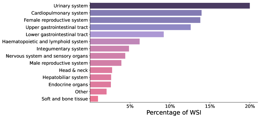
Tissue location: The tissue originates from 14 organ systems (Figure 2) comprising 58 different tissue types (examples for the granularity are lung, heart, adrenal gland, pancreas, oral cavity, bone marrow).
-
•
Labs: The slides were created in over different locations and labs in the EU and US.
-
•
Staining: To the best of our knowledge, we are the first to combine H&E and non-H&E stainings. Our dataset contains 3 broad staining categories: H&E (70%), IHC (10%), and other (20%). Subdividing each staining category further results in 97 unique staining types. Examples are H&E, ER, PR, PD-L1, HER-2, Giemsa, PAS, Gomori, and others.
-
•
Scanning: 6 different scanner types were used to scan the slides. This includes scanner types such as Roche Ventana DP600, Leica Aperio GT 450, and 3DHISTECH PANNORAMIC 1000. Scanning magnifications were typically 20x, 40x, and 80x.
-
•
Preparation: The dataset contains both formalin-fixed-paraffin-embedded (FFPE) and fresh-frozen (FF) tissue samples.
From each slide we extract patches within given tissue boundaries derived from an in-house model. The patch size is pixels at mpp. This results in million image patches in total for further processing and training.
3.2 Slide Grouping and Tissue Clustering
Histological slides typically come with metadata available for data curation. Based on expert input as well as the sample’s lab of origin, tissue type, and staining, all slides were assigned to one of 26 groups. These groups are selected to maximize homogeneity within the groups and heterogeneity across the groups. During training we sample patches uniformly from these slide groups.
In contrast, individual tissue patches typically have no tissue specific metadata available. For optimizing patch sampling during training, we therefore create clusters based on computer vision image features and pathologist expertise. First, we extract 36 standard computer vision image features for all 791 million image patches. These include image statistics such as mean and standard deviation for individual channels in common color modes such as RGB, LAB, HSV, and - tailored towards digital pathology - HED [35]. In contrast to [8], we do not use deep network features to reduce the computational burden. Next, we further reduce computational overhead by sub-sampling 500 patches for each slide and perform k-means clustering () on the respective image features. We propagate the labels of the clustering via K-NN to assign a cluster label to each of the 791 million patches. Finally, board-certified pathologists filter and aggregate the 100 image clusters assigning a description and a sampling probability (importance weight) to each cluster. This resulted in 9 distinct human-interpretable tissue clusters and 751 million image patches after filtering irrelevant clusters. During training, we sample patches according to the sampling probability of the clusters. Within the clusters, we sample equally from the 26 slide groups.
3.3 Data Augmentation
In pathology, it is known that staining and scanning outputs vary between labs and even within the same lab over a given time period. Consequently in histopathology studies, staining and scanner information can produce spurious correlations and so-called “Clever Hans” effects [36] when it is correlated with label information [1]. To address this shortcoming, we transfer and augment stain and scanner color profiles between patches in addition to the standard color augmentations in the view generation process of DINOv2. For each view, we pick a random other patch in the batch and normalize the image to the slide color statistics of the selected patch. This discourages the model from exploiting staining and scanner color features for learning representations.
3.4 DINOv2
We build upon the official DINOv2 implementation [8] with registers [38]. DINOv2 learns representations via self-distillation. As [31] has shown that self-supervised histopathology models can benefit from pre-training on natural images, we initialize the student model with the distilled DINOv2 version pre-trained on LVD-142M [8]. This has shown to improve performance in smaller experimental setups. We train a ViT-L/14 with a batch size of 960 on 16 A100-40GB GPUs for 625k iterations.
4 Evaluation
We evaluate our model on a number of public and internal benchmarks.
4.1 Tasks
The benchmarks contain 8 different patch-based downstream tasks covering different tissue types, prediction tasks, and staining protocols.
PCAM
The PatchCamelyon dataset consists of 327,680 H&E images ( pixels at 10x magnification) extracted from scans of lymph node sections of breast cancer. Each image is annotated with a binary label indicating presence of metastatic tissue. An image is labeled as metastatic tissue when one pixel was annotated as tumor [39, 40].
MHIST
The MHIST dataset consists of 3,152 fixed-size H&E images ( pixels at 8x magnification) of colorectal polyps. The task is to differentiate between hyperplastic polyp (HP) and sessile serrated adenoma (SSA). Each image is labeled according to the majority vote of seven pathologists. HPs are typically benign, while SSAs are precancerous lesions that can turn into cancer if left untreated and require sooner follow-up examinations [41].
CRC-100K
The dataset contains 107,180 H&E images ( pixels at 20x magnification) extracted from colorectal cancer scans. The task is to predict from each image the following 9 tissue classes: adipose, background, debris, lymphocytes, mucus, smooth muscle, normal colon mucosa, cancer-associated stroma, and colorectal adenocarcinoma epithelium. We use the unnormalized variant of the training dataset in our experiments (NCT-CRC-HE-100K-NONORM) [42, 43].
TIL Det.
The pan-cancer tumor-infiltrating lymphocyte (TIL) detection dataset [44, 45] consists of 304,097 (209,221 training and 56,275 test) H&E images ( pixels at 0.5mpp) extracted from scans of FFPE sections comprising 23 different cancer types from TCGA. The task is to predict TIL positive images. A TIL count higher than two indicates a positive image.
MSI CRC
MSI STAD
IHC (4 classes)
The task is to predict the cell type at the center of an image patch ( pixels at 0.5mpp). The four classes are carcinoma cell, lymphocyte, macrophage, and other. This internal benchmark dataset consists of 11,269, 3,218, and 1,609 board-certified pathologist annotated image patches taken from a larger corpora, split into train, validation, and test set respectively. The data is from 42 DAB-stained IHC slides of different cancer types, tissue locations and labs.
HE (8 classes)
The task is to predict the cell type at the center of an image patch ( pixels at 0.5mpp). The eight classes are carcinoma cell, epithelial cell, plasma cell, fibroblast, lymphocyte, granulocyte, macrophage, and other. This internal benchmark dataset consists of 84,017, 6,695, and 14,532 board-certified pathologist annotated image patches taken from a larger corpora, split into train, validation, and test set respectively. The data is from 361 NSCLC slides of different tissue locations and labs.
PCAM, MHIST, CRC-100K benchmarks are an out-of-distribution validation for our model as the data was generated by separate labs. The internal HE and IHC tasks are an out-of-sample validation for our model as we carefully separated cases from the pre-training and task datasets. For TIL Det., MSI CRC, MSI STAD there is a potential overlap between pre-training and task data, but we assume the impact to be negligible. Please note, similarly for CTransPath [15] and Phikon [16] it is unclear if pre-training and TIL Det., MSI CRC, MSI STAD task data overlap.
4.2 Results
We evaluated all public benchmarks with linear probing and internal benchmarks with linear probing and finetuning. In line with related work [18], we do not use any augmentations on public benchmarks, while on the internal benchmarks, we use color augmentations during training. During linear probing, we resize the images to 224px and fit a linear classifier with l2-regularization on the CLS token [8] representations. The l2-regularization parameter is selected based on a hold-out validation set (10% of the official training set or the official validation set, if existing). During finetuning we extend the CLS token with another linear layer and use mini-batch stochastic gradient descent with a logistics loss.
| Model | PCAM | MHIST | CRC-100K | TIL Det. | MSI CRC | MSI STAD | Avg(1-3) | Avg(1-6) |
|---|---|---|---|---|---|---|---|---|
| ResNet50 ImageNet | 0.818 | 0.783 | 0.838 | 0.915 | 0.653 | 0.664 | 0.813 | 0.779 |
| ViT-L/16 ImageNet | 0.852 | 0.792 | 0.843 | 0.924 | 0.669 | 0.671 | 0.829 | 0.792 |
| DINO ViT-S/8 [30] | 0.901 * | 0.779 * | 0.896 | 0.943 | 0.745 | 0.756 | 0.859 | 0.837 |
| CTransPath [15] | 0.858 * | 0.789 * | 0.879 * | 0.930 | 0.694 | 0.726 | 0.842 | 0.813 |
| Phikon [16] | 0.909 * | 0.793 * | 0.895 * | 0.946 | 0.733 | 0.751 | 0.866 | 0.838 |
| Virchow [18] | 0.927 * | 0.827 * | 0.948 * | - | - | - | 0.901 | - |
| Ours: RudolfV | 0.928 | 0.827 | 0.960 | 0.939 | 0.766 | 0.768 | 0.905 | 0.865 |
The results of the public benchmarks in Table 1 show that our proposed approach yields better performance on all tasks except on the TIL detection task, where Phikon [16] yields best performance. The comparison covers all pathology SSL models that are publicly available at the time of publication. Virchow [18] is not publicly available and could therefore not be included in all benchmarks 111We compare to the results in version 4 of the Virchow preprint [18]. While version 5 does not indicate a change in approach or model, it changes the evaluation procedure of all tasks and is thus not directly comparable anymore. Changes can be observed here: https://arxiv.org/abs/2309.07778..
We can observe a large performance gain compared to conventional ImageNet initialized models highlighting that in-domain pre-training can give a significant performance improvement. Staining and patch color are a confounding factor in task CRC-100K [1]. Our model outperforms all other models on this task, which can be an indication that stain augmentation helps to improve the stain invariance of foundation models.
| IHC (4 classes) | HE (8 classes) | |||
|---|---|---|---|---|
| Model | Finetuning | Linear | Finetuning | Linear |
| ResNet50 ImageNet | 0.6507 | 0.2914 | 0.6112 | 0.2143 |
| ViT-L/16 ImageNet | 0.8237 | 0.4234 | 0.7352 | 0.3305 |
| Phikon [16] | 0.8357 | 0.6373 | 0.8361 | 0.607 |
| Ours: RudolfV | 0.8745 | 0.6567 | 0.8864 | 0.7352 |
Table 2 shows evaluation results on internal benchmarks. Our proposed approach yields better results, both, for linear probing and for finetuning. For linear probing the model is up to 13 p.p. and for finetuning it is up to 5 p.p. better than the second best model.
5 Discussion and Future Work
This work shows that careful integration of pathological domain knowledge can lead to substantial performance gains with the - at the time of publication - best performing pathology foundation model, despite using an order of magnitude less slides and a model with less parameters than competing models. We assume that scaling the amount and diversity of data and the number of model parameters will further increase the performance of our approach. In follow-up work we will extend this manuscript with additional benchmarks, ablation studies, and increased data and model size.
We would like to note that while the present work is a machine learning focused study, we expect the results to be of great usefulness in future diagnostic real world settings and biomedical research. Future research will therefore explore exposing RudolfV to assisting clinical routine diagnostics in histopathology and more complex tasks including multimodal modeling.
Acknowledgement
We would like to thank the teams at Aignostics for supporting this work in many ways. Without their ground work this work would not have been possible.
The results shown here are in whole or part based upon data generated by the TCGA Research Network: https://www.cancer.gov/tcga.
References
- [1] Frederick Klauschen, Jonas Dippel, Philipp Keyl, Philipp Jurmeister, Michael Bockmayr, Andreas Mock, Oliver Buchstab, Maximilian Alber, Lukas Ruff, Grégoire Montavon, et al. Toward explainable artificial intelligence for precision pathology. Annual Review of Pathology: Mechanisms of Disease, 19:541–570, 2024.
- [2] James A Diao, Jason K Wang, Wan Fung Chui, Victoria Mountain, Sai Chowdary Gullapally, Ramprakash Srinivasan, Richard N Mitchell, Benjamin Glass, Sara Hoffman, Sudha K Rao, et al. Human-interpretable image features derived from densely mapped cancer pathology slides predict diverse molecular phenotypes. Nature communications, 12(1):1613, 2021.
- [3] Patricia Raciti, Jillian Sue, Juan A Retamero, Rodrigo Ceballos, Ran Godrich, Jeremy D Kunz, Adam Casson, Dilip Thiagarajan, Zahra Ebrahimzadeh, Julian Viret, et al. Clinical validation of artificial intelligence–augmented pathology diagnosis demonstrates significant gains in diagnostic accuracy in prostate cancer detection. Archives of Pathology & Laboratory Medicine, 147(10):1178–1185, 2023.
- [4] Alexander Binder, Michael Bockmayr, Miriam Hägele, Stephan Wienert, Daniel Heim, Katharina Hellweg, Masaru Ishii, Albrecht Stenzinger, Andreas Hocke, Carsten Denkert, et al. Morphological and molecular breast cancer profiling through explainable machine learning. Nature Machine Intelligence, 3(4):355–366, 2021.
- [5] Philipp Keyl, Michael Bockmayr, Daniel Heim, Gabriel Dernbach, Grégoire Montavon, Klaus-Robert Müller, and Frederick Klauschen. Patient-level proteomic network prediction by explainable artificial intelligence. NPJ Precision Oncology, 6(1):35, 2022.
- [6] Rishi Bommasani, Drew A Hudson, Ehsan Adeli, Russ Altman, Simran Arora, Sydney von Arx, Michael S Bernstein, Jeannette Bohg, Antoine Bosselut, Emma Brunskill, et al. On the opportunities and risks of foundation models. arXiv preprint arXiv:2108.07258, 2021.
- [7] Ting Chen, Simon Kornblith, Mohammad Norouzi, and Geoffrey Hinton. A simple framework for contrastive learning of visual representations. In International conference on machine learning, pages 1597–1607. PMLR, 2020.
- [8] Maxime Oquab, Timothée Darcet, Théo Moutakanni, Huy Vo, Marc Szafraniec, Vasil Khalidov, Pierre Fernandez, Daniel Haziza, Francisco Massa, Alaaeldin El-Nouby, et al. Dinov2: Learning robust visual features without supervision. arXiv preprint arXiv:2304.07193, 2023.
- [9] Kaiming He, Xinlei Chen, Saining Xie, Yanghao Li, Piotr Dollár, and Ross Girshick. Masked autoencoders are scalable vision learners. In Proceedings of the IEEE/CVF conference on computer vision and pattern recognition, pages 16000–16009, 2022.
- [10] Rayan Krishnan, Pranav Rajpurkar, and Eric J Topol. Self-supervised learning in medicine and healthcare. Nature Biomedical Engineering, 6(12):1346–1352, 2022.
- [11] Alec Radford, Jeffrey Wu, Rewon Child, David Luan, Dario Amodei, Ilya Sutskever, et al. Language models are unsupervised multitask learners. OpenAI blog, 1(8):9, 2019.
- [12] Tom Brown, Benjamin Mann, Nick Ryder, Melanie Subbiah, Jared D Kaplan, Prafulla Dhariwal, Arvind Neelakantan, Pranav Shyam, Girish Sastry, Amanda Askell, et al. Language models are few-shot learners. Advances in neural information processing systems, 33:1877–1901, 2020.
- [13] OpenAI. Gpt-4 technical report. arXiv preprint arXiv:2303.08774, 2023.
- [14] Tao Tu, Shekoofeh Azizi, Danny Driess, Mike Schaekermann, Mohamed Amin, Pi-Chuan Chang, Andrew Carroll, Chuck Lau, Ryutaro Tanno, Ira Ktena, et al. Towards generalist biomedical ai. arXiv preprint arXiv:2307.14334, 2023.
- [15] Xiyue Wang, Sen Yang, Jun Zhang, Minghui Wang, Jing Zhang, Wei Yang, Junzhou Huang, and Xiao Han. Transformer-based unsupervised contrastive learning for histopathological image classification. Medical image analysis, 81:102559, 2022.
- [16] Alexandre Filiot, Ridouane Ghermi, Antoine Olivier, Paul Jacob, Lucas Fidon, Alice Mac Kain, Charlie Saillard, and Jean-Baptiste Schiratti. Scaling self-supervised learning for histopathology with masked image modeling. medRxiv, pages 2023–07, 2023.
- [17] Richard J Chen, Tong Ding, Ming Y Lu, Drew FK Williamson, Guillaume Jaume, Bowen Chen, Andrew Zhang, Daniel Shao, Andrew H Song, Muhammad Shaban, et al. A general-purpose self-supervised model for computational pathology. arXiv preprint arXiv:2308.15474, 2023.
- [18] Eugene Vorontsov, Alican Bozkurt, Adam Casson, George Shaikovski, Michal Zelechowski, Siqi Liu, Philippe Mathieu, Alexander van Eck, Donghun Lee, Julian Viret, et al. Virchow: A million-slide digital pathology foundation model. arXiv preprint arXiv:2309.07778, 2023.
- [19] Rudolf Virchow. Cellular pathology as based upon physiological and pathological histology… 1863.
- [20] David M Reese. Fundamentals–rudolf virchow and modern medicine. Western journal of medicine, 169(2):105, 1998.
- [21] Myron Schultz. Rudolf virchow. Emerging infectious diseases, 14(9):1480, 2008.
- [22] Constantin Goschler. Rudolf Virchow: Mediziner-Anthropologe-Politiker. Böhlau Köln, 2021.
- [23] Ozan Ciga, Tony Xu, and Anne Louise Martel. Self supervised contrastive learning for digital histopathology. Machine Learning with Applications, 7:100198, 2022.
- [24] Ting Chen, Simon Kornblith, Kevin Swersky, Mohammad Norouzi, and Geoffrey E Hinton. Big self-supervised models are strong semi-supervised learners. Advances in neural information processing systems, 33:22243–22255, 2020.
- [25] Richard J Chen, Chengkuan Chen, Yicong Li, Tiffany Y Chen, Andrew D Trister, Rahul G Krishnan, and Faisal Mahmood. Scaling vision transformers to gigapixel images via hierarchical self-supervised learning. In Proceedings of the IEEE/CVF Conference on Computer Vision and Pattern Recognition, pages 16144–16155, 2022.
- [26] Mathilde Caron, Hugo Touvron, Ishan Misra, Hervé Jégou, Julien Mairal, Piotr Bojanowski, and Armand Joulin. Emerging properties in self-supervised vision transformers. In Proceedings of the IEEE/CVF international conference on computer vision, pages 9650–9660, 2021.
- [27] Alexey Dosovitskiy, Lucas Beyer, Alexander Kolesnikov, Dirk Weissenborn, Xiaohua Zhai, Thomas Unterthiner, Mostafa Dehghani, Matthias Minderer, Georg Heigold, Sylvain Gelly, et al. An image is worth 16x16 words: Transformers for image recognition at scale. In International Conference on Learning Representations, 2020.
- [28] Richard J Chen and Rahul G Krishnan. Self-supervised vision transformers learn visual concepts in histopathology. Learning Meaningful Representations of Life, NeurIPS 2021, 2021.
- [29] Ze Liu, Yutong Lin, Yue Cao, Han Hu, Yixuan Wei, Zheng Zhang, Stephen Lin, and Baining Guo. Swin transformer: Hierarchical vision transformer using shifted windows. In Proceedings of the IEEE/CVF international conference on computer vision, pages 10012–10022, 2021.
- [30] Mingu Kang, Heon Song, Seonwook Park, Donggeun Yoo, and Sérgio Pereira. Benchmarking self-supervised learning on diverse pathology datasets. In Proceedings of the IEEE/CVF Conference on Computer Vision and Pattern Recognition, pages 3344–3354, 2023.
- [31] Shekoofeh Azizi, Laura Culp, Jan Freyberg, Basil Mustafa, Sebastien Baur, Simon Kornblith, Ting Chen, Nenad Tomasev, Jovana Mitrović, Patricia Strachan, et al. Robust and data-efficient generalization of self-supervised machine learning for diagnostic imaging. Nature Biomedical Engineering, pages 1–24, 2023.
- [32] Gabriele Campanella, Ricky Kwan, Eugene Fluder, Jennifer Zeng, Aryeh Stock, Brandon Veremis, Alexandros D Polydorides, Cyrus Hedvat, Adam Schoenfeld, Chad Vanderbilt, et al. Computational pathology at health system scale–self-supervised foundation models from three billion images. arXiv preprint arXiv:2310.07033, 2023.
- [33] Yonglong Tian, Olivier J Henaff, and Aäron van den Oord. Divide and contrast: Self-supervised learning from uncurated data. In Proceedings of the IEEE/CVF International Conference on Computer Vision, pages 10063–10074, 2021.
- [34] OpenAI: Josh Achiam et al. GPT-4 Technical Report, 2023.
- [35] Arnout C Ruifrok, Dennis A Johnston, et al. Quantification of histochemical staining by color deconvolution. Analytical and quantitative cytology and histology, 23(4):291–299, 2001.
- [36] Sebastian Lapuschkin, Stephan Wäldchen, Alexander Binder, Grégoire Montavon, Wojciech Samek, and Klaus-Robert Müller. Unmasking clever hans predictors and assessing what machines really learn. Nature communications, 10(1):1096, 2019.
- [37] Khrystyna Faryna, Jeroen Van der Laak, and Geert Litjens. Tailoring automated data augmentation to h&e-stained histopathology. In Medical Imaging with Deep Learning, 2021.
- [38] Timothée Darcet, Maxime Oquab, Julien Mairal, and Piotr Bojanowski. Vision transformers need registers. arXiv preprint arXiv:2309.16588, 2023.
- [39] Bastiaan S Veeling, Jasper Linmans, Jim Winkens, Taco Cohen, and Max Welling. Rotation equivariant CNNs for digital pathology. June 2018.
- [40] Babak Ehteshami Bejnordi, Mitko Veta, Paul Johannes Van Diest, Bram Van Ginneken, Nico Karssemeijer, Geert Litjens, Jeroen AWM Van Der Laak, Meyke Hermsen, Quirine F Manson, Maschenka Balkenhol, et al. Diagnostic assessment of deep learning algorithms for detection of lymph node metastases in women with breast cancer. Jama, 318(22):2199–2210, 2017.
- [41] Jerry Wei, Arief Suriawinata, Bing Ren, Xiaoying Liu, Mikhail Lisovsky, Louis Vaickus, Charles Brown, Michael Baker, Naofumi Tomita, Lorenzo Torresani, et al. A petri dish for histopathology image analysis. In Artificial Intelligence in Medicine: 19th International Conference on Artificial Intelligence in Medicine, AIME 2021, Virtual Event, June 15–18, 2021, Proceedings, pages 11–24. Springer, 2021.
- [42] Jakob Nikolas Kather, Johannes Krisam, Pornpimol Charoentong, Tom Luedde, Esther Herpel, Cleo-Aron Weis, Timo Gaiser, Alexander Marx, Nektarios A Valous, Dyke Ferber, et al. Predicting survival from colorectal cancer histology slides using deep learning: A retrospective multicenter study. PLoS medicine, 16(1):e1002730, 2019.
- [43] Jakob Nikolas Kather, Niels Halama, and Alexander Marx. 100,000 histological images of human colorectal cancer and healthy tissue, May 2018.
- [44] Jakub R Kaczmarzyk, Shahira Abousamra, Tahsin Kurc, Rajarsi Gupta, and Joel Saltz. Dataset for tumor infiltrating lymphocyte classification (304,097 image patches from TCGA), June 2022.
- [45] Jakub R Kaczmarzyk, Rajarsi Gupta, Tahsin M Kurc, Shahira Abousamra, Joel H Saltz, and Peter K Koo. Champkit: a framework for rapid evaluation of deep neural networks for patch-based histopathology classification. Computer Methods and Programs in Biomedicine, page 107631, 2023.
- [46] Jakob Nikolas Kather, Alexander T Pearson, Niels Halama, Dirk Jäger, Jeremias Krause, Sven H Loosen, Alexander Marx, Peter Boor, Frank Tacke, Ulf Peter Neumann, et al. Deep learning can predict microsatellite instability directly from histology in gastrointestinal cancer. Nature medicine, 25(7):1054–1056, 2019.
- [47] Jakob Nikolas Kather. Histological images for MSI vs. MSS classification in gastrointestinal cancer, FFPE samples, February 2019.