Supplementary Information for
Scalable 3D Reconstruction From Single Particle X-Ray Diffraction Images Based on Online Machine Learning
This PDF file includes:
Supplementary Note 1: Ablation study
Supplementary Note 2: Replication scheme on PR772
Supplementary Note 3: Datasets
Supplementary Note 4: Qualitative reconstruction results on all synthetic datasets
Supplementary References
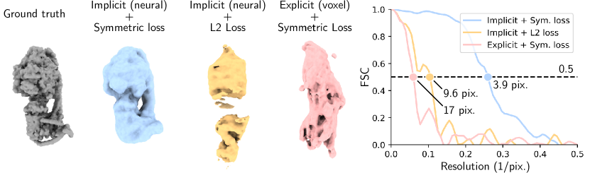
Supplementary Note 1: Ablation study
We conduct an ablation study and report the results in Fig. 1. Specifically, we analyze the importance of the implicit neural representation () by replacing it with an explicit representation () (i.e., a voxel grid) and that of the symmetric loss function by replacing it with a naive L2 loss
| (1) |
where . Our results show that both components are necessary for converging to the correct density.
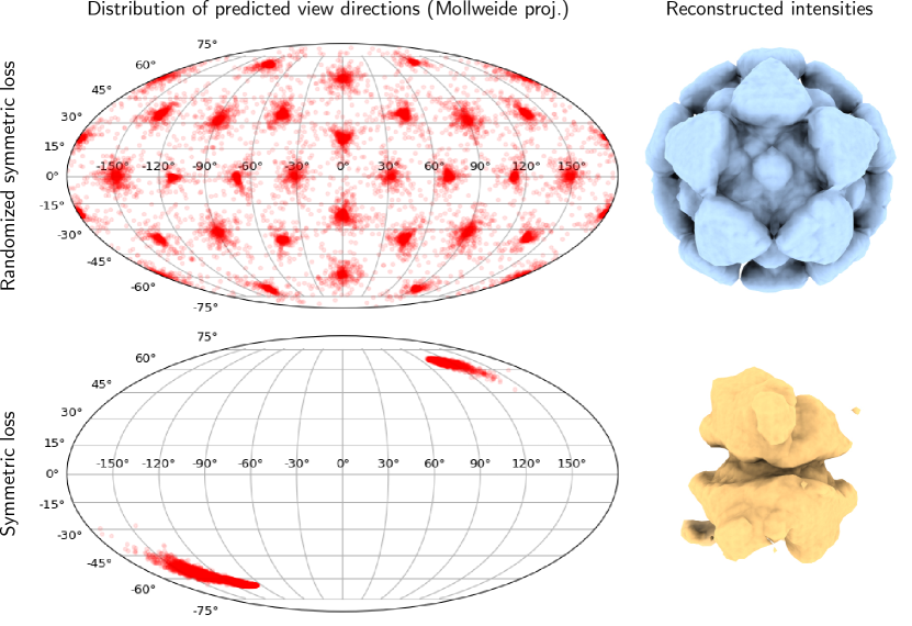
Supplementary Note 2: Replication scheme on PR772
We illustrate the importance of the randomized symmetric loss in Fig. 2. With the randomized symmetric loss, predicted poses are randomly rotated by a symmetry rotation of the icosahedron. This effectively enables the intensity volume to be supervised everywhere in , although the encoder only maps images to a subset of .
Supplementary Note 3: Datasets
We simulate all synthetic datasets using the Skopi software package [2]. All the simulation parameters, as well as the statistics of the synthetic and experimental datasets, are detailed in Table 1.
| ID | Type | # Images | Res. (pix.) | Beam Fluence | Photons per Frame | Corner Res. |
|---|---|---|---|---|---|---|
| 7R4X [3] | Synthetic | 50K, 500K, 5M | 128 128 | 8.0 Å | ||
| 6J5I [4] | Synthetic | 50K, 500K, 5M | 128 128 | 8.0 Å | ||
| 5O60 [5] | Synthetic | 5M | 128 128 | 8.0 Å | ||
| 5O60 | Synthetic | 5M | 128 128 | 8.0 Å | ||
| PR772 [1] | Experimental | 14,772 | 260 257 | 83 Å |
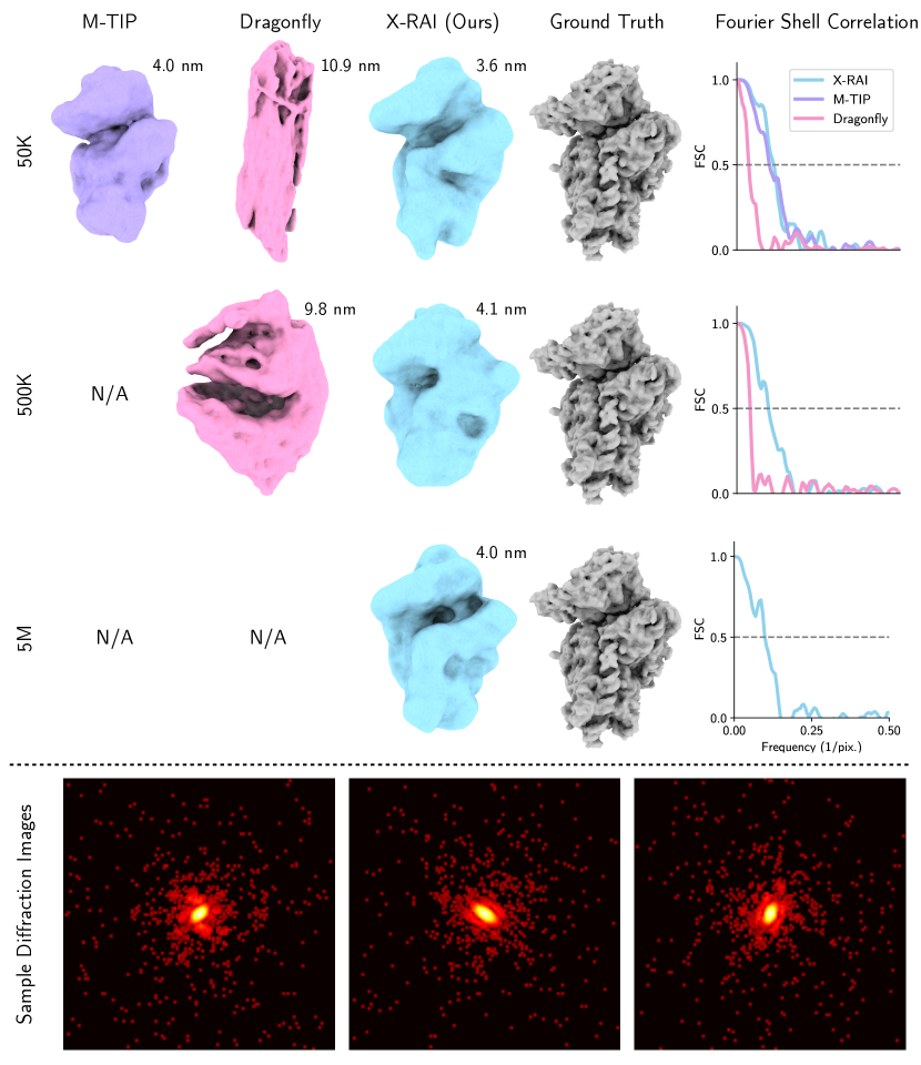
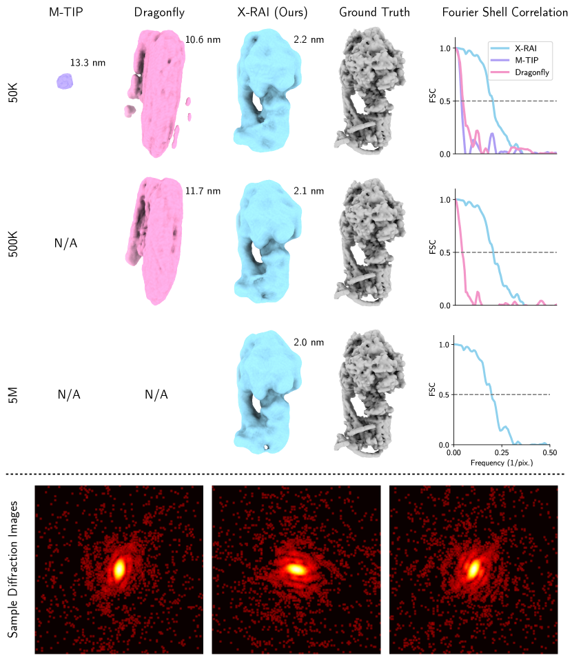
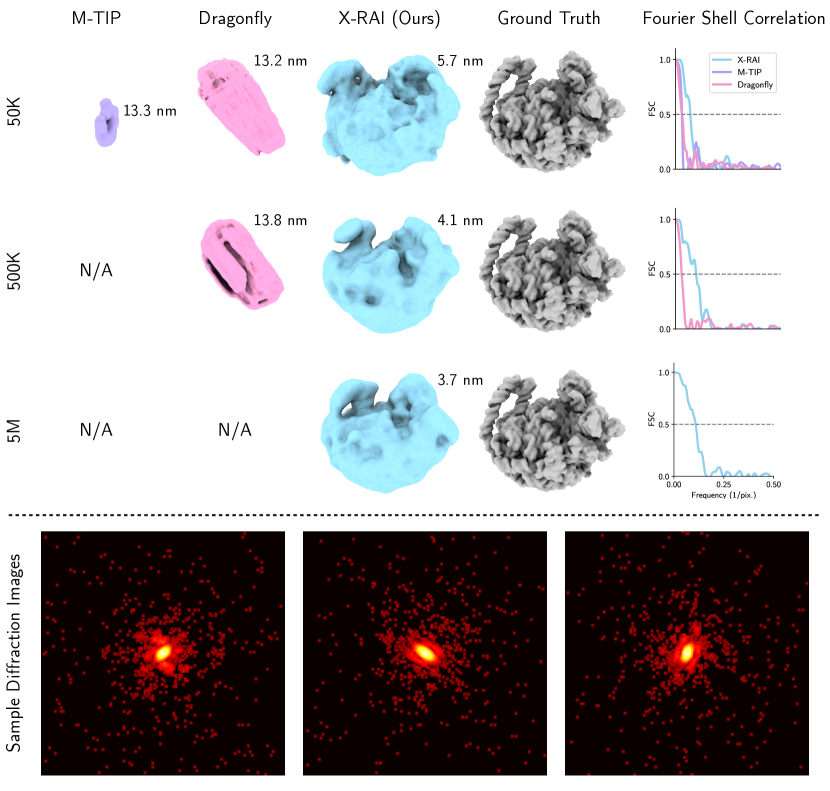
Supplementary Note 4: Qualitative reconstruction results on all synthetic datasets
We present comprehensive reconstruction results on all of the synthetic datasets shown in Table 1 of the main paper. M-TIP fails to reconstruct datasets containing 500 thousand or 5 million images within the allotted time of 48 hours, while Dragonfly times out when processing 5 million images. The results are shown in figures 3, 4, and 5.
Supplementary References
- [1] Hemanth KN Reddy et al. “Coherent soft X-ray diffraction imaging of coliphage PR772 at the Linac coherent light source” In Scientific data 4.1 Nature Publishing Group, 2017, pp. 1–9
- [2] Ariana Peck et al. “Skopi: a simulation package for diffractive imaging of noncrystalline biomolecules” In Journal of Applied Crystallography 55.4 International Union of Crystallography, 2022, pp. 1002–1010
- [3] Simone Pellegrino, Kyle C Dent, Tobias Spikes and Alan J Warren “Cryo-EM reconstruction of the human 40S ribosomal subunit at 2.15 Å resolution” In Nucleic Acids Research 51.8 Oxford University Press, 2023, pp. 4043–4054
- [4] Jinke Gu et al. “Cryo-EM structure of the mammalian ATP synthase tetramer bound with inhibitory protein IF1” In Science 364.6445 American Association for the Advancement of Science, 2019, pp. 1068–1075
- [5] Jendrik Hentschel et al. “The complete structure of the Mycobacterium smegmatis 70S ribosome” In Cell reports 20.1 Elsevier, 2017, pp. 149–160