Optical Transmission Enhancement of Ionic Crystals via Superionic Fluoride Transfer: Growing VUV-Transparent Radioactive Crystals
Abstract
The first nuclear excited state in \ch^229Th is a candidate for implementing a nuclear clock. Doping \ch^229Th into ionic crystals such as \chCaF2 is expected to suppress non-radiative decay, enabling nuclear spectroscopy and the realization of a solid-state optical clock. Yet, the inherent radioactivity of \ch^229Th prohibits the growth of high-quality single crystals with high \ch^229Th concentration; radiolysis causes fluoride loss, increasing absorption at . We overcome this roadblock by annealing \ch^229Th doped \chCaF2 at in \chCF4. The technique presented here allows to adjust the fluoride content without crystal melting, preserving its single-crystal structure. Superionic state annealing ensures rapid fluoride distribution, creating fully transparent and radiation-hard crystals. This approach enables control over the charge state of dopants which can be used in deep UV optics, laser crystals, scintillators, and nuclear clocks.
Thorium-229 (\ch^229Th), possessing an and approximately 600-second lifetime first nuclear excited state (isomer state), enables high-precision vacuum ultraviolet (VUV) laser spectroscopy Seiferle et al. (2019); Kraemer et al. (2023); Masuda et al. (2019). The anomalously low energy of this excited state offers the potential for the construction of an optical clock based on a nuclear transition Peik and Tamm (2003); Beeks et al. (2021). The structure of the nuclear levels is governed by both Coulomb and nuclear forces Hayes and Friar (2007). This allows probing of these forces via nuclear spectroscopy, paving the way for new fundamental research: For example the search for dark matter, or potential drifts in the fine-structure constant Peik et al. (2021); Fadeev et al. (2020).
^229Th is required to be in a 3+ or higher charge state to suppress the non-radiative decay Peik and Okhapkin (2015). The possibility of trapping charged \ch^229Th in a solid-state matrix offers an alternative to ion traps. In the solid, the nucleus is isolated due to the small interaction with its chemical environment Kazakov et al. (2012). Ionic crystals such as \chCaF2 are an excellent choice as host material for the \ch^229Th based nuclear clock Hehlen et al. (2013). The ionic character of these crystals naturally forces the \ch^229Th into a 4+ charged state, substituting the calcium (\chCa^2+) cation Pimon et al. (2022), and their large band gaps make them transparent to wavelengths around or Rubloff (1972). Ionic crystals such as oxides and fluorides display good scintillator properties and are resistant to VUV radiation, making them suitable to host the radioactive \ch^229Th and observe its radiative decay YANAGIDA (2018); Kraemer et al. (2023). In this experiment, calcium fluoride (\chCaF2) was chosen as the host material due to its excellent scintillator properties Rodnyi (1997), simple cubic structure Vil , large electronic and optical bandgap Rubloff (1972), and an unchanged optical bandgap after thorium Beeks and Schumm (2022) doping. Both \ch^232Th and \ch^229Th were used to grow single crystalline Th:\chCaF2 using the vertical gradient freeze method Beeks et al. (2023).
The process of growing \chCaF2 in a vacuum environment leads to a fluoride ion (\chF-) deficit due to thermal dissociation of \chCaF2 and its reaction with the residual water in the system Recker and Leckebusch (1971); Bollmann (1980). This sequence leads to the formation of a non-stoichiometric or fluoride-deficient crystal. To counterbalance the loss in fluoride ions, the crystal tends to generate metallic Ca nanoparticles Beeks and Schumm (2022), which possess the capacity to absorb and scatter light Rix (2011), especially within the VUV and optical range. Non-stoichiometry in all ionic crystals leads to changes in the configuration: either change of charge state of cations or formation of metal colloids Hughes and Jain (1979). Although this variation in configuration has been thoroughly examined in oxides like \chCeO2 Li et al. (2021); Barth et al. (2016), it has not been extensively studied for fluorides and there is a lack of common terminology, understanding and means of controlling the compositions Cirillo and Wright (1987); Dubois et al. (2011); Wang et al. (2005); Antonyak et al. (2017); Angervaks et al. (2018).
We study the impact of the Th:\chCaF2 crystal composition on its VUV transmission and the electronic structure of the dopant sites. Due to the \ch^229Th radioactivity, radiolysis becomes the major cause of the fluorine (\chF2) loss during the growth phase Beeks et al. (2023). This substantial \chF2 loss modifies the electronic structure of the \chCaF2 crystal, resulting in a consequential change in its absorption profile Beeks et al. (2023). The large resulting VUV absorption would make such a \ch^229Th:\chCaF2 unsuitable for a nuclear optical clock.
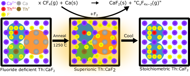
The change of dopant configuration and absorption profile due to composition was first observed in the work of Cirillo et al. (1987) Cirillo and Wright (1987), where adding \chF2 to \chEu^2+:\chCaF2 changed the charge state of Eu and absorption profile of the crystal. We developed a novel and safer experimental method that does not require toxic \chF2 gas to add fluoride ions to already grown, single-crystalline, Th doped \chCaF2 and still dramatically improve the transmission profile. We use an induction heated carbon crucible to anneal Th:CaF2 at above its superionic temperature, but below its melting temperature, in a carbon tetrafluoride (\chCF4) atmosphere.
The crystal is placed in the center of an induction coil in a carbon crucible, after which the system is evacuated to approximately . The chamber is then filled with \chCF4. In the heating step, the carbon crucible is heated using the induction coil (, heating rate ) while the walls of the vacuum system are water-cooled, creating a steep temperature gradient. The temperature gradient causes the \chCF4 gas to be reactive only at the crystal surface and inert at the vacuum system walls, which significantly reduces safety concerns while ensuring the efficiency of the process. After annealing for 1 hour while holding the temperature, the system is cooled down (cooling rate ) and at room temperature the \chCF4 is replaced by \chN2. A schematic representation of the process is shown in Figure 1.
Above the superionic transition temperature of \chCaF2 ( Gillan (1986)), but below the melting temperature, fluoride anions exhibit high mobility while calcium cations remain immobile. The mobility of fluoride ions ensures their uniform distribution throughout the bulk crystal.
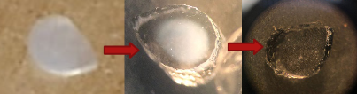
To produce a heavily fluoride deficient crystal, we first performed superionic annealing in vacuum instead of a \chCF4 atmosphere on a \ch^232Th:\chCaF2 crystal. Excessive fluorine loss from the crystal during vacuum superionic annealing for 24 hours has turned an initially transparent crystal into a cloudy and opaque one (see Figure 2, left). Annealing in an argon atmosphere was also performed, producing a similar but less deficient crystal. The formation of calcium metal colloids can explain the opacity Rix (2011). In the following phase, which consists of \chCF4 annealing in two cycles of one hour each, the crystal regains its visible transparency (see Figure 2, middle and right).
Following the fluorination treatment, we observed a significant improvement in the VUV transparency of the \ch^229Th:\chCaF2 crystals, as shown in Figure 3. Note that this crystal was fully opaque around 8 eV (150 nm) and hence unusable for nuclear laser spectroscopy directly after growth. After several cycles, the absorption was lower as compared to the non-radioactive \ch^232Th doped crystal, indicating \chCF4 could improve its absorption profile as well. The absorption spectrum was recorded using a McPherson 204/302 VUV spectrometer, as described in the work of Beeks et al. Beeks and Schumm (2022). As verified by gamma spectroscopy, the annealing process did not cause any quantifiable loss of radioactivity, indicating no noticeable reduction of the 229Th concentration.
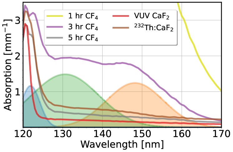
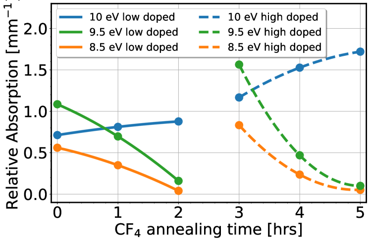
To quantitatively describe the VUV absorption profile over the time of fluorination, we identify three absorption lines (assuming gaussian profile) in Figure 3. We attribute the absorption to the \chTh^4+ charge transfer state Nickerson et al. (2020, 2021), the absorption to \chTh ions in a different surrounding, and the broad absorption to \chCa metallic colloids Rix (2011); Ryskin et al. (2017). The absorption is likely caused by a change in the surrounding of the Th ion: either the change of charge state of the Th atom to neutral/1+/2+/3+ due to the fluoride deficiency, or reaction of the non-stoichiometric \chCaF2 with oxygen in the air thereby replacing the \chF atoms surrounding \chTh by \chO atoms. Investigations of the absorption line are ongoing. In Figure 4 it can be seen that in low and high-doped crystals the absorption increases with cycle number, indicating an increase in \chTh^4+ with fully charge-compensated surrounding. The other two absorption lines assigned to Ca colloids and Th in a different surrounding both diminish. Effectively the electronic structure of the Th dopants is manipulated by adding fluoride ions to the deficient crystal.
Alternative methods of fluorination of low-doped \ch^229Th:\chCaF2 crystals were also tested. Highly fluoride deficient crystals (opaque) were treated using 3 different methods.
The first method was annealing in an \chF2 atmosphere. A Mg sample holder was passivated for two days at (heating rate: , cooling rate: ) in an \chF2 flow (20 % \chF2 in \chN2 atmosphere, ). The \ch^229Th:\chCaF2 crystal was placed in the sample holder and was fluorinated for five days at (heating rate: , cooling rate: ) in an \chF2 flow (20 % \chF2 in \chN2 atmosphere, ).
The second method was treatment using an \chNF3 plasma. In this method a Ni sample holder was passivated in a fluorine plasma at room temperature for one hour. The \ch^229Th:\chCaF2 crystal was placed in the sample holder and was fluorinated in a fluorine plasma at room temperature for three hours. \chNF3 was used as feeding gas with a flow rate of .
The third method was treatment in a \chF2 filled autoclave. In this method a Ni sample holder was passivated for one day at and approximately (heating rate: , cooling rate: , 20% \chF2 in Ar atmosphere). The \ch^229Th:\chCaF2 crystal was placed in the Ni sample holder and was high-pressure fluorinated at and approximately for five days (heating rate: , cooling rate: , 50% \chF2 in Ar atmosphere).
The resulting crystals still displayed high absorption as can be seen in Figure 5. The 5 days of \chF2 annealing at displayed results similar to 1 hour of \chCF4 annealing at .
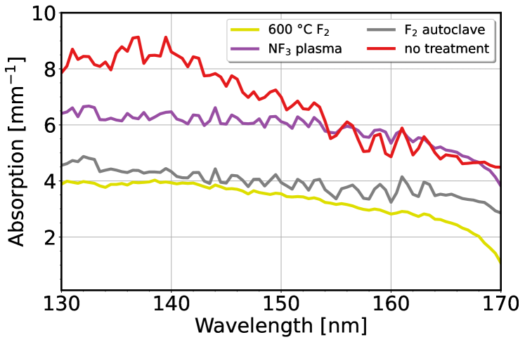
Compared to alternative treatments, only superionic annealing under \chCF4 atmosphere demonstrated a significant improvement in the VUV transmission (compare Figure 3 with Figure 5). Consequently, it can be concluded that the superionic state of \chCaF2 allows for efficient and rapid homogeneous distribution of the acquired fluoride ions. This contrasts with other methods where the solid phase of the crystal prevents fluoride ion diffusion through the crystal.
This report demonstrates the enhancement of optical transmission in ionic crystals through superionic fluoride transfer. This superionic state substantially decreases the treatment duration. Furthermore, our findings suggest the possibility of controlling the dopant surrounding in \chCaF2 by the addition or removal of \chF-, although the removal process carries a potential risk of creating \chCa metallic nanoparticles. We have developed a simple and safe method of fluorine manipulation in fluoride ionic crystals. Utilizing this method, we are able to fabricate highly transparent, heavily doped \ch^229Th:\chCaF2 crystals for a solid-state nuclear clock. The manipulation of the electronic structure brings the potential for advancements in optics, scintillator and laser crystal development by optimizing light absorption and emission through dopant and cation surrounding control (e.g., \chEu:\chCaF2).
.1 Acknowledgements
Acknowledgements.
This work is part of the thorium nuclear clock project that has received funding from the European Research Council (ERC) under the European Union’s Horizon 2020 research and innovation programme (Grant Agreement No. 856415). The research was supported by the Austrian Science Fund (FWF) Projects: I5971 (REThorIC) and P 33627 (NQRclock).References
- Seiferle et al. (2019) B. Seiferle, L. von der Wense, P. V. Bilous, I. Amersdorffer, C. Lemell, F. Libisch, S. Stellmer, T. Schumm, C. E. Düllmann, A. Pálffy, and P. G. Thirolf, Nature 573, 243 (2019).
- Kraemer et al. (2023) S. Kraemer, J. Moens, M. Athanasakis-Kaklamanakis, S. Bara, K. Beeks, P. Chhetri, K. Chrysalidis, A. Claessens, T. E. Cocolios, J. G. Correia, et al., Nature 617, 706 (2023).
- Masuda et al. (2019) T. Masuda, A. Yoshimi, A. Fujieda, H. Fujimoto, H. Haba, H. Hara, T. Hiraki, H. Kaino, Y. Kasamatsu, S. Kitao, K. Konashi, Y. Miyamoto, K. Okai, S. Okubo, N. Sasao, M. Seto, T. Schumm, Y. Shigekawa, K. Suzuki, S. Stellmer, K. Tamasaku, S. Uetake, M. Watanabe, T. Watanabe, Y. Yasuda, A. Yamaguchi, Y. Yoda, T. Yokokita, M. Yoshimura, and K. Yoshimura, Nature 573, 238 (2019).
- Peik and Tamm (2003) E. Peik and C. Tamm, Europhysics Letters 61, 181 (2003).
- Beeks et al. (2021) K. Beeks, T. Sikorsky, T. Schumm, J. Thielking, M. V. Okhapkin, and E. Peik, Nature Reviews Physics , 1 (2021).
- Hayes and Friar (2007) A. Hayes and J. Friar, Physics Letters B 650, 229 (2007).
- Peik et al. (2021) E. Peik, T. Schumm, M. S. Safronova, A. Pálffy, J. Weitenberg, and P. G. Thirolf, Quantum Science and Technology 6, 034002 (2021).
- Fadeev et al. (2020) P. Fadeev, J. C. Berengut, and V. V. Flambaum, Phys. Rev. A 102, 052833 (2020).
- Peik and Okhapkin (2015) E. Peik and M. Okhapkin, Comptes Rendus Physique, Comptes Rendus Physique 16, 516 (2015), arXiv:1502.07322v1 .
- Kazakov et al. (2012) G. A. Kazakov, A. N. Litvinov, V. I. Romanenko, L. P. Yatsenko, A. V. Romanenko, M. Schreitl, G. Winkler, and T. Schumm, New Journal of Physics 14, 083019 (2012).
- Hehlen et al. (2013) M. P. Hehlen, R. R. Greco, W. G. Rellergert, S. T. Sullivan, D. DeMille, R. A. Jackson, E. R. Hudson, and J. R. Torgerson, Journal of Luminescence 133, 91 (2013), 16th International Conference on Luminescence ICL’11.
- Pimon et al. (2022) M. Pimon, A. Grüneis, P. Mohn, and T. Schumm, Crystals 12, 1128 (2022).
- Rubloff (1972) G. W. Rubloff, Physical review B 5, 662 (1972).
- YANAGIDA (2018) T. YANAGIDA, Proceedings of the Japan Academy, Series B 94, 75 (2018).
- Rodnyi (1997) P. A. Rodnyi, Physical processes in inorganic scintillators, Vol. 14 (CRC press, 1997).
- (16) “Caf2 crystal structure: Datasheet from “pauling file multinaries edition – 2012” in springermaterials,” Copyright 2016 Springer-Verlag Berlin Heidelberg & Material Phases Data System (MPDS), Switzerland & National Institute for Materials Science (NIMS), Japan.
- Beeks and Schumm (2022) K. Beeks and T. Schumm, The Nuclear Excitation of Thorium-229 in the CaF2 Environment, Ph.D. thesis, Wien (2022).
- Beeks et al. (2023) K. Beeks, T. Sikorsky, V. Rosecker, M. Pressler, F. Schaden, D. Werban, N. Hosseini, L. Rudischer, F. Schneider, P. Berwian, et al., Scientific Reports 13, 3897 (2023).
- Dessovic et al. (2014) P. Dessovic, P. Mohn, R. Jackson, G. Winkler, M. Schreitl, G. Kazakov, and T. Schumm, Journal of Physics: Condensed Matter 26, 105402 (2014).
- Gillan (1986) M. Gillan, Journal of Physics C: Solid State Physics 19, 3391 (1986).
- Recker and Leckebusch (1971) K. Recker and R. Leckebusch, Journal of Crystal Growth 9, 274 (1971).
- Bollmann (1980) W. Bollmann, physica status solidi (a) 57, 601 (1980).
- Rix (2011) S. Rix, Radiation-induced defects in calcium fluoride and their influence on material properties under 193 nm laser irradiation, Ph.D. thesis, Mainz, Univ., Diss., 2011 (2011).
- Hughes and Jain (1979) A. Hughes and S. Jain, Advances in Physics 28, 717 (1979).
- Li et al. (2021) Q. Li, L. Song, Z. Liang, M. Sun, T. Wu, B. Huang, F. Luo, Y. Du, and C.-H. Yan, Advanced Energy and Sustainability Research 2, 2000063 (2021).
- Barth et al. (2016) C. Barth, C. Laffon, R. Olbrich, A. Ranguis, P. Parent, and M. Reichling, Scientific Reports 6, 21165 (2016).
- Cirillo and Wright (1987) K. M. Cirillo and J. C. Wright, Journal of crystal growth 85, 453 (1987).
- Dubois et al. (2011) M. Dubois, B. Dieudonne, A. Mesbah, P. Bonnet, M. El-Ghozzi, G. Renaudin, and D. Avignant, Journal of Solid State Chemistry 184, 220 (2011).
- Wang et al. (2005) F. Wang, X. Fan, D. Pi, and M. Wang, Solid state communications 133, 775 (2005).
- Antonyak et al. (2017) O. Antonyak, Z. Khapko, and M. Chylii, Radiation Effects and Defects in Solids 172, 456 (2017).
- Angervaks et al. (2018) A. E. Angervaks, A. V. Veniaminov, M. V. Stolyarchuk, V. E. Vasilev, I. Kudryavtseva, P. P. Fedorov, and A. I. Ryskin, J. Opt. Soc. Am. B 35, 1288 (2018).
- Nickerson et al. (2020) B. S. Nickerson, M. Pimon, P. V. Bilous, J. Gugler, K. Beeks, T. Sikorsky, P. Mohn, T. Schumm, and A. Pálffy, Phys. Rev. Lett. 125, 032501 (2020).
- Nickerson et al. (2021) B. S. Nickerson, M. Pimon, P. V. Bilous, J. Gugler, G. A. Kazakov, T. Sikorsky, K. Beeks, A. Grüneis, T. Schumm, and A. Pálffy, Physical Review A 103, 053120 (2021).
- Ryskin et al. (2017) A. I. Ryskin, P. P. Fedorov, N. T. Bagraev, A. Lushchik, E. Vasil’chenko, A. E. Angervaks, and I. Kudryavtseva, Journal of Fluorine Chemistry 200, 109 (2017).