ipr]Univ. Rennes, CNRS, IPR - UMR 6251, Rennes, 35000, France
Heterogeneous structure and dynamics of water in a hydrated collagen microfibril
Abstract
Collagen, type I in particular, is the protein of choice by Nature to design structural materials. Its hierarchical fibrillar structure confers to collagen its outstanding mechanical properties. Nonetheless, fibrillar collagen may rather be viewed as a composite material made of protein, macromolecules (such as glycosaminoglycans and proteoglycans) and water. Yet, the properties of water and the fine interactions of water with the protein constituent of these nanocomposites have only received limited attention. Here, we propose to investigate in-depth water structure and dynamics confined within the microfibril crystal structure of collagen type I to establish its impact on the properties of collagen. We found that the properties of water vary strongly with the level of hydration of the microfibril, and spatially along the long axis of the crystal, namely moving from the so-called gap region to the so-called overlap region. In short, at low hydration, water acts as a glue between protein chains; while at high hydration, water acts as a lubricant. Beyond self-assembly and properties of fibrillar collagen, such heterogeneous structure and anisotropic dynamics may control its biomineralization and the properties of biological tissues such as bone.
1 Keywords
collagen type I, nanoconfined water, hydration, molecular dynamics, collagen self-assembly, biomineralisation
2 Introduction
Collagen is ubiquitous in the animal world, from fishes to mammals, it is the protein of choice of organisms to design structural materials. Collagen type I is the most occurring type of collagen among the 28 co-existing collagen sequences, it is found in tendons, bone and cornea among others. Collagen-based tissues feature a wide-range of outstanding mechanical properties (elastic, viscous, fracture) which are tuned modifying the hierarchical fibrillar structure of collagen self-assemblies. Tuning of properties often relies on the association of collagen with additional organic (e.g. in tendon, ligaments) or inorganic molecules (e.g. in bone). The specific, amino acid structure, dictating the self-assembly of tropocollagen molecules into this intricate fibrillar architecture is widely accepted as a key ingredient of the mechanical properties of the tissues made of collagen type I. One significantly abundant collagen self-assembled structure is the collagen fibril which consists of twisted bundles of aligned tropocollagen molecules. The fibrillar structure of collagen itself is mostly correlated with its characteristic repetitive sequence of amino acids. The presence of additional molecules is also key to the stability of these fibrillar self-assemblies, macromolecules in particular, such as proteoglycans or glycosaminoglycans, cross-links and last but not least water molecules.
Hydration is known to be key for collagen stability 1, and the structure of water molecules within the the collagen fibril, around and in between tropocollagen molecules, has been characterised using nuclear magnetic resonance 2. The mechanical properties of collagen fibrils such as Young’s modulus can drop up to one order of magnitude depending on their water content 3, 4, 5, and upon drying fibrils become extensively brittle 6. Overall, nanoscale water molecules within the fibril mediate forces between tropocollagen molecules 7, 8. However, intricacies associated with controlling water content with collagen fibrils experimentally have limited access to quantitative analysis of the influence of hydration. And beyond structural aspects, water dynamics within the fibril have received no or at most very little attention. Yet, both largely affect conformation and functionalisation of proteins 9. To that extent, a precise and quantitative analysis of the effect of hydration, that is water structure and dynamics, on the molecular structural and mechanical properties of collagen fibrils is still lacking. Such understanding is key to understand the emergence of physical properties of collagen fibrils and collagen-based tissues.
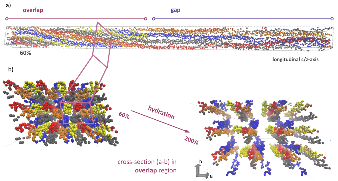
We here propose a thorough investigation of the water structure and dynamics within the microfibril crystal which constitutes a representative unit of the collagen fibril 10. Our investigation relies on long all-atom molecular dynamics simulation of the hydration of the collagen microfibril, from the water-to-protein mass ratio of tendon (approx. %) to the supposed onset of the fibril disassembly (approx. %). And we focus on providing spatially-resolved details about water structure (radial distribution function, hydrogen bonds) and dynamics (self-diffusion coefficient), and protein structure throughout the microfibril crystal, and in particular in the characteristic overlap and gap regions (see Figure 1.a).
3 Methods
Molecular dynamics simulations of the hydrated collagen microfibril are performed using the massively parallel NAMD package 11. The potentials describing amino acid atoms interactions are modeled and parameterised with the Generic Amber Force Field (GAFF) 12 which has been extensively used for the simulation of protein structure and functionalisation. Meanwhile the potentials describing the interactions involving water atoms rely on the TIP3P force field 13, we use a rigid water model for computational efficiency, as we do not intend to focus on high-frequency bond and angle vibrational modes. We perform ensemble MD simulations for better precision of the thermodynamic averages, the ensemble features replicas with different randomly initialised velocity of the protein atoms.
The position of the protein backbone atoms is set according to the microfibril crystallographic structure obtained by 10 using X-ray fiber diffraction on native rat tail tendon in situ. The structure labelled 3HR2 containing the position of the atoms can be found on the Protein Data Bank 14. The molecular system is set up using periodic boundary conditions and the microfibril crystals triclinic box 10. We modified amino acids from the 3HR2 structure to match the exact sequence of human collagen type I as found in the single chains’ sequences denoted NP_000079 (for alpha-1(I) helices) and NP_000080 (for the alpha-2(I) helices). Nonetheless, we preserved hydroxyproline amino acids in the sequence of the chains according to the original structure. Last, we added negatively charged chlorine ions to neutralise the system, which interactions are parameterised using GAFF.
Once the initial structure of the protein is set, we proceed to hydration of the molecular system in two stages following procedures found in the literature 15, 16. First, we add water molecules such as to stabilise the volume of the microfibril crystal characterised by X-ray crystallography. This is assumed to be the amount of water present in the tendon at physiological conditions. Second, we iteratively add a fixed amount of water molecules up to a hydration level equal to approximately % water-to-protein mass ratio at which we expect disassembly of the microfibril. Water molecules are inserted at random location in the microfibril crystal such that all water molecules are distant of the protein atoms from at least nm and of the other water molecules from at least nm. This is the standard addWater procedure found in the AmberTools suite 17. After each addition of water molecules, whether it is to stabilise or to progressively hydrate the crystal, we perform the following equilibration simulation in three stages: (i) a iterations molecular mechanics minimisation of the potential energy with fixed protein backbone atoms; (ii) a ns molecular dynamics simulation at fixed volume and at temperature set at 298 K (NVT ensemble) using the Langevin thermostat (damping coefficient: fs), and with fixed protein backbone atoms; and (iii) a ns molecular dynamics simulation at pressure set at MPa and at temperature set at K (NPT ensemble) using respectively the isotropic Berendsen barostat (pressure relaxation time: fs) and the Langevin thermostat (damping coefficient: fs).
Note that for both molecular dynamics simulations, assuming that the molecular system is at equilibrium, we draw the initial velocities of the atoms randomly from the Maxwell–Boltzmann distribution parameterised at K.
The first stage of the hydration process is achieved in a trial-and-error fashion, we insert a given number of water molecules in the microfibril crystal and we check whether the volume of the crystal remains stable at nm3, the volume obtained experimentally 10. We found that water molecules is the water content which stabilises the microfibril crystal. Such water content is approximately equivalent to Streeter and Leeuw 15 and % higher than to Gautieri et al. 16. One of the main difference with the later molecular model is the use of the force field, Gautieri et al. 16 used the GROMOS 43a1 force field from the GROMACS 4.0 code 18.
After the first stage, our equilibrated molecular system features a water-to-protein mass ratio about %. The subsequent second stage of the hydration process is achieved iteratively adding a fixed amount of water molecules up to % ( water molecules). The iterative procedure follows three stages: (i) an insertion of water molecules; (ii) the aforementioned equilibration simulation workflow; and (iii) an additional molecular dynamics simulation at pressure set at MPa and at temperature set at K (NPT ensemble) using respectively the anisotropic Berendsen barostat (pressure relaxation time: fs) and the Langevin thermostat (damping coefficient: fs), until the long dimension of the microfibril relaxes to its characteristic experimental length nm.
These steps are repeated until reaching the desired hydration level. Note that the simulation in stage (iii) using an anisotropic barostat, allowing non-proportional deformation of the microfibril crystal, is necessary to recover the observed characteristic length of the microfibril crystal, the so-called D-spacing, approximately nm 19, 20, 21. This stage systematically lasts less than ps. The molecular model of the hydrated microfibril is shown in Figure 1.
The verification of the equilibration of the initial molecular system (energy and volume, see figure S1.a), as well as the evolution of the dimensions of the microfibril crystal at different hydration levels (see figure S1.b) are thoroughly illustrated in the supplementary information.
4 Results
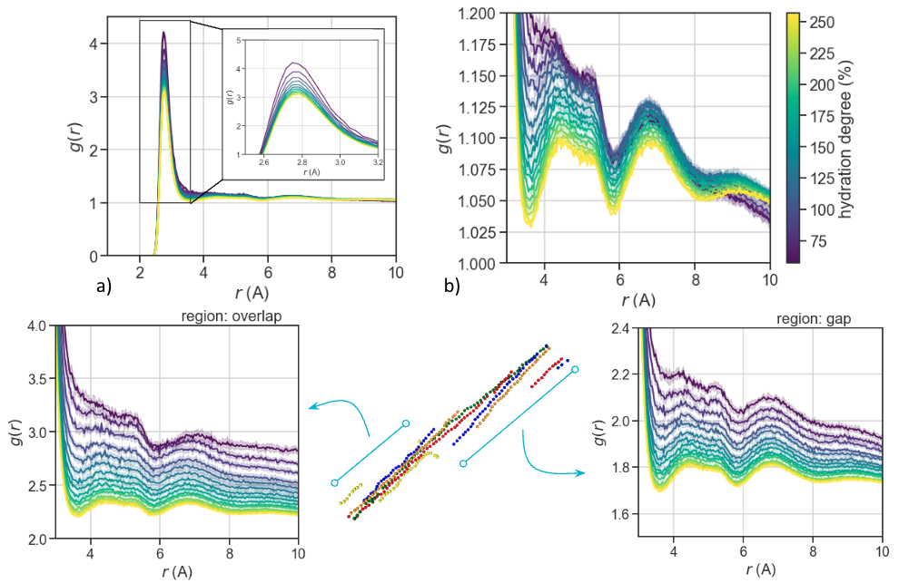
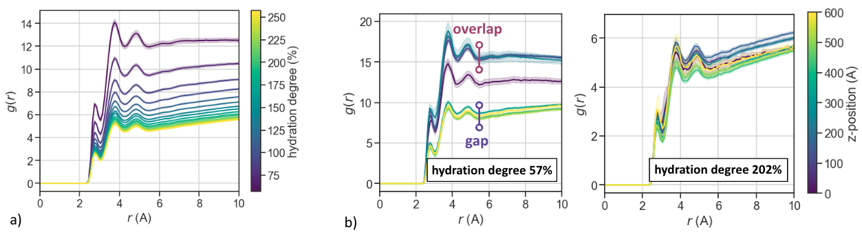
We first focus on the structure of water throughout the microfibril, to that extent we compute the radial distribution function of the oxygen atoms from water molecules (see Figure 2). The structure of water molecules on average over the whole microfibril crystal, but also over the overlap and over the gap regions becomes more organised for higher hydration levels (see Figure 2.b,c,d, respectively). A clear separation of the first and the second peak appears beyond % hydration. The second peak (around nm and ending at nm) and the third peak ( nm and ending at nm) of appear progressively sharper. Besides, we observe a decrease of amplitude and shift of the first peak with increasing hydration (from to Å) (see inset in Figure 2.a). Such an an increase of the distance between neighbouring water molecules can be associated with a decrease in confinement and the swelling of the microfibril (see Figure 1.b). Water molecules are getting organised in a more structured but less dense packing. Swelling can also be observed on the evolution of of the backbone atoms of the microfibril protein (see supp. figure S2). The structure of the microfibril displays three clear peaks at low hydration, which gradually shift toward higher values. This implies longer tropocollagen molecules interdistances. Further, the peaks become rounder with hydration. Unlike the water molecules structure, the microfibril protein structure grows more disorganised with increasing hydration level.
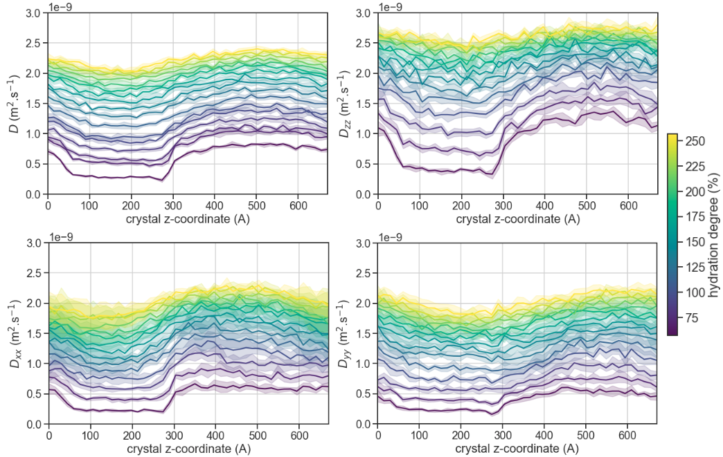
We then focus on the dynamics of water, the self-diffusion coefficient of water computed from mean squared displacement (MSD) of individual water molecules within the collagen microfibril is used for analysis (see Figure 3):
| (1) |
where is the number of water molecules, is their coordinates and the lag time from .
We found a linear evolution of the MSD with the lag time, independently of the probed location in the microfibril, supporting a standard water diffusion regime (see figure S3). The self-diffusion coefficients are computed using the slope of the evolution of the MSD (denoted ) or directional (denoted , and ) MSD from Einstein’s relation. The regression score of the linear fit of the evolution of the MSD with the time lag are used to compute self-diffusion coefficient estimation error (see top left, Figure 3), showing high precision of the computed values. We observe an overall diffusion of water smaller than that of bulk water. Although, bulk water diffusion values are recovered at high hydration levels (%) for . In the direction, we even encounter higher self-diffusion than in bulk water (above m2.s-1), peaking at almost m2.s-1 for high hydration levels ( %). We witness higher water self-diffusion in the gap region in comparison to the overlap region, systematically, independently of the probed direction, at low hydration levels. The difference in self-diffusion coefficient can reach % at lowest hydration levels, varying from to m2.s-1. The difference between the overlap and gap regions is inversely correlated with the hydration level. Only along the direction is the difference preserved at high hydration, in all other directions the difference vanishes.
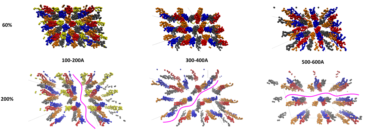
Meanwhile, we inspect qualitatively the evolution of the structure of the tropocollagen molecules within cross sections of the microfibril crystal for various -axis positions at low (%) and high (%) hydration levels (see Figure 4). As we removed water molecules from the visualisation, voids correspond to the locations where water molecules localises. At low hydration, water molecules appear to localise in the gap area (i.e. between and Åalong the z-axis, see top centre and top right insets), in small and non percolating (discontinuous) volumes. In contrast, at high hydration, water localises in continuous planes (pink) which cross the entire crystal unit cell orthogonally to the cross section, rotating around the long -axis (see bottom insets). These quantitative observation tend to support the earlier observation that the microfibrils swells with hydration, displaying water two-dimensional water transport channels.
Last feature of the water structure, we investigate the cylindrical distribution of water molecules around tropocollagen molecules (see Figure 5). We observe three distinct peaks which magnitude gradually decays away as hydration increases (see Figure 5.a). At low hydration, the three peaks are clearly observed in the core of the overlap region ( to nm) while they appear fainter in the gap region ( to nm) (see Figure 5.b). At high hydration, the amplitude of the three peaks is smaller in both regions, but it has to be noted that the decrease is less important in the gap region (see Figure 5.c).
We now turn to the analysis of hydrogen bonding. We investigate hydrogen bonding to obtain information about the stability of the hydrated collagen microfibril (see Figure 6). A hydrogen bond is found when a hydrogen atom is found within a Å distance from a donor-atom candidate and a Å distance acceptor-atom candidate and when the triplet of atoms form an angle larger than . We report an in-depth analysis of hydrogen bonds whereby a distinction is made between bonds depending on the type of acceptor and donor molecules, whether it is a water molecule (w) or a protein (p). The analysis of hydrogen bonds involving at least one water molecule is reported in Figure 6.a,b. Overall, the count of hydrogen bonds between two water molecules dominates and increases linearly with the hydration level, that is with the number of water molecules added in the microfibril crystal (see Figure6.b). In comparison, the number of hydrogen bonds between water molecules and the amino acids of the collagen protein remains stable. Further, we observe a stable number of hydrogen bonds per water molecules in the system (see Figure 6.a) around . This is less than the number of hydrogen bonds per water molecule in bulk water which is about 24.
We also focus on hydrogen bonds formed within and at the surface of the protein (see Figure 6.c,d). We observe that the number of hydrogen bonds in between protein amino acids (p-p) fluctuates but remains rather constant throughout the hydration process (see inset in Figure 6.c). These protein-protein hydrogen bonds are formed almost exclusively within -helix chains and between triplets of helices forming tropocollagen molecules. And rather rarely between distinct tropocollagen molecules. The stability of the protein-protein hydrogen bonds count demonstrates that the individual tropocollagen structure is not altered by the hydration level. We confirm this showing that the survival of protein-protein hydrogen bonds is much longer than hydrogen bonds involving at least one water molecule (see Figure S2.b). Further, the root mean squared displacement (RMSD) of the collagen protein backbone atoms (-carbon atoms) remains stable throughout the hydration process (see Figure S4). Now, observing the hydrogen bonds that involve at least one amino acid atom, the evolution with the hydration level is not as much stable (see Figure 6.c), a peak or a plateau can be seen around % hydration. We looked whether this variation in hydrogen bond count held in the gap and overlap regions of the crystal (see Figure 6.d). The bond count appeared rather stable in the overlap region up to % hydration, while a decay is observed in the gap region at much lower hydration. The destabilisation of the hydrogen bond network and therefore the disassembly of the microfibril seems to initiate in the gap region around % hydration.

5 Discussions
5.1 water structure and dynamics
Hydration dictates the structure and dynamics of water molecules. This structure becomes more ordered as hydration increases as shown by the narrower peaks in radial distribution functions. The broader second peak, hardly discernible from the first peak, at low hydration implies that the structure of water is far from bulk structure, whereby water displays medium-range order associated with three peaks 25 and tetrahedral organisation 26, 27. Medium-range order is only recovered at high hydration level (beyond % hydration) indicating localisation of bulk water between protein molecules. This water-water organisation may imply that water molecules are initially found in layers confined between tropocollagen molecules, but only at low hydration. Looking at the organisation of water molecules around tropocollagen molecules, we do also observe three peaks which the two first correspond to experimentally observed peaks at Å and Å 28. This organisation led to the water mono-layer model surrounding tropocollagen molecules proposed by 2. Nonetheless, the amplitude of these peaks decays with the hydration level, supporting the recovery of bulk water structure and dynamics at high hydration.
The diffusion of water in the microfibril crystal is overall normal (see figure S3), and in particular in the direction. Yet, subdiffusive regimes may be expected as they have been reported at the interface with proteins using neutron scattering experiments 29. Subdiffusive regimes correspond to with 30. Looking in more detail at the diffusion exponents in different locations of the microfibril crystal along the -axis and at varying hydration levels (see respectively figures S3.a and S3.b), we found that in the overlap region, in particular close to the dense region where telopeptides localise, and at low hydration level, the diffusion exponent drops. The exponent decreases below when focusing on the MSD along the or the axes. Therefore, in conditions associated with high confinement we do observe anomalous diffusion. This is consistent with former computational results of water dynamics implying that anomalous diffusion at the interface with proteins decreases when protein molecules regain mobility 31. Further, similarly to the recovery of bulk water medium-range order, the self-diffusion coefficient of bulk water is recovered in the microfibril at high hydration level. Last, in the dense overlap region, at low hydration level, water self-diffusion is as low as for measurements in supercooled water below K 32.
5.2 hydrated microfibril stability
Several observations made on the diffusion of water molecules, hydrogen bonding but most importantly the structure of the protein component can be related with the stability of the hydrated collagen microfibril. The insertion of water molecules in the crystal modulate the tropocollagen intermolecular interactions.
The variation of hydrogen bond count on tropocollagen molecules can be associated with the destabilisation of the water molecules organisation surrounding the protein chains. Indeed, we observe a decrease in the amplitude of the peaks corresponding to water layered structure around tropocollagen molecules with hydration (see Figure S5.a).
This can be related with the increased diffusion of water molecules and more fundamentally with the loss of confinement within the microfibril crystal. Both may lead to the destabilisation or the decrease survival probability of hydrogen bonds between tropocollagen and water molecules. The hydration level between % and % of water to collagen mass ratio maximises hydrogen bonding on tropocollagen molecules. This hydration range is optimal for force transfer between tropocollagen molecules mediated by water bridges. Interestingly, this hydration level is close to the physiological hydration level (about %) of collagen fibers in rat tail tendon 33.
Overall, higher hydration levels yield lower hydrogen bond counts, higher water diffusion coefficients and less stable water structure around tropocollagen molecules. This reinforces former observations stating that water molecules act at times (at low and medium hydration levels) as a "glue" and at times (at high hydration levels) as a "lubricant" between tropocollagen molecules 34. Note that in the overlap region the destabilisation is more significant and progressive than in the gap region.
Speculating on the hydrated microfibril mechanics, elastic properties are expected to vary specifically with hydration. More precisely, high hydrogen bonding on the tropocollagen molecules at low to moderate hydration (% to %) will reduce sliding between the triple helices and induce highest bending modulus. Meanwhile, reduce water diffusion up to % hydration will impede water escape during longitudinal (-axis) stretching of the microfibril, rendering it incompressible, and therefore maximise it Young modulus.
5.3 self-assembly and biomineralisation
Water structure and dynamics within the collagen microfibril can provide insights into the self-assembly. Assuming that the self-assembly of the microfibril is an equilibrium process, we can argue that it disassembles at hydration levels beyond %. We infer this from (i) the recovery of bulk water structure and dynamics beyond this hydration level. The disassembly is last observed in the overlap region, where the water self-diffusion reaches similar values to the gap region. Independently of the number of tropocollagen molecules, bulk water properties are recovered throughout the microfibril at these high hydration levels. Further, we infer disassembly from (ii) the hydrogen bond count between water and protein atoms which decreases beyond % hydration, while (iii) the layered organisation of water around tropocollagen molecules begins to disapper. Last, (iv) the tropocollagen intermolecular distance reveals that beyond % short and long range order is lost, only one broad peak is observed around nm, which may imply that the quasi-hexagonal packing is lost 35.
Water dynamics may also enlighten us about the biominerlisation of the microfibril. The water dynamics are highly anisotropic in the collagen crystal. Even more so, in the gap region. While diffusion along the -axis largely dominates at low and moderate hydration levels, the rat tail tendon physiological hydration, diffusion also varies between orthogonal and axes. Diffusion along the -axis is higher than along the -axis. We hypothesise that this anisotropy of the diffusion coefficient could induce an heterogeneous mineral ion diffusion within the microfibril, and therefore be responsible for the anisotropic shape of the hydroxyapatite crystals found in mineralised collagen, often reported as two-dimension platelets 36. Subsequently, the assumption that higher diffusion leads to higher transport of mineral ions, the mineral platelets should preferably aligned with the -axis of the microfibril crystal. And the second largest dimension of the platelets should be along the -axis, which is aligned with the radius of collagen fibrils 37. Consequently, the hydroxyapatite platelets should be oriented radially within mineralised collagen fibrils, favoring observations of interwoven organic and mineral phases 38, but disagreeing with hypothesis of randomly oriented platelets in the collagen fibril cross-section 36.
6 Conclusions
We built a molecular model of the hydration of the collagen microfibril. We investigated the variations of water structure, water dynamics and hydrogen bonding with hydration and with spatial resolution. We found that water is tightly confined at low hydration levels, whereby water molecules are organised in mono-layers and exhibit slow diffusion, while at high hydration levels water molecules recover bulk water properties. Overall, the role of water confined in the collagen microfibril changes with hydration, reinforcing interactions between tropocollagen molecules below % hydration while favouring sliding and disassembly beyond. Our findings highlight that sharp control over hydration of collagen fibrils enables organisms to switch from assembly to disassembly and to tune the mechanical properties of the assembled tissue. In turn, our molecular dynamics simulations provide new insights to understand the versatility of collagen as the building block for structural materials in Nature.
7 Acknowledgements
We acknowledge funding support from the Fondation ARC through its postdoctoral fellowship program. The authors gratefully acknowledge the UCL Advanced Research Computing centre (www.ucl.ac.uk/advanced-research-computing) for providing computing time on the Kathleen supercomputer.
8 Supporting Information
The supporting information contains supplementary figures: (i) the evolution of the water diffusion exponent to characterise the diffusion regime in the collagen microfibril; (ii) the radial distribution of protein backbone atoms as well as the lifetime of protein hydrogen bonds to characterise the structure of the protein phase of the microfibril; (iii) the variation in water structure around the tropocollagen backbones to characterise the water-protein interface. The data serves as a validation of the molecular model.
9 Data availability
The data that support the findings of this study are available from the corresponding author upon reasonable request.
10 Ethics declaration
The Authors declare no Competing Financial or Non-Financial Interests.
References
- Mogilner et al. 2002 Mogilner, I. G.; Ruderman, G.; Grigera, J. R. Collagen stability, hydration and native state. Journal of Molecular Graphics and Modelling 2002, 21, 209–213
- Fullerton and Amurao 2006 Fullerton, G. D.; Amurao, M. R. Evidence that collagen and tendon have monolayer water coverage in the native state. Cell Biology International 2006, 30, 56–65, _eprint: https://onlinelibrary.wiley.com/doi/pdf/10.1016/j.cellbi.2005.09.008
- Harley et al. 1977 Harley, R.; James, D.; Miller, A.; White, J. W. Phonons and the elastic moduli of collagen and muscle. Nature 1977, 267, 285–287, Number: 5608 Publisher: Nature Publishing Group
- Wenger et al. 2007 Wenger, M. P. E.; Bozec, L.; Horton, M. A.; Mesquida, P. Mechanical Properties of Collagen Fibrils. Biophysical Journal 2007, 93, 1255–1263
- Grant et al. 2008 Grant, C. A.; Brockwell, D. J.; Radford, S. E.; Thomson, N. H. Effects of hydration on the mechanical response of individual collagen fibrils. Applied Physics Letters 2008, 92, 233902, Publisher: American Institute of Physics
- Shen et al. 2010 Shen, Z. L.; Dodge, M. R.; Kahn, H.; Ballarini, R.; Eppell, S. J. In Vitro Fracture Testing of Submicron Diameter Collagen Fibril Specimens. Biophysical Journal 2010, 99, 1986–1995
- Leikin et al. 1997 Leikin, S.; Parsegian, V. A.; Yang, W.-H.; Walrafen, G. E. Raman spectral evidence for hydration forces between collagen triple helices. Proceedings of the National Academy of Sciences 1997, 94, 11312–11317, Publisher: Proceedings of the National Academy of Sciences
- Gautieri et al. 2012 Gautieri, A.; Pate, M. I.; Vesentini, S.; Redaelli, A.; Buehler, M. J. Hydration and distance dependence of intermolecular shearing between collagen molecules in a model microfibril. Journal of Biomechanics 2012, 45, 2079–2083
- Laage et al. 2017 Laage, D.; Elsaesser, T.; Hynes, J. T. Water Dynamics in the Hydration Shells of Biomolecules. Chemical Reviews 2017, 117, 10694–10725, Publisher: American Chemical Society
- Orgel et al. 2006 Orgel, J. P. R. O.; Irving, T. C.; Miller, A.; Wess, T. J. Microfibrillar structure of type I collagen in situ. Proceedings of the National Academy of Sciences 2006, 103, 9001–9005
- Phillips et al. 2020 Phillips, J. C. et al. Scalable molecular dynamics on CPU and GPU architectures with NAMD. The Journal of Chemical Physics 2020, 153, 044130
- Wang et al. 2004 Wang, J.; Wolf, R. M.; Caldwell, J. W.; Kollman, P. A.; Case, D. A. Development and testing of a general amber force field. Journal of Computational Chemistry 2004, 25, 1157–1174, _eprint: https://onlinelibrary.wiley.com/doi/pdf/10.1002/jcc.20035
- Jorgensen et al. 1983 Jorgensen, W. L.; Chandrasekhar, J.; Madura, J. D.; Impey, R. W.; Klein, M. L. Comparison of simple potential functions for simulating liquid water. The Journal of Chemical Physics 1983, 79, 926–935
- Berman et al. 2000 Berman, H. M.; Westbrook, J.; Feng, Z.; Gilliland, G.; Bhat, T. N.; Weissig, H.; Shindyalov, I. N.; Bourne, P. E. The Protein Data Bank. Nucleic Acids Research 2000, 28, 235–242
- Streeter and Leeuw 2011 Streeter, I.; Leeuw, N. H. d. A molecular dynamics study of the interprotein interactions in collagen fibrils. Soft Matter 2011, 7, 3373–3382, Publisher: Royal Society of Chemistry
- Gautieri et al. 2011 Gautieri, A.; Vesentini, S.; Redaelli, A.; Buehler, M. J. Hierarchical Structure and Nanomechanics of Collagen Microfibrils from the Atomistic Scale Up. Nano Letters 2011, 11, 757–766, Publisher: American Chemical Society
- Case et al. 2005 Case, D. A.; Cheatham III, T. E.; Darden, T.; Gohlke, H.; Luo, R.; Merz Jr., K. M.; Onufriev, A.; Simmerling, C.; Wang, B.; Woods, R. J. The Amber biomolecular simulation programs. Journal of Computational Chemistry 2005, 26, 1668–1688, _eprint: https://onlinelibrary.wiley.com/doi/pdf/10.1002/jcc.20290
- Berendsen et al. 1995 Berendsen, H.; van der Spoel, D.; van Drunen, R. GROMACS: A message-passing parallel molecular dynamics implementation. Computer Physics Communications 1995, 91, 43–56
- Hodge et al. 1960 Hodge, A. J.; Highberger, J. H.; Deffner, G. G. J.; Schmitt, F. O. The effects of proteases on the tropocollagen macromolecule and on its aggregation properties*. Proceedings of the National Academy of Sciences 1960, 46, 197–206, Publisher: Proceedings of the National Academy of Sciences
- Tromans et al. 1962 Tromans, W. J.; Horne, R. W.; Gresham, G. A.; Bailey, A. J. Electron microscope studies on the structure of collagen fibrils by negative staining. Zeitschrift für Zellforschung und Mikroskopische Anatomie 1962, 58, 798–802
- Olsen 1963 Olsen, B. R. Electron microscope studies on collagen. Zeitschrift für Zellforschung und Mikroskopische Anatomie 1963, 59, 184–198
- Michaud-Agrawal et al. 2011 Michaud-Agrawal, N.; Denning, E. J.; Woolf, T. B.; Beckstein, O. MDAnalysis: A toolkit for the analysis of molecular dynamics simulations. Journal of Computational Chemistry 2011, 32, 2319–2327
- Richard J. Gowers et al. 2016 Richard J. Gowers,; Max Linke,; Jonathan Barnoud,; Tyler J. E. Reddy,; Manuel N. Melo,; Sean L. Seyler,; Jan Domański,; David L. Dotson,; Sébastien Buchoux,; Ian M. Kenney,; Oliver Beckstein, MDAnalysis: A Python Package for the Rapid Analysis of Molecular Dynamics Simulations. Proceedings of the 15th Python in Science Conference. 2016; pp 98 – 105
- Smith et al. 2004 Smith, J. D.; Cappa, C. D.; Wilson, K. R.; Messer, B. M.; Cohen, R. C.; Saykally, R. J. Energetics of Hydrogen Bond Network Rearrangements in Liquid Water. Science 2004, 306, 851–853
- Wang et al. 2011 Wang, J.; Román-Pérez, G.; Soler, J. M.; Artacho, E.; Fernández-Serra, M.-V. Density, structure, and dynamics of water: The effect of van der Waals interactions. The Journal of Chemical Physics 2011, 134, 024516, Publisher: American Institute of Physics
- Rahman and Stillinger 2003 Rahman, A.; Stillinger, F. H. Molecular Dynamics Study of Liquid Water. The Journal of Chemical Physics 2003, 55, 3336–3359
- Renou et al. 2014 Renou, R.; Szymczyk, A.; Ghoufi, A. Water confinement in nanoporous silica materials. The Journal of Chemical Physics 2014, 140, 044704
- Berisio et al. 2002 Berisio, R.; Vitagliano, L.; Mazzarella, L.; Zagari, A. Crystal structure of the collagen triple helix model [(Pro-Pro-Gly)10]3. Protein Science 2002, 11, 262–270, _eprint: https://onlinelibrary.wiley.com/doi/pdf/10.1110/ps.32602
- Doster and Settles 2005 Doster, W.; Settles, M. Protein–water displacement distributions. Biochimica et Biophysica Acta (BBA) - Proteins and Proteomics 2005, 1749, 173–186
- Gallo and Rovere 2003 Gallo, P.; Rovere, M. Anomalous dynamics of confined water at low hydration. Journal of Physics: Condensed Matter 2003, 15, 7625
- Pizzitutti et al. 2007 Pizzitutti, F.; Marchi, M.; Sterpone, F.; Rossky, P. J. How Protein Surfaces Induce Anomalous Dynamics of Hydration Water. The Journal of Physical Chemistry B 2007, 111, 7584–7590, Publisher: American Chemical Society
- Pruppacher 1972 Pruppacher, H. R. Self-Diffusion Coefficient of Supercooled Water. The Journal of Chemical Physics 1972, 56, 101–107, Publisher: American Institute of Physics
- Fullerton et al. 1985 Fullerton, G. D.; Cameron, I. L.; Ord, V. A. Orientation of tendons in the magnetic field and its effect on T2 relaxation times. Radiology 1985, 155, 433–435, Publisher: Radiological Society of North America
- Zhang et al. 2007 Zhang, D.; Chippada, U.; Jordan, K. Effect of the Structural Water on the Mechanical Properties of Collagen-like Microfibrils: A Molecular Dynamics Study. Annals of Biomedical Engineering 2007, 35, 1216–1230
- Zhou et al. 2016 Zhou, H.-W.; Burger, C.; Wang, H.; Hsiao, B. S.; Chu, B.; Graham, L. The supramolecular structure of bone: X-ray scattering analysis and lateral structure modeling. Acta Crystallographica Section D: Structural Biology 2016, 72, 986–996, Number: 9 Publisher: International Union of Crystallography
- Xu et al. 2020 Xu, Y. et al. Intermolecular channels direct crystal orientation in mineralized collagen. Nature Communications 2020, 11, 5068, Number: 1 Publisher: Nature Publishing Group
- Perumal et al. 2008 Perumal, S.; Antipova, O.; Orgel, J. P. R. O. Collagen fibril architecture, domain organization, and triple-helical conformation govern its proteolysis. Proceedings of the National Academy of Sciences 2008, 105, 2824–2829, Publisher: National Academy of Sciences Section: Biological Sciences
- Reznikov et al. 2018 Reznikov, N.; Bilton, M.; Lari, L.; Stevens, M. M.; Kroger, R. Fractal-like hierarchical organization of bone begins at the nanoscale. Science 2018, 360