Short-term memory effects in the phototactic behavior of microalgae
Abstract
Phototaxis, the directed motion in response to a light stimulus, is crucial for motile microorganisms that rely on photosynthesis, such as the unicellular microalga Chlamydomonas reinhardtii. It is well known that microalgae adapt to ambient light stimuli. On time scales of several dozen minutes, when stimulated long enough, the response of the microalga evolves as if the light intensity were decreasing [Mayer, Nature (1968)]. Here, we show experimentally that microalgae also have a short-term memory, on the time scale of a couple of minutes, which is the opposite of adaptation. At these short time scales, when stimulated consecutively, the response of C. reinhardtii evolves as if the light intensity were increasing. Our experimental results are rationalized by the introduction of a simplified model of phototaxis. Memory comes from the interplay between an internal biochemical time scale and the time scale of the stimulus; as such, these memory effects are likely to be widespread in phototactic microorganisms.
I Introduction
Phototaxis, the directed motion of organisms in response to a light stimulus, is widespread both in prokaryotes and single-cell eukaryotes [1]. One of the cellular models for eukaryotic phototaxis is the microalga Chlamydomonas reinhardtii, which responds to blue-green light [2, 3]. When light hits the eyespot of the microalga, it induces photocurrents, whose amplitude depend on the light intensity. These photocurrents then induce flagellar currents which change the beating pattern of the flagella, leading to reorientation and eventually phototaxis [3, 4].
At low light intensities, wild-type C. reinhardtii cells swim towards the light, while they swim away from the light at high light intensities. This corresponds to positive and negative phototaxis, respectively [5]. What sets the change in phototactic behavior? The answer to this question is not fully settled [3].
Several biochemical parameters were found to affect the sign of phototaxis of C. reinhardtii, such as the amount of calcium ions in the surrounding medium [6, 7, 8], photosynthetic activity of the microalgae [9], the amount of intracellular reactive oxygen species [10], or the phosphorylation of channelrhodopsin-1, one of the photoreceptors of C. reinhardtii [11]. It is also known that the history of the alga plays a role in its phototactic response: like many unicellular organisms [12, 13, 14], C. reinhardtii adapt to their environment. A naive cell population, kept in the dark, exhibits negative phototaxis in response to an intense light stimulus; the same cell population, exposed to the same intense light stimulus, undergoes positive phototaxis when it has been previously exposed to light for a couple dozens minutes [15]. This change in phototactic behavior of a population depending on the history of irradiation is consistent with results obtained at the single-cell level by Rüffer and Nultsch [4], who monitored the change in beating of the two flagella of C. reinhardtii in response to increasing and decreasing light-stimuli. Such a history-dependent change in the phototactic response occurs on long time scales, of the order of a couple dozen minutes.
Here, we show experimentally that the change in phototactic behavior also depends on the recent history of the cell, where the time scale is of the order of a couple minutes. At these short time scales, the algae exhibit a behavior that is the exact opposite of the long-term adaptation: they integrate consecutive signals over time. When subjected to two consecutive, closely spaced identical stimuli, an alga essentially adds up the second stimulus to the first one, and acts as if the second stimulus were of higher intensity than the first one. Such a signal integration has, to the best of our knowledge, never been observed in the phototactic response of microalgae. Our results are rationalized by the introduction of a simplified model of phototaxis. The memory emerges in the model from the interplay between two time scales, the time between successive stimuli and the relaxation time of an inner biochemical process. Since the model is generic, similar short-term memory effects are likely to be widespread in other organisms that experience phototaxis.
II Methods
II.1 Culture preparation
C. reinhardtii strain CC-125 (Chlamydomonas Resource Center, University of Minnesota, MN, USA) were cultured on a solid medium prepared with Tris-Acetate-Phosphate (Gibco™ TAP, ThermoFischer Scientific, France) and agar every 4 weeks, to keep the strain motile, responsive to light and prevent as much as possible the cells from sticking to the walls of the devices. For experiments, colonies of C. reinhardtii were picked from solid cultures and recultured in liquid TAP. The cultures were placed in an agitating incubator at , under a day/light cycle of 14h/10h with an illuminance of and at a fixed temperature of . The cells reached maximum motile cell density within 3 days [16], after which a solution of swimming C. reinhardtii was obtained.
Before being used for experiments, the cultures underwent a series of centrifugation steps. This enabled to concentrate the algal solution, get rid of low-motility and dead algae, and of cellular debris. First, 45 mL of the liquid culture were centrifuged at 1057 for 10 minutes. Then, 39 mL of the supernatant was removed, and the bottom 6 mL were centrifuged at 73 for 2 minutes. The supernatant, containing the motile cells, was kept and centrifuged again at 285 for 5 minutes. The amount of supernatant was adjusted to obtain the desired algae final concentration.
II.2 Experimental setup
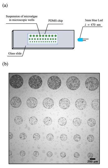
An array of shallow cylindrical wells was made in polydimethylsiloxane (PDMS, Dow-Corning Sylgard 184), using standard soft lithography techniques [17]. The wells had a depth of and diameters ranging from to . The PDMS was rendered hydrophilic by plasma cleaning. Then, a drop of of the algal solution was deposited on the PDMS. The device was closed with a plasma-activated glass slide by applying gentle pressure.
The trapped algae were observed under a Nikon TI microscope, using a 4x objective. Images were recorded at 10 fps with a CMOS camera (Hamamatsu ORCA-Flash4.0 LT, Hamamatsu Photonics, France). A long-pass red filter with a cut-on wavelength (Newport RG645) was placed between the microscope’s light source and the microwells. A 3 mm blue LED with a wavelength and a light intensity of (Planète Leds, France) was placed on the microscope’s plate, approximately away from the microfluidic chip, on the same plane. The LED operating voltage range was between and and its operating current was . The light intensity was tuned by varying the applied voltage. An illustration of the setup is shown in Fig. 1.
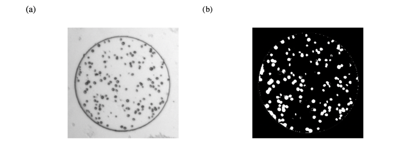
The algae concentration was determined in each well from images captured at the beginning of the experiments, before exposition to a light stimulus. Initially, the algae swam randomly and were distributed homogeneously in each well. They were identified by thresholding the images, and the total area they occupied in each well was determined. The concentration in algae was then defined as the fraction of surface area occupied by the algae in the well.
II.3 Measuring the local light intensity
We estimated the local light intensity inside a well using the number of photons reaching the sensor of the camera. First, we determined the camera offset value by blocking off all the light to the camera and taking a 16-bit image. The spatial average intensity in grey value of all the pixels in the image was the camera’s offset. Then, a 16-bit setup image of the sample was taken at the current experimental condition, with the light of the microscope turned off and the blue LED light on. The camera offset value was then subtracted from each pixel in the setup image. The grey values were first converted to number of electrons by dividing each pixel in the image by the conversion gain of the camera. The electrons were converted to photons by dividing the number of electrons from each pixel by the quantum efficiency (QE) of the sensor at . Last, the local light intensity inside a well was computed as the average number of photons within the well.
III Results
III.1 First experimental results
The experiments consist in exposing a population of C. reinhardtii to a directional source of light.
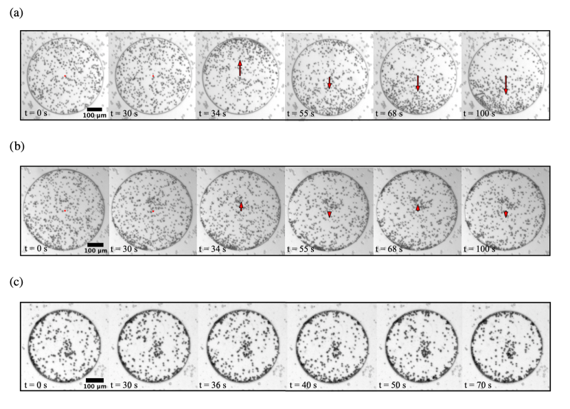
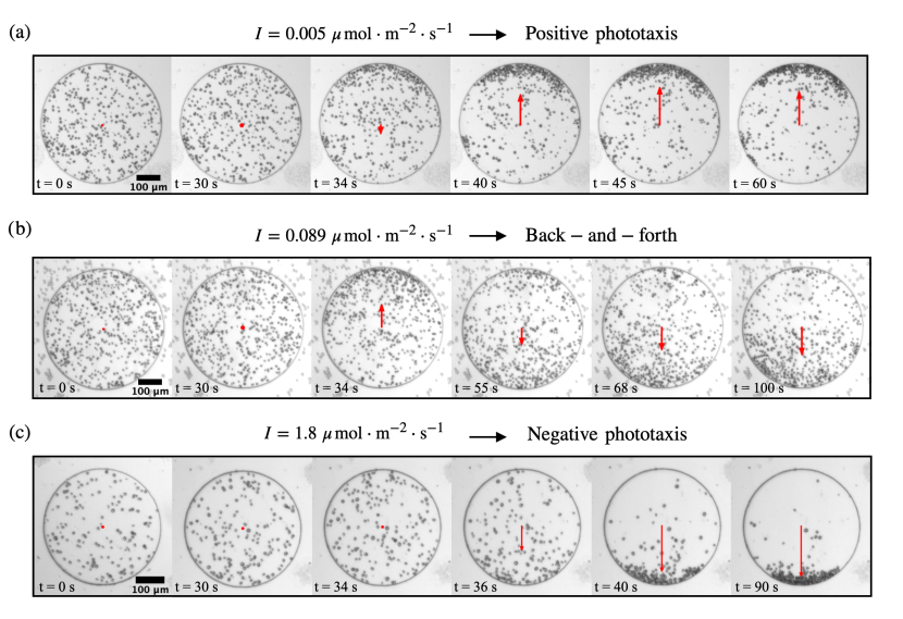
When a population of microalgae is exposed to a light intensity lower than , the algae systematically exhibit positive phototaxis and swim towards the light source. Within to seconds of stimulation, most of the algae have accumulated at the boundary facing the light, see snapshots in Fig. 2a. The algae remain accumulated at the boundary during at least one minute, time after which the imaging was stopped. When the light intensity is higher than , the algae always exhibit negative phototaxis and accumulate at the boundary opposing the light source, see snapshots in Fig. 2c. These results are well known in the literature although the precise value of the threshold intensities varies from experiment to experiment, see e.g. [18, 19].
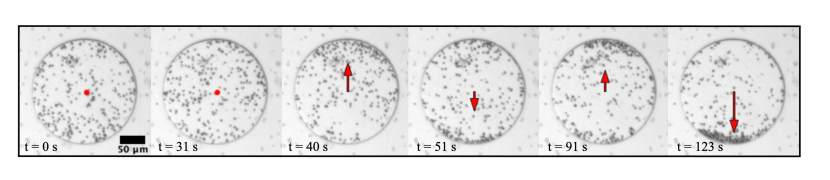
In between these two light intensities, there is a transition regime, where the phototactic behavior is hard to reproduce, despite keeping all experimental parameters identical: the microalgae sometimes exhibit a transient positive phototaxis followed by a negative phototaxis (see Fig. 2b), but can also display purely positive phototaxis, or purely negative phototaxis (see later in text).
The aim of our work is to better understand the parameters responsible for this variety of algal behaviors in the transition regime. We investigate the effects of well diameter, algae concentration, light intensity, and finally history of the algae, on the phototactic behavior.
III.2 Quantification of the phototactic behavior
To quantify the phototactic behavior of a microalgae population constrained in its well, we binarize the experimental images using a simple Otsu threshold on pixel intensities. This leads to images where the algae are white, on a black background (see Supp. Fig. 1). In each image, we then calculate the center of mass of the white pixels, corresponding to the center of mass of the population. The position of the center of mass is tracked over time, and eventually normalized to the radius of the well. A value of the center of mass (resp. ) corresponds to all algae accumulating at the boundary of the well facing (resp. opposite from) the light.
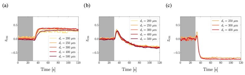
In one experimental run, the phototactic response of 15 – 30 wells containing microalgae is quantified. The field of view contains wells with at least 3 different diameters, and there are at least 4 wells of each diameter, see Fig. 1b. We start with an experiment where the algae are at the same concentration in all wells, and do not find any impact of the well diameter on the phototactic behavior of populations of C. reinhardtii: for all diameters, in one given experiment, the algae exhibit the same behavior, as shown by the evolution of the center of mass of the population, see Fig. 3. Note that the center of mass never reaches . This is mainly due to a fraction of the algae not responding to light, see Supp. Fig. 2. A second-order effect is that algae take space, and so the center of mass of the population can never reach .
The time scales for pure positive and pure negative phototaxis are different: positive phototaxis leads to an accumulation on the side of the light source within (mean std. deviation) after the stimulus is turned on. The time scale for negative phototaxis is slightly longer: . In the case where there is a back-and-forth between positive and negative phototaxis, the first accumulation occurs within while the second accumulation is within . Negative phototaxis always occurs on a time scale longer than positive phototaxis, irrespective of the behavior (back-and-forth or not) of the population.
III.3 Effect of cell concentration
One possibility for the alternating phototactic behavior would be to invoke screening of the light by the algae. We would then expect that at high algae concentration, the algae closest to the light source screen the light intensity, so that the algae further away from the light source see a dimmer light. This could result in a seemingly biphasic behavior, with, at high light intensities, algae close to the light source showing negative phototaxis, and algae further away from the light source exhibiting positive phototaxis.
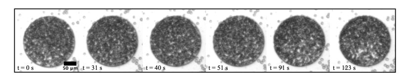
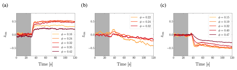
To test this hypothesis, we enclosed microalgae at concentrations to in the microwells, and monitored the evolution of their center of mass. The concentration is defined as the surface fraction of algae in a microwell, see Methods. For , the well is essentially packed with algae, which impedes their motion and so leads to the center of mass not moving even during the light stimulus, see Supp. Fig. 3. For , we do not see any effect of the concentration on the sign of phototaxis: the phototactic behavior depends solely on the light intensity, see Fig. 4. There is however an effect of concentration on the position of the center of mass : when the concentration is higher, the algae take more space and the center of mass of the population gets closer to the center of the well, see Fig. 4a and c.
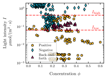
The experiments were reproduced over a range of light intensities and algae concentrations, in more than 300 wells. In each well, the behavior was quantified as positive phototaxis, negative phototaxis, or back-and-forth. The resulting phase diagram of phototactic behavior as a function of light intensity and algae concentration shows no clear influence of the algae concentration on the phototactic behavior, see Fig. 5.
III.4 Effect of history
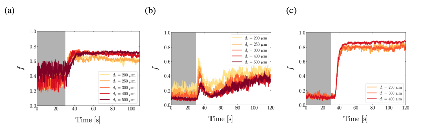
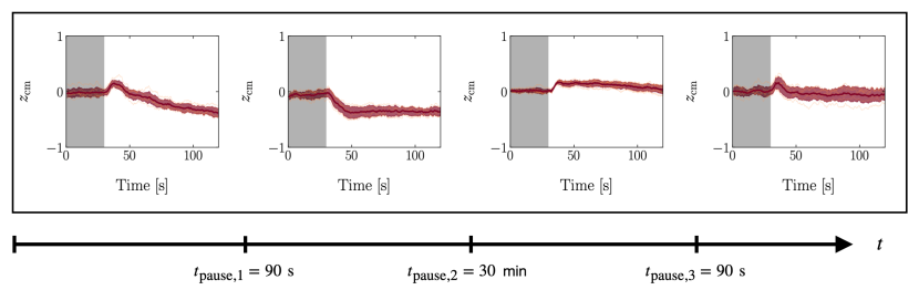
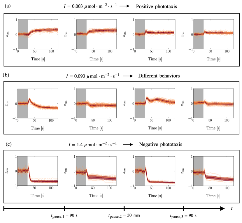
We finally investigated the effect of history on the behavior of the algae. To do so, a population of algae was kept in the dark for one hour and then exposed to a given light stimulus during 90 seconds. The light was then turned off during 90 seconds, after which the exact same stimulus was re-applied. At low light intensities, the qualitative behavior of algae does not change between the two stimuli: the algae always exhibit positive phototaxis, see the first two graphs in Fig. 6a. At high light intensities, the algae always exhibit negative phototaxis, see the first two graphs in Fig. 6c. At intermediate light intensities however, the behavior of the algae changes between the two stimuli: in the first stimulus, the algae exhibit a back-and-forth motion, first towards the light and then away from it. During the second stimulus, the algae show negative phototaxis, see the first two graphs in Fig. 6b.
To understand whether the change in behavior was permanent, the algae were allowed to rest in the dark for 30 minutes after the end of the second stimulus. Then, the same protocol was followed: a third stimulus, of the same intensity as the two previous ones, was applied. After 90 seconds in the dark, the algae were restimulated with a fourth, identical stimulus. In the case of low and high light intensity, the qualitative response of the algae was the same for the third and fourth stimuli as for the two first ones, see the last two graphs in Fig. 6a,c. At intermediate light intensities, the algae showed positive phototaxis during the third stimulus, and then qualitatively changed their behavior during the fourth stimulus to exhibit slightly negative phototaxis, see the last two graphs in Fig. 6b. This last response to light, averaged over 31 wells, is quite subtle. Indeed, after several stimuli, most of the algae stick to the wells, a likely effect of light stimulation [20]. These stuck algae move very slowly by gliding [21], see Supp. Fig. 4. The center of mass of the population then largely reflects the position of the stuck algae. The motile algae however do exhibit negative phototaxis, see Supp. Fig. 4.
Note that the quantitative response of the algae, as measured by the position of the center of mass , differs between the four stimuli – even in the case of low and high light intensities. For example, at low light intensities (positive phototaxis), the shift in the center of mass is identical for stimuli 1 and 2. The shift is less pronounced for stimuli 3 and 4, see Fig. 6a. In contrast, in the case of negative phototaxis, the shift of the center of mass is identical for stimuli 1 and 3, as well as for stimuli 2 and 4, see Fig. 6c: there, resting in the dark for 30 minutes seems to reset the phototactic behavior.
What is important in our case however is the qualitative response: the algae can change phototactic behavior at intermediate light intensities due to the stimuli they previously experienced. The change in behavior is not an adaptive response, which would make the algae show first negative phototaxis, then positive phototaxis in response to the same stimulus. It rather corresponds to an integration of the signal over time. This effect explains the variety of behaviors observed in the transition regime between positive and negative phototaxis: one cannot predict the phototactic response of an alga without knowing its history.
IV Model
We now propose a simplified model to describe our experimental observations. We call the light intensity within a well, and an intracellular biochemical species responsible for the sign of phototaxis. The total concentration of is conserved and is called . This species exists in two states: active (with concentration ) and inactive (with concentration ), such that .
We assume the transition from inactive to active state depends on the light intensity , while the reverse transition occurs at a constant rate :
| (1) |
where is the reaction rate at which is converted into . When the concentration of active molecules is large (resp. small), the algae undergo negative (resp. positive) phototaxis, which can be summarized as:
| (2) |
where is the position of an alga, is the threshold concentration at which cells transition from positive to negative phototaxis, and is the characteristic speed of the alga. Equations (3) and (5) constitute a minimal model of phototaxis.
In the remaining part of this study, all simulations parameters are fixed except for the light intensity . We normalize the concentrations of so that , and place the algae in a well of radius . The position of the alga is then renormalized by this radius to obtain a number between -1 and 1. The light intensities are expressed, as in the experiment, as a flux of photons in . We choose , , , and . Initially, the algae are naive () and . A detailed description of the choice of the model parameters can be found in the Supplementary Material.
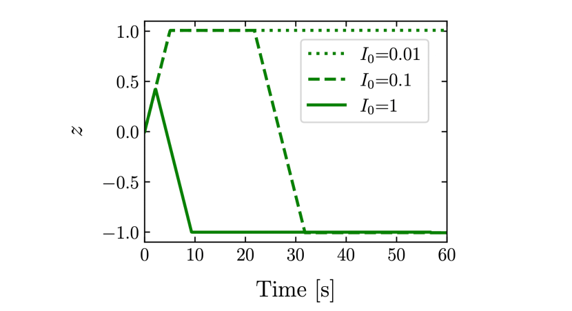
At low light intensities (), the amount of activated chemical species is lower than the threshold , and the resulting behavior is purely positive phototaxis. At intermediate light intensities (), the usage rate and relaxation rate are of similar orders of magnitude, and the behavior can alternate between positive and negative phototaxis, resulting in a back-and-forth motion of the algae. Finally, at high light intensities (), all molecules are instantly converted to an active state, and cannot relax back, resulting in negative phototaxis, see Fig. 7. Note that, at high light intensities, the simulations show an initial small positive phototactic effect; this is similar to what is observed in experiments, see Fig. 6c.
Apart from the qualitative agreement between the different phototactic behaviours, the model also reproduces well the different experimental time scales. Positive phototaxis leads to accumulation within , which depends solely on the swimming speed of the alga. Accumulation due to negative phototaxis occurs on a longer time scale, , due to the initial small positive phototactic effect. The back-and-forth motion leads to accumulation within , a consequence of the addition of three time scales: the time to swim to , the time to produce enough activated species to overcome the threshold , and the time to eventually swim to .
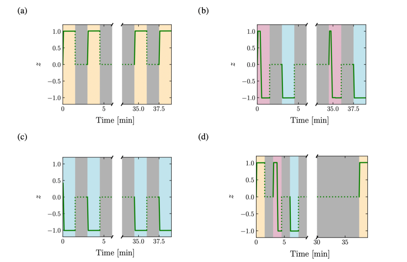
We now simulate the application of successive stimuli of , as in the experiments. Between two stimuli, the alga position is reset to 0. Indeed, in the experiments, the algae repopulate homogeneously the well after one minute without light stimulus, and so the center of mass of the population goes back to 0 before the next experiment. At low () and high () light intensities, applying the same stimulus multiple times does not change the phototactic behavior, independently of the time between stimuli, see Fig. 8a,c.
At intermediate light intensity (), we find that the phototactic behavior depends on the number of stimuli applied, their duration, and on the timing between stimuli. At the beginning of the first simulation, algae are in their naive state. When the stimulus is applied, the number of active molecules increases but is initially lower than the threshold , which leads to positive phototaxis. When the amount of active molecules increases above the threshold value , the phototactic behavior reverts during the experiment, and leads to the overall back-and-forth behavior. The light is then turned off for a duration . After this rest time, the amount of active molecules has decayed but is still above , so that when the algae are restimulated, they display purely negative phototaxis. We then remove the light stimulus for , allowing almost all active molecules to revert back to their inactive state. When the light is switched back on during the third experiment, the algae exhibit the back-and-forth motion: positive phototaxis at the beginning of the experiment, negative phototaxis once the threshold amount of active molecules is reached. Finally, the light is turned off once more for a duration . After this rest time, the amount of active molecules is still above , and the algae display purely negative phototaxis in the last simulation. Simulations results can be seen in Fig. 8b, where the pause times are illustrated with gray-shaded areas.
A prediction of the model is that algae can switch behavior successively from positive phototaxis, to back-and-forth, to negative phototaxis before reverting back to positive phototaxis. Such is the case for three consecutive stimuli of spaced by and a pause of before the fourth stimulus, see Fig. 8d. This switch through all phototactic behaviors requires a precise tuning of both the light intensity and the waiting times, which is experimentally tedious. We were however able to experimentally observe the switch from positive to back-and-forth behavior and from back-and-forth to negative behavior, see Supp. Fig. 5.
V Discussion
We have shown experimentally that the change in phototactic behavior of C. reinhardtii can be due to its recent history of illumination. When exposed to two closely spaced, identical stimuli, the algae act as if they were time-integrating the stimuli. This is visible when the stimulus is close to that delimiting positive and negative phototaxis. Then, the algae show two different behaviors in response to the two consecutive stimuli: first a back-and-forth response, then a negative phototaxis. When the stimuli are sufficiently spaced apart, the algae have had time to relax to a basal state, and show the same qualitative response to the two stimuli, see Fig. 6. In our experiments, we find this time of complete relaxation to be approximately equal to 30 minutes.
A simplified model of phototaxis explains this memory effect by the introduction of a time-scale of relaxation in the inner biochemistry of the cell. When the time-scale of relaxation is larger than the time between two consecutive stimuli, the alga integrates the signal and can display different behaviors in response to the two identical stimuli, see Fig. 8. The time needed to return to the basal state should be of order , which underlies our choice of for the relaxation time constant. Our experimental data are consistent with an inner relaxation time in the range to 20 minutes. What could be the biochemical species corresponding to the intracellular species introduced in the model? It is very likely that our simplified model compounds the dynamics of multiple biochemical species, activated sequentially in a signalling cascade. Still, it is known that the photoreceptors for phototaxis in C. reinhardtii are channelrhodopsins, that become phosphorylated within milliseconds after the application of a light stimulus. A high level of phosphorylated channelrhodopsin-1 (ChR1) in the cell correlates with a change from positive to negative phototaxis [11], much like a high level of activated corresponds to negative phototaxis in our model. Moreover, the time scale of dephosphorylation of ChR1 in C. reinhardtii has been shown to be of the order of to 15 minutes [11], which is compatible with our value of . A possibility is therefore to identify with ChR1.
A more physical model of phototaxis was recently introduced by Leptos et al. to describe the response of C. reinhardtii to light [22]. In their model, Leptos et al. take into account the rotation of the microalga around its axis while swimming, and the resulting oscillating light intensity seen by the eyespot. The consecutive step-up and step-down stimuli sensed by the eyespot during one body rotation lead to a realignment of the trajectory towards the light source. Our model averages out the body rotation of the microalgae and describes the behavior of the microorganism at a more coarse-grained time-scale. Another difference is that the model of Leptos et al. does not take into account the fact that phototaxis changes sign when the light intensity increases. This change of phototactic behavior requires the introduction of a threshold value in the model, below (resp. above) which phototaxis is positive (resp. negative). Such a threshold is incorporated in our simplified model.
Note that the threshold in the model, introduced as a simple means to obtain a change in behavior between positive and negative phototaxis at different light intensities, leads to the back-and-forth behavior at intermediate light intensities. The existence of this threshold also enables to explain why the time scale of negative phototaxis is longer than that of positive phototaxis. Negative phototaxis occurs when the amount of active species overcomes a threshold ; this takes time, during which the alga exhibits positive phototaxis. Only when do the algae show negative phototaxis, and accumulate at one boundary of the well. The small initial positive phototactic response predicted by the model is in agreement with experimental data. The model also predicts that it is possible to switch the phototactic behavior of algae from positive phototaxis to back-and-forth to negative phototaxis, using three consecutive identical stimuli, by adjusting the waiting times between the stimuli and choosing carefully the stimulus intensity, see Fig. 8d. While we were not able to find the experimental conditions corresponding to this theoretical prediction, we were able to experimentally observe the switch from back-and-forth to negative phototaxis, and from positive phototaxis to back-and-forth in a couple experiments, see Supp. Fig. 5.
While the model successfully reproduces the phototactic behavior and the time scales at play, it fails to reproduce two experimental observations. First, in a couple experiments, multiple consecutive back-and-forth of the center of mass were observed at intermediate intensities, see Supp. Fig. 6. Second, in experiments with consecutive stimuli at intermediate light intensities, algae in the third experiment (after a waiting time of 30 minutes) display positive phototaxis instead of the expected back-and-forth behavior observed during the first experiment, see Fig. 6b and Supp. Fig. 5. Both behaviors can be obtained in the model by supposing that the threshold increases with the time spent under a light stimulus, and so depends on the history of the microorganism. Such an increasing threshold would also enable to recover the traditional adaptative behavior at longer time scales [15]. This implies having a third equation describing the time-evolution of , thus introducing another time constant into the problem.
In the lab, the memory effects reported here imply that it is not possible to repeat the same phototaxis experiments on a given batch of cells and expect similar outcomes. Such an effect was already known in the case of long-term adaptation [15]. The data reported here show that even in the case of short-term experiments, the behavior is not necessarily reproducible. To study phototaxis quantitatively, experimental repeats should be carried out with different batches of naive cells. Having identified this short time-scale, our experiments pave the way for the study of phototaxis in the presence of time-varying light stimuli, much as the algae are subject to in their natural environment. There, light is constantly fluctuating. The short-term memory could then be a beneficial way to integrate a light signal randomly interrupted by the shadow of other motile organisms or objects.
Acknowledgments
We thank David Gonzalez-Rodriguez for insightful comments on an early version of the model, Maxime Simon for preliminary experiments, Salome Guttierez-Ramos for help with microfabrication, and Caroline Frot for precious technical support. This work was supported by “Investissements d’Avenir” LabEx PALM (ANR-10-LABX-0039-PALM) and by PEPS “Mécanique du futur”.
References
- Jékely [2009] G. Jékely, Philosophical Transactions of the Royal Society B: Biological Sciences 364, 2795 (2009).
- Foster and Smyth [1980] K. W. Foster and R. D. Smyth, Microbiological reviews 44, 572 (1980).
- Hegemann and Berthold [2009] P. Hegemann and P. Berthold, in The Chlamydomonas Sourcebook (Elsevier, 2009) pp. 395–429.
- Rüffer and Nultsch [1991] U. Rüffer and W. Nultsch, Cell motility and the cytoskeleton 18, 269 (1991).
- Feinleib and Curry [1971] M. E. H. Feinleib and G. M. Curry, Physiologia Plantarum 25, 346 (1971).
- Nultsch et al. [1986] W. Nultsch, J. Pfau, and R. Dolle, Archives of microbiology 144, 393 (1986).
- Morel-Laurens [1987] N. Morel-Laurens, Photochemistry and Photobiology 45, 119 (1987).
- Dolle et al. [1987] R. Dolle, J. Pfau, and W. Nultsch, Journal of plant physiology 126, 467 (1987).
- Takahashi and Watanabe [1993] T. Takahashi and M. Watanabe, FEBS Letters 336, 516 (1993).
- Wakabayashi et al. [2011] K.-i. Wakabayashi, Y. Misawa, S. Mochiji, and R. Kamiya, Proceedings of the National Academy of Sciences 108, 11280 (2011).
- Böhm et al. [2019] M. Böhm, D. Boness, E. Fantisch, H. Erhard, J. Frauenholz, Z. Kowalzyk, N. Marcinkowski, S. Kateriya, P. Hegemann, and G. Kreimer, The Plant Cell 31, 886 (2019).
- Macnab and Koshland Jr [1972] R. M. Macnab and D. E. Koshland Jr, Proceedings of the National Academy of Sciences 69, 2509 (1972).
- Berg and Tedesco [1975] H. C. Berg and P. Tedesco, Proceedings of the National Academy of Sciences 72, 3235 (1975).
- Van Haastert [1983] P. Van Haastert, The Journal of cell biology 96, 1559 (1983).
- Mayer [1968] A. Mayer, Nature 217, 875 (1968).
- Harris et al. [2009] E. Harris, D. Stern, and G. Witman, The chlamydomonas sourcebook, vol. 1 san diego (2009).
- Xia and Whitesides [1998] Y. Xia and G. M. Whitesides, Annual review of materials science 28, 153 (1998).
- Ueki et al. [2016] N. Ueki, T. Ide, S. Mochiji, Y. Kobayashi, R. Tokutsu, N. Ohnishi, K. Yamaguchi, S. Shigenobu, K. Tanaka, J. Minagawa, et al., Proceedings of the National Academy of Sciences 113, 5299 (2016).
- Ramamonjy et al. [2022] A. Ramamonjy, J. Dervaux, and P. Brunet, Physical Review Letters 128, 258101 (2022).
- Kreis et al. [2018] C. T. Kreis, M. Le Blay, C. Linne, M. M. Makowski, and O. Bäumchen, Nature Physics 14, 45 (2018).
- Till et al. [2022] S. Till, F. Ebmeier, A. A. Fragkopoulos, M. G. Mazza, and O. Bäumchen, Physical Review Research 4, L042046 (2022).
- Leptos et al. [2023] K. C. Leptos, M. Chioccioli, S. Furlan, A. I. Pesci, and R. E. Goldstein, Physical Review E 107 (2023).
Supplementary Material
.1 Determining the parameters of the simplified phototactic model
The dynamics of the inner biochemical species in our simplified phototaxis model evolve according to:
| (3) |
where is the reaction rate at which the inactive species of concentration is converted into the active species of concentration . The total concentration is conserved and is called . The transition from inactive to active state depends on the light intensity , while the reverse transition occurs at a constant rate .
The solution to this equation writes
| (4) |
We use for simplicity. To obtain time scales close to the experimental time scales, we choose . Indeed, we know experimentally that there is a memory effect when stimulating a second time after a pause of a couple minutes, so . We also know that the phototactic behavior is reset after 30 minutes, so .
Then, we assume the position of the alga evolves according to:
| (5) |
where is the threshold concentration at which cells transition from positive to negative phototaxis, and is the characteristic speed of the alga. We know experimentally that .
Two parameters now need to be determined, for which there is no direct experimental input: and . To estimate them, we use the fact that, at , the algae exhibit a back-and-forth behavior, with negative phototaxis occuring at . We then know that at this intensity, , and so:
| (6) |
For each value of there is therefore a corresponding value of that enables to obtain the back-and-forth behavior at . We choose arbitrarily , so that for . This determines .
.2 Supplementary figures