UniChest: Conquer-and-Divide Pre-training for Multi-Source Chest X-Ray Classification
Abstract
Vision-Language Pre-training (VLP) that utilizes the multi-modal information to promote the training efficiency and effectiveness, has achieved great success in vision recognition of natural domains and shown promise in medical imaging diagnosis for the Chest X-Rays (CXRs). However, current works mainly pay attention to the exploration on single dataset of CXRs, which locks the potential of this powerful paradigm on larger hybrid of multi-source CXRs datasets. We identify that although blending samples from the diverse sources offers the advantages to improve the model generalization, it is still challenging to maintain the consistent superiority for the task of each source due to the existing heterogeneity among sources. To handle this dilemma, we design a Conquer-and-Divide pre-training framework, termed as UniChest, aiming to make full use of the collaboration benefit of multiple sources of CXRs while reducing the negative influence of the source heterogeneity. Specially, the “Conquer" stage in UniChest encourages the model to sufficiently capture multi-source common patterns, and the “Divide" stage helps squeeze personalized patterns into different small experts (query networks). We conduct thorough experiments on many benchmarks, e.g., ChestX-ray14, CheXpert, Vindr-CXR, Shenzhen, Open-I and SIIM-ACR Pneumothorax, verifying the effectiveness of UniChest over a range of baselines, and release our codes and pre-training models at https://github.com/Elfenreigen/UniChest.
1 Introduction
Chest X-Ray (CXR) in screening chest diseases is essential to detect and control their fatal infectious impact on human lives in broad countries [25]. To reduce the labor costs, deep learning techniques have become prevalent for machine-assisted CXR diagnosis, driving medical imaging recognition into the new era [35]. Specially, with the rapid development of pre-training models, extensive studies have been conducted and shown promise in a wide range of tasks and domains [38], drawing increasing attention in medical community.
Recently, Vision-Language Pre-training techniques has significantly improved the performance of machine-aided CXR disease diagnosis [5, 36, 40, 44]. Some studies even have shown the potential of surpassing the experienced radiologists in diagnosing some chest diseases [44]. Besides, in combination with medical domain-specific knowledge, these pretrained models exhibit more reasonable explanations in the lesion grounding [40, 44]. Nevertheless, it is worth noting that these works for CXR VLP only consider pre-training on a single-source dataset e.g., MIMIC-CXR of about 300K samples [22]. Recalling the practice of GPT [10] or CLIP [32] that utilizes billions of multi-source samples, single-source VLP inherently induces drawbacks in the disease coverage and representativeness [22], especially under real-world medical applications.
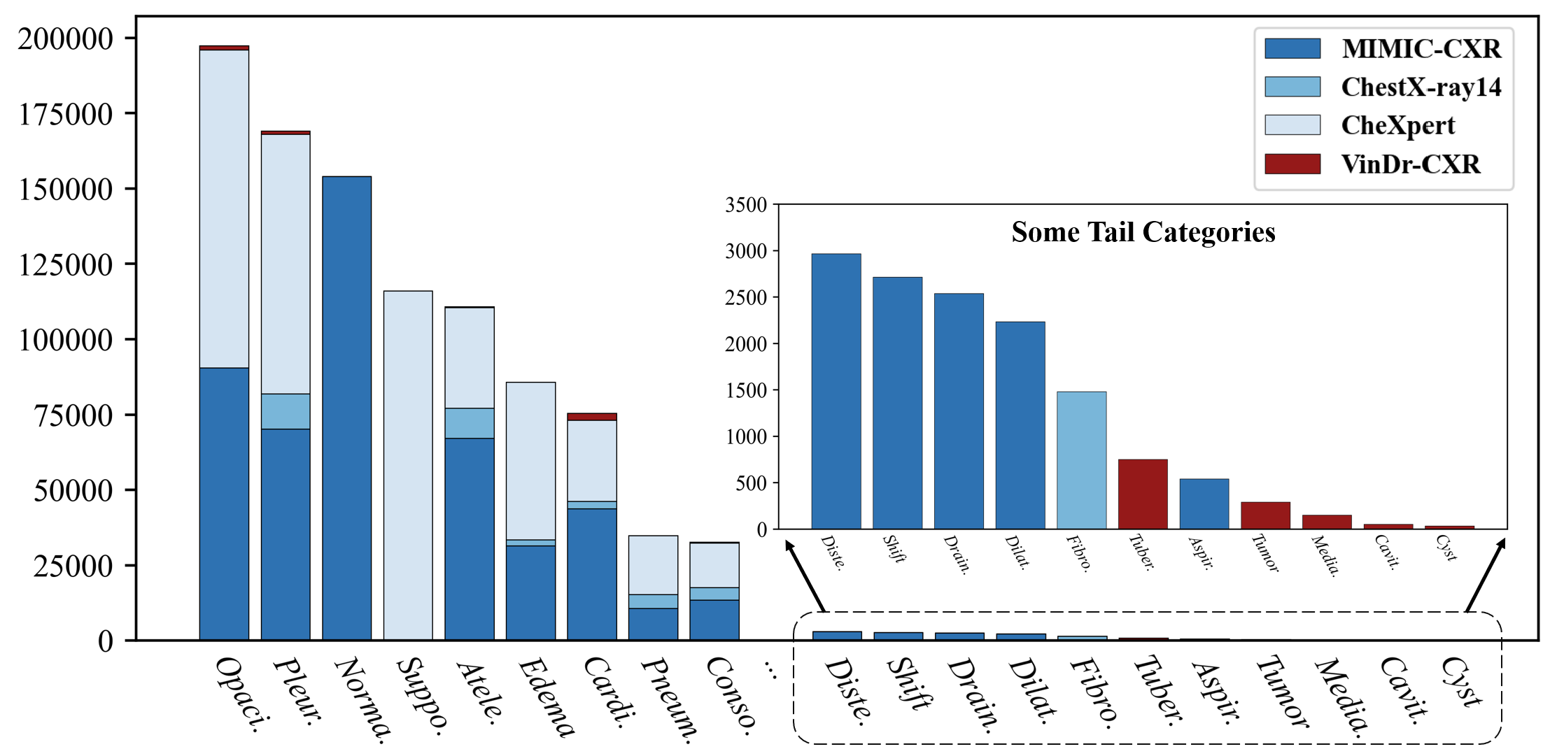
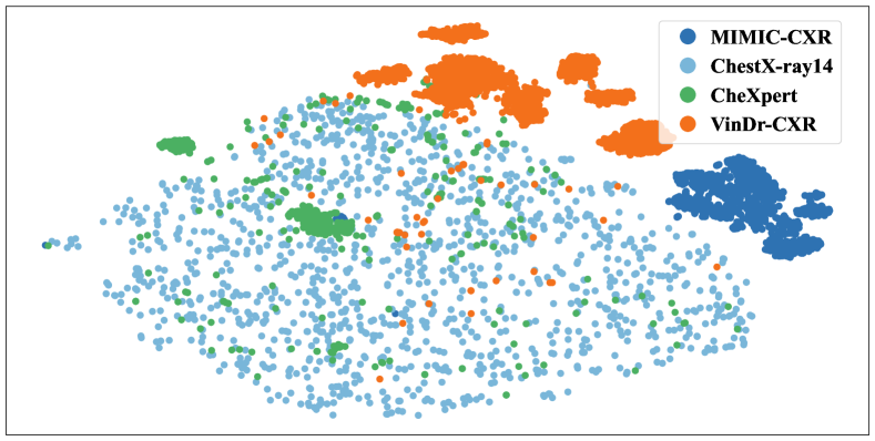
Motivated by the above limitation, we explore building a more powerful pre-training framework by leveraging multi-source CXR data. Generally, the label space union of multiple sources can help expand the coverage of the disease categories, particularly for rare diseases. Besides, samples from different sources with diverse radiation equipment, collection standards and population distributions, may complement each other [42], which helps enhance the generalization ability of pretrained models. Unfortunately, we argue that it is still very challenging to effectively utilize multi-source CXRs, as the source heterogeneity (shown in Fig. 1) also exacerbates the complexity of the CXR disease data, which could impair the holistic improvement for all source tasks during pre-training.
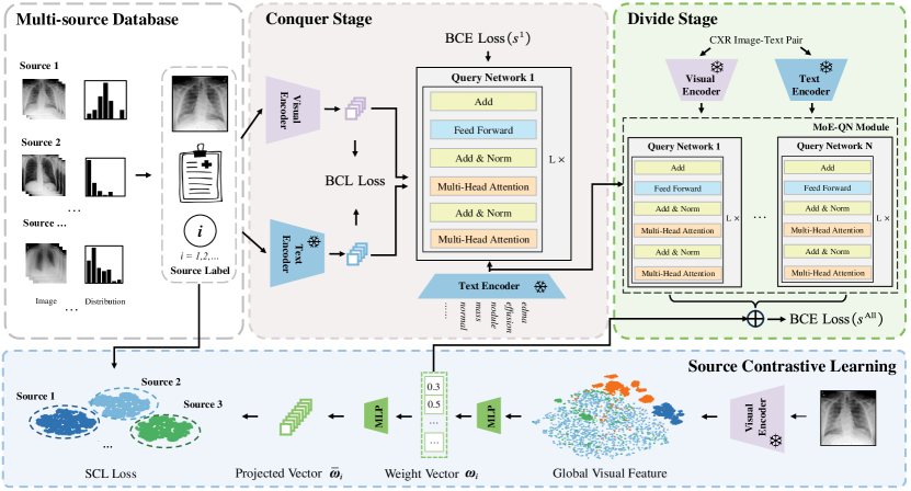
To address this dilemma, we propose a Conquer-and-Divide pre-training framework, termed as UniChest, which maintains the merits of multi-source data collaboration and simultaneously weakens the negative effect of the source heterogeneity by a proper training isolation. Specifically, an the “Conquer" stage of UniChest, we promote the capture of multi-source common patterns at first in parts of the model, which focuses on enhancing the feature extraction abilities. Then, at the “Divide" stage of UniChest, we introduce a mixture of experts warmed up from previous stage, together with the guidance of source contrastive learning, to squeeze the source-specific patterns into different experts, which reduces the interference among sources during pre-training. With this framework, we can achieve better efficiency and effectiveness for pre-training on the large-scale multi-source CXR data. In a nutshell, the contribution of this work can be summarized as follows:
-
•
We are among the first attempts to explore pre-training on the multi-source CXR data and propose a Conquer-and-Divide pre-training framework to overcome the dilemma induced by the source heterogeneity in scaling up data.
-
•
We design a mixture of deep query networks together with a source contrastive learning loss to squeeze source-specific patterns into separate components, promoting a harmonious multi-source collaboration for pre-training.
- •
2 Related Works
2.1 Deep Learning for Chest X-Ray Disease Diagnosis
Considering the labor and repeatability of human experts in CXR diagnosis, it is possible to find the computer-aided solutions powered by deep learning [37, 15, 6, 24]. Thereby, extensive CNN-based methods for CXRs have been explored in recent years [30]. For instance, Khoiriyah [23] built a network comprising of three convolutional layers and three connected layers, showing remarkable performance in automatic pneumonia detection. The enhancement by transfer learning further reduced the training cost and improved the generalization performance [41, 26]. Specifically, several studies utilized models pre-trained on the natural domain e.g., ImageNet [12] as initialization and finetuned the last layer [8]. In addition, post-hoc techniques can be also very beneficial to enhance the stability and accuracy of the CXR diagnosis. For example, some explorations [14, 9] focus on the model ensemble, i.e., directly summarizing the multiple outputs of a series of models as the final prediction, achieving the remarkable performance.
2.2 Vision-Language Pre-training in Medical Domain
Recently, Vision-Language Pre-training (VLP) models have achieved impressive success in natural domain [3, 21, 27], which then drives many extensions in the medical area and improves the ability of machine-aided medical applications. ConVIRT [45] made the first attempt to integrate VLP into medical models, which follows the two-stream paradigm with the bidirectional contrastive learning. GLoRIA [17] explored the fine-grained information contained in the image and report, proposing a framework for learning both global and regional representations of two modalities. BioVIL [5] focused on the representation of radiological reports and proposed a radiology-specific text encoder along with the classical VLP paradigm. CheXzero [36] retrained a pre-trained CLIP model [32] on the CXR data and showed considerable improvement. MedKLIP [40] extracted entities from reports and converted them to the medical-specific knowledge descriptions, which enhanced the model reasoning ability. KAD [44] built up a medical knowledge graph to fine-tune text encoder and performed the image-text contrastive learning with paired chest X-ray pairs, showing state-of-the-art capability on common benchmarks. However, all these works mentioned above are pre-trained on a single dataset from the identical source, which overlooks the non-negligible heterogeneity problem in their corresponding application on the multi-source CXR data.
3 Methodology
In this section, we first present the problem formulation and the motivation of our study. Then, we will introduce the basic model design that consists of modality-specific backbones and the MoE-QN module. Finally, we provide detailed descriptions and analysis of the Conquer-and-Divide pre-training stages.
3.1 Problem Formulation
Assuming that we have a training set of N samples collected from the multiple sources, , where denotes the CXR image, is the label set indicating what diseases are found for , means the source identity, and denotes the report of . Note that, for those samples that have no reports, we convert to correspondingly. Our goal is to train a vision-language pre-training model on the given multi-source data , which can accurately diagnose the chest diseases. Specifically, in the inference phase, the model can estimate the diseases in the given set for any CXR image.
3.2 Motivation
Generally, scaling up CXR samples for pre-training should be useful to improve generalization as discussed in the previous sections. However, we should point out that the resulting multi-source samples, on one hand, enjoy the better diversity w.r.t. training samples and label space, yet on the other hand, suffer from the non-negligible source heterogeneity issue as shown in Fig. 1. Physically, the locales and time frames of source samples can be quite diverse, as ChestX-ray14 [39] contains the samples captured from 1992 to 2015 in the U.S. while VinDr-CXR [31] consists of CXRs in Vietnam from 2018 to 2020. Despite visually similar CXRs in human eyes, the model can actually react very differently in the face of some imperceptive factors like light circumstances and instrument qualities [33]. From the t-SNE visualization in Fig. 1, we can find that the distributions of various sources exhibit the distinct disparities.
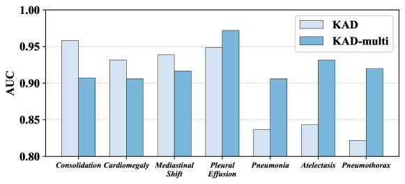
To further under the influence of the source heterogeneity, we implement the straightforward pre-training on the hybrid multi-source data and compare its performance with the pre-training on single source. Specifically, we adopt the current state-of-the-art (SOTA) KAD [44] to conduct the pre-retraining on MIMIC-CXR dataset, and compare with the pre-training on the multiple sources (termed as KAD-multi). As shown in Fig. 3, KAD-multi does not achieve the consistent improvement, and on some certain diseases, KAD-multi even significantly lags behind the vanilla KAD.
With this observation, we re-think the early VLP paradigm that is naively applied in scaling up CXR data and overlooks the source heterogeneity issue. To handle this dilemma, we actually should allow the pre-training to capture the multi-source common patterns and simultaneously can maintain the source-specific patterns, which motivates us to incorporate the philosophy of “Conquer" and “Divide" design for pre-training.
3.3 Conquer-and-Divide Pre-training
In this part, we describe the proposed Conquer-and-Divide pre-training framework, which includes the model architecture, the loss design and the training schedules, detailed as follows.
3.3.1 The Model Architecture
We follow the prevalent vision-language pre-training paradigm with the proper tailored design to train a diagnosis model, which consists of two modality-specific encoders and one modality-interaction module. Note that, this is different from the classical supervised framework that directly maps the input image space to the label space. The merits are tri-fold: First, we can inject more knowledge into the label space via the textual encoder, which is richer in semantics than the naive one/multi-hot label vector. The second is that we can incorporate the prior knowledge to promote the learning efficiency of vision encoder, when the textual description for medical images is available, e.g., the report information of samples from MIMIC. Finally, VLP allows us to achieve better generalization for open-set categories by means of the powered textual encoder. In the following, we concretely describe the architecture design of each component in our UniChest.
Given a sample , we take the ResNet-50 as the visual backbone to encode and adopt the output of the -th residual block as the image representation [40], denoting as . For the report information , RadGraph [20] is used to extract key entities and filter the irrelevant words. When reports containing more than one sentence, we extract entities sentence by sentence, and use [SEP] token as the separation between different entities111For CXR samples without reports, we use their corresponding labels as the input entity and also take [SEP] token to separate the multiple labels.. After the entity extraction, we use PubMedBERT [13] pre-trained on Unified Medical Language System (UMLS) data [4] as textual backbone for generating text representation [44]. For clarity, we summarize the modality-specific encoding process with equations as below,
| (1) |
For the disease prediction, we introduce a MoE-QN Module, namely, mixture of query networks, where each query network consists of a few transformer decoder layers. The MoE-QN module plays the role of overcoming the source heterogeneity issues by squeezing the source-specific patterns into different query networks, but its training should carefully follow our Conquer-and-Divide schedules, which will be discussed in the subsequent sections. For each query network, we take the fine-grained visual representation as Key and Value, and utilize the textual representation of the disease set (similarly encoded by ) as Query. The output from the sequential transformer decoder will be fed to one MLP layer to obtain the prediction of each Query Network,
| (2) |
where is the total class number of all sources and is the number of query networks. Then, we transform of all query networks to the total prediction by an automatic linear combination, which will be described at the “Divide" stage.
3.3.2 The Training at the “Conquer" Stage
As discussed in the earlier sections, the source heterogeneity issue can be the core bottleneck to deteriorate the performance of pre-training for the consistent improvement for each source task. Therefore, at this “Conquer" stage, we first encourage the model to capture multi-source common patterns as many as possible. In brief, the training loss of this stage involves two parts, the image-text bidirectional contrastive loss and the multi-label cross-entropy loss w.r.t. the first query network prediction. Taking a batch of samples as an example, emphasizes the alignment between the global visual representation and the textual representation, which could be formulated as follows,
| (3) |
where is the average pooling of along the first two dimensions so that we reduce it to the same dimension of , and is temperature with the default setting 1.0 following [7]. Regarding , we only enforce the supervision on the first query network and compute the multi-label cross-entropy loss at this stage. Note that, samples in some domains may have smaller label space than the union label space of all sources. In this case, we neglect the computation between prediction and missing classes of these samples, even if it may belong to them (but are unobserved). Finally, we sum up and (only involves the first query network) as the overall loss for each mini-batch at the “Conquer" stage as follows
| (4) |
3.3.3 The Training at the “Divide" Stage
Previous stage follows the classical pre-training spirit, where we treat multiple sources equally, and expect that the model can sufficiently learn the multi-source common patterns. At the “Divide" phase, we mainly explore to mediate the negative impact induced by the source heterogeneity. Concretely, on the basis of the warming-up during the “Conquer" stage, we train all the query networks with freezing all the other parts of the model. Here, the multi-label cross-entropy loss will be implemented on the ensemble prediction from , unlike using in Eq. (4). The final prediction is characterized by the following equation
| (5) |
where is the hyperparameter to balance the training of the first query network and the remaining query networks, and is the learnable weight to summarize the contribution of the source-specific modules for the diagnosis prediction. Note that, the intuition behind Eq. (5) is to inherit the training gains at the “Conquer" stage by , and simultaneously squeeze the source-specific patterns to the other query networks by .
Source Contrastive Learning. In Eq. (5), we introduce the learnable weights to incorporate the source-specific patterns into the other query networks. However, without any guidance, it is challenging to achieve this goal by optimization. Here, we introduce a source contrastive learning to promote the desire. Specifically, we transform the global visual representation into a simplex via one MLP layer as the weight vector, namely, . Then, is projected into a higher dimension space by another MLP layer , denoted as , to perform a source contrastive learning with the guidance of source id in the following
| (6) |
Above contrastive learning helps us to learn the similar weight vector for the samples from the same source, which makes the source-specific patterns learned by the similar query networks. Under this mechanism, we naturally learn an automatic optimal assignment for the remaining query networks to overcome the source heterogeneity issue. Then, with Eq. (5) and Eq. (6), the overall loss at the “Divide" stage can be formulated as
| (7) |
| Purpose | Dataset | # Samples | # Diseases | Region | Collection Institution | Time Scope |
| Pre-training | MIMIC-CXR | 348900 | 41 | Northeast USA | MIT | 2011-2016 |
| ChestX-ray14 | 98637 | 14 | Northeast USA | NIH | 1992-2015 | |
| CheXpert | 223414 | 14 | Western USA | Stanford Hospital | 2002-2017 | |
| VinDr-CXR | 15000 | 28 | Vietnam | VinBrain | 2018-2020 | |
| Multi-CXR | 685951 | 60 | USA & Vietnam | MIT, etc. | 1992-2020 | |
| Downstream | Shenzhen | 662 | 1 | Southeast China | Guangdong Medical College | Sep. 2012 |
| Open-I | 3547 | 14 | USA | NLM | / | |
| SIIM-ACR | 2135 | 1 | Northeast USA | Kaggle | / | |
| PadChest | 39053 | 193 | Spain | University of Alicante | 2009-2017 | |
| ChestX-Det10 | 542 | / | Northeast USA | Deepwise | 1992-2015 |
| Method | Mean | Ate. | Car. | Eff. | Inf. | Mas. | Nod. | Pne. | Pne. | Con. | Ede. | Emp. | Fib. | Thi. | Her. |
| ConVIRT | 0.808 | 0.771 | 0.867 | 0.825 | 0.703 | 0.818 | 0.761 | 0.722 | 0.857 | 0.747 | 0.854 | 0.901 | 0.809 | 0.771 | 0.909 |
| GLoRIA | 0.800 | 0.760 | 0.855 | 0.818 | 0.700 | 0.814 | 0.749 | 0.715 | 0.828 | 0.739 | 0.832 | 0.887 | 0.813 | 0.767 | 0.921 |
| BioViL | 0.800 | 0.765 | 0.871 | 0.824 | 0.697 | 0.819 | 0.752 | 0.710 | 0.845 | 0.742 | 0.842 | 0.871 | 0.821 | 0.759 | 0.888 |
| MedKLIP | 0.801 | 0.764 | 0.849 | 0.823 | 0.697 | 0.820 | 0.747 | 0.712 | 0.839 | 0.751 | 0.848 | 0.879 | 0.817 | 0.777 | 0.892 |
| KAD | 0.825 | 0.785 | 0.897 | 0.840 | 0.713 | 0.836 | 0.771 | 0.740 | 0.874 | 0.753 | 0.860 | 0.916 | 0.829 | 0.778 | 0.961 |
| UniChest | 0.858 | 0.823 | 0.922 | 0.861 | 0.739 | 0.850 | 0.778 | 0.777 | 0.933 | 0.795 | 0.893 | 0.958 | 0.877 | 0.837 | 0.977 |
3.3.4 Difference from the pre-training with fine-tuning
The proposed Conquer-and-Divide pre-training is intrinsically different from the ordinary fine-tuning after pre-training, although both of them are a two-stage process. First, the “Divide" training of UniChest is towards all multi-source data, while the fine-tuning after pre-training is narrowed down to a single-source scenario. This leads to the distinction in the generalization ability of two frameworks, where UniChest is significantly stronger than pre-training with fine-tuning as supported by Table 2. Second, the “Divide" stage is to properly mediate the heterogeneity patterns in a broad sense, which does not mean these patterns are not related to the generalization. Conversely, the downstream task can still apply the ordinary fine-tuning ways to leverage these patterns by automatic activated weights for diagnosis, and achieve the fast adaptation. Finally, when it comes to the experimental comparison, we mainly focus on the zero-shot performance of pre-training, instead of the performance by the fine-tuning, which follows the prevalent pre-training spirit that tries to avoid the expensive tuning cost for downstream tasks as much as possible.
4 Experiments
In this section, we will introduce the datasets for pre-training and downstream tasks, evaluation metrics and implementation details will also be detailedly described. At last, we will present the experimental results of our proposed method compared with other baselines.
4.1 Datasets
We combine some common CXR datasets containing MIMIC-CXR [22], ChestX-ray14 [39], CheXpert [18] and VinDr-CXR [31] for pre-training, namely Multi-CXR, which yields a grand total of 685,951 Chest X-Ray images.
For evaluating the generalization performance of the pre-trained model, we adopt Shenzhen [19], Open-I [11], SIIM-ACR Pneumothorax [1] and PadChest [11] for zero-shot classification and ChestX-Det10 [28] which is the subset of ChestX-ray14 for intuitive lesion grounding. More detailed statistics of pre-training and downstream datasets can be found in Table 1. We can observe that significant diversity exists in the collection time, population distribution, disease category coverage and sample scales among different datasets.
4.2 Implementation Details
The pre-training process of UniChest is conducted on a single NVIDIA A100 GPU for 30 epochs in the first “Conquer” stage and 20 epochs in the second “Divide” stage. The starting checkpoint for the second stage is the one that performed the best on the ChestX-ray14 validation set. The depth of each query network in MoE-QN Module is set to 4. The hyperparameter which weights the first query network during the “Divide” stage is set to be and the total number of query network is in default. We set the training batch size as 32 and resize input images as 512 512. We adopt the AdamW optimizer in conjunction with the cosine annealing scheduler for managing the learning rate, where the initial learning rate is . To perform data augmentation, we additionally utilize the Fourier amplitude mixup method, which has been demonstrated to be effective for medical imaging data [43, 29]. Besides, we utilize the ASL loss [34] as . These techniques have demonstrated promising outcomes in the analysis of CXR data.
| Dataset | ChestX-ray14 | CheXpert | VinDr-CXR | |||||||||
| Method | aAUC | aF1 | aACC | mAP | aAUC | aF1 | aACC | mAP | aAUC | aF1 | aACC | mAP |
| ConVIRT | 0.5804 | 0.1623 | 0.5385 | 0.1008 | 0.6640 | 0.3609 | 0.7232 | 0.2990 | 0.6803 | 0.1748 | 0.8233 | 0.1129 |
| GLoRIA | 0.6099 | 0.1803 | 0.5616 | 0.1220 | 0.6889 | 0.4094 | 0.7472 | 0.3580 | 0.6775 | 0.1953 | 0.7796 | 0.1380 |
| BioViL | 0.5766 | 0.1561 | 0.5842 | 0.0948 | 0.5769 | 0.3080 | 0.6446 | 0.2157 | 0.5856 | 0.1507 | 0.7726 | 0.1048 |
| CheXzero | 0.6872 | 0.2205 | 0.7698 | 0.1597 | 0.7537 | 0.4488 | 0.8136 | 0.4069 | 0.7407 | 0.2354 | 0.8562 | 0.1713 |
| MedKLIP | 0.7233 | 0.2541 | 0.7946 | 0.1860 | 0.8389 | 0.5385 | 0.8672 | 0.5130 | 0.7770 | 0.2433 | 0.8517 | 0.1897 |
| KAD | 0.7933 | 0.3363 | 0.8639 | 0.2746 | 0.8165 | 0.5467 | 0.7835 | 0.5200 | 0.7599 | 0.2960 | 0.8724 | 0.2315 |
| KAD-CXR14 | 0.8380 | 0.4006 | 0.8913 | 0.3401 | 0.7455 | 0.4296 | 0.7658 | 0.3901 | 0.7711 | 0.2399 | 0.8410 | 0.1740 |
| KAD-CXP | 0.7014 | 0.2444 | 0.7744 | 0.1767 | 0.8804 | 0.5964 | 0.8846 | 0.5606 | 0.7382 | 0.2256 | 0.8101 | 0.1619 |
| KAD-VC | 0.6372 | 0.1881 | 0.6776 | 0.1275 | 0.7228 | 0.4161 | 0.7811 | 0.3285 | 0.8621 | 0.3839 | 0.9418 | 0.3255 |
| MedKLIP-multi | 0.7915 | 0.2994 | 0.8691 | 0.2313 | 0.7010 | 0.4244 | 0.7395 | 0.3410 | 0.7879 | 0.2802 | 0.9167 | 0.2196 |
| KAD-multi | 0.8431 | 0.4077 | 0.8905 | 0.3457 | 0.8819 | 0.5912 | 0.8713 | 0.5626 | 0.8716 | 0.3698 | 0.9390 | 0.3202 |
| UniChest | 0.8584 | 0.4293 | 0.8999 | 0.3797 | 0.9005 | 0.6446 | 0.8912 | 0.6328 | 0.8807 | 0.4028 | 0.9501 | 0.3520 |
| Dataset | Shenzhen | Open-I | SIIM-ACR Pneumothorax | |||||||||
| Method | AUC | F1 | ACC | AP | aAUC | aF1 | aACC | mAP | AUC | F1 | ACC | AP |
| ConVIRT | 0.7166 | 0.6908 | 0.6375 | 0.7619 | 0.6265 | 0.1855 | 0.7504 | 0.1115 | 0.6356 | 0.4322 | 0.4778 | 0.2948 |
| GLoRIA | 0.5831 | 0.6733 | 0.5060 | 0.6001 | 0.6449 | 0.2218 | 0.8134 | 0.1535 | 0.5342 | 0.3770 | 0.4047 | 0.2084 |
| BioViL | 0.6227 | 0.6872 | 0.5544 | 0.6174 | 0.5669 | 0.1515 | 0.6966 | 0.0941 | 0.4678 | 0.3109 | 0.1836 | 0.1761 |
| CheXzero | 0.7801 | 0.7302 | 0.6692 | 0.7969 | 0.6887 | 0.2429 | 0.8689 | 0.1850 | 0.6879 | 0.4722 | 0.5588 | 0.3240 |
| MedKLIP | 0.7909 | 0.7741 | 0.6906 | 0.7986 | 0.7219 | 0.2897 | 0.9048 | 0.2119 | 0.8924 | 0.6833 | 0.8428 | 0.6869 |
| KAD | 0.8856 | 0.8474 | 0.7893 | 0.8141 | 0.7346 | 0.3248 | 0.8808 | 0.2603 | 0.8967 | 0.6480 | 0.8385 | 0.6093 |
| KAD-CXR14 | 0.8764 | 0.8037 | 0.8000 | 0.8947 | 0.6986 | 0.2676 | 0.8617 | 0.1856 | 0.9162 | 0.6561 | 0.8348 | 0.6872 |
| KAD-CXP | 0.7204 | 0.6793 | 0.6031 | 0.7611 | 0.7701 | 0.3314 | 0.9011 | 0.2631 | 0.9132 | 0.6554 | 0.8456 | 0.6771 |
| KAD-VC | 0.8832 | 0.8448 | 0.8578 | 0.9186 | 0.7012 | 0.2112 | 0.8468 | 0.1503 | 0.8002 | 0.4995 | 0.7661 | 0.4012 |
| MedKLIP-multi | 0.8431 | 0.7780 | 0.7855 | 0.8817 | 0.6440 | 0.2270 | 0.7943 | 0.1469 | 0.8958 | 0.6888 | 0.8412 | 0.6853 |
| KAD-multi | 0.8794 | 0.8419 | 0.8547 | 0.9180 | 0.7736 | 0.3409 | 0.9065 | 0.2749 | 0.9016 | 0.6515 | 0.8395 | 0.5847 |
| UniChest | 0.9499 | 0.8906 | 0.8913 | 0.9598 | 0.7830 | 0.3520 | 0.9104 | 0.2877 | 0.9260 | 0.6928 | 0.8807 | 0.6929 |
4.3 Evaluation Metrics
Some common metrics for multi-label classification are adopted to evaluate the model performance, i.e., area under curve (AUC), F1 score, accuracy (ACC) and average precision (AP) for each category and their average value, namely, average AUC (aAUC), average F1 (aF1), average accuracy (aACC) and mean average precision (mAP), for comprehensive comparison. Following the strategy of MedKLIP [40], the final binary threshold of each category prediction is the value when the maximum F1 score is achieved and ACC metric also adopts this threshold.
4.4 Baselines
We consider a wide range of baselines in Chest X-Ray pre-training, including ConVIRT [45], GLoRIA [17], BioVIL [5], CheXzero [36], MedKLIP [40], and KAD [44].
ConVIRT trains two modality-specific encoders by bidirectional contrastive loss to learn visual representations. GLoRIA utilizes both global and fine-grained features for medical VLP. BioVIL proposes a radiology-specific text encoder for the subsequent classical pipeline of VLP. CheXzero retrains one CLIP model with a corpus of the medical domain. MedKLIP designs one novel entity extraction and transition module to inject domain-specific knowledge into the process of VLP. KAD incorporates a medical knowledge graph to further improve the capability of current VLP models in Chest X-Ray, showing SOTA performance on some common public benchmarks.
4.5 Results
In this part, we provide the results of in-domain evaluation on pre-training datasets and zero-shot evaluation on downstream datasets. Additionally, we showcase the lesion grounding capability through intuitive examples.
4.5.1 In-domain evaluation on pre-training datasets
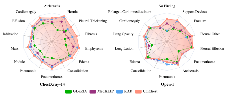
We compare UniChest with other baselines on the test set of pre-training datasets in Table 3. UniChest demonstrates a notable improvement in various evaluation metrics compared to the best baseline models. Specifically, for ChestX-ray14, UniChest shows a significant improvement of 6.51%, 9.30%, 3.60% and 10.51% respectively as to aAUC, aACC, aF1 and mAP than the best baseline. In terms of fine-grained classification for each category, UniChest demonstrates persistent gains as shown in Fig, 4 (left). For CheXpert, UniChest outperforms 6.16%, 9.79%, 2.40% and 11.28%. In the case of VinDr-CXR, UniChest surpasses the best baselines by 12.08%, 10.68%, 7.77% and 12.05%. By introducing multi-source CXR datasets and the Conquer-and-Divide pre-training framework, the diagnosis ability of VLP model has been significantly enhanced.
To mitigate the impact of inconsistent training data, we also conduct variants of the baselines that utilize the same training data with UniChest. These variants include models trained on single-source data (i.e., KAD-CXR14, KAD-CXP, and KAD-VC in Table 3) as well as models trained on multi-source data (i.e., MedKLIP-multi and KAD-multi). KAD-CXR14 outperforms KAD on ChestX-ray14 with improvements of 4.47% in aAUC, 6.43% in aACC, 2.74% in aF1, and 6.55% in mAP. However, it falls short compared to UniChest, which achieves better results by 2.04%, 2.87%, 0.86%, and 3.96% respectively. KAD-CXP and KAD-VC also demonstrate similar performance. Moreover, it is worth noting that while the single dataset-based KAD models may perform reasonably well on their corresponding datasets, their generalization capability to other datasets is limited. This is evident in the case of KAD-VC’s performance on ChestX-ray14, where it may not generalize effectively. For the baseline variants that utilize multi-source data, MedKLIP-multi and KAD-multi, their overall performance exceeds that of their vanilla methods and achieves comparable or even better results than using only the corresponding single-source data. However, these two methods of direct data replenishment lag behind our proposed UniChest with a substantial margin. Generally speaking, the comparison with these variants validates the necessity and importance of developing a multi-source CXR foundational model and demonstrates the effectiveness of our framework design.
4.5.2 Zero-shot classification evaluation
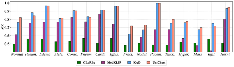
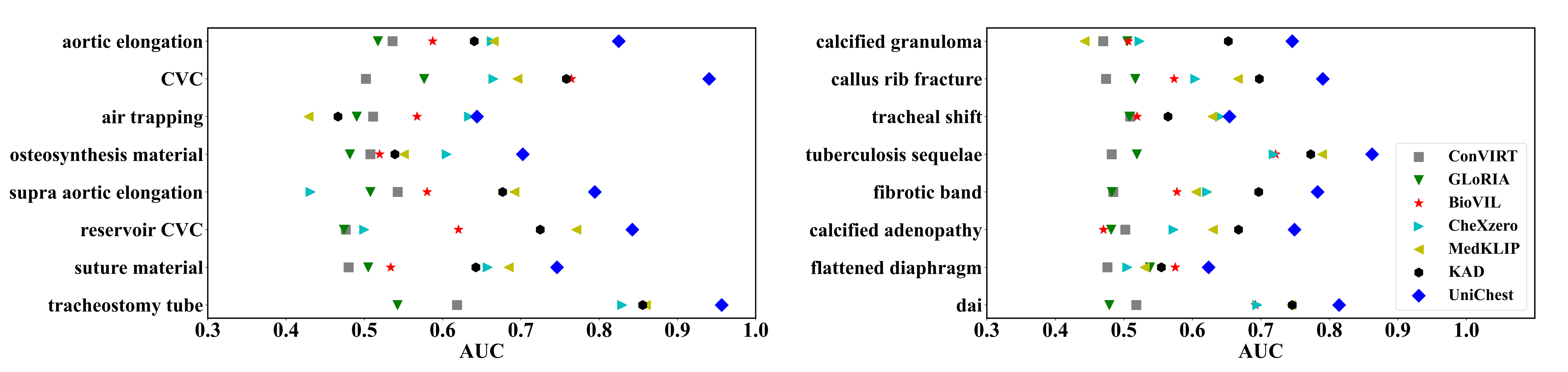
We conduct the zero-shot evaluation to assess the generalization ability of CXR pre-training models. The results in Table 4 demonstrate the superiority of UniChest across various datasets. In the case of the Shenzhen dataset, UniChest surpasses the best baseline KAD by 6.43%, 4.32%, 10.20% and 14.57% respectively as to AUC, ACC, F1 and AP. For the SIIM-ACR Pneumothorax dataset, UniChest outperforms 2.93%, 0.95%, 3.79% and 0.60% respectively compared with the best baseline. As to Open-I dataset, UniChest achieves significant improvements by 4.84%, 2.72%, 0.56% and 2.74%. As indicated by the per-category evaluation in Fig. 4 (right), UniChest achieves performance improvements across the majority of the categories. For instance, the classification ability is sharply consolidated of No Finding, Pleural Other and Support Devices with a margin of over 3%. For PadChest, UniChest obtains an average AUC of 0.8403 for 16 seen categories during pre-training, surpassing KAD by 1.51%. Specifically, the diagnosis capability of 11 classes achieves SOTA as shown in Fig. 5, among which the improvements of fracture, nodule and pleural thickening are around or over 5%. For unseen pathologies in Fig. 6, UniChest also showcases superior performance in the given 16 diseases , demonstrating its value in rare disease diagnosis. Besides, it is worth noting that the multi-source data baseline variants (e.g., KAD-multi) exhibit relatively modest and inconsistent improvements compared to their vanilla methods and single-source data baseline variants (e.g., KAD-CXR14), which emphasizes the necessity of our method design in enhancing the model’s generalization ability. In summary, the results in Table 4 validate the significance of the Conquer-and-Divide pre-training framework for multi-source CXR samples, which indicates its potential in assisting clinical human diagnosis and highlights its unignorable value.
4.5.3 Qualitative grounding visualization
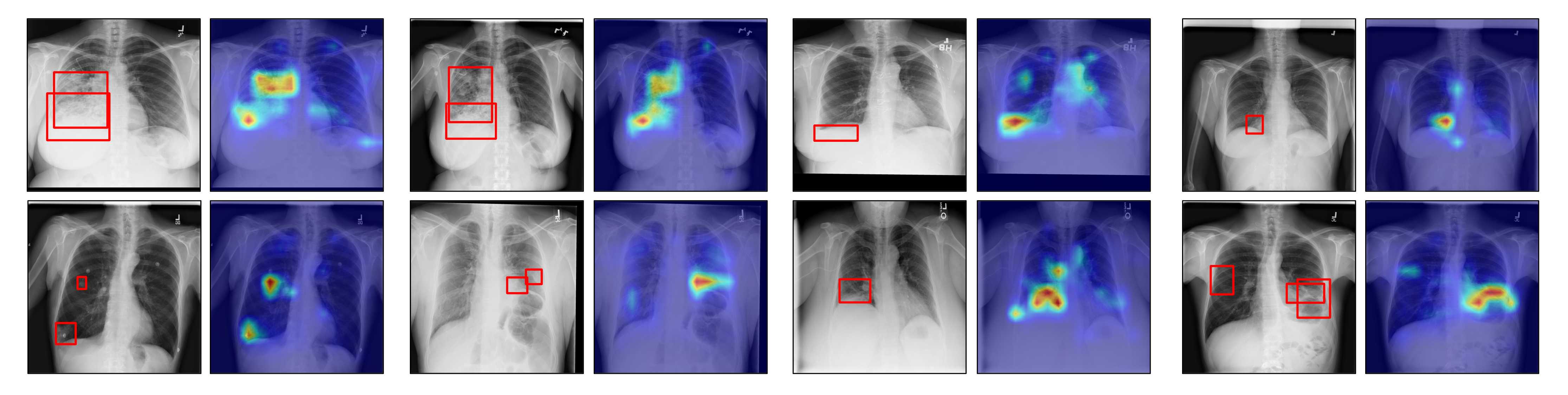
In Fig. 7, we present several examples of lesion grounding on ChestX-Det10 by UniChest. To provide an intuitive visualization, we generate spectrum heatmaps on the original CXR images based on the MoE-QN Module’s regional cross-attention maps in transformer decoder layers. By comparing the model-detected lesions with the bounding boxes annotated by expert clinicians, we observe a strong alignment between model findings and the diagnoses made by experts, demonstrating the reasoning ability and interpretability of UniChest.
| Dataset | Shenzhen | SIIM-ACR Pneumothorax | ChestX-ray14 | |||||||||
| Method | AUC | F1 | ACC | AP | AUC | F1 | ACC | AP | aAUC | aF1 | aACC | mAP |
| KAD (single-source SOTA) | 0.8856 | 0.8474 | 0.7893 | 0.8141 | 0.8967 | 0.6833 | 0.8428 | 0.6869 | 0.7933 | 0.3363 | 0.8639 | 0.2746 |
| UniChest (only Conquer stage) | 0.9013 | 0.8558 | 0.8594 | 0.9313 | 0.9019 | 0.6450 | 0.8494 | 0.6601 | 0.8468 | 0.4143 | 0.8957 | 0.3543 |
| UniChest (with equal weights) | 0.9463 | 0.8851 | 0.8838 | 0.9508 | 0.8687 | 0.5867 | 0.8234 | 0.5507 | 0.8528 | 0.4250 | 0.8971 | 0.3722 |
| UniChest | 0.9499 | 0.8906 | 0.8913 | 0.9598 | 0.9260 | 0.6928 | 0.8807 | 0.6929 | 0.8584 | 0.4293 | 0.8999 | 0.3797 |
5 Ablation Study
5.1 The Effectiveness of Conquer-and-Divide Framework
5.1.1 On “Conquer" stage
Data replenishment from multiple sources is an intractable issue mentioned in Section 3. We compare the impact of incorporating multi-source training data in the “Conquer” stage of our UniChest on the diagnostic performance, as displayed in the second row of Table 5. It turns out that simply augmenting the training data with multiple sources does not consistently yield significant performance gains compared to using a single source, as shown in the first row of the table. According to Table 5, explicit data replenishment improves the performance on ChestX-ray14 by 6.08%, aligning with the training data distribution. However, the performance of the zero-shot evaluation varies between the two inference sets. For the Shenzhen dataset, the influence of the “Conquer” stage has a significantly positive effect, as all four metrics experience considerable improvement. In the case of SIIM-ACR Pneumothorax, the AUC and ACC values are comparable to previous state-of-the-art approaches, but the other two metrics decrease by approximately 3.26%.
5.1.2 On “Divide" stage
A comparison of the second and fourth rows of Table 5 reveals that UniChest significantly outperforms the “Conquer” stage in terms of zero-shot generalization by 4.86% (AUC), 3.48% (F1), 3.19% (ACC), and 2.85% (AP) for Shenzhen, and 2.41% (AUC), 4.78% (F1), 3.13% (ACC), and 3.28% (AP) for SIIM-ACR Pneumothorax. To further present the effectiveness of the soft-gating mechanism and in the MoE-QN structure during the “Divide” stage, we present the results of removing the weight generation process and replacing it with equal weights in the third row of Table 5. As shown on SIIM-ACR Pneumothorax, our mechanism significantly enhances zero-shot generalization.
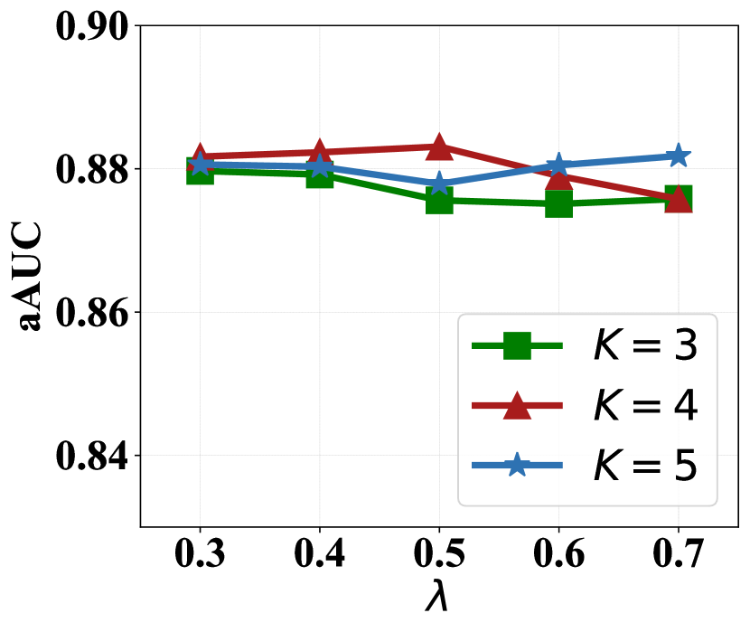
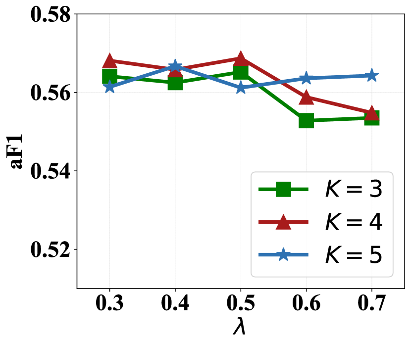
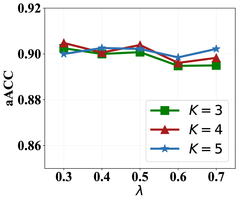
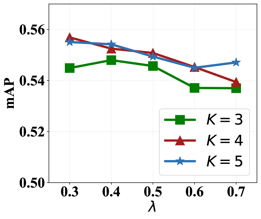
5.2 The Robustness under Different Hyper-parameters
The main hyper-parameters in our UniChest framework are the weight of the first query network during the “Divide” stage, and the total number of query networks . In the previous sections, we set the default values of as and as . In Fig. 8, we present results for various combinations of and , showcasing the average numerical outcomes across six different datasets, including ChestX-ray14, CheXpert, VinDr-CXR, Shenzhen, SIIM-ACR Pneumothorax, and Open-I. For instance, the default setting achieves best numerical results as to aAUC and aF1, while the range of variation for the maximum and minimum values of aAUC and aACC are below 1%, which validates the robustness of our method under different hyper-parameter combinations.
5.3 Performance under Different Backbones
We also explore the influence of modality backbones in the whole architecture. Firstly, we substitute ResNet-50 with DenseNet-121 [16] for the visual backbone and then replace the default fine-tuned PubMedBERT with ClinicalBERT [2] as textual encoder. As shown in Table 6, we notice the average numerical outcomes across six different datasets of the three settings are comparable, demonstrating the insensitivity of backbone selection.
6 Discussion and Conclusion
In this paper, we propose a novel Conquer-and-Divide pre-training framework for multi-source Chest X-Rays, namely UniChest, which is among the first attempts to fuse and utilize CXR samples from various origins harmonically. Our method effectively balances the benefits of multi-source data while minimizing the negative impacts the inter-source heterogeneity brings. In the “Conquer” stage, UniChest enhances feature extraction by sharing model components across different sources. In the “Divide” stage, it employs a mixture of deep query modules and utilizes a novel source-contrastive learning loss to isolate source-specific patterns, further reducing cross-source interference. Through extensive experiments on a range of benchmark datasets, we show the robustness and effectiveness of UniChest under diverse settings in CXR diagnosis.
| Method | aAUC | aF1 | aACC | mAP |
| w/ DenseNet-121 | 0.8794 | 0.5531 | 0.9078 | 0.5410 |
| w/ ClinicalBERT | 0.8849 | 0.5643 | 0.8997 | 0.5427 |
| UniChest | 0.8831 | 0.5687 | 0.9039 | 0.5508 |
Despite its powerful performance, UniChest still has some limitations in its design and application. Firstly, similar to MedKLIP and KAD, UniChest is limited to generating coarse-grained grounded heatmaps by utilizing cross-attention maps, which falls short of meeting the requirements for precise pixel-level segmentation. Therefore, the development of a comprehensive universal CXR model that combines both classification and fine-grained lesion grounding is a promising avenue for benefiting the medical community, which would not only offer efficiency but also ensure trustworthiness in daily applications. Secondly, although the Conquer-and-Divide framework is precise and meaningful and we have proposed one implementation paradigm within this framework. There is room for further exploration of other concrete frameworks that align with this spirit. We hope that our UniChest will inspire the exploration and utilization of multi-source CXRs.
References
- [1] Society for imaging informatics in medicine: Siim-acr pneumothorax segmentation. https://www.kaggle.com/c/siim-acr-pneumothorax-segmentation. 2019.
- Alsentzer et al. [2019] Emily Alsentzer, John Murphy, William Boag, Wei-Hung Weng, Di Jin, Tristan Naumann, and Matthew McDermott. Publicly available clinical BERT embeddings. In Proceedings of the 2nd Clinical Natural Language Processing Workshop, pages 72–78. Association for Computational Linguistics, June 2019.
- Bianchi et al. [2021] Federico Bianchi, Giuseppe Attanasio, Raphael Pisoni, Silvia Terragni, Gabriele Sarti, and Sri Lakshmi. Contrastive language-image pre-training for the italian language. arXiv preprint arXiv:2108.08688, 2021.
- Bodenreider [2004] Olivier Bodenreider. The unified medical language system (umls): integrating biomedical terminology. Nucleic acids research, 32(suppl_1):D267–D270, 2004.
- Boecking et al. [2022] Benedikt Boecking, Naoto Usuyama, Shruthi Bannur, Daniel C Castro, Anton Schwaighofer, Stephanie Hyland, Maria Wetscherek, Tristan Naumann, Aditya Nori, Javier Alvarez-Valle, et al. Making the most of text semantics to improve biomedical vision–language processing. In ECCV, pages 1–21, 2022.
- Chan et al. [2020] Heang-Ping Chan, Lubomir M Hadjiiski, and Ravi K Samala. Computer-aided diagnosis in the era of deep learning. Medical physics, 47(5):e218–e227, 2020.
- Chen et al. [2020] Ting Chen, Simon Kornblith, Mohammad Norouzi, and Geoffrey Hinton. A simple framework for contrastive learning of visual representations. In International conference on machine learning, pages 1597–1607. PMLR, 2020.
- Choudhuri and Paul [2021] Rudrajit Choudhuri and Amit Paul. Multi class image classification for detection of diseases using chest x ray images. In INDIACom, pages 769–773. IEEE, 2021.
- Chouhan et al. [2020] Vikash Chouhan, Sanjay Kumar Singh, Aditya Khamparia, Deepak Gupta, Prayag Tiwari, Catarina Moreira, Robertas Damaševičius, and Victor Hugo C De Albuquerque. A novel transfer learning based approach for pneumonia detection in chest x-ray images. Applied Sciences, 10(2):559, 2020.
- Dale [2021] Robert Dale. Gpt-3: What’s it good for? Natural Language Engineering, 27(1):113–118, 2021.
- Demner-Fushman et al. [2016] Dina Demner-Fushman, Marc D Kohli, Marc B Rosenman, Sonya E Shooshan, Laritza Rodriguez, Sameer Antani, George R Thoma, and Clement J McDonald. Preparing a collection of radiology examinations for distribution and retrieval. Journal of the American Medical Informatics Association, 23(2):304–310, 2016.
- Deng et al. [2009] Jia Deng, Wei Dong, Richard Socher, Li-Jia Li, Kai Li, and Li Fei-Fei. Imagenet: A large-scale hierarchical image database. In CVPR, pages 248–255. Ieee, 2009.
- Gu et al. [2021] Yu Gu, Robert Tinn, Hao Cheng, Michael Lucas, Naoto Usuyama, Xiaodong Liu, Tristan Naumann, Jianfeng Gao, and Hoifung Poon. Domain-specific language model pretraining for biomedical natural language processing. ACM Transactions on Computing for Healthcare, 3(1):1–23, 2021.
- Hashmi et al. [2020] Mohammad Farukh Hashmi, Satyarth Katiyar, Avinash G Keskar, Neeraj Dhanraj Bokde, and Zong Woo Geem. Efficient pneumonia detection in chest xray images using deep transfer learning. Diagnostics, 10(6):417, 2020.
- Hosny et al. [2018] Ahmed Hosny, Chintan Parmar, John Quackenbush, Lawrence H Schwartz, and Hugo JWL Aerts. Artificial intelligence in radiology. Nature Reviews Cancer, 18(8):500–510, 2018.
- Huang et al. [2017] Gao Huang, Zhuang Liu, Laurens Van Der Maaten, and Kilian Q Weinberger. Densely connected convolutional networks. In CVPR, pages 4700–4708, 2017.
- Huang et al. [2021] Shih-Cheng Huang, Liyue Shen, Matthew P Lungren, and Serena Yeung. Gloria: A multimodal global-local representation learning framework for label-efficient medical image recognition. In ICCV, pages 3942–3951, 2021.
- Irvin et al. [2019] Jeremy Irvin, Pranav Rajpurkar, Michael Ko, Yifan Yu, Silviana Ciurea-Ilcus, Chris Chute, Henrik Marklund, Behzad Haghgoo, Robyn Ball, Katie Shpanskaya, et al. Chexpert: A large chest radiograph dataset with uncertainty labels and expert comparison. In AAAI, volume 33, pages 590–597, 2019.
- Jaeger et al. [2014] Stefan Jaeger, Sema Candemir, Sameer Antani, Yì-Xiáng J Wáng, Pu-Xuan Lu, and George Thoma. Two public chest x-ray datasets for computer-aided screening of pulmonary diseases. Quantitative imaging in medicine and surgery, 4(6):475, 2014.
- Jain et al. [2021] Saahil Jain, Ashwin Agrawal, Adriel Saporta, Steven Truong, Tan Bui, Pierre Chambon, Yuhao Zhang, Matthew P Lungren, Andrew Y Ng, Curtis Langlotz, et al. Radgraph: Extracting clinical entities and relations from radiology reports. In NeurIPS Datasets and Benchmarks Track, 2021.
- Jia et al. [2021] Chao Jia, Yinfei Yang, Ye Xia, Yi-Ting Chen, Zarana Parekh, Hieu Pham, Quoc Le, Yun-Hsuan Sung, Zhen Li, and Tom Duerig. Scaling up visual and vision-language representation learning with noisy text supervision. In ICML, pages 4904–4916. PMLR, 2021.
- Johnson et al. [2019] Alistair EW Johnson, Tom J Pollard, Nathaniel R Greenbaum, Matthew P Lungren, Chih-ying Deng, Yifan Peng, Zhiyong Lu, Roger G Mark, Seth J Berkowitz, and Steven Horng. Mimic-cxr-jpg, a large publicly available database of labeled chest radiographs. arXiv preprint arXiv:1901.07042, 2019.
- Khoiriyah et al. [2020] Septy Aminatul Khoiriyah et al. Convolutional neural network for automatic pneumonia detection in chest radiography. In IES, pages 476–480. IEEE, 2020.
- Kieu et al. [2020] Stefanus Tao Hwa Kieu, Abdullah Bade, Mohd Hanafi Ahmad Hijazi, and Hoshang Kolivand. A survey of deep learning for lung disease detection on medical images: state-of-the-art, taxonomy, issues and future directions. Journal of imaging, 6(12):131, 2020.
- Kim and Kim [2020] Junghyun Kim and Kwan Hyoung Kim. Role of chest radiographs in early lung cancer detection. Translational lung cancer research, 9(3):522, 2020.
- Lee et al. [2020] Sang Won Lee, Yueh-Ting Chiu, Philip Brudnicki, Audrey M Bischoff, Angus Jelinek, Jenny Zijun Wang, Danielle R Bogdanowicz, Andrew F Laine, Jia Guo, and Helen H Lu. Darwin’s neural network: Ai-based strategies for rapid and scalable cell and coronavirus screening. arXiv preprint arXiv:2007.11653, 2020.
- Li et al. [2021] Junnan Li, Ramprasaath Selvaraju, Akhilesh Gotmare, Shafiq Joty, Caiming Xiong, and Steven Chu Hong Hoi. Align before fuse: Vision and language representation learning with momentum distillation. NeurIPS, 34:9694–9705, 2021.
- Liu et al. [2020] Jingyu Liu, Jie Lian, and Yizhou Yu. Chestx-det10: chest x-ray dataset on detection of thoracic abnormalities. arXiv preprint arXiv:2006.10550, 2020.
- Liu et al. [2021] Quande Liu, Cheng Chen, Jing Qin, Qi Dou, and Pheng-Ann Heng. Feddg: Federated domain generalization on medical image segmentation via episodic learning in continuous frequency space. In CVPR, pages 1013–1023, 2021. doi: 10.1109/CVPR46437.2021.00107.
- Meedeniya et al. [2022] Dulani Meedeniya, Hashara Kumarasinghe, Shammi Kolonne, Chamodi Fernando, Isabel De la Torre Díez, and Goncalo Marques. Chest x-ray analysis empowered with deep learning: A systematic review. Applied Soft Computing, page 109319, 2022.
- Nguyen et al. [2022] Ha Q Nguyen, Khanh Lam, Linh T Le, Hieu H Pham, Dat Q Tran, Dung B Nguyen, Dung D Le, Chi M Pham, Hang TT Tong, Diep H Dinh, et al. Vindr-cxr: An open dataset of chest x-rays with radiologist’s annotations. Scientific Data, 9(1):429, 2022.
- Radford et al. [2021] Alec Radford, Jong Wook Kim, Chris Hallacy, Aditya Ramesh, Gabriel Goh, Sandhini Agarwal, Girish Sastry, Amanda Askell, Pamela Mishkin, Jack Clark, et al. Learning transferable visual models from natural language supervision. In ICML, pages 8748–8763. PMLR, 2021.
- Rammuni Silva and Fernando [2022] Ravidu Suien Rammuni Silva and Pumudu Fernando. Effective utilization of multiple convolutional neural networks for chest x-ray classification. SN Computer Science, 3(6):492, 2022.
- Ridnik et al. [2021] Tal Ridnik, Emanuel Ben-Baruch, Nadav Zamir, Asaf Noy, Itamar Friedman, Matan Protter, and Lihi Zelnik-Manor. Asymmetric loss for multi-label classification. In ICCV, pages 82–91, 2021.
- Shin et al. [2023] Hyun Joo Shin, Kyunghwa Han, Leeha Ryu, and Eun-Kyung Kim. The impact of artificial intelligence on the reading times of radiologists for chest radiographs. NPJ Digital Medicine, 6(1):82, 2023.
- Tiu et al. [2022] Ekin Tiu, Ellie Talius, Pujan Patel, Curtis P Langlotz, Andrew Y Ng, and Pranav Rajpurkar. Expert-level detection of pathologies from unannotated chest x-ray images via self-supervised learning. Nature Biomedical Engineering, pages 1–8, 2022.
- Van Ginneken et al. [2001] Bram Van Ginneken, BM Ter Haar Romeny, and Max A Viergever. Computer-aided diagnosis in chest radiography: a survey. IEEE Transactions on medical imaging, 20(12):1228–1241, 2001.
- Wang et al. [2023] Wenhai Wang, Jifeng Dai, Zhe Chen, Zhenhang Huang, Zhiqi Li, Xizhou Zhu, Xiaowei Hu, Tong Lu, Lewei Lu, Hongsheng Li, Xiaogang Wang, and Yu Qiao. Internimage: Exploring large-scale vision foundation models with deformable convolutions. In CVPR, pages 14408–14419, June 2023.
- Wang et al. [2017] Xiaosong Wang, Yifan Peng, Le Lu, Zhiyong Lu, Mohammadhadi Bagheri, and Ronald M Summers. Chestx-ray8: Hospital-scale chest x-ray database and benchmarks on weakly-supervised classification and localization of common thorax diseases. In CVPR, pages 2097–2106, 2017.
- Wu et al. [2023] Chaoyi Wu, Xiaoman Zhang, Ya Zhang, Yanfeng Wang, and Weidi Xie. Medklip: Medical knowledge enhanced language-image pre-training for x-ray diagnosis. In ICCV, pages 21372–21383, October 2023.
- Yang et al. [2020] Qiang Yang, Yu Zhang, Wenyuan Dai, and Sinno Jialin Pan. Transfer learning. Cambridge University Press, 2020.
- Yang et al. [2022] Yuzhe Yang, Hao Wang, and Dina Katabi. On multi-domain long-tailed recognition, imbalanced domain generalization and beyond. In ECCV, pages 57–75. Springer, 2022.
- Zhang et al. [2022a] Ruipeng Zhang, Qinwei Xu, Chaoqin Huang, Ya Zhang, and Yanfeng Wang. Semi-supervised domain generalization for medical image analysis. In ISBI, pages 1–5, 2022a.
- Zhang et al. [2023] Xiaoman Zhang, Chaoyi Wu, Ya Zhang, Weidi Xie, and Yanfeng Wang. Knowledge-enhanced visual-language pre-training on chest radiology images. Nature Communications, 14(1):4542, 2023.
- Zhang et al. [2022b] Yuhao Zhang, Hang Jiang, Yasuhide Miura, Christopher D Manning, and Curtis P Langlotz. Contrastive learning of medical visual representations from paired images and text. In Machine Learning for Healthcare, 2022b.