Insulator-to-metal Mott transition facilitated by lattice deformation in monolayer -RuCl3 on graphite
Abstract
Creating heterostructures with graphene/graphite is a practical method for charge-doping -RuCl3, but not sufficient to cause the insulator-to-metal transition. In this study, detailed scanning tunneling microscopy/spectroscopy measurements on -RuCl3 with various lattice deformations reveal that both in-plane and out-of-plane lattice distortions may collapse the Mott-gap in the case of monolayer -RuCl3 in proximity to graphite, but have little impact on its bulk form alone. In the Mott-Hubbard framework, the transition is attributed to the lattice distortion-facilitated substantial modulation of the electron correlation parameter. Observation of the orbital textures on a highly compressed monolayer -RuCl3 flake on graphite provides valuable evidence that electrons are efficiently transferred from the heterointerface into Cl3 orbitals under the lattice distortion. It is believed that the splitting of Ru bands within the trigonal distortion of Ru-Cl-Ru octahedra bonds generated the electrons transfer pathways. The increase of the Cl3 states enhance the hopping integral in the Mott-Hubbard bands, resulting in the Mott-transition. These findings suggest a new route for implementing the insulator-to-metal transition upon doping in -RuCl3 by deforming the lattice in addition to the formation of heterostructure.
I INTRODUCTION
Mott-transition in materials with strong electron correlations is commonly referred to as a discontinuous phase transition (first-order), during which both spin and charge undergo substantial qualitative changes [1, 2, 3]. It ensures many intriguing physics such as high-temperature superconductivity (HTSC), strange metal phase, charge density wave (CDW), and other symmetry broken states [4, 5, 6, 7]. Cuprates that exhibit HTSC and charge-order in the vicinity of Mott-transition are an established instance [8]. Under sufficiently strong spin-orbital-coupling (SOC), Mott insulators can exhibit quantum spin liquid (QSL) phase with topological order and fractional spinon excitation [9, 10, 11, 12], providing a conductive environment for investigating a continuous Mott-transition (second-order phase transition) with more intriguing physics [13, 14]. By virtue of spin-dependent interactions among spin-half moments [15, 16, 17], the Kitaev honeycomb model admitting an exact QSL state and non-Abelian Majorana spinons stands out as a remarkable example. Numerous theoretical studies [18, 19, 20] have predicted that the Mott-transition in such a Kitaev QSL system could result in a rich phase diagram containing p-wave topological superconducting phase. Charge-doping is regarded as an efficient method for approaching such a Mott-transition. Hitherto, a carrier doped phase of the proximate Kitaev QSL material -RuCl3 has been studied by multiple research groups utilizing various strategies [21, 22, 23, 24]. Comprising heterostructures of graphene/graphite appear to be the most advantageous among the options reported to date, duo to the ability to prevent the introduction of crystal defects. Experimental evidences have provided empirical support for the notion that electrons, possessing a magnitude of 1013 cm-2, can be transferred from graphene to -RuCl3, resulting in an equal amount of holes doping in graphene [23, 25, 26, 27, 28]. However, the insulator-to-metal transition accompanied by the emergence of a well-defined Fermi surface (FS) has not been observed in -RuCl3 yet, impeding further exploration of the potential superconductivity in this system.
In our recent study [29], we reported that the in-gap states of -RuCl3 in proximity to graphite sensitively experienced a notable augmentation upon the introduction of lattice distortion, which aligns with the theoretical anticipation that straining the lattice may be a powerful potential method for approaching to the Mott-transition in -RuCl3 [30, 31, 25]. Here, we use scanning tunneling microscopy/spectroscopy (STM/STS) to further explore the electronic properties of -RuCl3 with lattice deformations and strain at liquid nitrogen temperature (77 K). The experimental findings offer a significant clarification that the imposition of strain, regardless of whether it is compressive or extensive, in-plane or out-of-plane, on the lattice of monolayer (ML)--RuCl3 transferred onto a graphite substrate, may result in the insulator-to-metal Mott transition, while it gives negligible impact on the bulk form of -RuCl3. Upon subjecting the ML--RuCl3 lattice to a uniform compression of 18%, we observe atomic-resolved orbital textures, which provide the evidence that newly arising of low-energy electronic states in the Cl3 orbitals are responsible for the Mott-transition. The proposed explanation posits that the lattice distortion has caused the splitting of the band, which provides the pathways for the electrons transferring from the heterointerface to the upper Cl3 orbitals. The new arising states effectively modulate the electron-correlations in Mott-Hubbard bands in -RuCl3. These findings provide a prospective route for the manifestation of FS, thereby affording an opportunity to explore the phenomenon of topological superconductivity in the vicinity of a Mott-transition within a Kitaev QSL.
II RESULTS AND DISCUSSION
A STM/STS on bulk -RuCl3 with film corrugations
-RuCl3 is an insulating 4d transition-metal halide with honeycomb layers composed of nearly ideal edge-sharing RuCl6 octahedra [32], which can be exfoliated into a two-dimensional ML with a thickness of 600 pm [33], as depicted schematically in Fig. 1a. As illustrated in the upper panel of Fig. 1b, the crystal field of RuCl6 octahedra splits the five-fold degenerate d levels of 4d5 Ru3+ into the doublet eg and the triplet t2g with a charge gap exceeding 2 eV. The t2g manifold composed of the dxy, dyz and dxz orbitals of Ru3+ hybridizes with the 3p (px, py, pz) orbitals of Cl- [34, 30]. Subsequently, the SOC (energy: soc) induces an additional splitting within the t2g manifold, resulting in a =1/2 doublet and a =3/2 quartet, characterized by an energy of soc/2. Consequently, in the hole picture, one hole is accommodated in the lower =1/2 Kramer’s doublet [17, 35], as schematically shown in Fig. 1b. Within the Mott-Hubbard framework, the =1/2 manifold undergoes a splitting due to the Coulomb interaction U, resulting in the formation of upper Hubbard band (UHB) and lower Hubbard band (LHB). It demonstrates that the Mott-gap can exceed 2 eV, when the value of U is set to be 4.5 eV during DFT calculations, as shown in the bottom panel of Fig. 1b, which aligns favorably with the Mott-gap measured via spectroscopy and photoemission spectroscopy on bulk -RuCl3 [29, 36]. The results imply that -RuCl3 is a highly correlated electron system.
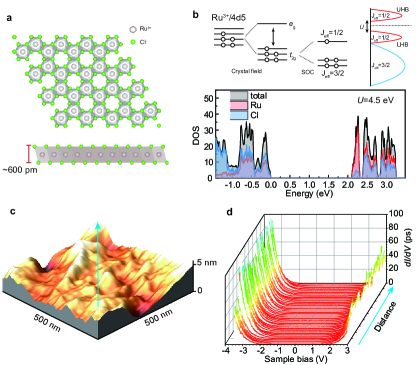
Several academic studies [37, 38] have demonstrated that the imposition of strain or pressure upon the crystal of a Mott insulator possesses the potential to cause an insulator-to-metal Mott-transition. Our prior research [29] has revealed that a minor distortion of lattice in ML--RuCl3 can substantially modify the electronic characteristic by causing the in-gap states within the Mott-gap. A question arises whether the applied strain or the lattice distortion possesses the capacity to trigger a Mott-transition within the bulk form of -RuCl3. In this study, we exfoliated the -RuCl3 thin flakes using the Scotch tapes from a bulk single crystal synthesized from commercially available RuCl3 powder by means of a vacuum sublimation. After repeatedly reducing the thickness of the film, we finally exfoliated a target -RuCl3 thin flakes using a thermal release tap in order to transfer to a fresh surface of highly oriented pyrolytic graphite (HOPG) substrate. After heating the sample to 120 oC for 20 seconds in an argon atmosphere in the glovebox, the -RuCl3 thin films were released on the graphite surface. Lattice deformation and strain of the -RuCl3 film are inevitably introduced during the transfer process. Then, the sample was directly transferred into an STM chamber and annealed at 280 oC in an ultra-high vacuum chamber (1E-10 Torr) for at least 2 hours for degassing and improving the contacting quality prior to STM/STS measurements. We measured the thicknesses of -RuCl3 flakes by a scanning topographic image at the step-edge and measured its height-profile to the graphite surface. We started by examining the condition of a thick -RuCl3 flake in presence of the pronounced film corrugations, as demonstrated by the STM morphology in Fig. 1c. Through the acquisition of dI/dV spectra along the designated path indicated by the blue arrow line in Fig. 1c, it ascertained that the large Mott-gap, measured approximately to be 2 eV, remains robust over the corrugations. In contrast to the case of ML--RuCl3 on graphite in our previous work where the in-gap states arise in the Mott-gap[29], the corrugations did not give rise to any in-gap states, as shown in Fig. 1d. This serves to validate the notion that a considerable crystal deformation is incapable of collapsing the strong Mott-Hubbard framework in bulk form -RuCl3. The viewpoint is also supported by previous calculations that the Mott-Hubbard band of -RuCl3 is hardly changed under a biaxial in-plane tensile strain of 8% [39].
B Mott-gap collapsed by the modest strain in ML--RuCl3 in proximity to graphite
Subsequently, our focus was redirected towards the characteristics exhibited by the ML--RuCl3 following its transfer onto the graphite. As depicted in the STM image in Fig. 2a, a selected region of ML--RuCl3 exhibits substantial film corrugations. The STM morphology was subjected to the fast Fourier transform (FFT), and the observable shifts of the Bragg peaks associated with the Ru and Cl atoms indicate that the out-of-plane corrugation has caused the in-plane distortion of the crystal lattices, as shown inset of Fig. 2a. Previous studies have revealed that the lattice morphology under STM within the sample bias (Vb) range of -3.0 to 1.5 V is dominated by Ru t2g and Cl pπ (pz) orbitals, and the resulting hybridization of these orbitals gives rise to the characteristic corner-shared Kagome pattern in STM images [40], which has been demonstrated in our recent experiment on bulk -RuCl3 [29]. Nevertheless, upon observation on ML--RuCl3, it was noted that the FFT-filtered image (as shown in Fig. 2b) did not exhibit the anticipated Kagome-like patterns. This suggests that forming heterostructure has exerted a significant influence on the orbital configurations in ML--RuCl3, a finding that seems to contradict the observations made on the epitaxial ML--RuCl3 on graphite [40].
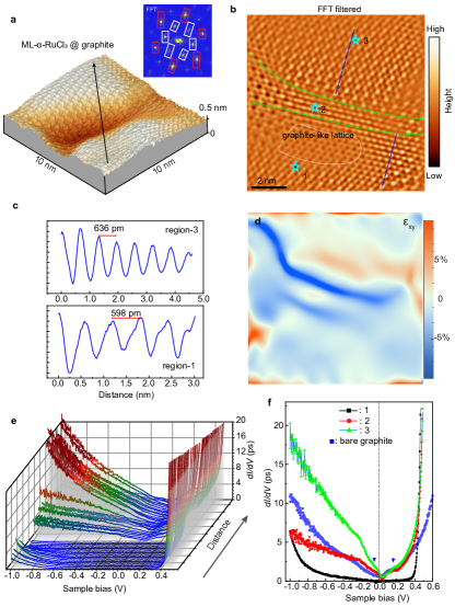
The film corrugation partitions the surface into three separate regions: region-1, characterized by a nearly strain-free lattice; region-2, where the lattice experiences compression; and region-3, where the lattice undergoes expansion, as seen in Fig. 2c. In region-1, an intriguing observation is the existence of an area exhibiting graphene-like lattices. This observation aligns with our previous research [29], which suggests the possibility of hetero-interfacial hybridization between -RuCl3 and graphite. The lattice constants for regions-1 and -3 were determined by extracting the height profile along the dashed lines, yielding the values of approximately 6 Å for region-1 and 6.4 Å for region-3. The strain distribution across the surface has been conducted utilizing the geometric phase analysis method [41], as shown in Fig. 2d. Subsequently, dI/dV spectra were obtained along the trajectory illustrated in Fig. 2a, encompassing the three aforementioned regions. The results display that the large Mott-gap maintains itself in region-1, whereas the Mott-gap collapses within the presence of a finite local density of states (LDOS) at the Fermi level in regions-2 and -3. Irrespective of whether the material experiences compressive or tensile strain, as depicted by the strain field maps in Fig. 2d, the findings suggest that the lattice distortions induce the emergence of FS in -RuCl3. Given the negligible impact of comparable lattice distortions on the bulk film, as depicted in Figs. 1c and d, it is highly indicative that the lattice distortions have effectively facilitated the emergence of in-gap states in ML--RuCl3 within the heterostructure.The observed phenomenon is consistance with the theoretical calculation that applying pressure on the -RuCl3/graphene heterostructure enables the interfacial charge transfer in -RuCl3 [27]. Through the quantitative analysis with regard to the spectra observed within the three defined regions in Fig. 2f, it was discovered that, despite the compressed region (region-2) displaying a reduced occupied LDOS in comparison to the region subjected to tensile strain (region-3), the line-shape of the spectra within both regions exhibited a remarkable similarity, rendering them nearly indiscernible. All these observed metallic dI/dV spectra exhibit a notable dip near the Fermi level, being reminiscent of the pseudogap observed in the charge-doped cuprate.
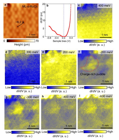
Then, we measured energy-resolved dI/dV maps on a ML--RuCl3 with its lattice under modest tension (3.5%), as depicted in Fig. 3a. Fig. 3b displays the average spectral curve, which has a nearly closed energy gap. Although the tensile strain has indeed resulted in an enhancement of the distinct states within the Mott-gap, our observations in dI/dV maps (figs. 3c to i) have not revealed any discernible contrasts in orbital texture. This outcome aligns with our previous findings on strain-free ML--RuCl3[29]. It is implied that although the electrons present in -RuCl3 possess equal magnitudes to the doped holes in graphite, approximately 1013 cm-2, these electrons have not occupied any orbitals within the energy range being examined by STS under the condition of modest lattice distortion. Consequently, it implies that the electrons in the in-gap states are itinerant or have been strongly dispersed during the tunneling process. Charge puddles can be seen in dI/dV maps that are acquired under negative energies, as shown in Figs. 3e-i. These charge puddles are thought to arise from the inhomogeneous distribution of the electrons, which aligns with the recent theoretical prediction that the spatial distribution of the doping electrons exhibits inhomogeneity in -RuCl3 [28]. Similar charge puddles are also frequently observed in charge-doped cuprates before the emergence of charge-ordered phase [42, 43, 44, 45].
C Energy dependent orbital textures in the extremely strained ML--RuCl3
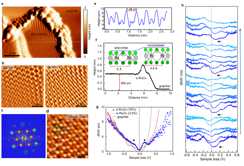
To further elucidate the intricate interplay among lattice deformation, charge transfer, and the insulator-to-metal transition within the ML--RuCl3 in proximity to graphite, we examined another extraordinary scenario wherein substantial biaxial strain (the lattice is shrunk over 18%) is unintentionally imposed upon a piece of ML--RuCl3 flake, as illustrated in Fig. 4a. Atomic-resolved STM images acquired at various sample biases, as depicted in Fig. 4b, show a lattice morphology that exhibits similar features when the lattice is subjected to modest tensile strain, as illustrated in Fig. 2c. The Kagome-like pattern as well as the trimer pattern expected in pristine -RuCl3 were also not detected at the low energy scale, which is a supplementary evidence notifying that the orbital configurations of ML--RuCl3 are indeed modified upon lattice deformation and being in proximity to graphite. Moreover, the examined flake also failed to exhibit a graphite-like lattice under a bias as small as 50 mV (Fig. 4b), which contradicts the observations depicted in strain-free scenarios [29]. Therefore, it indicates that the electron tunneling procedure through the heterostructure has been more seriously modified.
The FFT image of the STM morphology displays the Bragg peaks corresponding to the Ru and Cl atoms, which maintains the hexagonal lattice symmetry under the strain, as depicted in Fig. 4c. By decreasing the distance between STM tip and the surface, hexagonal Ru atoms of the -RuCl3 lattice becomes clear, as shown in Fig. 4d. Following the STM profile, Fig. 4e showcases that the lattice constant is 4.8 Å in this strained flake, which elucidates that the lattice is subjected to a substantial compression, exceeding -18%. The application of such a large in-plane compressive strain amplifies the out-of-plane buckling. Thickness of the present flake (500 pm), as displayed in Fig. 4f, is greater than that of the strain-free flake (350 pm) obtained in our previous investigation [29]. It can be imagined that the significant compression in the lattice results from the thermal treatment employed during the fabrication of the heterostructure. As seen in the averaged dI/dV spectrum of the compressed lattice, as shown in Fig. 4g, the Mott-gap is thoroughly collapsed. It results in an emergence of a similar pseudogap-like feature (within the range of 20 meV) near the Fermi level, akin to the ones observed in Fig. 2f. These empirical observations suggest that the application of strains may exert a systematic influence on the Mott-transition. Spatially resolved dI/dV spectra along the blue arrow line in Fig. 4d exhibit repetitive modulation of the line-shapes that aligns with the lattice’s symmetry, as depicted in Fig. 4h. This observation implies the existence of distinct orbital textures that align with the lattice geometry, potentially unveiling insights into the underlying mechanism of the strain facilitated Mott-transition.
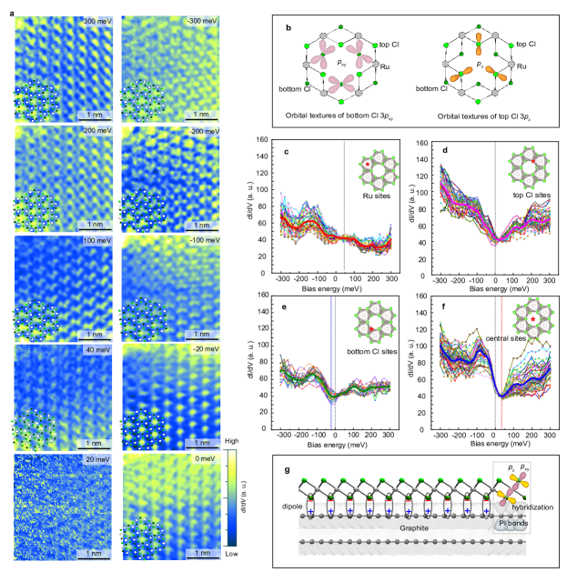
Therefore, we conducted an analysis of energy-resolved dI/dV mappings within the defined region depicted in Fig. 4d, encompassing an energy range spanning from 300 mV to -300 mV, as visually represented in Fig. 5a. In contrast to the depicted scenario in Fig. 3, wherein the orbital texture appears indistinct, the observed region showcases remarkably discernible energy-dependent and atomically-resolved orbital textures. This observation indicates that these low-energy surface electron states reside in specified orbitals within the system. The obtained dI/dV maps within the energy range reveal a remarkably diminished LDOS on Ru sites, in constrast to the pronounced concentration of high intensity in the proximity of Cl atoms. The manifestation of a weak orbital texture arising from the hybridization of Ru and Cl orbitals (t2g-pz hybridization) is confined to an energy magnitude of 40 meV. The fact that the LDOS at Cl sites exhibited a network-like pattern at the energy levels exceeding 200 meV is concurrent with a depletion of Ru sites, indicating that the pattern is predominantly composed by the Cl3 orbitals. Two distinct patterns of orbital textures are discernible when examining the occupied states. In the vicinity of Fermi level, precisely at 0 and -20 meV of the dI/dV maps, the LDOSs exhibit pronounced intensity patterns situated in the central region of the hexagonal lattice. In the absence of solid atomic entities occupying the central positions within -RuCl3 lattice, we hypothesize that the central LDOSs are predominantly generated from the bonds present on the graphite surface. Below -20 meV, the configuration of a three-pointed star within the hexagonal lattice can be observed. It can be unequivocally attributed to the 3pz lobes of the topmost Cl atoms, since they are primarily detected by STM tip, and contribute the three-pointed star patterns to the orbital textures, as illustrated in Fig. 5b.
The aforementioned observations demonstrate that the Cl3p orbitals are the primary source of the textures at a low energies, which is paradoxical given that the low-energy electronic configuration of the -RuCl3 could be dominated by the degenerate and bands from the Ru4d orbitals, whereas the Cl3p orbital electrons predominantly occupy the valence band below -2 eV [33, 40, 36]. In fact, the current observations are in agreement with the theoretical computations, which indicate that the electrons transferred from the heterointerface have a greater affinity for bonding to the Cl sites [28]. It suggests that the observed electron states in the orbital texture are the result of charge transfer in the heterostructure. In addition, the strain in ML--RuCl3 has altered the electron distribution from the heterointerface to the Cl orbitals as a result of the lattice distortion.
The spatially-averaged LDOS was then collected at distinct locations, specifically the Ru sites, the top and bottom Cl sites, and the sites in the center of the hexagons, as depicted in Figs. 5c to f. The LDOS curves observed at the Ru sites exhibit a nearly flat profile with a relatively reduced intensity (Fig. 5c). This feature implies that the Coulomb reputation still wishes to preserve the Mott-gap in band, and also explains the depletion morphology observed on the Ru sites in dI/dV maps in Fig. 5a. The spectral data collected from the top and bottom Cl sites as well as the center of the hexagons all exhibit a pseudogap-like feature in the spectral curves with a minimum value near the Fermi level (as depicted in Figs. 5d and f). It is noteworthy that the curves originating from the bottom Cl sites present a comparatively lower intensity due to the increased tunneling distance and a negative shift of its minimum point. The spectra measured at the centers of the hexagons exhibit an asymmetric line shape, with the occupied states being significantly higher than the empty ones. In addition, as depicted in Fig. 5f, a positive shift of the spectral minimum is observed. As was previously mentioned, the central points have potential to provide information regarding graphite. Comparing the dI/dV spectra to those of bare graphite, the positive shift of the minimum can be interpreted as evidence of hole-doping, which is consistent with previous findings that the graphite in the heterostructure is hole doped [24, 46, 23, 25]. The negative shift of the spectral minimum at bottom Cl sites is consistent with the band calculations [28] that the low-energy bands of -RuCl3 shift downward in the heterostructure. As depicted schematically in Fig. 5g, an opposite shift of the spatial spectra suggests the presence of an interfacial dipole layer between -RuCl3 and graphene [46]. The hybridization between Cl3 and orbitals of graphite adequately accounts for the formation of interface dipole.
This extreme case with significant lattice compression allows us to visualize the orbital structures in ML--RuCl3 in close proximity to graphite and provides information regarding the interfacial dipoles. However, it remains unclear how the lattice distortion could cause the Mott-Hubbard band to collapse. It is essential to note, even though a large amount of electrons is transferred from graphite to strain-free ML--RuCl3 in the heterostructure, the substantial Mott-gap (2 eV)[29] may effectively impede the movements of electrons from heterointerface into the Mott-Hubbard band. Therefore, forming the dipole layer is resonable, as depicted in Fig. 5f. Such a propose can accurately interpret the findings in our previous studies that the in-gap states were observable as the probing tip approaching to the surface [29], and that the orbital textures on the top surface were indistinct (Fig. 3). However, by referring to the Jahn-Teller theorem and the discussion in a recent study concerning on epitaxially grown -RuCl3 on graphite [40], any lattice distortion to prolong (shorten) the Ru-Ru bonds within widening (reducing) the Ru-Cl-Ru angles would cause the trigonal distortion of the Ru-Cl octahedra, as depicted schematically in Fig. 4f (inset). Such distortion will cause the splitting of t2g manifold into a1g singlet and e doublet, while preserving the hybridization with the orbitals of both the top and bottom Cl atoms. Despite the fact that -RuCl3 bulk and ML remain in the Mott insulating state under t2g splitting [47, 39], the distribution of doped electrons -RuCl3 may be ultimately altered. Therefore, it is proposed with sufficient adequacy that the electrons residing at the bottom Cl layer may exhibit a higher propensity to transfer into the top Cl orbitals via the band splitting and the hybridization of -- and --. The redistribution of electrons concurrently decreases the parameter ( is the hopping integral), or breaks the half-filled band in the Mott-Hubbard framework, which collapses the Mott-gap as shown in the dI/dV spectra in the strained ML--RuCl3.
III CONCLUSION
In conclusion, we reported the observation of lattice-distortion facilitated Mott-gap collapse in ML--RuCl3 in the heterostructure involving graphite. We clarified that the identical film deformation failed to change the Mott-gap in bulk -RuCl3. Important information was provided by the energy-resolved orbital textures by conducting STM/STS measurements on an ML--RuCl3 flake experiencing an extremely large compressed lattice strain. It was proven that a majority of charges for collapsing the Mott-gap reside in the Cl3 orbitals. In order to comprehend the physical mechanism, a model was proposed that the strain causes the splitting of band into and orbitals, and the orbital hybridization of ( and ) with the Clpz greatly modify the distribution of electrons those are transferred from graphite. Part of electrons previously accumulated at the herterointerface (bottom Cl layer) are transferred to the upper Cl layers, via the -- and -- pathways under lattice deformation, resulting in the Mott transition in the contexts of the doped Mott-Hubbard bands. The results will provide avenues for investigating the topological superconductivity in the vicinity of a Mott-transition within a Kitaev QSL candidate.
Acknowledgements.
This work was supported by National Basic Research and Development plan of China (Grants No. 2019YFA0308400), the National Natural Science Foundation of China (Grants No. U2032204, 12174027, 62174093), and the Strategic Priority Research Program of the Chinese Academy of Sciences (Grants No. XDB28000000 and XDB33030000).References
- Imada et al. [1998] M. Imada, A. Fujimori, and Y. Tokura, Review of Modern Physics 70, 1039 (1998).
- Sordi et al. [2011] G. Sordi, K. Haule, and A.-M. S. Tremblay, Physical Review B 84, 075161 (2011).
- Chatzieleftheriou et al. [2023] M. Chatzieleftheriou, A. Kowalski, M. Berović, A. Amaricci, M. Capone, L. De Leo, G. Sangiovanni, and L. de’ Medici, Physical Review Letters 130, 066401 (2023), publisher: American Physical Society.
- Yee and Balents [2015] C.-H. Yee and L. Balents, Physical Review X 5, 021007 (2015).
- Hanaguri et al. [2004] T. Hanaguri, C. Lupien, Y. Kohsaka, D.-H. Lee, M. Azuma, M. Takano, H. Takagi, and J. C. Davis, Nature 430, 1001 (2004), number: 7003 Publisher: Nature Publishing Group.
- da Silva Neto et al. [2014] E. H. da Silva Neto, P. Aynajian, A. Frano, R. Comin, E. Schierle, E. Weschke, A. Gyenis, J. Wen, J. Schneeloch, Z. Xu, S. Ono, G. Gu, M. Le Tacon, and A. Yazdani, Science 343, 393 (2014), publisher: American Association for the Advancement of Science.
- Sakurai et al. [2011] Y. Sakurai, M. Itou, B. Barbiellini, P. E. Mijnarends, R. S. Markiewicz, S. Kaprzyk, J.-M. Gillet, S. Wakimoto, M. Fujita, S. Basak, Y. J. Wang, W. Al-Sawai, H. Lin, A. Bansil, and K. Yamada, Science 332, 698 (2011), publisher: American Association for the Advancement of Science.
- Lee et al. [2006] P. A. Lee, N. Nagaosa, and X.-G. Wen, Reviews of Modern Physics 78, 17 (2006), publisher: American Physical Society.
- Pesin and Balents [2010] D. Pesin and L. Balents, Nature Physics 6, 376 (2010), number: 5 Publisher: Nature Publishing Group.
- Broholm et al. [2020] C. Broholm, R. J. Cava, S. A. Kivelson, D. G. Nocera, M. R. Norman, and T. Senthil, Science 367, eaay0668 (2020), publisher: American Association for the Advancement of Science Section: Review.
- Savary and Balents [2016] L. Savary and L. Balents, Reports on Progress in Physics 80, 016502 (2016), publisher: IOP Publishing.
- Zhou et al. [2017] Y. Zhou, K. Kanoda, and T.-K. Ng, Reviews of Modern Physics 89, 025003 (2017), publisher: American Physical Society.
- Pustogow et al. [2018] A. Pustogow, M. Bories, A. Löhle, R. Rösslhuber, E. Zhukova, B. Gorshunov, S. Tomić, J. A. Schlueter, R. Hübner, T. Hiramatsu, Y. Yoshida, G. Saito, R. Kato, T.-H. Lee, V. Dobrosavljević, S. Fratini, and M. Dressel, Nature Materials 17, 773 (2018), number: 9 Publisher: Nature Publishing Group.
- Furukawa et al. [2018] T. Furukawa, K. Kobashi, Y. Kurosaki, K. Miyagawa, and K. Kanoda, Nature Communications 9, 307 (2018), number: 1 Publisher: Nature Publishing Group.
- Kitaev [2006] A. Kitaev, Annals of Physics 321, 2 (2006), arXiv: cond-mat/0506438.
- Gohlke et al. [2018] M. Gohlke, G. Wachtel, Y. Yamaji, F. Pollmann, and Y. B. Kim, Physical Review B 97, 075126 (2018), publisher: American Physical Society.
- Takagi et al. [2019] H. Takagi, T. Takayama, G. Jackeli, G. Khaliullin, and S. E. Nagler, Nature Reviews Physics 1, 264 (2019), number: 4 Publisher: Nature Publishing Group.
- Zhang and Liu [2021] S.-M. Zhang and Z.-X. Liu, Physical Review B 104, 115108 (2021), publisher: American Physical Society.
- You et al. [2012] Y.-Z. You, I. Kimchi, and A. Vishwanath, Physical Review B 86, 085145 (2012), publisher: American Physical Society.
- Halász et al. [2014] G. B. Halász, J. T. Chalker, and R. Moessner, Physical Review B 90, 035145 (2014), publisher: American Physical Society.
- Jo et al. [2021] M.-k. Jo, H. Heo, J.-H. Lee, S. Choi, A. Kim, H. B. Jeong, H. Y. Jeong, J. M. Yuk, D. Eom, J. Jahng, E. S. Lee, I.-y. Jung, S. R. Cho, J. Kim, S. Cho, K. Kang, and S. Song, ACS Nano 15, 18113 (2021), publisher: American Chemical Society.
- Zhou et al. [2016] X. Zhou, H. Li, J. A. Waugh, S. Parham, H.-S. Kim, J. A. Sears, A. Gomes, H.-Y. Kee, Y.-J. Kim, and D. S. Dessau, Physical Review B 94, 161106(R) (2016), publisher: American Physical Society.
- Zhou et al. [2019] B. Zhou, J. Balgley, P. Lampen-Kelley, J.-Q. Yan, D. G. Mandrus, and E. A. Henriksen, Physical Review B 100, 165426 (2019), publisher: American Physical Society.
- Rizzo et al. [2020] D. J. Rizzo, B. S. Jessen, Z. Sun, F. L. Ruta, J. Zhang, J.-Q. Yan, L. Xian, A. S. McLeod, M. E. Berkowitz, K. Watanabe, T. Taniguchi, S. E. Nagler, D. G. Mandrus, A. Rubio, M. M. Fogler, A. J. Millis, J. C. Hone, C. R. Dean, and D. N. Basov, Nano Letters 20, 8438 (2020), publisher: American Chemical Society.
- Biswas et al. [2019] S. Biswas, Y. Li, S. M. Winter, J. Knolle, and R. Valentí, Physical Review Letters 123, 237201 (2019), publisher: American Physical Society.
- Mashhadi et al. [2019] S. Mashhadi, Y. Kim, J. Kim, D. Weber, T. Taniguchi, K. Watanabe, N. Park, B. Lotsch, J. H. Smet, M. Burghard, and K. Kern, Nano Letters 19, 4659 (2019), publisher: American Chemical Society.
- Gerber et al. [2020] E. Gerber, Y. Yao, T. A. Arias, and E.-A. Kim, Physical Review Letters 124, 106804 (2020), publisher: American Physical Society.
- Souza et al. [2022] P. H. Souza, D. P. d. A. Deus, W. H. Brito, and R. H. Miwa, Physical Review B 106, 155118 (2022), publisher: American Physical Society.
- Zheng et al. [2023] X. Zheng, K. Jia, J. Ren, C. Yang, X. Wu, Y. Shi, K. Tanigaki, and R.-R. Du, Physical Review B 107, 195107 (2023), publisher: American Physical Society.
- Iyikanat et al. [2018] F. Iyikanat, M. Yagmurcukardes, R. T. Senger, and H. Sahin, Journal of Materials Chemistry C 6, 2019 (2018), publisher: The Royal Society of Chemistry.
- Kaib et al. [2021] D. A. S. Kaib, S. Biswas, K. Riedl, S. M. Winter, and R. Valentí, Physical Review B 103, L140402 (2021), publisher: American Physical Society.
- Plumb et al. [2014] K. W. Plumb, J. P. Clancy, L. J. Sandilands, V. V. Shankar, Y. F. Hu, K. S. Burch, H.-Y. Kee, and Y.-J. Kim, Physical Review B 90, 041112(R) (2014), publisher: American Physical Society.
- Lu et al. [2023] W. Lu, H. Lee, J. Cha, J. Zhang, and I. Chung, Angewandte Chemie 135, e202219344 (2023), publisher: Wiley Onlin Library.
- Koitzsch et al. [2016] A. Koitzsch, C. Habenicht, E. Müller, M. Knupfer, B. Büchner, H. C. Kandpal, J. van den Brink, D. Nowak, A. Isaeva, and T. Doert, Phys. Rev. Lett. 117, 126403 (2016).
- Jackeli and Khaliullin [2009] G. Jackeli and G. Khaliullin, Physical Review Letters 102, 017205 (2009), publisher: American Physical Society.
- Sinn et al. [2016] S. Sinn, C. H. Kim, B. H. Kim, K. D. Lee, C. J. Won, J. S. Oh, M. Han, Y. J. Chang, N. Hur, H. Sato, B.-G. Park, C. Kim, H.-D. Kim, and T. W. Noh, Scientific Reports 6, 39544 (2016), number: 1 Publisher: Nature Publishing Group.
- Bu et al. [2019] K. Bu, W. Zhang, Y. Fei, Z. Wu, Y. Zheng, J. Gao, X. Luo, Y.-P. Sun, and Y. Yin, Communications Physics 2, 1 (2019), number: 1 Publisher: Nature Publishing Group.
- Dymkowski and Ederer [2014] K. Dymkowski and C. Ederer, Physical Review B 89, 161109(R) (2014), publisher: American Physical Society.
- Vatansever et al. [2019] E. Vatansever, S. Sarikurt, F. Ersan, Y. Kadioglu, O. Üzengi Aktürk, Y. Yüksel, C. Ataca, E. Aktürk, and U. Akinci, Journal of Applied Physics 125, 083903 (2019).
- Wang et al. [2022] Z. Wang, L. Liu, M. Zhao, H. Zheng, K. Yang, C. Wang, F. Yang, H. Wu, and C. Gao, Quantum Frontiers 1, 16 (2022).
- Hÿtch et al. [1998] M. J. Hÿtch, E. Snoeck, and R. Kilaas, Ultramicroscopy 74, 131 (1998).
- Cai et al. [2016] P. Cai, W. Ruan, Y. Peng, C. Ye, X. Li, Z. Hao, X. Zhou, D.-H. Lee, and Y. Wang, Nature Physics 12, 1047 (2016), number: 11 Publisher: Nature Publishing Group.
- Battisti et al. [2017] I. Battisti, K. M. Bastiaans, V. Fedoseev, A. de la Torre, N. Iliopoulos, A. Tamai, E. C. Hunter, R. S. Perry, J. Zaanen, F. Baumberger, and M. P. Allan, Nature Physics 13, 21 (2017), number: 1 Publisher: Nature Publishing Group.
- da Silva Neto et al. [2016] E. H. da Silva Neto, B. Yu, M. Minola, R. Sutarto, E. Schierle, F. Boschini, M. Zonno, M. Bluschke, J. Higgins, Y. Li, G. Yu, E. Weschke, F. He, M. Le Tacon, R. L. Greene, M. Greven, G. A. Sawatzky, B. Keimer, and A. Damascelli, Science advances 2, e1600782 (2016).
- Frano et al. [2020] A. Frano, S. Blanco-Canosa, B. Keimer, and R. J. Birgeneau, Journal of Physics: Condensed Matter 32, 374005 (2020), publisher: IOP Publishing.
- Rossi et al. [2023] A. Rossi, C. Johnson, J. Balgley, J. C. Thomas, L. Francaviglia, R. Dettori, A. K. Schmid, K. Watanabe, T. Taniguchi, M. Cothrine, D. G. Mandrus, C. Jozwiak, A. Bostwick, E. A. Henriksen, A. Weber-Bargioni, and E. Rotenberg, Nano Letters 23, 8000 (2023), publisher: American Chemical Society.
- Liu et al. [2023] L. Liu, K. Yang, G. Wang, D. Lu, Y. Ma, and H. Wu, Phys. Rev. B 107, 165134 (2023).