: Knowledge distillation via Attention Supervision, and Symmetrical structure guiding for Polyp Segmentation
Abstract
Polyp segmentation, a contentious issue in medical imaging, has seen numerous proposed methods aimed at improving the quality of segmented masks. Currently, state-of-the-art techniques yield impressive results. However, the sheer size of these models poses challenges for practical industry applications. To address this, we present a Knowledge Distillation framework, incorporating attention supervision and the symmetrical guiding method. This framework is designed to facilitate knowledge transfer from a teacher model to a more compact student model with fewer parameters. Our experimental evaluation of the framework assesses its effectiveness in enabling the student model to acquire knowledge from the teacher efficiently. Additionally, our method serves to prevent the student model from incorporating redundant features that could lead to inaccurate predictions. Consequently, our method, boasting approximately 5 million parameters, achieves competitive results comparable to the state-of-the-art approaches. The implementation can be found at: https://github.com/huyquoctrinh/KDAS3
Index Terms— Polyp segmentation, Knowledge Distillation, Symmetrical Guiding, Attention Supervision, deep learning
1 Introduction
Colorectal Cancer (CRC) stands as one of the most perilous diseases worldwide, emerging as a prevalent affliction affecting approximately one-third of the global population. To help the efficient treatment, the early diagnosis of CRC and Polyp segmentation tools are proposed to support the early treatment while this tool can help to localize the polyp region.
In recent years, numerous methods have been proposed to address this issue. In 2015, UNet [1] was initially introduced as an efficient approach for segmenting polyps in the early stages. Subsequent methods, such as ResUNet [2], ResUNet++ [3], UNet++ [4], and DDANet [5], followed suit. In 2021, a novel polyp segmentation framework called Polyp-PVT was introduced, utilizing the Pyramid Vision Transformer backbone. This method demonstrates strong generalization capabilities, effectively capturing global information while focusing on potential polyps in the image. Although the state-of-the-art model yields impressive results, its substantial model weight poses challenges for industry implementation.
To address this issue, we present , a Knowledge Distillation framework employing learning through Attention maps in the supervision branches. However, a gap exists in Knowledge Distillation when applied to segmentation—specifically, the absence of regional learning in the student model, leading to redundant feature acquisition. To mitigate this problem, we propose guiding the learning of the student model through the Symmetrical structure from the lower stages of the model. The Symmetrical structure’s performance lies in its ability to focus on and emphasize regions containing polyps. This focus enables the model to learn implicit and potential information from the deeper layers of the teacher model, consequently reducing redundant regions in the segmented mask.
For a fair comparison, our method is implemented with the baseline from Polyp-PVT [6] with the student model as the PVTV2-B0 backbone (approximate 5M parameters) and the teacher model from the public weight of Polyp-PVT [6]. The resulting model is benchmarked in five different datasets are Kvasir-SEG dataset, the ClinicDB dataset, the Colon-DB dataset, the CVC-300 dataset, and the Etis dataset. In addition, experiments are conducted to analyze the effectiveness of Symmetrical guiding. From State-of-the-art, our student model obtains competitive results with the lightweight model.
To sum up, our contributions include four folds: (1) We propose a novel Knowledge Distillation framework base on the Attention mechanism and the Supervision concept, and the symmetrical structure guiding for Polyp Segmentation; (2) We propose the symmetrical structure guiding modules, which can emphasize the polyp region, and reduce the learning on the redundant feature, from then, the student model can focus on learning the potential polyp region that is captured by the teacher model; (3) The effectiveness and the advantages of our methods are benchmarked in five different datasets. The model achieves a competitive result with state-of-the-art while the number of parameters is significantly smaller; (4) The two popular Knowledge Distillation methods are also implemented and well-tested in the Polyp Segmentation domain to have the comparison with our method.
This paper is organized as follows: in Section 2, we briefly review existing methods related to this research. Then we propose our methods in Section 3. Experiments setup are in Section 4. Results of the experiment and the discussion are in Section 5. Finally, we present the conclusion in Section 6.
2 Related Work
Knowledge Distillation is the method that makes the student model learn from the teacher and was introduced to be worked in 2015 by Hinton et al. [7] with Kullback-Leibler divergence loss (well known as KL divergence). Regarding the medical image, although previous methods achieve State-of-the-art results, they have more limitations when deployed in production. Almost top-tier methods such as Meta-Polyp [8], Polyp2Seg [9], and Polyp-PVT [6] use the big backbone, and they seem to be not friendly in the cost computation. However, if these methods change to the smaller backbone for training, it will probably reduce the generalization of the model in the out-of-distribution dataset. From this point, Knowledge Distillation is the efficient method to leverage the knowledge from the teacher model is the State-of-the-art and the student model, which is the model with a tiny backbone. The objective when using this method is to minimize the knowledge distribution gap between the teacher model and the student model, as a result, the student model can perform as well as the teacher model. Particularly, in the Polyp segmentation task, the State-of-the-art model achieves stable results, this is the reason why Knowledge Distillation is necessary to optimize the computation cost and can leverage this problem into reality.
3 Method
3.1 General description
As demonstrated in Figure.1, this framework includes two branches, the first branch is for the teacher model, and the other branch is for the student model. The input images with shape with B representing the batch size, input will be forwarded by both the teacher and the student while the teacher model is frozen for the inference, and the student model will not be frozen for the student learning. The supervision output will be used for the knowledge distillation while the distribution loss between the two models will be calculated to minimize the gap between the distribution of teacher and student. Moreover, we propose a Symmetrical Guiding block for guiding the learning from the student with the knowledge from the teacher.
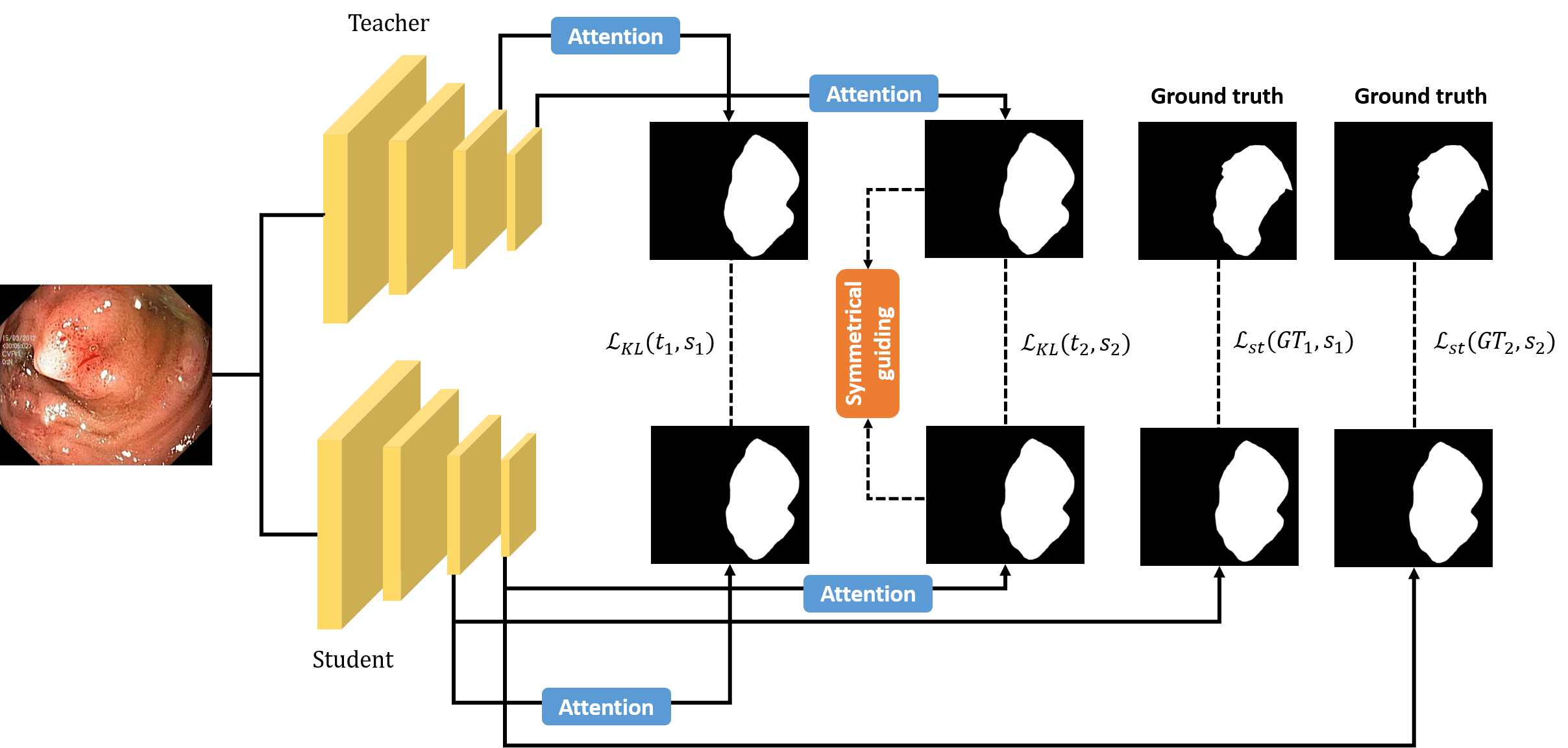
3.2 Symmetrical guiding
To guide the student in learning the better polyp region, and enhance the boundary result from the student, the symmetrical guiding modules are proposed to guide the student model in learning the synthesized activation mask and help to remove the redundant part of the mask. This module includes two inputs: the output activation map of the teacher, denote , and the output activation map of the student, denote . For each input, symmetrical structures are created following Equation.1.
| (1) |
Where represents the Symmetrical structure output for each teacher and student, the operation denotes the Adaptive Average Pooling operation which helps eliminate the topological constraint of this structure.
The sum between it and its transposed is initially employed to create a symmetrical structure which , this structure is a cube that contains the information from multiple outputs.
For the learning stage, the Kullback-Leiber divergence loss is implemented following the Hinton et al [7] to optimize the parameters of the student model for minimizing the distance between two distributions of the output symmetrical structure of student and teacher.
| (3) |
In Equation.3, values represent symmetrical structures, is the number of images in a batch data, and value is the temperature of the loss function.
3.3 Attention Supervision Knowledge Distillation
The KL divergence method can make the model focus on redundant knowledge which can lead to the redundant mask that is generated from the student model. To answer this question, the self-attention knowledge distillation [10] is employed for each supervision output. The scale dot product attention mechanism will help transform the predicted mask into the attention mask in which the student and teacher have the same attention distribution, and no parameters increasing through this transform. To minimize the gap of distribution between the student model, and the teacher model, the KL-divergence is implemented following the Equation.4:
| (4) |
Where N represents the number of images, is for the KL divergence, and denotes the attention map of each supervision output from the teacher model and the student model, and represents for the temperature of this loss function.
4 Experiments
4.1 Dataset
To conduct the fair comparison, the experiment’s dataset follows the merged dataset from the PraNet [11] experiment for the training stage which includes 900 samples from Kvasir-SEG [12] and 550 samples from CVC-ClinicDB [13]. The remaining images of Kvasir-SEG [12] and CVC-ClinicDB [13] with three unseen datasets such as ColonDB [14], CVC-300 [15] and ETIS [16] are used for benchmarking our method.
4.2 Implementation detail
In our implementation, we based our experiment base on the Polyp-PVT baseline [6]. The Pytorch framework is used with the Tesla V100 32GB for training. The image is resized to , and the setting for the batch size is 16. The AdamW optimizer is used with the learning rate . The best weight is collected after 120 epochs with the training time about 2 hours. For the testing, the image is resized to for the testing phase. The temperature for training setup is set at 2. The Jaccard loss function used to supervise our model in the training process is formulated as follows:
| (5) |
The Jaccard Loss, which is shown in Equation.5, is also called the Intersection over Union (IOU) metric [17]. The true label is denoted as , while the predicted label is represented as . Both labels are expressed as one-hot vectors, indicating the classes with a length equivalent to the number of classes, denoted as . The smoothing factor is set to 0.7 in our method. Note that we do not use deep supervision techniques to train our model.
Three evaluation common metrics are adopted for the evaluation: mean Dice (mDice), mean IoU (mIoU), and Mean Absolute Error (MAE). The higher value is better for mDice as well as mIoU, and the lower is better for MAE.
5 Result
5.1 Performance Comparisons
To evaluate the effectiveness of our model, we compare our PVTV0 distilled model (about 5M parameters) with several methods that have a higher number of parameters, including UNet [1] (2015, 7.6M), UNet++ [4] (2018, 9M), SFA [18] (2021), PraNet [11] (2019, 32.55M), MSNet [19] (2021, 29.74), HarDNet-CPS [20] (2023) and PEFNet [21] (2023, 27.98M). Since the setting datasets of PEFNet [21] are different, we retrain both methods on the same setting in PraNet [11] for a fair comparison.
5.2 Qualitative result
As presented in Table 2, our Knowledge Distillation framework contributes to the tiny model achieving superior performance in the Etis and ColonDB datasets. Although the results in the three remaining datasets are slightly lower, they remain competitive with previous methods. These findings highlight our model’s generalization in unseen domains, despite the significantly lower number of parameters in the distilled model compared to existing methods.
5.3 Ablation study
To evaluate the effectiveness of the symmetrical guiding, we analyze the quantitative contribution of the Knowledge Distillation method with the model training from the original, Knowledge Distillation with KL divergence, Attention-KD method, and the Attention-KD with symmetrical guiding, which is shown in Table 1.
According to the empirical findings, when the model is trained without Knowledge Distillation, the dice score yields 0.717. When the model is trained based on the Knowledge Distillation with KL divergence, the result improves gradually to 0.747, with the Attention KD, it improves to 0.735. By incorporating the full pipeline with symmetrical guiding, the dice score experiences a substantial boost, reaching 0.755.
| Model | |||
|---|---|---|---|
| Baseline | 0.642 | 0.717 | 0.026 |
| KL divergence | 0.675 | 0.747 | 0.016 |
| Attention-KD | 0.649 | 0.735 | 0.024 |
| Attention-KD + SMG | 0.677 | 0.755 | 0.015 |
The ablation study results demonstrate our assumption that the symmetrical guiding can guide the model to focus on the key feature of the object and prevent the model from learning the redundant feature, which leads to the better performance of our distilled model.
| Methods | ||||
|---|---|---|---|---|
| Kvasir | UNet[1] | 0.756 | 0.821 | 0.055 |
| UNet++ [4] | 0.743 | 0.820 | 0.048 | |
| SFA [18] | 0.611 | 0.723 | 0.075 | |
| PraNet [11] | 0.840 | 0.898 | 0.030 | |
| MSNet [19] | 0.862 | 0.907 | 0.028 | |
| PEFNet [21] | 0.833 | 0.892 | 0.029 | |
| HarDNet-CPS [20] | 0.856 | 0.911 | 0.025 | |
| Our | 0.848 | 0.913 | 0.027 | |
| ClinicDB | UNet [1] | 0.767 | 0.824 | 0.019 |
| UNet++ [4] | 0.729 | 0.794 | 0.022 | |
| SFA [18] | 0.607 | 0.700 | 0.042 | |
| PraNet [11] | 0.849 | 0.899 | 0.009 | |
| MSNet [19] | 0.879 | 0.921 | 0.008 | |
| PEFNet [21] | 0.814 | 0.866 | 0.010 | |
| HarDNet-CPS [20] | 0.887 | 0.917 | 0.008 | |
| Our | 0.872 | 0.925 | 0.008 | |
| CVC-300 | UNet [1] | 0.639 | 0.717 | 0.022 |
| UNet++ [4] | 0.636 | 0.687 | 0.018 | |
| SFA [18] | 0.329 | 0.467 | 0.065 | |
| PraNet [11] | 0.797 | 0.871 | 0.010 | |
| MSNet [19] | 0.807 | 0.869 | 0.010 | |
| PEFNet [21] | 0.797 | 0.871 | 0.010 | |
| HarDNet-CPS [20] | 0.826 | 0.891 | 0.008 | |
| Our | 0.798 | 0.875 | 0.008 | |
| Colon-DB | UNet [1] | 0.449 | 0.519 | 0.061 |
| UNet++ [4] | 0.410 | 0.483 | 0.064 | |
| SFA [18] | 0.347 | 0.469 | 0.094 | |
| PraNet [11] | 0.640 | 0.712 | 0.043 | |
| MSNet [19] | 0.678 | 0.755 | 0.041 | |
| PEFNet [21] | 0.638 | 0.710 | 0.036 | |
| HarDNet-CPS [20] | 0.658 | 0.729 | 0.037 | |
| Our | 0.679 | 0.759 | 0.032 | |
| ETIS | UNet [1] | 0.343 | 0.406 | 0.036 |
| UNet++ [4] | 0.344 | 0.401 | 0.035 | |
| SFA [18] | 0.217 | 0.297 | 0.109 | |
| PraNet [11] | 0.567 | 0.628 | 0.031 | |
| MSNet [19] | 0.664 | 0.719 | 0.020 | |
| PEFNet [21] | 0.572 | 0.636 | 0.019 | |
| HarDNet-CPS [20] | 0.619 | 0.69 | 0.008 | |
| Our | 0.677 | 0.755 | 0.015 |
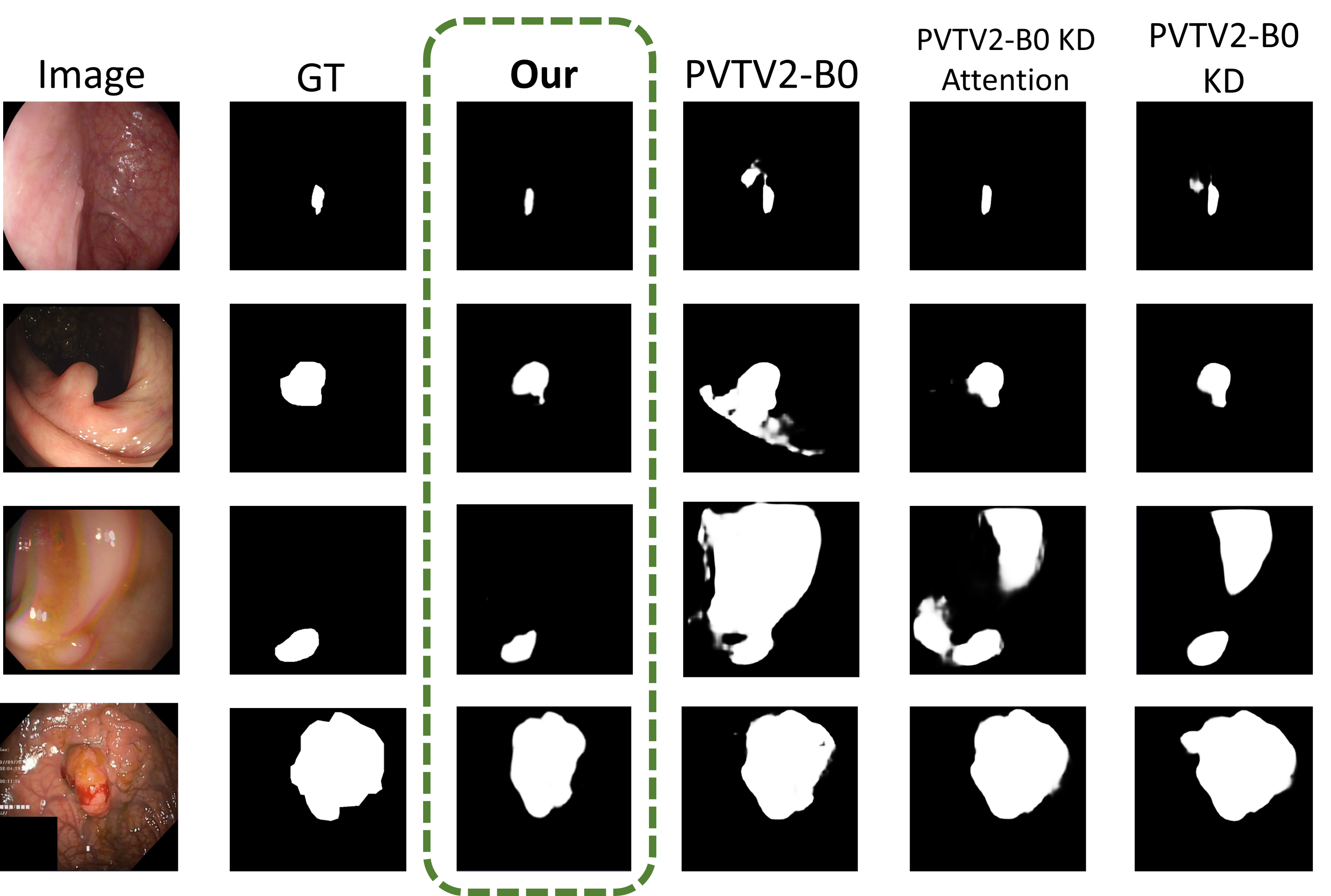
5.4 Qualitative visualization
In Figure 2, we perform the qualitative visualization for several knowledge distillation methods with the training model from the PVT V2 B0 backbone. It can be seen that our method can cover more accurate polyp regions, and it can ignore the redundant region that appears in the other methods.
6 Conclusion
In this paper, we propose the Knowledge distillation framework via Attention Supervision, and Symmetrical structure guiding for effective transfer of knowledge from the teacher model to the student model which helps to train an effective segmentation model in the industry environment. The symmetrical structure guiding is proposed and evaluated can solve the problem of the redundant feature from the learning process, and the Knowledge Distillation framework is demonstrated to help the light model achieve similar performance to the heavy model.
We believe that this method will enable the segmentation model to save the computation cost and keep the merits from the heavy model. Although there are some limitations to our proposed framework, this is a promising method that can help the segmentation model be deployed in the product environment.
References
- [1] O. Ronneberger, P. Fischer, and T. Brox, “U-Net: Convolutional Networks for Biomedical Image Segmentation,” in MICCAI, 2015.
- [2] F. I. Diakogiannis, F. Waldner, P. Caccetta, and C. Wu, “ResUNet-a: A deep learning framework for semantic segmentation of remotely sensed data,” ISPRS Journal of Photogrammetry and Remote Sensing, 2020.
- [3] D. Jha, P. H. Smedsrud, M. A. Riegler, D. Johansen, T. De Lange, P. Halvorsen, and H. D. Johansen, “ResUNet++: An Advanced Architecture for Medical Image Segmentation,” in ISM, 2019.
- [4] Z. Zhou, M. M. Rahman Siddiquee, N. Tajbakhsh, and J. Liang, “UNet++: A Nested U-Net Architecture for Medical Image Segmentation,” in Deep Learning in Medical Image Analysis and Multimodal Learning for Clinical Decision Support, 2018.
- [5] N. K. Tomar, D. Jha, S. Ali, H. D. Johansen, D. Johansen, M. A. Riegler, and P. Halvorsen, “Ddanet: Dual decoder attention network for automatic polyp segmentation,” in Pattern Recognition. ICPR International Workshops and Challenges, 2021.
- [6] D. Bo, W. Wenhai, F. Deng-Ping, L. Jinpeng, F. Huazhu, and S. Ling, “Polyp-PVT: Polyp Segmentation with PyramidVision Transformers,” CAAI AIR, 2023.
- [7] G. Hinton, O. Vinyals, and J. Dean, “Distilling the knowledge in a neural network,” arXiv preprint arXiv:1503.02531, 2015.
- [8] Q. Trinh, “Meta-polyp: A baseline for efficient polyp segmentation,” in 2023 IEEE 36th International Symposium on Computer-Based Medical Systems (CBMS). Los Alamitos, CA, USA: IEEE Computer Society, jun 2023, pp. 742–747. [Online]. Available: https://doi.ieeecomputersociety.org/10.1109/CBMS58004.2023.00312
- [9] V. Mandujano-Cornejo and J. A. Montoya-Zegarra, “Polyp2seg: Improved polyp segmentation with vision transformer,” in MICCAI, 2022.
- [10] W. Wang, F. Wei, L. Dong, H. Bao, N. Yang, and M. Zhou, “Minilm: Deep self-attention distillation for task-agnostic compression of pre-trained transformers,” Advances in Neural Information Processing Systems, vol. 33, pp. 5776–5788, 2020.
- [11] D.-P. Fan, G.-P. Ji, T. Zhou, G. Chen, H. Fu, J. Shen, and L. Shao, “Pranet: Parallel reverse attention network for polyp segmentation,” in MICCAI, 2020.
- [12] D. Jha, P. H. Smedsrud, M. A. Riegler, P. Halvorsen, T. d. Lange, D. Johansen, and H. D. Johansen, “Kvasir-SEG: A Segmented Polyp Dataset,” in Multimedia Modeling, 2020.
- [13] J. Bernal, F. J. Sánchez, G. Fernández-Esparrach, D. Gil, C. Rodríguez, and F. Vilariño, “WM-DOVA maps for accurate polyp highlighting in colonoscopy: Validation vs. saliency maps from physicians,” CMIG, pp. 99–111, 2015.
- [14] N. Tajbakhsh, S. R. Gurudu, and J. Liang, “Automated Polyp Detection in Colonoscopy Videos Using Shape and Context Information,” TMI, pp. 630–644, 2016.
- [15] D. Vázquez, J. Bernal, F. J. Sánchez, G. Fernández-Esparrach, A. M. López, A. Romero, M. Drozdzal, and A. C. Courville, “A Benchmark for Endoluminal Scene Segmentation of Colonoscopy Images,” Journal of Healthcare Engineering, 2017.
- [16] J. S. Silva, A. Histace, O. Romain, X. Dray, and B. Granado, “Towards embedded detection of polyps in WCE images for early diagnosis of colorectal cancer,” IJCARS, pp. 283–293, 2014.
- [17] J. Bertels, T. Eelbode, M. Berman, D. Vandermeulen, F. Maes, R. Bisschops, and M. B. Blaschko, “Optimizing the dice score and jaccard index for medical image segmentation: Theory and practice,” in MICCAI, 2019.
- [18] Y. Fang, C. Chen, Y. Yuan, and K.-y. Tong, “Selective Feature Aggregation Network with Area-Boundary Constraints for Polyp Segmentation,” in MICCAI, 2019.
- [19] X. Zhao, L. Zhang, and H. Lu, “Automatic Polyp Segmentation via Multi-scale Subtraction Network,” in MICCAI, 2021.
- [20] T. Yu and Q. Wu, “Hardnet-cps: Colorectal polyp segmentation based on harmonic densely united network,” Biomedical Signal Processing and Control, vol. 85, p. 104953, 2023.
- [21] T.-H. Nguyen-Mau, Q.-H. Trinh, N.-T. Bui, P.-T. V. Thi, M.-V. Nguyen, X.-N. Cao, M.-T. Tran, and H.-D. Nguyen, “PEFNet: Positional Embedding Feature for Polyp Segmentation,” in MultiMedia Modeling, 2023.