Chemically Active Wetting
Abstract
Wetting of liquid droplets on passive surfaces is ubiquitous in our daily lives, and the governing physical laws are well-understood. When surfaces become active, however, the governing laws of wetting remain elusive. Here we propose chemically active wetting as a new class of active systems where the surface is active due to a binding process that is maintained away from equilibrium. We derive the corresponding non-equilibrium thermodynamic theory and show that active binding fundamentally changes the wetting behavior, leading to steady, non-equilibrium states with droplet shapes reminiscent of a pancake or a mushroom. The origin of such anomalous shapes can be explained by mapping to electrostatics, where pairs of binding sinks and sources correspond to electrostatic dipoles along the triple line. This is an example of a more general analogy, where localized chemical activity gives rise to a multipole field of the chemical potential. The underlying physics is relevant for cells, where droplet-forming proteins can bind to membranes accompanied by the turnover of biological fuels.
From water droplets spreading on glass surfaces to raindrops rolling off plant leaves, wetting phenomena are ubiquitous in our daily lives. On macroscopics scales, the laws of wetting on passive surfaces are well-understood. The shape of a wetted droplet follows a spherical cap and the contact angle between the cap and the surface is governed by the law of Young-Dupré relating the surface tensions at the triple line [1, 2, 3, 4]. The stationary shape of a wetted drop can however deviate from a spherical cap in the presence of gravitation [5], visco-plasticity [6] and heterogeneous or patterned surfaces [7].
Wetting phenomena are not limited to solid surfaces in the macroscopic world; they also manifest at mesoscopic scales on biological surfaces such as membranes. Micrometer-sized coacervate droplets wet lipid bilayer surfaces and the contact angle follows the law of Young-Dupré [8]. Wetting interactions on such scales can even deform membrane vesicles [9, 10, 11], give rise to a large variety of complex droplet and vesicle shapes [12] and modulate lipid packing in the membrane [12]. In cells, wetting of biomolecular condensates occurs on membrane surfaces of organelles [13, 14, 15] and the cell’s membrane [16, 17, 18, 19, 20]. A key property of membranes is that molecules, in particular droplet components, can bind to specific receptors embedded in the membrane. In cells, binding is often active with a chemical activity that maintains binding away from equilibrium. This additional activity is typically supplied by biological fuels such as ATP or GTP [21, 22, 23].
Active biophysical systems exhibit a rich set of phenomena [24, 25, 26, 27]. Chemically active drops can divide [28, 29, 30], form liquid shells [31, 32, 33], and suppress coarsening [34, 31, 35]. The mismatch of chemical and phase equilibrium leads to spatial fluxes of the components even in steady state [36]. How fluxes that are driven by active binding processes affect wetting remains elusive.
To understand the interplay between active binding and membrane wetting, we propose a new class of active systems, chemically active wetting, and derive the corresponding non-equilibrium thermodynamic theory. We draw an analogy to the field of electrostatics suggesting that the triple line acts as a source multi-pole. The resulting fluxes deform the spherical cap-like droplet at equilibrium to shapes reminiscent of a pancake or a mushroom at non-equilibrium steady state.
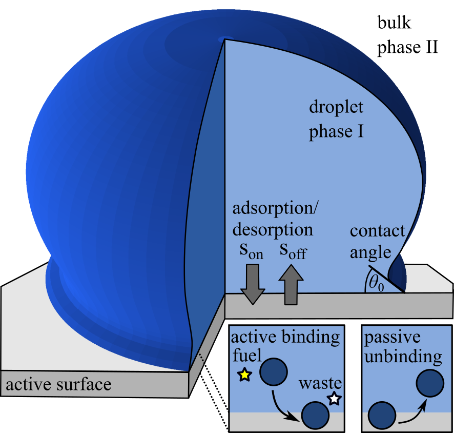
Theory of active wetting: We consider a binary solute-solvent mixture that can phase-separate in the bulk and which is in contact with a membrane surface (Fig. 1). In a finite system, a droplet-phase rich in solutes can coexist with a dilute phase in the bulk and wet the surface, enclosing a local contact angle . Moreover, the solutes are able to bind to the membrane with a rate governed by the net desorption flux (Eq. (10)). This binding process can be passive settling at binding equilibrium, or active, involving an additional external free energy .
The dynamics of active wetting can be described by a continuum theory where the fields of volume fraction in the bulk and the area fraction in the membrane are determined by the conservation laws:
| (1a) | ||||
| (1b) | ||||
| where and are to the diffusive fluxes in bulk and membrane. The gradient vector in the membrane plane is denoted by . | ||||
The contact angle implies a boundary condition for the bulk volume fraction at the membrane surface
| (1c) |
with the normal vector of the membrane . The contact angle is linked to the coupling free energy between bulk and membrane surface via ; more details are discussed in SI, section III.B.
For solutes being conserved in membrane and bulk, the net desorption flux is related to the normal component of diffusive bulk flux at the membrane surface:
| (1d) |
with and are the molecular volume and molecular area, respectively.
The net desorption flux is composed of the difference between an unbinding and a binding flux, . In passive systems, the two fluxes are linked by the detailed-balance of the rates, , with the chemical potentials in bulk, , and membrane, . To make the surface ‘active’, binding is maintained away from chemical equilibrium corresponding to , we introduce an external free energy , such that
| (1e) |
To ensure that this active system cannot be mapped on a passive system (i.e., by redefining the internal free energy for a constant ), the external free energy has to be phase-dependent. For simplicity, we choose , where denotes the activity parameter and is the bulk volume fraction at the membrane surface. Note that our choice of corresponds to a system where the magnitude of the external free energy inside the dense phase is the larger compared to the droplet surrounding. We consider positive and negative which describe the tendency to enrich or deplete the membrane surface by active binding.
The diffusive fluxes in bulk and membrane,
| (1f) | ||||
| (1g) |
are driven by gradients in the bulk and membrane chemical potential and , respectively, where and denote respective kinetic coefficients. Their volume and area fraction dependence is given in the methods section.
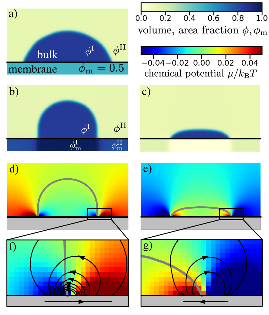
For a passive surface without any active binding processes (), the steady-state solution of Eqs. (1) corresponds to thermodynamic equilibrium. It is characterized by a homogeneous chemical potential that is identical between bulk and membrane implying that the diffusive fluxes and , and the binding flux are each zero. In this case, the wetted droplet takes the shape of a spherical cap and the contact angle fulfills the law of Young-Dupré (Fig. 2a).
When maintaining binding away from chemical equilibrium (), we find a non-equilibrium steady state with position-dependent chemical potentials that drive diffusive fluxes in the membrane and the bulk. Such fluxes are most pronounced near the triple line. Interestingly, these localized fluxes strongly affect the shape of wetted droplets, leading to deviations from a pronounced spherical cap (Fig. 2b-g). Depending on the value of the external free energy (), we find shapes that are qualitatively different from a passive system with the contact line expanding or contracting relative to the passive case. For a chemically active surface, we observe droplet shapes that are reminiscent of a pancake or a mushroom, respectively.
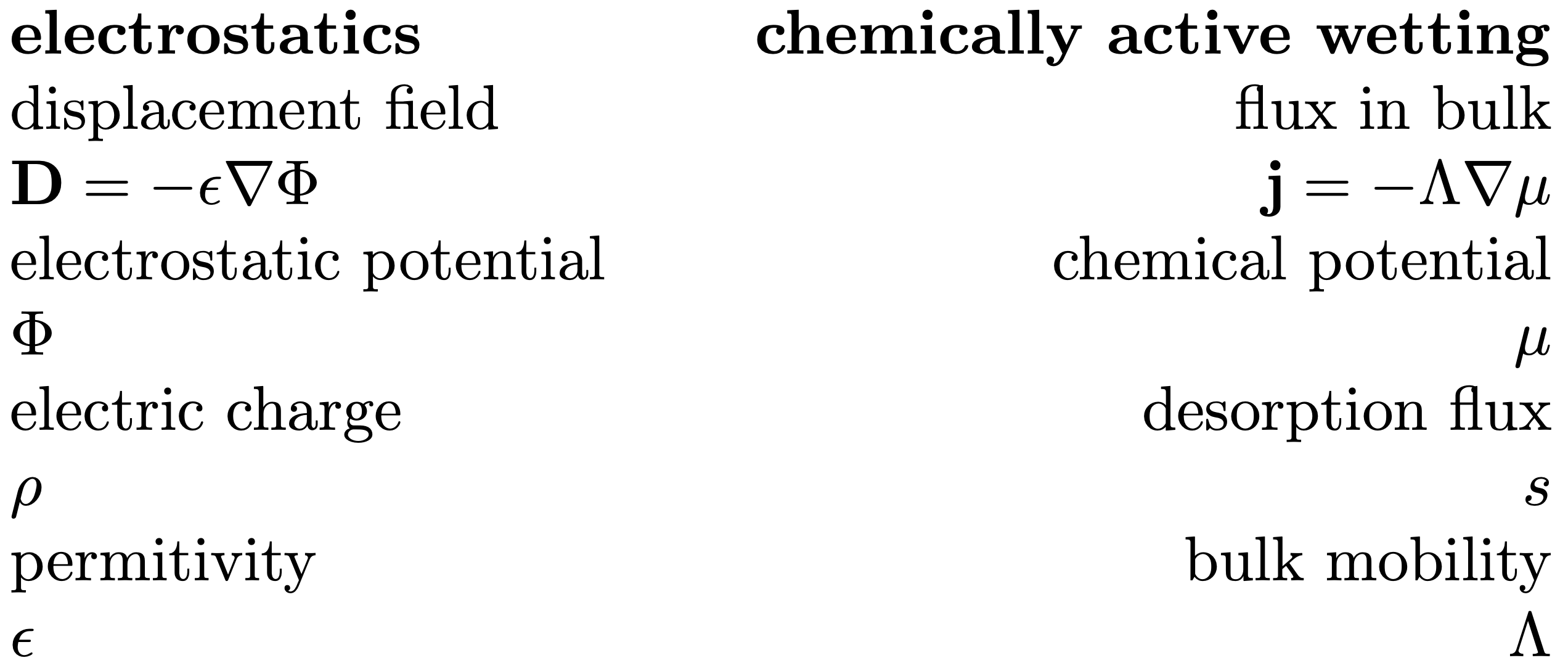
Mapping on electrostatics: The shape of wetting droplets on an active surface can be understood by drawing an analogy to electrostatics. To this end, we consider a charge free, linear dielectric medium adjacent to a non-conducting, non-polarizable medium. The interface is heterogeneously charged with a charge area density . According to Gauss’s law, the displacement field fulfills in the absence of free charges and at the interface (for more details, see SI IV). Comparing the electrostatic equations with the dynamic equations for active wetting Eqs. (1) at steady state (, ) suggests an mapping between electrostatics and active wetting, which is depicted in Table 3. Specifically, the net desorption flux generates a position-dependent chemical potential in the same way as a charge density gives rise to an electrostatic potential . Therefore, the far field of the chemical potential corresponds to the electrostatic potential field of a multipole.
To illustrate the mapping to electrostatics further, we consider a two-dimension system for simplicity. In this case, a two-dimensional droplet interacts with a one-dimensional membrane or equivalently, a two-dimensional electrostatic potential resulting from a one-dimensional line charge density. The three dimensional case is discussed in the SI, section V.
The multi-pole generated by the binding flux gives rise to a chemical potential profile which is governed to leading order by two dipoles positioned at . For constant mobility , this chemical potential can be written as (SI, section VA):
| (2) |
This chemical potential profile corresponds to the superposition of two dipole moments with opposite orientation and magnitude . The two dipoles have a distance from each other. In Eq. (2), and denote the lateral and horizontal coordinates and is a constant offset, that acts as a Lagrange multiplier to ensure a symmetric droplet shape.
The chemical potential profile Eq. (2) can be derived from the multipole moments of the binding flux . The monopole has to vanish due to particle conservation in a stationary system. The dipole moment of the whole active surface also vanishes due to the mirror symmetry of the droplet, which is formally written as . Thus, the first non-vanishing moment is the quadrupole moment. The quadrupole moment is generated by two oppositely oriented dipoles of equal magnitude
| (3) |
that are placed at , with the dipole position given as
| (4) |
Using the magnitude of the dipole moments and its positions, we obtain the potential profile is given in Eq. (2).
The dipole moment is caused by the mismatch of the membrane area fractions and adjacent to the dense and dilute bulk phase. To estimate , we describe the bulk droplet in the limit of a sharp interface at which local equilibrium holds leading the dense and dilute equilibrium values and . We fix the bulk chemical potential to be constant. Far from the contact line, the system becomes homogeneous even in the active case. The lateral diffusive membrane flux must therefore vanish. Subsequently the binding flux vanishes as well, which implies . This results in the following relationship:
| (5) |
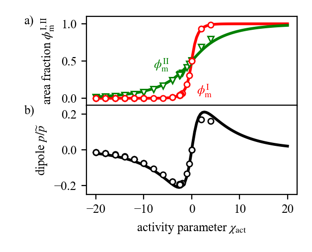
where is a function of . The values of that we determine based on Eq. (5) agree well with the simulation results (Fig. 4a). Furthermore, using an the sharp interface model, we find an analytic approximation for the dipole moment (see SI Sec.VI B for details)
| (6) |
where denote the diffusion constants in a surface of area fraction . Fig. 4b) shows that the analytic results obtained from the sharp interface model agree well with the numerical solution of the continuum model (Eq. (1)). We see that the magnitude of exhibits a maximum around and vanishes if the magnitude of increases. The dipole vanishes for large since the surface in both domains I and II gets either depleted ( for negative ), or fully occupied ( for positive ). Thus, in both cases, the difference between becomes small leading to a vanishing magnitude of the dipole moment according to Eq. (6).
Droplet shapes on active surfaces: The droplet shape is determined by the position-dependent chemical potential that results from the active binding processes with the surface. Note that there are no chemical reactions in the bulk. Therefore, we can consider the droplet interface between the dense droplet phase and the dilute phase to be at local equilibrium, implying a Gibbs-Thomson relation [37]. For a binary mixture described by a symmetric free energy density, the mean curvature is proportional to the chemical potential, , with , the surface tension of the planar interface. Using an arc length parameterization with the arc length , the mean curvature , and as the angle to the horizontal -axis, we find the following shape equations:
| (7a) | ||||
| (7b) | ||||
| (7c) | ||||
| with the boundary conditions | ||||
| (7d) | ||||
| (7e) | ||||
with denoting the mid point of the droplet interface and is the position of the triple line. For a given , the offset of the chemical potential in Eq. (2) has to be adjusted to match the boundary condition at . We note that the area, i.e. the two-dimensional volume, can be specified instead of . In this case, is a free parameter and acts as a Lagrange multiplier of the volume.
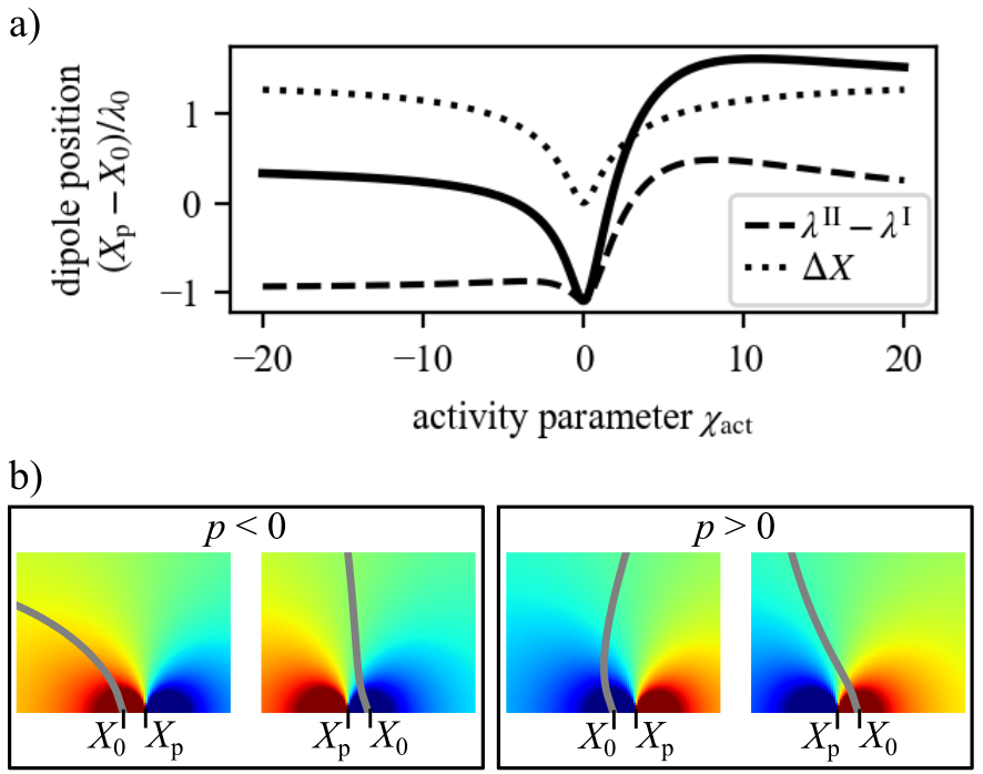
The activity parameter affects the position of the dipole
| (8) |
relative to the triple line at by a symmetric contribution and an in general asymmetric contribution from the reaction-diffusion length scales (Fig. 5a); see SI, section VIB for the expressions of and . The dipole can be deflected to the left or the right of the triple line. The asymmetry of this deflection with the activity parameter results from reaction rate coefficients and diffusivities depending on volume and area fractions that vary between the domains I and II (Fig. 5a). The changes in the droplet position are accompanied by pronounced changes in droplet shape in the vicinity of the triple line (Fig. 5b). The shape is calculated by solving Eqs. (7) with the chemical potential Eq. (2); more details are given in SI, section VI.A. We find a rather flat, pancake-like drop with a positive local curvature at the triple line when and have different signs. Once both have the same sign, the drop has a negative curvature at the triple line leading to mushroom shapes.
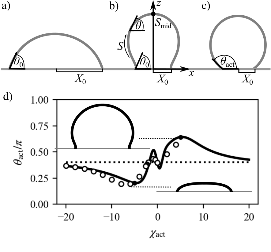
The effects on droplet shape by the active surface can be characterized by the active contact angle :
| (9) |
where is the area of the droplet, i.e., the two-dimensional equivalent of the droplet volume. The active contact angle becomes the local contact angle when the droplet wets a passive surface leading to a circular cap shape.
The active contact angle and thus the droplet shape is controlled by the activity parameter . For large and negative , is decreased, indicating a pancake shape while for large and positive , the active contact angle in enhanced corresponding to a mushroom shape (Fig. 6d). The results of the sharp interface model (solid line) agree well with the numerical calculations for a continuous interface (open circles).
Active droplet wetting in experiments: An open question is how to experimentally realize an active system where wetting and, thereby, the droplet shape can be controlled by active binding processes to a surface. The essential ingredient is a chemical component that can form droplets and bind to a surface. Binding and unbinding need to occur in a cyclic fashion and at similar rates. Moreover, binding and unbinding have to be maintained away from chemical equilibrium, for example, via a hydrolyzing fuel component that is chemo-stated. Moreover, the binding rate coefficients need to be fast enough such that the reaction length scales are small and localize well around the triple line. In this case, pronounced changes in droplet shape are expected. To be specific by the numbers, according to our model, pronounced shape changes occur for an activity parameter (Fig. (6)(d)), corresponding to a reaction-diffusion scale relative to droplet size . This case could be realized for example by a surface diffusion constant, , of and a binding rate in the order of . Thus, we propose a system that uses a ATP-driven phosphatase/kinase cycle to remove/donate a phosphate group to a phase separating component and thereby controls binding [38]. The turn-over of ATP enables to actively regulate binding by changing the ATP concentration and thereby control the shape of wetted droplets experimentally.
Conclusion: Our work shows that wetting on an active surface is significantly different from wetting on passive surfaces. We propose a novel class of active systems where an active surface that is maintained away from equilibrium by a binding process that breaks the detailed balance of the rates. We find that this binding process leads at steady-state to flux loops near the triple line. While for a passive surface, the shape of wetted droplets is a spherical cap with a minimal surface area, flux loops adjacent to active surfaces deflect the triple line where all three phases coexist. This results in droplet shapes reminiscent of a pancake or a mushroom. A striking property is that the lower dimensional active surface can strongly affect the shape of the higher dimensional droplet.
In the quest for understanding the complexities of non-equilibrium thermodynamics, drawing analogies to the field of electrostatics revealed the underlying principles governing dynamic processes and non-equilibrium thermodynamics across diverse scales [39, 40]. In our work, we establish a conceptual mapping between non-equilibrium chemical systems and materials with electrical properties. This conceptual mapping provides insights into the relationship between non-equilibrium chemical systems and passive materials with electrical properties.
Our findings of shapes that significantly deviate from a spherical cap suggest that active wetting can deform and alter the structural integrity of deformable membranes. We expect that such deformations can arise from the induced flux loops localized at the triple line acting as a local pump. Furthermore, such fluxes may drive membrane shape remodeling, including changes in membrane topology. Such changes would provide a gateway for biomolecular transport. Wetting on active surfaces and the associated transport phenomena thus may have implications for a variety of cellular processes, including membrane budding [41], and vesicle rupture [42].
Acknowledgments: We thank A. Honigmann and A. Hyman for fruitful discussions on the topics of wetting in cells and G. Bartolucci for helpful comments on the subject. We thank H. Vuijk for the critical comments on the manuscript. C. Weber acknowledges the European Research Council (ERC) under the European Union’s Horizon 2020 research and innovation programme (“Fuelled Life” with Grant agreement No. 949021) and the SPP 2191 “Molecular Mechanisms of Functional Phase Separation” of the German Science Foundation. F. Jülicher acknowledges funding by the Volkswagen Foundation.
Methods: We describe chemically active binding such that the binding flux becomes stronger or weaker than the passive system. In contrast, the unbinding flux remains unchanged, which leads to the following representation of the net desorption flux
| (10) |
with an intrinsic binding rate, the Boltzmann constant and the temperature. In an experimental setting, our model corresponds to a scenario where fuel, which drives the active binding process, partitions into the droplet and where fuel is continuously supplied from a reservoir while waste products are cleared sufficiently fast.
To study the wetting behavior, we describe both the bulk and the membrane by a Flory-Huggins free energy density, with
| (11) |
in bulk and
| (12) |
in the membrane and , the Flory-Huggins interaction parameters. The free energy of the system, which is composed of the bulk with volume and the membrane now reads
| (13) |
with and characterising the free energy cost for spatial inhomogeneities. The last term in Eq. (13) denotes the binding energy between bulk and membrane. For simplicity, we restrict ourselves to a coupling that is linear in the bulk volume fraction at the surface, , with a constant binding energy per unit area . The chemical potential in bulk and membrane are obtained from the free energy as and . We model the mobility coefficients as in bulk and in the membrane, with constant , . Furthermore, the volume fraction is subjected to the boundary condition
| (14) |
at the the surface.
We define the reaction diffusion length scale as a characteristic length scale of our system.
‡ These authors contributed equally to this work.
References
- Young [1805] T. Young, An essay on the cohesion of fluids, Phil. Trans. R. Soc. 95, 65 (1805).
- Dupré and Dupré [1869] A. Dupré and P. Dupré, Théorie mécanique de la chaleur (Gauthier-Villars, 1869).
- de Gennes [1985] P. G. de Gennes, Wetting: statics and dynamics, Rev. Mod. Phys. 57, 827 (1985).
- Gennes et al. [2004] P.-G. Gennes, F. Brochard-Wyart, D. Quéré, et al., Capillarity and wetting phenomena: drops, bubbles, pearls, waves (Springer, 2004).
- Dević et al. [2019] I. Dević, J. Encarnación Escobar, and D. Lohse, Equilibrium drop shapes on a tilted substrate with a chemical step, Langmuir 35, 3880 (2019).
- Martouzet et al. [2021] G. Martouzet, L. Jørgensen, Y. Pelet, A.-L. Biance, and C. Barentin, Dynamic arrest during the spreading of a yield stress fluid drop, Phys. Rev. Fluids 6, 044006 (2021).
- Wu et al. [2022] Y. Wu, M. Kuzina, F. Wang, M. Reischl, M. Selzer, B. Nestler, and P. A. Levkin, Equilibrium droplet shapes on chemically patterned surfaces: theoretical calculation, phase-field simulation, and experiments, J. Colloid Interface Sci. 606, 1077 (2022).
- Kusumaatmaja et al. [2009] H. Kusumaatmaja, Y. Li, R. Dimova, and R. Lipowsky, Intrinsic contact angle of aqueous phases at membranes and vesicles, Phys. Rev. Lett. 103, 238103 (2009).
- Liao et al. [2019] Y.-C. Liao, M. S. Fernandopulle, G. Wang, H. Choi, L. Hao, C. M. Drerup, R. Patel, S. Qamar, J. Nixon-Abell, Y. Shen, W. Meadows, M. Vendruscolo, T. P. Knowles, M. Nelson, M. A. Czekalska, G. Musteikyte, M. A. Gachechiladze, C. A. Stephens, H. A. Pasolli, L. R. Forrest, P. St George-Hyslop, J. Lippincott-Schwartz, and M. E. Ward, Rna granules hitchhike on lysosomes for long-distance transport, using annexin a11 as a molecular tether, Cell 179, 147 (2019).
- Agudo-Canalejo et al. [2021] J. Agudo-Canalejo, S. W. Schultz, H. Chino, S. M. Migliano, C. Saito, I. Koyama-Honda, H. Stenmark, A. Brech, A. I. May, N. Mizushima, and R. L. Knorr, Wetting regulates autophagy of phase-separated compartments and the cytosol, Nature 591, 142 (2021).
- Lu et al. [2022] T. Lu, S. Liese, L. Schoenmakers, C. A. Weber, H. Suzuki, W. T. S. Huck, and E. Spruijt, Endocytosis of coacervates into liposomes, J. Am. Chem. Soc. 144, 13451 (2022).
- Mangiarotti et al. [2023a] A. Mangiarotti, M. Siri, N. Tam, Z. Zhao, L. Malacrida, and R. Dimova, Biomolecular condensates modulate membrane lipid packing and hydration, Nature Communications 14 (2023a).
- Brangwynne et al. [2009] C. P. Brangwynne, C. R. Eckmann, D. S. Courson, A. Rybarska, C. Hoege, J. Gharakhani, F. Jülicher, and A. A. Hyman, Germline p granules are liquid droplets that localize by controlled dissolution/condensation, Science 324, 1729 (2009).
- Zhao and Zhang [2020] Y. Zhao and H. Zhang, Phase separation in membrane biology: The interplay between membrane-bound organelles and membraneless condensates., Dev Cell 55, 30 (2020).
- Kusumaatmaja et al. [2021] H. Kusumaatmaja, A. I. May, and R. L. Knorr, Intracellular wetting mediates contacts between liquid compartments and membrane-bound organelles, Journal of Cell Biology 220, e202103175 (2021).
- Beutel et al. [2019] O. Beutel, R. Maraspini, K. Pombo-Garcia, C. Martin-Lemaitre, and A. Honigmann, Phase separation of zonula occludens proteins drives formation of tight junctions, Cell 179, 923 (2019).
- Zhao et al. [2021] X. Zhao, G. Bartolucci, A. Honigmann, F. Jülicher, and C. A. Weber, Thermodynamics of wetting, prewetting and surface phase transitions with surface binding, New J. Phys. 23, 123003 (2021).
- Mangiarotti et al. [2023b] A. Mangiarotti, N. Chen, Z. Zhao, R. Lipowsky, and R. Dimova, Wetting and complex remodeling of membranes by biomolecular condensates, Nature Communications 14, 2809 (2023b).
- Pombo-García et al. [2022] K. Pombo-García, C. Martin-Lemaitre, and A. Honigmann, Wetting of junctional condensates along the apical interface promotes tight junction belt formation, bioRxiv , 2022 (2022).
- Sun et al. [2023] D. Sun, X. Zhao, T. Wiegand, G. Bartolucci, C. Martin-Lemaitre, S. W. Grill, A. A. Hyman, C. Weber, and A. Honigmann, Assembly of tight junction belts by surface condensation and actin elongation, bioRxiv , 2023 (2023).
- Moser et al. [2009] M. Moser, K. R. Legate, R. Zent, and R. Fässler, The tail of integrins, talin, and kindlins, Science 324, 895 (2009).
- Christie et al. [2013] M. P. Christie, P. Simerska, F. E.-C. Jen, M. P. Jennings, and I. Toth, Liposomes for improved enzymatic glycosylation of lipid-modified lactose enkephalin, ChemPlusChem 78, 793 (2013).
- Harrington et al. [2021] L. Harrington, J. M. Fletcher, T. Heermann, D. N. Woolfson, and P. Schwille, De novo design of a reversible phosphorylation-dependent switch for membrane targeting, Nat. Commun. 12, 1472 (2021).
- Wurtz and Lee [2018] J. D. Wurtz and C. F. Lee, Chemical-reaction-controlled phase separated drops: Formation, size selection, and coarsening, Phys. Rev. Lett. 120, 078102 (2018).
- Berry et al. [2018] J. Berry, C. P. Brangwynne, and M. Haataja, Physical principles of intracellular organization via active and passive phase transitions, Reports on Progress in Physics 81, 046601 (2018).
- Weber et al. [2019] C. A. Weber, D. Zwicker, F. Jülicher, and C. F. Lee, Physics of active emulsions, Rep. Prog. Phys. 82, 064601 (2019).
- Ziethen et al. [2023] N. Ziethen, J. Kirschbaum, and D. Zwicker, Nucleation of chemically active droplets, Phys. Rev. Lett. 130, 248201 (2023).
- Zwicker et al. [2017] D. Zwicker, R. Seyboldt, C. A. Weber, A. A. Hyman, and F. Jülicher, Growth and division of active droplets provides a model for protocells, Nat. Phys. 13, 408 (2017).
- Seyboldt and Jülicher [2018] R. Seyboldt and F. Jülicher, Role of hydrodynamic flows in chemically driven droplet division, New J. Phys. 20, 105010 (2018).
- Bauermann et al. [2022a] J. Bauermann, C. A. Weber, and F. Jülicher, Energy and matter supply for active droplets, Ann. Phys. 534, 2200132 (2022a).
- Bartolucci et al. [2021] G. Bartolucci, O. Adame-Arana, X. Zhao, and C. A. Weber, Econtrolling composition of coexisting phases via molecular transitions, Biophys. J. 120, 4682 (2021).
- Bergmann et al. [2023] A. M. Bergmann, J. Bauermann, G. Bartolucci, C. Donau, M. Stasi, A.-L. Holtmannspoetter, F. Julicher, C. A. Weber, and J. Boekhoven, Liquid spherical shells are a non-equilibrium steady state, bioRxiv , 2023 (2023).
- Bauermann et al. [2023] J. Bauermann, G. Bartolucci, J. Boekhoven, C. A. Weber, and F. Jülicher, Formation of liquid shells in active droplet systems 10.48550/arXiv.2306.10852 (2023).
- Zwicker et al. [2015] D. Zwicker, A. A. Hyman, and F. Jülicher, Suppression of ostwald ripening in active emulsions, Physical Review E 92, 012317 (2015).
- Kirschbaum and Zwicker [2021] J. Kirschbaum and D. Zwicker, Controlling biomolecular condensates via chemical reactions, Journal of The Royal Society Interface 18, 20210255 (2021).
- Bauermann et al. [2022b] J. Bauermann, S. Laha, P. M. McCall, F. Jülicher, and C. A. Weber, Chemical kinetics and mass action in coexisting phases, Journal of the American Chemical Society 144, 19294 (2022b).
- Bray [1994] A. Bray, Theory of phase-ordering kinetics, Advances in Physics 43, 357 (1994), https://doi.org/10.1080/00018739400101505 .
- Case et al. [2019] L. B. Case, J. A. Ditlev, and M. K. Rosen, Regulation of transmembrane signaling by phase separation, Annual review of biophysics 48, 465 (2019).
- Tsori and de Gennes [2004] Y. Tsori and P.-G. de Gennes, Self-trapping of a single bacterium in its own chemoattractant, EPL 66, 599 (2004).
- Deviri and Safran [2021] D. Deviri and S. A. Safran, Physical theory of biological noise buffering by multicomponent phase separation, Proc. Natl. Acad. Sci. U.S.A. 118, e2100099118 (2021).
- Vutukuri et al. [2020] R. Vutukuri, M. Hoore, C. Abaurrea Velasco, L. Buren, A. Dutto, T. Auth, D. Fedosov, G. Gompper, and J. Vermant, Active particles induce large shape deformations in giant lipid vesicles, Nature 586, 52 (2020).
- Penič et al. [2020] S. Penič, L. Mesarec, M. Fošnarič, L. Mrówczyńska, H. Hägerstrand, V. Kralj-Iglič, and A. Iglič, Budding and fission of membrane vesicles: A mini review, Frontiers in Physics 8, 10.3389/fphy.2020.00342 (2020).