GenEM: Physics-Informed Generative Cryo-Electron Microscopy
Abstract
In the past decade, deep conditional generative models have revolutionized the generation of realistic images, extending their application from entertainment to scientific domains. Single-particle cryo-electron microscopy (cryo-EM) is crucial in resolving near-atomic resolution 3D structures of proteins, such as the SARS-COV-2 spike protein. To achieve high-resolution reconstruction, AI models for particle picking and pose estimation have been adopted. However, their performance is still limited as they lack high-quality annotated datasets. To address this, we introduce physics-informed generative cryo-electron microscopy (GenEM), which for the first time integrates physical-based cryo-EM simulation with a generative unpaired noise translation to generate physically correct synthetic cryo-EM datasets with realistic noises. Initially, GenEM simulates the cryo-EM imaging process based on a virtual specimen. To generate realistic noises, we leverage an unpaired noise translation via contrastive learning with a novel mask-guided sampling scheme. Extensive experiments show that GenEM is capable of generating realistic cryo-EM images. The generated dataset can further enhance particle picking and pose estimation models, eventually improving the reconstruction resolution. We will release our code and annotated synthetic datasets.
1 Introduction
Over the past decade, deep generative models ranging from earlier variational auto-encoders (VAEs) [24] and generative adversarial networks (GANs) [13] to the more recent diffusion models [14] have shown successes in realistic image generation. With inputs as simple as textual descriptions, techniques such as Stable Diffusion [38] can generate photographic quality images far richer than one can capture. Additional controls [49] can be imposed to produce tailored visual effects, e.g., theatrical lighting [35], specific perspectives [30], human poses [52], etc. In fact, the successes of image generation have gone way beyond visual pleasantness, stimulating significant advances in scientific explorations. Examples include brain magnetic resonance imaging (MRI) to computational tomography (CT) translation [46], X-ray image generation [41], cosmological N-body simulation [50], etc. All have long relied on expensive imaging apparatus.
Different from “entertainment” applications that have largely focused on perceptual soundness, generation techniques for scientific imaging should faithfully follow the physical process: the generated results would eventually be applied to downstream tasks such as medical image diagnosis and protein structure reconstruction, where physical correctness overweighs visual pleasantness. In this work, we extend image generation to a specific biomolecular imaging technique, single-particle cryo-electron microscopy (cryo-EM) to improve the performance of its downstream tasks. Cryo-EM aims to recover the near-atomic resolution 3D structure of proteins, with its latest application in recovering the SARS-COV-2 spike protein [47] structure to guide drug development.
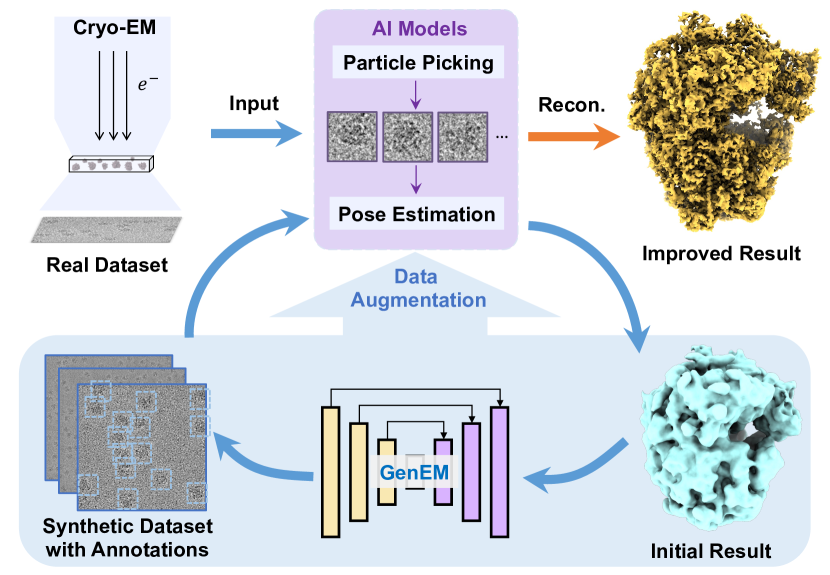
As shown in Figure 1, the cryo-EM pipeline starts with capturing 2D projection images, termed as micrographs, of flash-frozen purified specimens. These contain hundreds of thousands of target particles with unknown but varied locations, poses, and shapes. High-resolution cryo-EM reconstruction, similar to multi-view stereo [12] in computer vision, requires accurately picked particles and precisely estimated poses. Recent AI models such as Topaz [4] for particle picking and CryoFIRE [27] for pose estimation have been widely used. Yet, their performances are limited due to the lack of high-quality training datasets with expert annotations, which are very labor-intensive and difficult to acquire. A potential solution is using ground-truth annotations as controls to generate realistic synthetic images. However, it is quite challenging to generate physically correct synthetic images with realistic cryo-EM noises, which aims to enhance particle picking and pose estimation models, eventually improving reconstruction resolution on real datasets. Existing physical simulations [51] are computationally intensive to emulate complex noises. State-of-the-art generative methods such as CycleGAN [55] and Contrastive Unpaired Translation (CUT) [33] lack consideration of physical constraints and cannot generate random realistic noises.
In this paper, we propose physics-informed generative cryo-EM (GenEM), the first approach that integrates physical-based cryo-EM simulation with a generative unpaired noise translation to produce realistic synthetic cryo-EM datasets with annotations. Our synthetic dataset can significantly enhance cryo-EM particle picking and pose estimation models. To introduce physical constraints, GenEM starts from initial 3D biomolecule structures that can be obtained from the initial reconstruction. Then GenEM simulates the real cryo-EM imaging process on a virtual specimen that contains a variety of target particles to obtain physical-based simulation results. Furthermore, real cryo-EM images exhibit complex noises induced by limited electron beam dosage and manufacturing errors. To generate realistic noises, we propose a novel unpaired noise translation technique via contrastive learning to transform an initial Gaussian noise to a more realistic noise learned from real cryo-EM images. Finally, we find that randomly sampled negative patches are likely semantically similar to the positive sample, conflicting with contrastive learning assumptions. GenEM hence adopts a particle-background mask to guide the sampling scheme, ensuring that negative and positive samples are semantically different. Therefore, we improve the training efficiency and performance via our novel mask-guided sampling scheme.
We evaluate the capabilities of our method in photo-realism, particle picking, and pose estimation across three real cryo-EM datasets. The results show that GenEM not only produces the most photo-realistic cryo-EM images but also significantly improves the reconstruction resolution of the target molecule by fine-tuning particle picking and pose estimation models. In terms of photo-realism, we measure the similarity between generated and real micrographs. The results show that our approach achieves the best realistic cryo-EM image generation. In particle picking, the model augmented with GenEM data demonstrates the highest increased accuracy and finally improves the resolution of reconstruction in all three datasets. In pose estimation, we develop an end-to-end training pipeline on synthetic datasets and evaluate them on real datasets. The pose estimation module trained with our data achieves the best reconstruction resolutions across all three datasets. GenEM shows significant potential for generative methods in applications of scientific fields like cryo-EM where high-quality annotated real datasets are exhaustively required yet infeasible to obtain.
2 Related Works
Our work aims to extend generation methods to the field of cryo-EM. We therefore only discuss the most relevant works in respective fields.
Cryo-EM Simulations. Theoretical simulation techniques, based on physical priors, combine atomic-level simulations with global projection to accurately compute electron scattering during the imaging process [51, 42, 39, 29, 31]. Traditional simulation methods such as InsilicoTEM [51] model the interaction between electrons by taking specimens as multi-slices to theoretically improve the algorithm’s interpretability. However, they typically require expensive computational resources and may require complex adjustments of parameters to achieve ideal results. Our work also includes an efficient physical-based simulation module to effectively present the structural information of particles. Additionally, we generate realistic noises via a novel unpaired noise translation technique.
Unpaired Image-to-image Translation. Deep generative models can be used to fill the gap between simulated projections and real images without paired data, known as unpaired image-to-image translation. The diffusion model has been recently introduced to this task, such as UNIT-DDPM [40]. Although the experiments demonstrate compelling results in low resolution, the current forms of these methods do not perform sufficiently well at high resolutions like , preventing them from further applications in cryo-EM image generation.
Compared with diffusion models, GAN-based methods are more efficient and lightweight for high-resolution image generation. CycleGAN [55] and DualGAN [48] introduce two generative adversarial networks to calculate cycle consistency losses, allowing for training on unpaired data [19, 25, 34, 22]. DiscoGAN [23] extended this idea to maintain consistency in both directions. UNIT [28] uses shared latent representations for unsupervised image-to-image translation, while MUNIT[16] extends this concept to achieve multi-modal translation. TraVeLGAN [2], DistanceGAN [3], and GcGAN [11] achieve this goal through various methods, making one-way translation possible while avoiding traditional cycle consistency. Recently, Contrastive Unpaired Translation (CUT) [33] introduces a new generative framework via contrastive learning [21, 43, 53]. The core idea of contrastive loss is to encourage the generated images to be consistent with the original images in a low-dimensional embedding space, thereby improving image consistency and quality. However, existing methods do not address the physical process of electron microscopy projection. Our method introduces a physical-based simulation to add physical constraints. Further, we propose a novel unpaired noise translation based on contrastive learning with a mask-guided sampling scheme.
Neural Noise Generation. In response to the constraints posed by basic synthetic noise models, extensive work has been dedicated to developing various generative noise models. These models aim to generate complex noise patterns to tackle the challenges associated with real-world image-denoising tasks or other downstream tasks. Some recent methods focus on signal-dependent noise modeling [26, 18, 15, 5], while others [1, 8, 9] use metadata such as camera parameters to generate noise with specific distributions. These studies aim to enhance the performance of image denoising and noise synthesis to meet the demands of real-world scenarios. However, the current methods for generating signal-dependent noise follow a deterministic process, lacking the ability to vary from a single content to multiple real images. We instead incorporate Gaussian noise with physically simulated images to introduce randomness to the networks, achieving the translation from simple Gaussian noise distribution to the complex randomness presented in real cryo-EM images.
3 Physics-informed Generative Cryo-EM
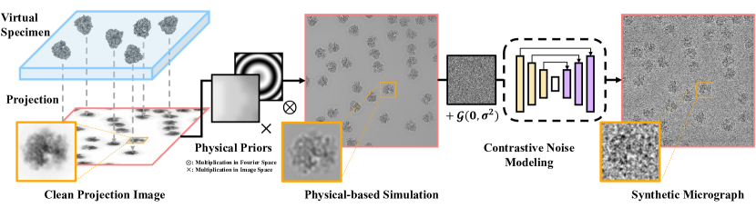
We propose the GenEM method, which for the first time integrates physics-informed generative cryo-EM image generation with a novel contrast noise generation technique, as shown in Figure 2. Our approach first prepares a virtual specimen containing a large number of uniformly distributed target protein structures (Section 3.1). Based on this, we emulate the cryo-EM imaging process to introduce physical constraints (Section 3.2). To generate realistic cryo-EM noises, we use an unpaired noise translation via contrastive learning (Section 4.1) with a novel mask-guided sampling scheme (Section 4.2). Finally, we detail our training scheme of GenEM (Section 4.3).

3.1 Virtual Specimen Preparation
To simulate the cryo-EM imaging process, we first prepare the virtual specimen, a large volume containing numerous low-resolution target particles. Particles inside the position, rotation, and conformation state of the specimen are randomly presented. To obtain an initial model of these particles, we employ cryoSPARC [36] for coarse particle picking and pose estimation. The picked particles with estimated poses then input to cryoDRGN [54] for continuous dynamic reconstruction of an initial 3D neural volume of density , where represents the spatial coordinates, denotes a high-dimensional vector in cryoDRGN representing particle conformation states.
During the construction of the virtual specimen , we place particles with random conformations into an initially empty virtual specimen. This placement involves randomly sampled rotation matrices and translations , which can be further used as ground-truth annotations. To prevent unrealistic overlapping due to random placement, particles are iteratively inserted. The inserted particle area in the virtual specimen is marked as occupied to avoid further placement. The virtual specimen can be expressed as:
| (1) |
where the number of particle , minimum and maximum are derived from real data to ensure content fidelity.
3.2 Emulating the Imaging Process
Based on a virtual specimen, we can mimic the cryo-EM imaging process. This process includes electron-specimen interaction, uneven ice thickness, and aberrations of cryo-EM “optical” devices. The electron-specimen interaction can be modeled as a projection of the specimen density using the weak phase object approximation [44] (WPOA). In the actual imaging process, we must consider the local attenuation of imaging intensity caused by uneven ice layer thickness. Thus, we utilize a re-implemented version of Icebreaker [32] to generate a weight map representing the local attenuation of electron maximum intensity. Lastly, we model the aberrations of optical devices as a point spread function , which can be estimated by CTFFIND4 [37] from cryo-EM images. Consequently, we can represent the 2D physical-based result as
| (2) |
4 Noise Generation via Contrastive Learning
Real cryo-electron microscopy (cryo-EM) images contain complex, random noises. Accurately generating realistic noises on synthetic cryo-EM images is crucial for downstream tasks. As shown in Figure 3, our method translates a simple Gaussian noise to a realistic cryo-EM noise via an unpaired noise translation (Section 4.1). In the training process, we introduce a mask-guided sampling scheme (Section 4.2) to improve training efficiency and performance.
4.1 Unpaired Noise Translation
The generation of noise can be modeled as an unpaired image-to-image translation task using a conditional generative model. The input, , learns realistic noises from real cryo-EM images , to produce synthetic cryo-EM images . The generative model includes a discriminator , and a generative function , where is a encoder and is a decoder.
The original contrastive learning method lacks randomness, producing only one electron microscopy noise image per physically simulated image, limiting the generation of varied synthetic datasets for downstream tasks. To overcome this, we introduced randomness in training process by adding zero-mean Gaussian random noise with variance to physical microscopy images, producing intermediate results , which is passed to generative function to produce . In all our experiments, we adjust the variance of Gaussian noise by signal-noise-ratio (SNR). Following this, we randomly select paired patches in and as the positive sample and query, respectively. We then choose patches in as negative samples. The encoder maps these to normalized vectors , , and , respectively. Our objective is to maximize the likelihood of selecting a positive sample by minimizing the cross-entropy loss:
| (3) | ||||
where is a temperature factor that scales the distance between the query and samples.
4.2 Mask-Guided Contrastive Learning
Randomly sampled negative patches are likely semantically similar to the positive sample, conflicting with contrastive learning assumptions [10]. To address this, we propose a mask-guided sampling scheme. We binarize a physical-based image to a particle-background mask , with denoting particle and denoting background. Guided by this mask, we randomly select the positive and the negative samples at locations where the mask labels are opposite, improving training efficiency and quality of results.
Similar to CUT [33], our method leverages multiple intermediate layers of to extract multi-scale features from input patches. Notably, we align the receptive field with particle size by selecting the ’s first layers. Feature map from each layer is then passed through compact two-layer MLP networks () to generate a feature , where is the -th layer. In all our experiments, we set to 5.
For particle-background mask locations, we define those on particles as , and those on the background as , where represents all locations with and represents all locations with . The feature of -th layer at a particle position is denoted as , and the set represented as . Similarly, for background positions, we denote the -th layer feature as , with the set represented as . Features encoded from the output are represented as , where . We propose a novel mask-guided Noise Contrastive Estimation (NCE) loss for contrastive learning:
| (4) | ||||
By minimizing MaskNCE loss, we effectively differentiate particle features and background features.
4.3 Training Scheme
We introduce randomness to the physical-based intermediate image by adding a random sampled Gaussian noise to a physical-based clean image. The intermediate image then inputs into the generative function to get a synthetic output image. Following the mask-guided sampling scheme, we extract multi-scale features to calculate MaskNCE loss, with query in the input domain, positive and negative in the output domain. Additionally, we introduce an adversarial loss function, , to encourage the network to produce more realistic simulated cryo-EM images, , as follows:
| (5) |
The overall training loss function is:
| (6) |
where is a experimental hyperparameter for adjusting loss weights.
5 Experiments
In this section, we evaluate our GenEM on a variety of challenging datasets. We run all experiments on a single NVIDIA GeForce RTX3090 GPU and it takes about 2 hours to train a GenEM with 200 epochs. We choose PatchGAN [17] as our discriminator and UNet implemented by [33] as our generator. During each training iteration, we resize the image resolution to . Guided by the particle-background mask, we evenly sample 256 queries on particles and 256 on the background, ensuring that their corresponding negative samples are located where the labels are opposite. The training dataset consists of 100 real images and 100 physical-based simulated results. We also do a data augmentation by rotating images by 90, 180, and 270 degrees.
Datasets. We evaluate GenEM on three challenging cryo-EM datasets. Each includes expert-curated particle picking results for high-resolution reconstruction, regarded as annotated particles. 1) T20S Proteasome (EMPIAR-10025) [6] contains 196 images and 49,954 annotated particles. The high density of these particles leads to occlusions, posing significant challenges in particle picking. 2) 80S Ribosome (EMPIAR-10028) [45] contains 1,081 images with 105,417 annotated particles. The complexity of these particles corresponding to a complex structure makes it difficult to estimate their poses. 3) Asymmetric Integrin (EMPIAR-10345) [7] includes 1,644 micrographs and 84,266 annotated particles with varied conformations, requiring more advanced processing for heterogeneous reconstruction.
5.1 Comparison
Similar to traditional image generation tasks, we begin with evaluating the similarity between our generated data and actual cryo-EM data. To validate our approach in the context of a cryo-EM data processing pipeline, we apply the generated data to two critical downstream tasks: particle picking and pose estimation. In the particle picking task, a state-of-the-art particle picking model augmented by GenEM data achieves the highest performance improvement, leading to better reconstruction resolution. In the pose estimation task, we train a cryo-EM pose estimation model with an augmented dataset. The results show that our model achieves the best reconstruction resolution on real datasets. Across all experiments, GenEM outperforms existing baselines both qualitatively and quantitatively.
Baselines. To evaluate the performance of GenEM compared to other existing methods, we select a traditional baseline approach by adding Gaussian noise to physically simulated results (denoted as Gaussian Noise). We also choose existing state-of-the-art unpaired image-to-image translation works, CycleGAN and CUT, as baselines to represent the performance of current deep generative models.
| best second-best | |||
| Metric | FID | ||
| Dataset | Proteasome | Ribosome | Integrin |
| Gaussian Noise | 89.87 | 177.01 | 174.94 |
| CycleGAN | 89.33 | 114.20 | 54.83 |
| CUT | 46.61 | 101.54 | 72.30 |
| Ours | 42.96 | 11.17 | 42.46 |
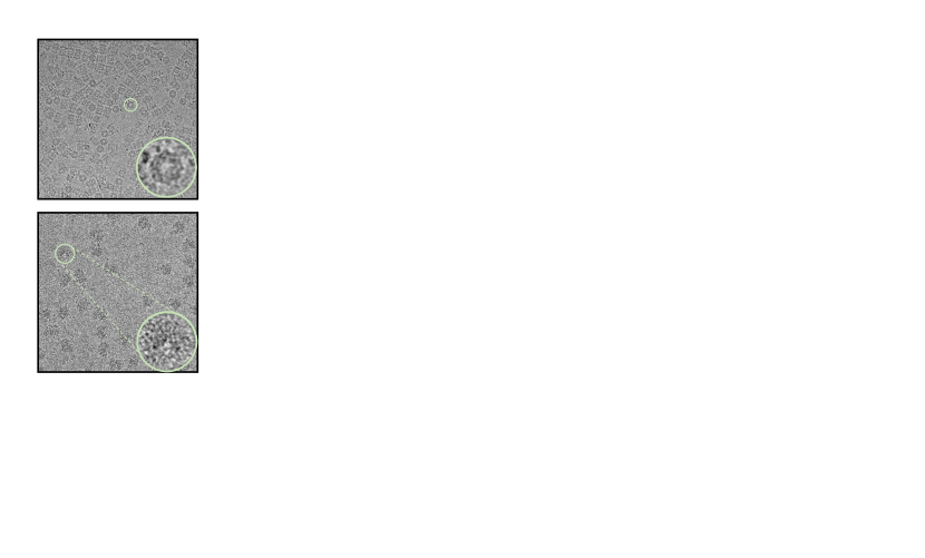
Photo-realism. We compare GenEM with several baselines in terms of the realism of synthesized cryo-EM images. As illustrated in Figure 4, our approach consistently achieves the best quality across all datasets in terms of overall noise distribution and the preservation of particle structural information. While Gaussian noise effectively preserves structural information, it fails to mimic authentic noise distributions. CycleGAN struggles with convergence on the Proteasome dataset and has severe artifacts on background regions on the Ribosome dataset due to the breakdown of its bijection assumption. CUT can achieve better photo-realism compared to CycleGAN but its performance is still limited due to its random sampling scheme. As shown in Table 1, we also employed Frechet Inception Distance (FID) which is widely used in generative tasks to measure the similarity between generated and real data. Our results significantly outperform existing baseline methods in all three datasets.
| best second-best | ||||||
|---|---|---|---|---|---|---|
| Metric | AUPRC | Res(Å) | ||||
| Dataset | Proteasome | Ribosome | Integrin | Proteasome | Ribosome | Integrin |
| Gaussian Noise | 0.471 | 0.485 | 0.259 | 2.50 | 3.76 | 7.63 |
| CycleGAN | 0.200 | 0.308 | 0.414 | 6.83 | 4.14 | 6.42 |
| CUT | 0.415 | 0.001 | 0.335 | 2.57 | 5.94 | 5.99 |
| Pre-trained Topaz | 0.302 | 0.679 | 0.526 | 2.59 | 3.28 | 5.08 |
| Topaz* | 0.599 | 0.749 | 0.650 | 2.53 | 3.27 | 5.36 |
| Ours | 0.490 | 0.797 | 0.606 | 2.48 | 3.25 | 4.85 |
| best second-best | |||
| Metric | Res(px) | ||
| Dataset | Proteasome | Ribosome | Integrin |
| Gaussian Noise | 2.97 | 6.00 | 6.86 |
| CycleGAN | 3.02 | 4.74 | 5.94 |
| CUT | 2.66 | 5.40 | 6.34 |
| Ours | 2.60 | 4.27 | 5.25 |
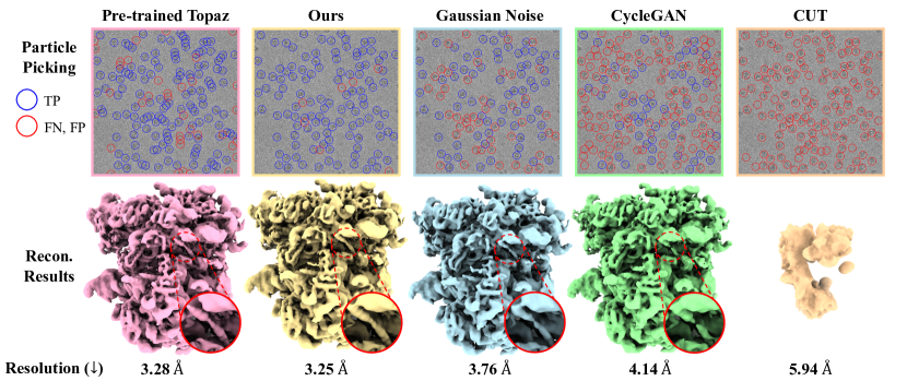
Particle Picking. We fine-tune a popular particle picking model, Pre-trained Topaz [4], with generated datasets with ground-truth annotations from different methods. As shown in Figure 5, we use the fine-tuned Topaz to pick particles on the Ribosome dataset. Our method outperforms the Pre-trained Topaz and other baselines in terms of visual picking results and reconstruction accuracy - our reconstruction result reveals the most detailed double helix structures, achieving the best resolution. While other baseline methods show a noticeable domain gap between their generated data and real data, reducing Topaz’s performance. Notably, although the baseline methods significantly impacted Topaz’s performance, the reconstruction results may be not markedly decreased due to the sufficient quantity of data.
Table 2 presents the quantitative particle picking results, where we utilized the Area Under the Precision-Recall Curve (AUPRC) metric. This metric is particularly suitable for datasets with incomplete positive labeling, a scenario evident in our manual annotations. Additionally, we include a version of Topaz that has been fine-tuned on roughly 1000-2000 particle annotations from 10 micrographs, referred to as Topaz*. The quantitative comparison results on AUPRC demonstrate that our method enhances Topaz’s performance and outperforms Topaz* with manual annotations on the Ribosome dataset. Taking each particle picking result as an input, cryoSPARC [36] reconstructs a 3D volume with its spatial resolution Res(Å). The quantitative comparison results on Res(Å) show that our approach consistently achieves the best resolution.
Pose Estimation. In the cryo-EM reconstruction pipeline, the accuracy of pose estimation is crucial for the resolution of the final reconstruction. Due to the unknown ground-truth particle poses in real cryo-EM images, existing cryo-EM reconstruction AI models, such as cryoFIRE [27], resort to self-supervised learning to predict particle poses directly from images during reconstruction. However, by leveraging the ground-truth particle pose information, we can supervise particle poses for the pre-trained pose estimation module of cryoFIRE. This approach improves the prediction of real particle poses, as shown in Table 3.
| best second-best | ||||
|---|---|---|---|---|
| Dataset | Ribosome | |||
| Task | Photo-realism | Particle Picking | Pose Estimation | |
| Metric | FID | AUPRC | Res(Å) | Res(px) |
| w/o Physical Priors | 16.63 | 0.789 | 3.29 | 4.88 |
| w/o Gaussian Noise | 50.74 | 0.735 | 3.31 | 4.24 |
| Third Layer Noise | 15.83 | 0.764 | 3.31 | 4.45 |
| Fifth Layer Noise | 21.30 | 0.777 | 3.26 | 4.46 |
| w/o Mask | 12.61 | 0.721 | 3.29 | 4.44 |
| Ours | 11.17 | 0.797 | 3.25 | 4.27 |
5.2 Ablation Study
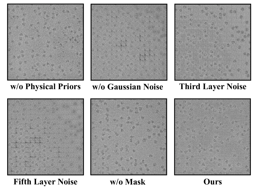
To generate realistic cryo-electron microscopy images, our method incorporates several key designs, including the integration of physical priors, the introduction of randomness in the generative model, and a mask-guided contrastive learning sampling strategy. We validated the effectiveness of these designs on the Ribosome dataset. First, we remove physical priors including ice thickness weighting and optical aberration simulation, denoted as w/o Physical Priors. We also remove Gaussian noise addition (w/o Gaussian Noise) to validate the effectiveness of introducing randomness. Further, we explore introducing randomness at the feature level by adding Gaussian noise at the third layer of the encoder (Third Layer Noise) and the fifth layer of the encoder (Fifth Layer Noise). Lastly, we remove the mask-guided sampling scheme (w/o Mask) to show how significantly it helps. As illustrated in Figure 6, the qualitative results show that removing modules or changing the position of Gaussian noise struggles insignificant CTF effects, severe artifacts on background noise, or content inconsistency. Quantitative results in Table 4 also show that our complete model consistently achieves the best or the second-best performance in all metrics.
6 Conclusion
Limitations. As the first trial to achieve authentic cryo-EM image generation with a novel combination of physical-based simulation and unpaired noise translation via contrastive learning, GenEM has some limitations. First, our physical-based simulation relies on an initial 3D model, which may be hard to obtain in some challenging cases, such as when the target molecule is too small to detect or is too flexible to estimate poses. Existing protein sequence prediction models like AlphaFold 2 [20] are helpful in giving an initial 3D structure. Also, GenEM lacks the generalization capability like changing input particles while still preserving realistic noises as we train GenEM per scene. In the future, we are willing to train a generalized version of GenEM to unlock more real applications in cryo-EM.
Conclusion. We have presented the first approach, GenEM, that marries physical-based simulation with a contrastive noise generation, to improve cryo-EM reconstruction results. To simulate a physical-based image, we first prepare a virtual specimen that contains enormous target particles, then we simulate the cryo-EM imaging process including projection, uneven ice thickness, and optical aberrations. Then we adopt an unpaired noise translation to transform simple Gaussian noises into complex realistic noises. Finally, we significantly improve the training efficiency and performance by proposing a novel mask-guided sampling scheme. Extensive experiments have shown that GenEM’s generated dataset with ground-truth annotations can effectively improve particle picking and pose estimation models, eventually improving reconstruction results. We believe that GenEM serves as a critical step for high-fidelity cryo-EM image generation, with essential cryo-EM applications such as particle picking and pose estimation.
References
- Abdelhamed et al. [2019] Abdelrahman Abdelhamed, Marcus A Brubaker, and Michael S Brown. Noise flow: Noise modeling with conditional normalizing flows. In Proceedings of the IEEE/CVF International Conference on Computer Vision, pages 3165–3173, 2019.
- Amodio and Krishnaswamy [2019] Matthew Amodio and Smita Krishnaswamy. Travelgan: Image-to-image translation by transformation vector learning. In Proceedings of the ieee/cvf conference on computer vision and pattern recognition, pages 8983–8992, 2019.
- Benaim and Wolf [2017] Sagie Benaim and Lior Wolf. One-sided unsupervised domain mapping. Advances in neural information processing systems, 30, 2017.
- Bepler et al. [2018] Tristan Bepler, Andrew Morin, Micah Rapp, Julia Brasch, Lawrence Shapiro, Alex J. Noble, and Bonnie Berger. Positive-unlabeled convolutional neural networks for particle picking in cryo-electron micrographs. Nature methods, 16:1153 – 1160, 2018.
- Cai et al. [2022] Yuanhao Cai, Xiaowan Hu, Haoqian Wang, Yulun Zhang, Hanspeter Pfister, and D. Wei. Learning to generate realistic noisy images via pixel-level noise-aware adversarial training. ArXiv, abs/2204.02844, 2022.
- Campbell et al. [2015] Melody G. Campbell, David Veesler, Anchi Cheng, Clinton S. Potter, and Bridget Carragher. 2.8 å resolution reconstruction of the thermoplasma acidophilum 20s proteasome using cryo-electron microscopy. eLife, 4, 2015.
- Campbell et al. [2020] Melody G. Campbell, Anthony Cormier, Saburo Ito, Robert I Seed, and Stephen L. Nishimura. Cryo-em reveals integrin-mediated tgf- activation without release from latent tgf-. Cell, 180:490–501.e16, 2020.
- Chang et al. [2020] Ke-Chi Chang, Ren Wang, Hung-Jin Lin, Yu-Lun Liu, Chia-Ping Chen, Yu-Lin Chang, and Hwann-Tzong Chen. Learning camera-aware noise models. In European Conference on Computer Vision, pages 343–358. Springer, 2020.
- Chen et al. [2019] Ting Chen, Xiaohua Zhai, Marvin Ritter, Mario Lucic, and Neil Houlsby. Self-supervised gans via auxiliary rotation loss. In Proceedings of the IEEE/CVF conference on computer vision and pattern recognition, pages 12154–12163, 2019.
- Chuang et al. [2020] Ching-Yao Chuang, Joshua Robinson, Yen-Chen Lin, Antonio Torralba, and Stefanie Jegelka. Debiased contrastive learning. Advances in neural information processing systems, 33:8765–8775, 2020.
- Fu et al. [2019] Huan Fu, Mingming Gong, Chaohui Wang, Kayhan Batmanghelich, Kun Zhang, and Dacheng Tao. Geometry-consistent generative adversarial networks for one-sided unsupervised domain mapping. In Proceedings of the IEEE/CVF Conference on Computer Vision and Pattern Recognition, pages 2427–2436, 2019.
- Furukawa and Hernández [2015] Yasutaka Furukawa and Carlos Hernández. 2015.
- Goodfellow et al. [2014] Ian Goodfellow, Jean Pouget-Abadie, Mehdi Mirza, Bing Xu, David Warde-Farley, Sherjil Ozair, Aaron Courville, and Yoshua Bengio. Generative adversarial nets. In Advances in Neural Information Processing Systems. Curran Associates, Inc., 2014.
- Ho et al. [2020] Jonathan Ho, Ajay Jain, and Pieter Abbeel. Denoising diffusion probabilistic models. In Advances in Neural Information Processing Systems, pages 6840–6851. Curran Associates, Inc., 2020.
- Hong et al. [2020] Zhiwei Hong, Xiaocheng Fan, Tao Jiang, and Jianxing Feng. End-to-end unpaired image denoising with conditional adversarial networks. In AAAI Conference on Artificial Intelligence, 2020.
- Huang et al. [2018] Xun Huang, Ming-Yu Liu, Serge Belongie, and Jan Kautz. Multimodal unsupervised image-to-image translation. In Proceedings of the European conference on computer vision (ECCV), pages 172–189, 2018.
- Isola et al. [2017] Phillip Isola, Jun-Yan Zhu, Tinghui Zhou, and Alexei A Efros. Image-to-image translation with conditional adversarial networks. In IEEE Conference on Computer Vision and Pattern Recognition (CVPR), 2017.
- Jang et al. [2021] Geonwoon Jang, Wooseok Lee, Sanghyun Son, and Kyoung Mu Lee. C2n: Practical generative noise modeling for real-world denoising. 2021 IEEE/CVF International Conference on Computer Vision (ICCV), pages 2330–2339, 2021.
- Johnson et al. [2016] Justin Johnson, Alexandre Alahi, and Li Fei-Fei. Perceptual losses for real-time style transfer and super-resolution. ArXiv, abs/1603.08155, 2016.
- Jumper et al. [2021] John Jumper, Richard Evans, Alexander Pritzel, Tim Green, Michael Figurnov, Olaf Ronneberger, Kathryn Tunyasuvunakool, Russ Bates, Augustin Žídek, Anna Potapenko, et al. Highly accurate protein structure prediction with alphafold. Nature, 596(7873):583–589, 2021.
- Jung et al. [2022] Chanyong Jung, Gihyun Kwon, and Jong-Chul Ye. Exploring patch-wise semantic relation for contrastive learning in image-to-image translation tasks. 2022 IEEE/CVF Conference on Computer Vision and Pattern Recognition (CVPR), pages 18239–18248, 2022.
- Kim et al. [2019] Junho Kim, Minjae Kim, Hyeonwoo Kang, and Kwanghee Lee. U-gat-it: Unsupervised generative attentional networks with adaptive layer-instance normalization for image-to-image translation. ArXiv, abs/1907.10830, 2019.
- Kim et al. [2017] Taeksoo Kim, Moonsu Cha, Hyunsoo Kim, Jung Kwon Lee, and Jiwon Kim. Learning to discover cross-domain relations with generative adversarial networks. In International conference on machine learning, pages 1857–1865. PMLR, 2017.
- Kingma and Welling [2013] Diederik P Kingma and Max Welling. Auto-encoding variational bayes. arXiv preprint arXiv:1312.6114, 2013.
- Lee et al. [2018] Hsin-Ying Lee, Hung-Yu Tseng, Jia-Bin Huang, Maneesh Kumar Singh, and Ming Yang. Diverse image-to-image translation via disentangled representations. ArXiv, abs/1808.00948, 2018.
- Lee and Kim [2022] Seunghwan Lee and Tae Hyun Kim. Noisetransfer: Image noise generation with contrastive embeddings. In Proceedings of the Asian Conference on Computer Vision, pages 3569–3585, 2022.
- Levy et al. [2022] Axel Levy, Gordon Wetzstein, Julien N. P. Martel, Frédéric Poitevin, and Ellen D. Zhong. Amortized inference for heterogeneous reconstruction in cryo-em. Advances in neural information processing systems, 35:13038–13049, 2022.
- Liu et al. [2017] Ming-Yu Liu, Thomas Breuel, and Jan Kautz. Unsupervised image-to-image translation networks. Advances in neural information processing systems, 30, 2017.
- Lobato and Van Dyck [2015] I Lobato and D Van Dyck. Multem: A new multislice program to perform accurate and fast electron diffraction and imaging simulations using graphics processing units with cuda. Ultramicroscopy, 156:9–17, 2015.
- Long et al. [2023] Xiaoxiao Long, Yuan-Chen Guo, Cheng Lin, Yuan Liu, Zhiyang Dou, Lingjie Liu, Yuexin Ma, Song-Hai Zhang, Marc Habermann, Christian Theobalt, and Wenping Wang. Wonder3d: Single image to 3d using cross-domain diffusion, 2023.
- Madsen and Susi [2020] Jacob Madsen and Toma Susi. abtem: Ab initio transmission electron microscopy image simulation. Microscopy and Microanalysis, 26(S2):448–450, 2020.
- Olek et al. [2022] Mateusz Olek, Kevin Cowtan, Donovan Webb, Yuriy Chaban, and Peijun Zhang. Icebreaker: Software for high-resolution single-particle cryo-em with non-uniform ice. Structure, 30(4):522–531, 2022.
- Park et al. [2020] Taesung Park, Alexei A Efros, Richard Zhang, and Jun-Yan Zhu. Contrastive learning for unpaired image-to-image translation. In Computer Vision–ECCV 2020: 16th European Conference, Glasgow, UK, August 23–28, 2020, Proceedings, Part IX 16, pages 319–345. Springer, 2020.
- Pfeiffer et al. [2019] Micha Pfeiffer, Isabel Funke, Maria Ruxandra Robu, Sebastian Bodenstedt, Leon Strenger, Sandy Engelhardt, Tobias Ross, Matthew J. Clarkson, Kurinchi S. Gurusamy, Brian R. Davidson, Lena Maier-Hein, Carina Riediger, Thilo Welsch, Jürgen Weitz, and Stefanie Speidel. Generating large labeled data sets for laparoscopic image processing tasks using unpaired image-to-image translation. In International Conference on Medical Image Computing and Computer-Assisted Intervention, 2019.
- Ponglertnapakorn et al. [2023] Puntawat Ponglertnapakorn, Nontawat Tritrong, and Supasorn Suwajanakorn. Difareli: Diffusion face relighting, 2023.
- Punjani et al. [2017] Ali Punjani, John L Rubinstein, David J Fleet, and Marcus A Brubaker. cryosparc: algorithms for rapid unsupervised cryo-em structure determination. Nature methods, 14(3):290–296, 2017.
- Rohou and Grigorieff [2015] Alexis Rohou and Nikolaus Grigorieff. Ctffind4: Fast and accurate defocus estimation from electron micrographs. Journal of structural biology, 192(2):216–221, 2015.
- Rombach et al. [2022] Robin Rombach, Andreas Blattmann, Dominik Lorenz, Patrick Esser, and Björn Ommer. High-resolution image synthesis with latent diffusion models. In Proceedings of the IEEE/CVF conference on computer vision and pattern recognition, pages 10684–10695, 2022.
- Rullgård et al. [2011] Hans Rullgård, L-G Öfverstedt, Sergey Masich, Bertil Daneholt, and Ozan Öktem. Simulation of transmission electron microscope images of biological specimens. Journal of microscopy, 243(3):234–256, 2011.
- Sasaki et al. [2021] Hiroshi Sasaki, Chris G Willcocks, and Toby P Breckon. Unit-ddpm: Unpaired image translation with denoising diffusion probabilistic models. arXiv preprint arXiv:2104.05358, 2021.
- Teixeira et al. [2018] Brian Teixeira, Vivek Singh, Terrence Chen, Kai Ma, Birgi Tamersoy, Yifan Wu, Elena Balashova, and Dorin Comaniciu. Generating synthetic x-ray images of a person from the surface geometry. In Proceedings of the IEEE Conference on Computer Vision and Pattern Recognition (CVPR), 2018.
- Vulović et al. [2013] Miloš Vulović, Raimond BG Ravelli, Lucas J van Vliet, Abraham J Koster, Ivan Lazić, Uwe Lücken, Hans Rullgård, Ozan Öktem, and Bernd Rieger. Image formation modeling in cryo-electron microscopy. Journal of structural biology, 183(1):19–32, 2013.
- Wang et al. [2021] Weilun Wang, Wen gang Zhou, Jianmin Bao, Dong Chen, and Houqiang Li. Instance-wise hard negative example generation for contrastive learning in unpaired image-to-image translation. 2021 IEEE/CVF International Conference on Computer Vision (ICCV), pages 14000–14009, 2021.
- Williams and Carter [1996] David B. Williams and C. Barry Carter. Transmission electron microscopy: A textbook for materials science. 1996.
- Wong et al. [2014] Wilson W. Wong, Xiao chen Bai, Alan Brown, Israel S. Fernández, Eric Hanssen, Melanie M Condron, Yan hong Tan, Jake Baum, and Sjors H. W. Scheres. Cryo-em structure of the plasmodium falciparum 80s ribosome bound to the anti-protozoan drug emetine. eLife, 3, 2014.
- Yang et al. [2020] Heran Yang, Jian Sun, Aaron Carass, Can Zhao, Junghoon Lee, Jerry L. Prince, and Zongben Xu. Unsupervised mr-to-ct synthesis using structure-constrained cyclegan. IEEE Transactions on Medical Imaging, 39(12):4249–4261, 2020.
- Yao et al. [2020] Hangping Yao, Yutong Song, Yong Chen, Nanping Wu, Jialu Xu, Chujie Sun, Jiaxing Zhang, Tianhao Weng, Zheyuan Zhang, Zhigang Wu, et al. Molecular architecture of the sars-cov-2 virus. Cell, 183(3):730–738, 2020.
- Yi et al. [2017] Zili Yi, Hao Zhang, Ping Tan, and Minglun Gong. Dualgan: Unsupervised dual learning for image-to-image translation. In Proceedings of the IEEE international conference on computer vision, pages 2849–2857, 2017.
- Zhang et al. [2023a] Lvmin Zhang, Anyi Rao, and Maneesh Agrawala. Adding conditional control to text-to-image diffusion models. In Proceedings of the IEEE/CVF International Conference on Computer Vision, pages 3836–3847, 2023a.
- Zhang et al. [2023b] Xiaowen Zhang, Patrick Lachance, Yueying Ni, Yin Li, Rupert AC Croft, Tiziana Di Matteo, Simeon Bird, and Yu Feng. Ai-assisted super-resolution cosmological simulations iii: Time evolution. arXiv preprint arXiv:2305.12222, 2023b.
- Zhang et al. [2020] Yue Zhang, R Tammaro, Peter J Peters, and RBG Ravelli. Could egg white lysozyme be solved by single particle cryo-em? Journal of Chemical Information and Modeling, 60(5):2605–2613, 2020.
- Zhao et al. [2022] Fuqiang Zhao, Wei Yang, Jiakai Zhang, Pei Lin, Yingliang Zhang, Jingyi Yu, and Lan Xu. Humannerf: Efficiently generated human radiance field from sparse inputs. In Proceedings of the IEEE/CVF Conference on Computer Vision and Pattern Recognition (CVPR), pages 7743–7753, 2022.
- Zheng et al. [2021] Chuanxia Zheng, Tat-Jen Cham, and Jianfei Cai. The spatially-correlative loss for various image translation tasks. 2021 IEEE/CVF Conference on Computer Vision and Pattern Recognition (CVPR), pages 16402–16412, 2021.
- Zhong et al. [2021] Ellen D Zhong, Tristan Bepler, Bonnie Berger, and Joseph H Davis. Cryodrgn: reconstruction of heterogeneous cryo-em structures using neural networks. Nature methods, 18(2):176–185, 2021.
- Zhu et al. [2017] Jun-Yan Zhu, Taesung Park, Phillip Isola, and Alexei A Efros. Unpaired image-to-image translation using cycle-consistent adversarial networks. In Proceedings of the IEEE international conference on computer vision, pages 2223–2232, 2017.