Automatic Report Generation for Histopathology images using pre-trained Vision Transformers and BERT
Abstract
Deep learning for histopathology has been successfully used for disease classification, image segmentation and more. However, combining image and text modalities using current state-of-the-art methods has been a challenge due to the high resolution of histopathology images. Automatic report generation for histopathology images is one such challenge. In this work, we show that using an existing pre-trained Vision Transformer in a two-step process of first using it to encode 4096x4096 sized patches of the Whole Slide Image (WSI) and then using it as the encoder and a pre-trained Bidirectional Encoder Representations from Transformers (BERT) model for language modeling-based decoder for report generation, we can build a fairly performant and portable report generation mechanism that takes into account the whole of the high resolution image, instead of just the patches. Our method allows us to not only generate and evaluate captions that describe the image, but also helps us classify the image into tissue types and the gender of the patient as well. Our best performing model achieves a 79.98% accuracy in Tissue Type classification and 66.36% accuracy in classifying the sex of the patient the tissue came from, with a BLEU-4 score of 0.5818 in our caption generation task.
Index Terms— computer vision, nlp, histopathology, deep learning, vision language models
1 Introduction
High resolution histopathology slides are a rich resource of information that current deep learning methods are able to exploit for various use cases like disease classification, cell segmentation and outcome prediction. However, as the images are very high resolution, usually in the range of 150,000x150,000px, they often require non-trivial modifications to existing state-of-the-art (SOTA) deep learning architectures to be used successfully. The most common method for handling these high resolution images is to patch the bigger image into smaller sized images that can be fed into Convolutional Neural Networks. For example, in a classification setting, this often works as a multiple instance learning problem, where each patch is given the same overall image label. A potential drawback to this is that patching can lead to removal of overall context from the whole slide image (WSI) that the model might need to learn to make the correct decision, unless handled properly.
Automatic report generation for histopathology images is an area of existing research that also suffers from the need for modifying SOTA image captioning architectures to fit researchers needs. Image captioning for histopathology helps us combine two rich sources of information, that is, high resolution WSIs and associated diagnostic reports that describe features of the image. In clinical settings, automatic report generation has been successfully used for X-ray images and claim to reduce the burden for radiologists by assisting them in describing the image [1]. Other use cases for automated image captioning in medical images can be image retrieval, as generated reports could be part of a searchable database, and encouraging standardized clinical ontologies by using words from a standard vocabulary to describe similar things. Therefore, automated image captioning for histopathology can be similarly useful for a wide variety of tasks that can assist physicians and radiologists in their tasks.
2 Related Work
Current research for histopathological image captioning focuses on CNN based encoder and Recurrent neural Network (RNN) based decoder architectures [2, 3, 4]. This is inspired by Show, attend and tell paper, that in particular has the capability of using the attention mechanism to focus on certain areas of the image to generate captions [5].
Using Imagenet pre-trained Convolutional Neural Network (CNN) encoders to encode smaller sized patches of the high resolution WSI has been successfully used in a variety of ways to essentially reduce the size of large dimensional WSIs to smaller and computationally manageable representations [6]. In recent years, a self-supervised Vision Transformer (ViT) based image representation learning mechanism called Hierarchical Image Pyramid Transformer (HIPT) has been proposed [7]. The self-supervised pre-training leverages DINO (distillation with no labels) at two levels, 256x256 sized patches and 4096x4096 sized patches. The authors show that this can then be leveraged for further downstream tasks like disease sub-typing and survival prediction, as the pre-trained ViT representations are now looking at the WSI at multiscale level [8].
A recent work by Zhang et al. uses a two-step process in which they first encode all patches of a WSI using a triplet loss based convolutional autoencoder and use the features from the bottleneck layer to cluster the patches into clusters [2]. In the second step they randomly sample the patches from each cluster, use a ImageNet pretrained ResNet-18 to extract -dimensional features for the -patches, then use attention pooling to reduce dimensional feature vector to and then feed into a LSTM decoder to generate captions. More recently, Gamper and Rajpoot describe the ARCH dataset which contains histopathology images extracted from textbooks and their associated descriptions, which they use for caption generation based pre-training task to generate an encoder that when used for downstream tasks like multiple instance learning shows promising results compared to other pre-trained encoders [3]. In Tsuneki and Kanavati [4], the authors use high resolution WSIs from a Japanese hospital system and associated translated text reports, for their automated captioning system. They use EfficientNetB3 [9] and DenseNet121 [10] pretrained on ImageNet dataset and extract features from the penultimate layer for 300x300 patches extracted from the WSI. They then use global average pooling and 3x3 average pooling to reduce the feature sizes and feed them into an RNN based decoder for generating their captions.
BERT is a transformer model pre-trained on a large corpus of unlabeled text data that is able to learn bidirectional representations for text and has been successfully used for various downstream tasks in like Question Answering (QA), Named Entity Recognition (NER) and Natural Language Inference (NLI) [11]. BioBERT and ClinicalBERT, trained on medical research articles and clinical notes data respectively, performed better on clinical downstream tasks than just BERT [12, 13].
More recently, pre-trained transformers have been successfully used for optical character recognition (OCR), that is, converting text in images to machine-readable text [14]. The authors were able to outperform state-of-the-art (SOTA) approaches for OCR using pre-trained Vision Transformers and transformer-based language models. The authors utilize the image representations from the vision transformers and, along with the context generated before, use it to predict the next tokens. A ”[BOS]” and ”[EOS]” tokens are appended at the beginning and end of the ground truth tokens. Note that using ”[BOS]” token shifts the sequence to the right by one place and is used to indicate start of generation.
Motivated by the success of pre-trained transformer models for such diverse downstream tasks, we propose a method that uses HIPT to encode WSIs that captures multi-level representations, and a BioClinicalBERT based decoder that is able to utilize powerful text representations to generate descriptions of the WSI.
3 Dataset
We get our imaging and associated text data from the Genotype-Tissue Expression (GTEx) portal111https://www.gtexportal.org/home/histologyPage, same as [2]. Our dataset consists of 9911 training samples, 972 validation samples and 984 testing samples. We do this because this is the only publicly available high resolution histology data with associated descriptions of each histology slide at the time of writing this paper that we could find. Data from Tsuneki and Kanavati [4] comes in the form of 300x300 patches222https://zenodo.org/record/6021442 and the ARCH dataset by Gamper and Rajpoot[3] are of mixed quality, magnifications, and resolutions that do not meet our criteria of working with high resolution Whole Slide Images which are the most likely form of data available in health systems.
Data in the GTEx portal comes in a tabular format with , and in separate columns. We create the description for each tissue in the following format:
this is a {tissue_type} tissue from a {sex} patient and it has {pathology_notes}.
An example caption would look like this: this is a small intestine - terminal ileum tissue from a male patient and it has 6 pieces, prominent lymphoid component in 4 of 6 pieces.
4 Method
4.1 WSI encoding using HIPT
To encode the whole WSI for caption generation, we use the HIPT (Hierarchical Image Pyramid Transformer) architecture as described in [7], where the authors first describe the self-supervised pre-training of multiscale Vision Transformers (ViT) using the DINO method for knowledge distillation pioneered by [8] on 10,678 WSIs. They then describe using this pre-trained multiscale ViT for downstream tasks like slide level classification, survival prediction and further using the unique attention maps generated by the ViTs for finding morphological phenotypes. They show that a HIPT based WSI level encoder outperforms current SOTA in multiple instance learning for histopathology classification, that is, CLAM-SB [15].
Here, we explain the steps taken to encode the WSI using HIPT as that will inform our use later when we use it for report generation. For -, is the size of the patch and is the size of the non overlapping tokens extracted from the or image following the notation described in [7].
-
1.
Create 4096x4096 patches for each WSI, taking care that each patch contains more than 50% tissue area.
-
2.
Initialize and freeze -16 and -256 subnetworks.
-
3.
For each patch, first extract the representation for each 256x256 sized patch from -16. Since a has 256 patches, each can now be represented using a vector .
-
4.
We also extract the representation for each patch from -256.
-
5.
We concatenate the and representations for each -patch such that each WSI is now represented with a vector .
Chen et al. [7] generously provide their pre-trained weights for -16 and -256 in a GitHub repository333https://github.com/mahmoodlab/HIPT/ that we utilize to generate multi-scale representations of our Whole Slide Image.
4.2 Caption Generation using BERT
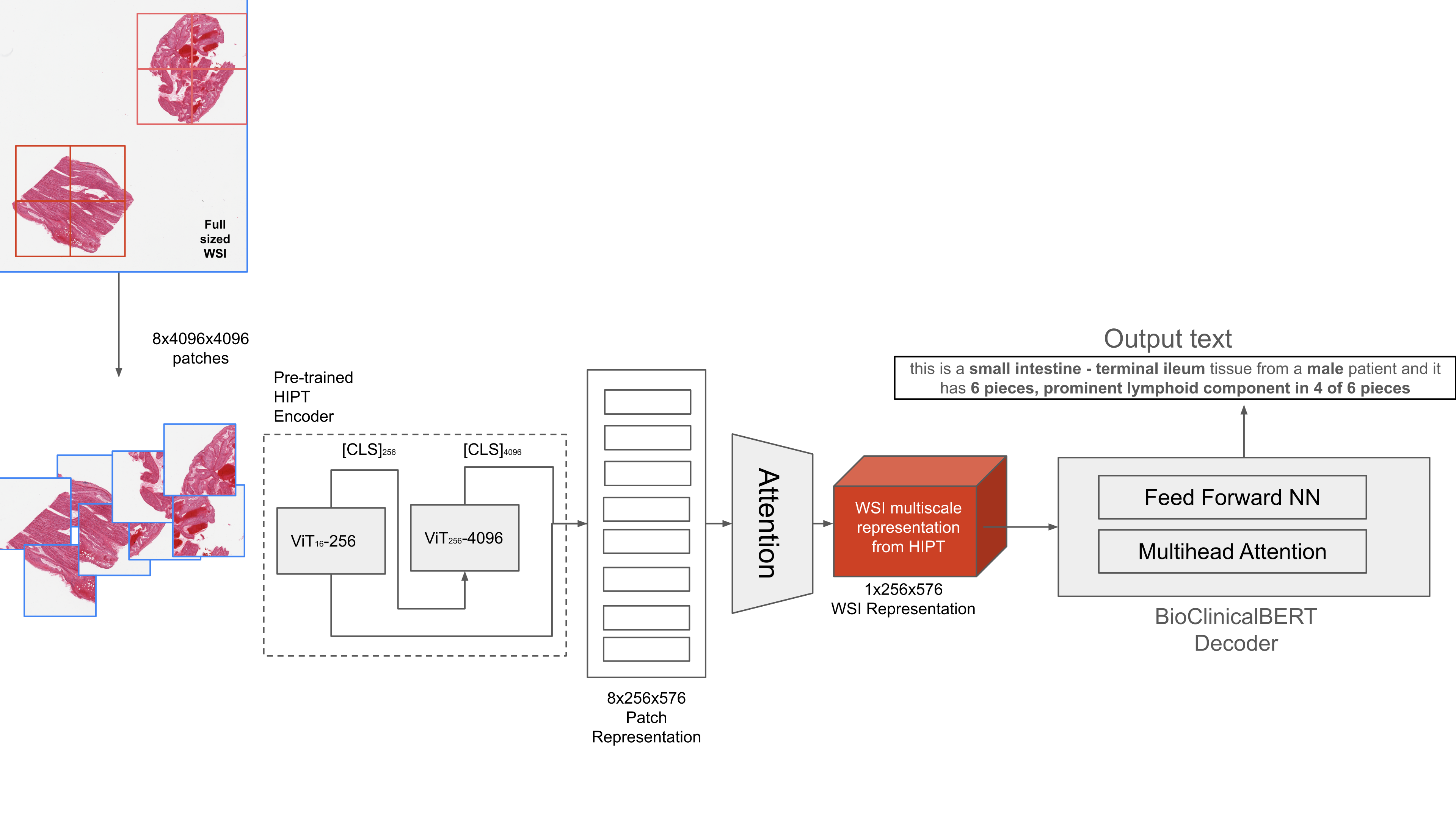
Here, we go over the steps to train the VisionEncoderDecoder Model for caption generation also illustrated in Figure 1.
-
1.
We generated in Section 4.1 that is able to represent all the -patches for each WSI. We feed this in to a trainable Attention layer to create a weighted representation of size . The HIPT representations simultaneously encode information at a cellular level from -16, which is looking at extremely tiny 16x16 tokens at 20X magnification and at the tissue region level where -256 is looking at 256x256 patches at the same magnification, which results in powerful representations.
-
2.
We feed this multi-level representation for each WSI into the ClinicalBioBERT based decoder following a similar process as [14].
4.3 Training
Our training process first requires us to extract for each WSI. We then use these extracted representations in an end to end training process that first encodes the WSI using HIPT and generates weighted representations using the attention layer, which is then fed into BERT for language modeling (BertLMHeadModel)444https://huggingface.co/docs/transformers/model_doc/bert-based decoder. The Cross Entropy loss function is used for training. We utilize PyTorch 1.12 and PyTorch Lightning 2.0.5 for our experiments (code available here). We use a batch size of 1, with gradient accumulation of 16. We use a learning rate of 2e-5 with the Adam optimizer and train for 10 epochs for every model and use the model weights for the epoch for which the validation loss was the lowest. We also incorporate value based gradient clipping to tackle the exploding gradient problem. All our models were trained on a single NVIDIA A100 GPU, and mixed precision training helped reduce training time from 10hrs to 2hrs. Both pre-trained vision transformers, that is, -16 and -256 were frozen during training and only the attention layer and non-linear projection layer with ReLU activation were trained. We unfroze different layers of the decoder to test which setup worked best according to our evaluation criteria.
4.4 Evaluation
We evaluate the efficacy of our captioning model using 3 metrics. We use the BLEU-4 score proposed by [16] to evaluate how closely the generated captions match the actual captions. We restrict ourselves BLEU scores as it is being used by most researchers in automatic caption generation for histopathology [2, 4]. We calculate the accuracy of generated to the actual and the accuracy of the generated vs actual . These 3 metrics, we believe, allow us to holistically evaluate our model.
5 Experiments
| Model | BLEU-4 | Tissue Acc. (%) | Gender Acc. (%) |
|---|---|---|---|
| HIPT-BERT | 0.5543 | 70.73 | 59.76 |
| HIPT-BioClinicalBERT | 0.5571 | 73.48 | 57.42 |
| HIPT-BioClinicalBERT (1 layer unfrozen) | 0.2319 | 58.74 | 49.49 |
| HIPT-BioClinicalBERT (2 layers unfrozen) | 0.5807 | 78.05 | 62.70 |
| HIPT-BioClinicalBERT (3 layers unfrozen) | 0.5784 | 80.79 | 56.30 |
| HIPT-BioClinicalBERT (4 layers unfrozen) | 0.5715 | 78.66 | 54.67 |
| HIPT-BioClinicalBERT (5 layers unfrozen)* | 0.5818 | 79.98 | 66.36 |
| HIPT-BioClinicalBERT (6 layers unfrozen) | 0.5652 | 79.47 | 62.40 |
| Tissue Sample ID | Actual Caption | Generated Caption |
|---|---|---|
| GTEX-145MO-0426 | this is a adipose - subcutaneous tissue from a male patient and it has 2 pieces, 5-10% fascia/vascular tissue, rep. delineated | this is a adipose - subcutaneous tissue from a male patient and it has 2 pieces ; 5 % fibrovascular content |
| GTEX-18A7A-0226 | this is a skin - sun exposed (lower leg) tissue from a female patient and it has 6 pieces; well trimmed; up to 10% dermal fat | this is a skin - sun exposed ( lower leg ) tissue from a male patient and it has 6 pieces ; well trimmed ; 10 % dermal fat |
| GTEX-14ABY-0326 | this is a adipose - subcutaneous tissue from a male patient and it has 2 pieces; 10% of fibrovascular component | this is a adipose - subcutaneous tissue from a male patient and it has 2 pieces ; 10 % fibrovascular content |
5.1 Vanilla BERT vs BioClinicalBERT
Our first experiment is designed to test the performance of vanilla BERT (bert-base-uncased) vs BioClinicalBERT to test the effect of domain specific pre-training on captioning performance. We freeze all layers except for the attention layer in the encoder and the final language modeling head in front of the BERT decoder. The results can be seen in Table 1. Here we see that HIPT-BioClinicalBERT slightly outperforms the HIPT-BERT combination of encoder-decoder in the BLEU-4 score and Tissue accuracy criteria. Therefore, we use the HIPT-BioClinicalBERT combination for further experiments. This also validates our theory that domain specific pre-training helps in downstream tasks related to that particular domain, although very slightly in our case.
5.2 Fine-tuning different layers
We sequentially unfreeze the last few layers of the BERT-based decoder to test which setting provides the best performance. We unfroze the last -layers where of the 12 total layers available. These results are also available in Table 1, where we can see that when the last 5 layers are unfrozen, we achieve the highest scores in 2 of our 3 metrics, that is, BLEU-4 and Gender accuracy with Tissue accuracy not far behind. Our aim in this experiment was to evaluate how many parameters needed to be trained in order to achieve the best result, and in our case, unfreezing the last 5 of 12 layers in the BERT decoder provided that.
We also see that our model struggles to classify which gender the tissue came from as we have 637 male and 347 female patients in our test set, which means we can achieve 64% accuracy by always guessing the tissue is coming from a male patient in our test set. We suspect this is due to class imbalance in our training data where we have 5072 vs 4839 male to female patient ratio. We also expect these results to improve once we add more data from female patients to our model and perform more involved fine-tuning with different learning rates, optimizers and learning rate schedules which requires further analysis.
6 Discussion
In this paper, we show that powerful pre-trained ViT based representations could be used to encode a very high resolution histology image slide for another downstream task, that is, successful automatic report generation. We have also validated that self supervised pre-training is helpful, as it lessens the burden of training data hungry models from scratch every time. Most previous methods for caption generation in histopathology required having a method to encode these high resolution images into manageable representations that could then be used for model learning, which can now successfully be replaced by powerful self-supervised pre-trained encoders.
Of the 37 different tissue types available in our test data, our model was able to correctly classify over 80% of them, which suggests that we can use successfully use the captioning model for multi-class classification, which is a valuable objective.
We present, in this paper, a method to utilize powerful pre-trained transformer models for automatic report generation for histopathology with an end-to-end training mechanism that to the best of our knowledge has not been proposed before. Transformer-based language models significantly outperform RNN-based models, as BioClinicalBERT being pre-trained on clinical notes and medical research articles encodes complex information that the RNN-based encoders used in previous papers on automatic image captioning for histopathology cannot. We believe this work broadens the scope of research in histopathology by introducing transformers in place of traditional CNN-RNN based encoder-decoder models for caption generation
Limitations: Our model struggles to correctly classify the gender of the patient that the tissue came from, which requires further analysis. We are also far behind state-of-the-art in image caption generation for regular images, as the reported BLEU scores show, and there is room for improvement in this area.
7 Acknowledgments
The work in the paper was partially supported by the National Center for Advancing Translational Science of the National Institutes of Health Award UL1TR003015/ KL2TR003016.
8 Compliance with ethical standards
This research study was conducted retrospectively using human subject data made available in open access available here. Ethical approval was not required as confirmed by the license attached with the open access data.
References
- [1] Hyeryun Park, Kyungmo Kim, Jooyoung Yoon, Seongkeun Park, and Jinwook Choi, “Feature difference makes sense: a medical image captioning model exploiting feature difference and tag information,” in Proceedings of the 58th Annual Meeting of the Association for Computational Linguistics: Student Research Workshop, 2020, pp. 95–102.
- [2] Renyu Zhang, Christopher Weber, Robert Grossman, and Aly A Khan, “Evaluating and interpreting caption prediction for histopathology images,” in Machine Learning for Healthcare Conference. PMLR, 2020, pp. 418–435.
- [3] Jevgenij Gamper and Nasir Rajpoot, “Multiple instance captioning: Learning representations from histopathology textbooks and articles,” in Proceedings of the IEEE/CVF conference on computer vision and pattern recognition, 2021, pp. 16549–16559.
- [4] Masayuki Tsuneki and Fahdi Kanavati, “Inference of captions from histopathological patches,” in International Conference on Medical Imaging with Deep Learning. PMLR, 2022, pp. 1235–1250.
- [5] Kelvin Xu, Jimmy Ba, Ryan Kiros, Kyunghyun Cho, Aaron Courville, Ruslan Salakhudinov, Rich Zemel, and Yoshua Bengio, “Show, attend and tell: Neural image caption generation with visual attention,” in International conference on machine learning. PMLR, 2015, pp. 2048–2057.
- [6] Chi-Long Chen, Chi-Chung Chen, Wei-Hsiang Yu, Szu-Hua Chen, Yu-Chan Chang, Tai-I Hsu, Michael Hsiao, Chao-Yuan Yeh, and Cheng-Yu Chen, “An annotation-free whole-slide training approach to pathological classification of lung cancer types using deep learning,” Nature communications, vol. 12, no. 1, pp. 1193, 2021.
- [7] Richard J Chen, Chengkuan Chen, Yicong Li, Tiffany Y Chen, Andrew D Trister, Rahul G Krishnan, and Faisal Mahmood, “Scaling vision transformers to gigapixel images via hierarchical self-supervised learning,” in Proceedings of the IEEE/CVF Conference on Computer Vision and Pattern Recognition, 2022, pp. 16144–16155.
- [8] Mathilde Caron, Hugo Touvron, Ishan Misra, Hervé Jégou, Julien Mairal, Piotr Bojanowski, and Armand Joulin, “Emerging properties in self-supervised vision transformers,” in Proceedings of the IEEE/CVF international conference on computer vision, 2021, pp. 9650–9660.
- [9] Mingxing Tan and Quoc Le, “Efficientnet: Rethinking model scaling for convolutional neural networks,” in International conference on machine learning. PMLR, 2019, pp. 6105–6114.
- [10] Gao Huang, Zhuang Liu, Laurens Van Der Maaten, and Kilian Q Weinberger, “Densely connected convolutional networks,” in Proceedings of the IEEE conference on computer vision and pattern recognition, 2017, pp. 4700–4708.
- [11] Jacob Devlin, Ming-Wei Chang, and Kenton Lee, “Google, kt, language, ai: Bert: pre-training of deep bidirectional transformers for language understanding,” in Proceedings of NAACL-HLT, 2019, pp. 4171–4186.
- [12] Jinhyuk Lee, Wonjin Yoon, Sungdong Kim, Donghyeon Kim, Sunkyu Kim, Chan Ho So, and Jaewoo Kang, “Biobert: a pre-trained biomedical language representation model for biomedical text mining,” Bioinformatics, vol. 36, no. 4, pp. 1234–1240, 2020.
- [13] Emily Alsentzer, John R Murphy, Willie Boag, Wei-Hung Weng, Di Jin, Tristan Naumann, and Matthew McDermott, “Publicly available clinical bert embeddings,” arXiv preprint arXiv:1904.03323, 2019.
- [14] Minghao Li, Tengchao Lv, Jingye Chen, Lei Cui, Yijuan Lu, Dinei Florencio, Cha Zhang, Zhoujun Li, and Furu Wei, “Trocr: Transformer-based optical character recognition with pre-trained models,” in Proceedings of the AAAI Conference on Artificial Intelligence, 2023, vol. 37, pp. 13094–13102.
- [15] Ming Y Lu, Drew FK Williamson, Tiffany Y Chen, Richard J Chen, Matteo Barbieri, and Faisal Mahmood, “Data-efficient and weakly supervised computational pathology on whole-slide images,” Nature biomedical engineering, vol. 5, no. 6, pp. 555–570, 2021.
- [16] Kishore Papineni, Salim Roukos, Todd Ward, and Wei-Jing Zhu, “Bleu: a method for automatic evaluation of machine translation,” in Proceedings of the 40th annual meeting of the Association for Computational Linguistics, 2002, pp. 311–318.