A Compact Implicit Neural Representation for Efficient Storage of
Massive 4D Functional Magnetic Resonance Imaging
Abstract
Functional Magnetic Resonance Imaging (fMRI) data is a kind of widely used four-dimensional biomedical data, demanding effective compression but presenting unique challenges for compression due to its intricate temporal dynamics, low signal-to-noise ratio, and complicated underlying redundancies. This paper reports a novel compression paradigm specifically tailored for fMRI data based on Implicit Neural Representation (INR). The proposed approach focuses on removing the various redundancies among the time series, including (i) conducting spatial correlation modeling for intra-region dynamics, (ii) decomposing reusable neuronal activation patterns, and using proper initialization together with nonlinear fusion to describe the inter-region similarity. The above scheme properly incorporates the unique features of fMRI data, and experimental results on publicly available datasets demonstrate the effectiveness of the proposed method, surpassing state-of-the-art algorithms in both conventional image quality evaluation metrics and fMRI downstream tasks. This work in this paper paves the way for sharing massive fMRI data at low bandwidth and high fidelity.
1 Introduction
Functional Magnetic Resonance Imaging (fMRI), as a widely available non-invasive imaging tool, has been widely used in cognitive neuroscience, clinical psychology, and psychiatry. As large-scale fMRI datasets continue to proliferate, coupled with the ever-growing demand in both clinical [53, 10, 38] and academic domains [15, 24, 40], the fMRI data constitutes a paramount proportion of massive biomedical data [51, 19, 12, 29]. For efficient storage and transmission, there is an urgent necessity for a high-quality data compression paradigm tailored specifically to fMRI.
fMRI data encompasses three spatial dimensions in addition to a temporal dimension, with the temporal dimension presenting intricate neural dynamics [23] and with a relatively low signal-to-noise ratio [11], which pose significant challenges for conventional biomedical data compression algorithms. Moreover, distinct from natural videos or ultrasound data, the redundancies in fMRI data predominantly reside within and between temporal signals of various brain regions [32, 52], rather than adjacent frames. This unique characteristic also challenges conventional compression algorithms and calls for new compressors that make extensive use of the intrinsic structure of fMRI data.
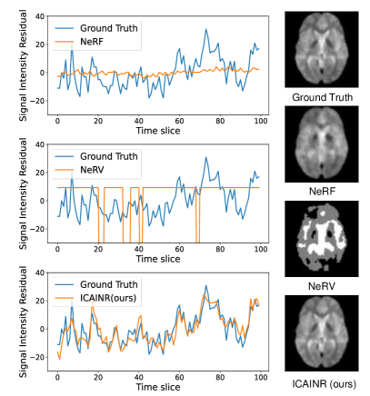
Implicit Neural Representation (INR) is becoming increasingly widespread in various domains, such as shape representation [18, 17, 31], scene rendering [33, 62, 57, 61], and image/video representation [47, 7, 6, 5]. Due to its inherent advantages in modeling internal data correlations [46], as well as the recent development of INR-based data compression algorithms [13, 6, 49, 14, 60, 58, 59] achieving comparable or even surpassing traditional compressors, INR stands out as the most cutting-edge and promising approach in the fields of data compression and deep learning. The success of INR-based compression on videos and three-dimensional biomedical data implies the feasibility but still cannot directly support its applications on fMRI data. First, it is straightforward but difficult to directly extend existing biomedical data compression techniques [61] to 4D fMRI data. When directly employing INR to model the mapping from spatio-temporal coordinates to the signal values, the intricate dynamics [23] and heavy noise [11] might degenerate the coding accuracy and result in poor encoding accuracy in the temporal dimension, as shown in Fig. 1. Second, the INR-based compression algorithms designed for conventional videos, such as NeRV, [6], focuses on eliminating redundancies in adjacent video frames, and is not applicable to fMRI structure. The comparison is shown in Fig. 1. In summary, one should design new compression techniques to incorporate the unique features of fMRI data, including the structure, redundancy, and imaging quality.
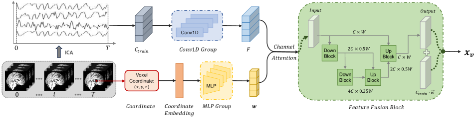
To achieve high-quality compression of 4D fMRI data, we propose an INR-based paradigm, leveraging INR’s powerful representation capability and the unique features of the fMRI data. First, we employ a neural network representing the coordinates-to-intensity mapping to model the correlations of neural dynamics within the same brain region, thereby eliminating redundancies. Next, we decompose the data into a set of reusable neuronal activation patterns and corresponding spatial distribution, effectively removing the among-region redundancies. Subsequently, we use INR to reduce redundancies underlying the spatial distribution of activation patterns and introduce a feature fusion network to simulate the integration of neuronal activation patterns in real fMRI data, thus enhancing the encoding precision. Finally, we introduce an initialization scheme for neuronal activation patterns based on Independent Component Analysis (ICA) to further decrease correlations between different neuronal activation patterns.
We demonstrate the largely superior performance on publicly available datasets to validate the proposed method. It’s worth noting that, in terms of evaluation metrics, in addition to the common metrics like PSNR and SSIM, which measure pixel-wise differences, we also introduced additional evaluation criteria widely used in fMRI data analysis as part of our assessment. Because fMRI data is primarily utilized for data analysis rather than diagnosis by observation, using the disparity between decompressed data and the original version in downstream tasks as an additional evaluation metric allows for a more comprehensive assessment of the compression algorithms.
In summary, the contributions of this paper can be outlined as follows:
-
•
Four-dimensional Data Compression Based on INR. Development of an INR-based compression paradigm tailored for the unique challenges of fMRI data.
-
•
Spatial Correlation Modeling of Local Neuronal Dynamics. A neural-network-based approach utilizing the spatial correlation to eliminate the redundancies within local brain regions.
-
•
Decomposition and Fusion of Neuronal Activation Patterns. A technique to describe fMRI dynamics with a nonlinear fusion of reusable activation patterns, minimizing the inter-regional redundancies.
-
•
Superb Compression Performance. We achieve advantageous data fidelity at high compression ratios, in terms of both image quality and downstream tasks.
2 Related Work
Data Compression.
Data compression plays a significant role in the storage and sharing of data. Over the past few decades, there has been rapid development in data compression algorithms, leading to tremendous successes with algorithms like JPEG[55], H.264[56], and H.265[50]. The techniques which have been adopted by these compressors such as discrete wavelet transform[22] and block-size motion compensation later became extensively applied in the field of data compression. In recent years, with the emergence of deep learning, data compression techniques based on deep learning have gained momentum, resulting in the development of various new compression algorithms[30, 1]. However, when it comes to high-dimensional data, such as four-dimensional data, there still lack proper compressors.
INR-based Compression.
Recent progress in data compression has introduced INR for more compact data representation, yielding promising results[6, 27, 13, 8]. Unlike traditional data compression algorithms that employ explicit encoding methods, INR-based data compression algorithms leverage the powerful information capacity of neural networks to implicitly store data information within network parameters, thereby achieving efficient compression. However, for the task of fMRI compression, existing INR-based data compression algorithms have certain limitations. Some algorithms focus only on spatial or temporal redundancies, making it difficult to extend them to four-dimensional datasets or achieve satisfactory compression results. Some INR-based video compression algorithms address both types of redundancies, but due to the higher demands for fidelity in medical image compression compared to natural images, and the increased complexity of fMRI data structures, the compression strategies employed by existing INR compression algorithms are challenging to apply. In order to more effectively preserve fMRI information, we have altered the data modeling approach of INR. Instead of directly modeling fMRI signals, we utilize INR to model the interrelationships among brain region activities.
High Dimension Medical Data Compression.
There is a lack of adequate compression methods for four-dimensional medical data. The technical routes of these algorithms can be roughly divided into two categories. The first category is based on motion compensation, which use motion vectors to model the difference between frames to reduce redundancies[37, 45, 43, 44]. However, unlike nature scene videos, the redundancies in fMRI data is concentrated between the temporal signals within and between various brain regions, rather than between adjacent frames[32, 52]. Therefore, using motion compensation does not effectively utilize the spatiotemporal redundancies of fMRI for compression. The second category is based on transform like wavelet transform.[25, 28, 39] The transform applied to images or signals is modified to higher dimensions and then applied to medical imaging data. This modification is usually a combination of 1D transform and 3D transform, leading to limited ability to sparsely capture more complex, higher-order discontinuities[4]. Our method is completely different from the traditional data compression algorithms in terms of spatiotemporal redundancies compression. We use the correlations between brain region activities in fMRI to perform redundancies compression, fully considering the characteristics of the data.
3 Method
3.1 Mathematical Representation of fMRI data
The fMRI data can be represented as a set of time series
| (1) |
Here represents the 3D spatial coordinates, and represents the fMRI signal time series at the corresponding spatial location
| (2) |
with respectively denoting the width, height, depth, and length of the time series in the fMRI data.
The principle of localization in brain function organization suggests that brain functions are carried out in a set of brain regions [32]. This implies that the brain space can be divided into several brain regions based on function, with similarities in neuronal activation within each brain region. Therefore, the set of time series for each brain region can be represented as
| (3) |
in which represents the sets of three-dimensional coordinates for the brain region labeled as , and represents the number of brain region labels. Therefore, we have
| (4) |
implying that can be represented as the union of .
3.2 Correlation Modeling with Spatial Coordinates
The overall structure of the network is shown in Fig. 2. Combined with the mathematical representation of fMRI, our neural network parameterization function is designed at a macro level as
| (5) |
Here represents the network parameters. The high similarities within the neural activation in a brain region mentioned above indicates that there exist high correlations between in the same , which matches with ’s advantageous modeling capability of local structures. In other words, if we use the neural network parameterization function mapping from spatial coordinates to the intensity, the local correlations of the function will be enhanced. This aligns well with the advantage of INR in modeling internal correlations, and can more effectively eliminate the redundancies of time-series within local brain regions.
3.3 Neuronal Activation Patterns Decomposition
The connectionism principle of brain function suggests that there exist correlations within the neuronal activation patterns in certain brain regions[32, 52]. At the same time, theoretically, the generation process of fMRI signals can be viewed as the output of a Linear Time-Invariant System (LTI System)[2, 3]:
| (6) |
Here, denotes the system generating fMRI signals, is the input stimulus, is the stimulus distribution of signal intensity, and is the neuronal activation pattern. Therefore, theoretically, can be decomposed into the weighted superposition of several time series characterizing neuronal activation patterns. Inspired by this prior, as shown in Fig. 2, we established a learnable matrix . Each row of the matrix is mapped to the feature space through Conv1d blocks to obtain the feature matrix . As such, the network can learn types of reusable neuronal activation patterns, and the correlations of neuronal activation patterns between brain regions can be modeled by the neural network, thereby eliminating the redundancies of time series between brain regions.
3.4 Modeling of Activation Pattern Distribution and Feature Fusion
While modeling several reusable neuronal activation patterns, we use INR groups to model their spatial distribution
| (7) |
and then concatenate the outputs into
| (8) |
Here function represents the th INR network with being its parameters. The components of represent the spatial distribution of each activation pattern. In this way, we can further eliminate the redundancies within the distribution of neuronal activities.
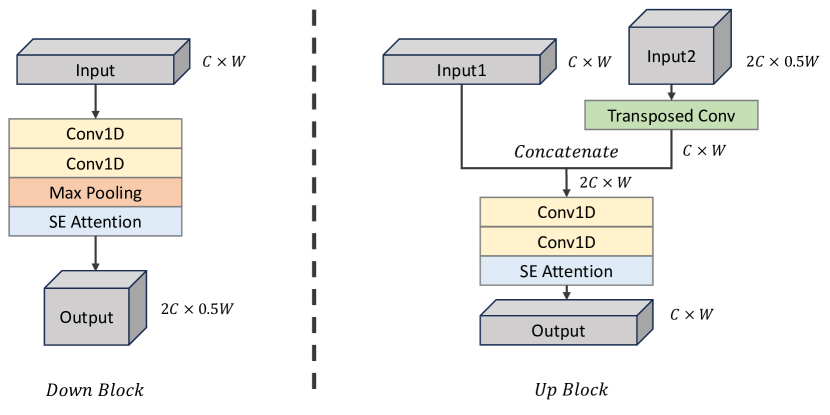
Subsequently, we use the Feature Fusion Block to fuse the features of various neuronal activation patterns. This module is composed of several downsampling and upsampling operations. The internal structures of the Down Block and Up Block are shown in Fig. 3. We concatenate the outputs of the Down Block and Up Block, which allows better utilization of the information contained in the outputs of each sampling module[41], ultimately modeling the target signal .
3.5 Activation Pattern Initialization by ICA
To enable the network to converge quickly, it is necessary to set good initialization to the learnable matrix . We initialize the matrix with a set of independent values to minimize the redundancies within each activation pattern. Specifically, we adopted the ICA algorithm, decomposing the original fMRI data into a set of time-series bases. The matrix form of ICA can be represented as , where is the original signal matrix and decomposed into a mixing matrix and a source matrix . Correspondingly, applying ICA to fMRI signals gets
| (9) |
In this equation, represents the fMRI time series, denotes the activation patterns with the vector describing their spatial distributions. These activation patterns will be used for the initialization of the matrix to minimize the dependency among matrix rows.
3.6 Overview of Network’s Workflow
Based on the above analysis, we provide a summary of the workflow of the network, as shown in Fig. 2. Firstly, we apply ICA to the fMRI data and use the obtained results to initialization the learnable matrix . When the training process begins, every row vector in will be the input of Conv1d blocks respectively, and the output will be concatenated into a feature map . Meanwhile, the coordinate will be embedded to serve as the input of MLPs. The output of MLPs are single weight numbers which are concatenated into the weight vector . During the process of Channel Attention, and will be integrated and serve as the input of the Feature Fusion Block. The Feature Fusion Block contains several Down Blocks and Up Blocks respectively for downsampling and upsampling, whose structures are shown in Fig. 3. Finally, a Conv1D block will be applied to the output of Feature Fusion Block and generate the output vector .
3.7 Model Compression
Model pruning, quantization, and entropy coding are prominent techniques in model compression[20].To speed up the training process, we do not apply model pruning in our network. Upon completion of training, we proceed to quantize the network parameters and subsequently use Huffman coding as the entropy coding method.
4 Experiments and Analysis
4.1 Implementation Details
We conducted experiments on four fMRI datasets collected for different downstream tasks: Three datasets are from OpenfMRI111These three datasets were obtained from the OpenfMRI database, with accession numbers ds000007, ds000101, ds00102, respectively., an open-source repository for the free and open sharing of fMRI datasets. The fourth dataset is the widely used Haxby dataset, a pioneering study of brain pattern recognition[21], which is with extended time series and suitable for the fMRI classification task.
During the training stage, we randomly selected fMRI data from the four datasets. For the first three datasets, for easier subsequent analysis, we uniformly preprocessed the fMRI data by aligning towards a standard brain template, resulting in voxel dimensions of 646448 and a time series length of 100. The Haxby dataset comes with a pre-matched mask, and do not perform registration which is unnecessary for the classification task. Due to its longer time series, we opted to address CUDA memory limitations by slicing the data in Haxby.
In the experiments, our MLP network was configured with 5 layers, and the frequency of the coordinate embedding was set to 10. We used JPEG to compress the mean of the data, and utilized the Adamax optimizer with an initial learning rate of 8e-4. The training epoch was set to 1500. We also utilized ICA’s output network pre-training. The compression ratio of the network can be adjusted by varying parameters such as the number of ICA components, average frame compression quality, count of the MLP parameters, number of convolution channels and network layers. All these parameters can be adjusted using stored YAML files.
| Method | ICAINR(ours) | H.264 | H.265 | JPEG | NeRF | NeRV | SIREN | SSF | DVC |
| Compression Ratio | 127.83* | 97.49 | 125.99# | 102.95 | 99.77 | 93.72 | 99.76 | 66.99 | 81.13 |
| PSNR(dB) | 79.31* | 65.37 | 61.49 | 56.14 | 67.63 | 48.01 | 69.09# | 33.03 | 57.09 |
| 1 - SSIM | 5.54E-5* | 1.96E-3 | 8.38E-4 | 2.74E-3 | 4.83E-4 | 2.16E-2 | 4.01E-4# | 9.39E-2 | 4.39E-4 |
| Mean of FLA Residual | 0.32* | 1.15 | 1.04 | 1.19 | 0.37# | 1.47 | 1.10 | 1.21 | 1.32 |
| Std of FLA Residual | 0.24* | 0.60 | 0.58 | 0.72 | 0.34# | 0.88 | 0.50 | 0.61 | 0.55 |
| Mean of FCA Residual | 0.09* | 0.32 | 0.37 | 0.33 | 0.18 | 0.16 | 0.12# | 0.50 | 0.29 |
| Std of FCA Residual | 0.04* | 0.22 | 0.28 | 0.14 | 0.12 | 0.11 | 0.08# | 0.35 | 0.24 |
4.2 Benchmark Methods and Evaluation Metrics
To demonstrate the advantages of the proposed approach, we comprehensively compare with eight state-of-the-art (SOTA) algorithms, which can be broadly categorized into three groups, including widely used traditional compression algorithms like JPEG[55], H.264[56], and H.265 (HEVC)[50], data-driven deep learning compression algorithms such as SSF[1] and DVC[30], and implicit neural representation-based compressors, including NeRV[6], NeRF[34], and SIREN[46]. For the competitors designed for 2D or 3D data, we compressed the 4D fMRI data slice by slice or concatenated the slices into 2D or 3D data.
We used the OpenCV implementation of JPEG and the FFmpeg implementation of H.264 and H.265. We set the compression ratio by calculating the corresponding bit rate. For DVC and SSF, which are data-driven methods, we changed the compression ratio by specifying the quality parameters and fine-tuned the pre-trained models provided by their authors. For SIREN and NeRF, we set the layer numbers of MLP to 7, and for NeRV, we specified the network parameters to achieve different compression ratios.
The evaluation metrics employed in our experiments can be divided into two parts. The first part involves traditional image quality evaluation metrics PSNR and SSIM, which are compared across various compression ratios. The second part pertains to downstream tasks based on fMRI data. We selected three downstream tasks, which are all classical and widely used methods in fMRI analysis. We believe that these tasks can provide a comprehensive evaluation of compression quality and fidelity, as they quantitatively measure the preservation of various informative cues within the data, including the information about stimuli-brain, intra-brain, and fMRI signal patterns, covering a significant portion of fMRI analysis. These downstream tasks encompass:
i) General linear model First Level Analysis (FLA): This task involves fitting and hypothesis testing of fMRI waveforms to compute the strength of association between specific stimuli/tasks and various brain regions[16], which is the most widely used in fMRI statistical parametric mapping[48]. We conduct FLA on three datasets and use the mean absolute difference between the statistical maps obtained from the original and compressed data for model evaluation.
ii) Brain regions Functional Connection Analysis (FCA): The brain connectome characteristics have offered valuable insights to explain the diversity of pathological conditions and behaviors across different peoples[35], and functional connection is one of the most widely used connectome characteristic, which is defined as the correlation coefficients between the voxel waveforms of different brain areas[42]. In this task, the same dataset was used as in the FLA task, with the MSDL atlas template provided in nilearn[54]. Again, we use the mean absolute difference between the correlation matrices from the ground truth and decompressed fMRI for performance evaluation.
iii) fMRI-based Classification Task (CT): Decoding or pattern recognition techniques is a significant part of fMRI analysis[21]. With the development of machine learning, there has been growing interest in decoding fMRI data by machine learning. We use a linear classifier based on SVM[9], which is most commonly used in fMRI classification[36], to do our experiment. In this task, We employed the Haxby dataset with various kinds of images serving as stimuli. We trained the SVM with the voxel waves, to distinguish stimuli images of the house and face. And we applied 10-folder cross-validation and used the cross-validation classification accuracy of the decompressed fMRI as the evaluation metric.
4.3 Experiment Results
PSNR & SSIM.
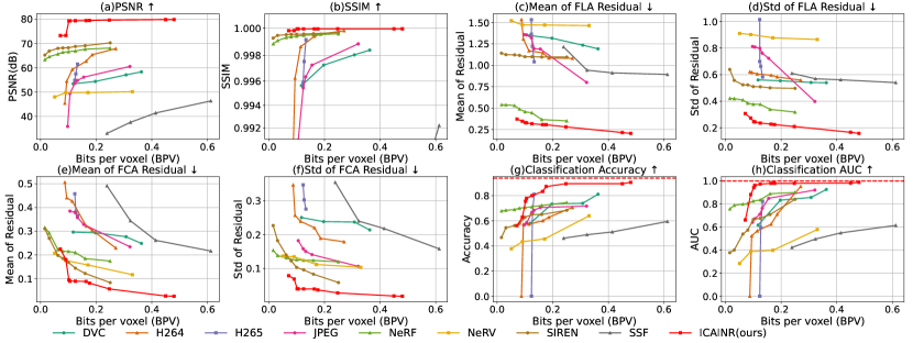
We first validate the data fidelity after fMRI compression, and compare our approach against existing compression algorithms. Here we use PSNR and SSIM as quantitative evaluation metrics, and provide the scores of different algorithms across varying compression ratios, as plotted in Fig. 4a and Fig. 4b. Notably, under similar compression ratios, our method consistently outperforms existing SOTA algorithms at both higher and lower compression ratios. When changing the compression ratio, some algorithms fluctuates, in contrast to the remarkably stable performance of our approach.
Visual Results.
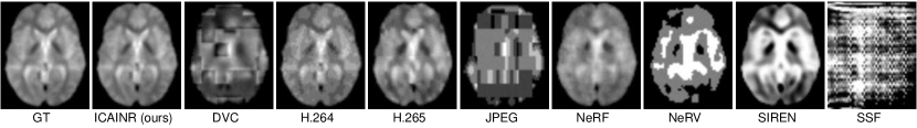
The visual comparison of the decompressed data by various algorithms at a fixed compression ratio (100) is shown in Fig. 5. Note that the actual achieved compression ratio of different algorithms differs slightly, are shown in Tab. 1, because one cannot specified the final compression ratio exactly. From the results, it is evident that despite yielding relatively high PSNR and SSIM values, many algorithms failed to deliver satisfactory visual quality. For instance, JPEG and DVC suffer from block effect[26], with noticeable fragmentation between adjacent image blocks. The INR-based algorithms, like NeRF, SIREN, and NeRV, sacrificed a considerable amount of high frequency details and thus show over-smoothness. Differently, other algorithms such as H.264 and H.265 exhibited noticeable noise, and it seems that SSF almost fail to model the fMRI data.
FLA Results.
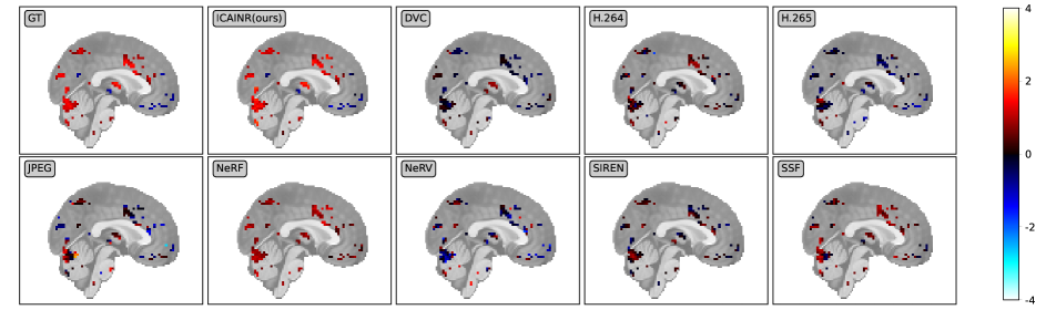
To evaluate the behaviors of our compressor and other competitors on FLA, We computed the mean and standard deviation of residue between the compressed fMRI data and ground truth FLA results, as plotted in Fig. 4c and Fig. 4d. The curves indicate that our method has the smallest residue and performance fluctuation under all compression ratios among these algorithms. Among other algorithms, NeRF can achieve results slightly inferior to ours by around 0.1 and 0.15 in terms of mean and standard deviation on average, while the performance of the remaining competitors are quite limited. Additionally, we display the visualized FLA obtained by various algorithms at a compression ratio about 100, as illustrated in Fig. 6. We can observe that our method exhibits high similarity to the ground truth, while others show significant differences. These results indicate that our method effectively preserves the activities caused by stimuli, which are one of the most important components of fMRI signals.
FCA Results.
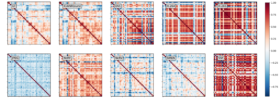
Regarding to the influence of compression on FCA, similar to FLA, we illustrated the mean and standard deviation of residual after decompression in Fig. 4e and Fig. 4f. The curves show that our method has much less information loss than other algorithms if the compression ratio is lower than 170, and the average loss approximates to zero when compression ratio is around 3040. When compression ratio is higher, the performance in mean residual of NeRF, SIREN, NeRV and our method is similar. From the the standard deviation, one can conclude that our approach is the lowest performance fluctuation among all the algorithms working at a similar compression ratio, and achieves stable performance (standard deviation close to 0) when the compression ratio is lower than 100. We also compare the visualized brain connectivity at a compression ratio of about 100 in Fig. 7, from which one can observe that our compressor presents the highest similarity to ground truth. These results show our capability of keeping the correlation information among brain regions.
CT Results.
To test the effectiveness of our compressor and advantageous over previous competitors in the successive classification task (CT), we calculated the 10-folder cross validation accuracy and AUC of the decompressed data compared to the original data before compression, as shown in Fig. 4g and Fig. 4h. The results demonstrate that our algorithm outperforms other methods at compression ratio lower than 170, and can achieve accuracy and AUC close to the ground truth, 93.89% and 0.9975 respectively, when the compression ratio reduces below 100. In other words, our compression preserves most of the distinctive features in the fMRI data volumes.
In summary, our algorithm showcases superior outcomes in terms of both PSNR, SSIM, as well as impressive score in downstream tasks, while some previous algorithms focus on PSNR and SSIM but suffer from degraded performance in successive analysis. This robust performance substantiates our algorithm’s capacity to effectively retain crucial information in fMRI, providing the necessity to take into account the physical meaning of fMRI data during compression and presenting the potential to perform processing and analysis on fMRI data at low bandwidth.
| Method | Compression Ratio | PSNR(dB) | 1 - SSIM | FLA Residual | FCA Residual |
| ICAINR(origin) | 173.14* | 78.74* | 2.28E-5* | 0.320.24* | 0.140.10* |
| ICAINR(without fusion) | 172.96# | 78.67# | 2.31E-5# | 0.330.25# | 0.140.10* |
| ICAINR(uniform init) | 164.96 | 78.60 | 2.36E-5 | 0.360.30 | 0.280.09 |
| ICAINR(normal init) | 165.33 | 78.17 | 2.56E-5 | 0.490.35 | 0.220.07 |
| SIREN(3D) | 160.51 | 72.93 | 6.06E-5 | 0.360.33 | 0.500.16 |
| SIREN(4D) | 159.06 | 70.77 | 1.04E-4 | 1.110.51 | 0.18 0.11# |
4.4 Ablation Studies
In this section, we quantitatively identify the contribution of the key modules in the proposed approach.
Decomposition of Neuronal Activation Patterns.
We compared SIREN, which is INR-based method modeling the data directly, with our method. We removed the Feature Fusion Block in our network to eliminate its impact. As listed in 2nd, 5th and 6th rows of Tab. 2), which showed better performance in terms of all metrics. The results show that using pattern decomposition is advantageous over directly modeling the 4D fMRI data with INR, because of the high complexity of fMRI signals and various underlying correlations.
Feature Fusion Block.
At a similar compression ratio, we compare the quality of the decompressed data before and after removing the feature fusion block. The results are shown in the 1st and 2nd rows of Tab. 2, demonstrating that the Feature Fusion Block achieves higher data fidelity of the target fMRI signals than naive linear modeling.
ICA Initialization.
To verify the effectiveness of ICA initialization, we compared the performance with the matrix uses ICA initialization, Uniform Initialization, and Normal Initialization 1st, 3rd and 4th rows in Tab. 2). The results, especially the performance in downstream tasks, show that ICA initialization can guide the network to model the signals better, especially the neural activation information and correlations.
5 Summary and Discussions
In this paper, we introduce a novel INR-based compression paradigm for fMRI data. Leveraging the strong representation capability of deep neural networks and fMRI’s unique characteristics, we compactly describe the neural activation patterns and their spatial distributions, which reduces both temporal and spatial redundancies in the raw recordings.
Advantages.
From the comprehensive experiments, our algorithm outperforms existing SOTAs in terms of data fidelity such as PSNR and SSIM, as well as evaluation metrics for downstream fMRI tasks, which meets the fidelity requirements of medical image compression. From the comprehensive experiments, our algorithm achieves a higher compression ratio and outperforms existing SOTA methods in terms of both image fidelity and downstream tasks. Notably, our study pioneers the application of INR-based compression methods to four-dimensional biomedical data. Furthermore, we present a novel INR-based compression framework, offering valuable insights for future research in INR-based biomedical image compression.
Limitations and Future Work.
As a preliminary research on INR-based fMRI compression, our approach can be extended in several aspects before becoming a mature compression tool. Firstly, as a deep learning-based compression algorithm, our approach takes longer time than conventional compressors. Therefore, we are exploring novel architectures and techniques, such as meta-learning, for fast model learning. Secondly, we use all the initial components obtained from ICA, but some of them contain little information about the neural activities. We are also working on strategies for extracting informative signals to avoid compressing unnecessary components and thus build a more compact compression. Thirdly, we adopt a widely used method for network compression, and there is room for designing more tailored model compression techniques for our network in the future.
References
- Agustsson et al. [2020] Eirikur Agustsson, David Minnen, Nick Johnston, Johannes Balle, Sung Jin Hwang, and George Toderici. Scale-space flow for end-to-end optimized video compression. In Proceedings of the IEEE/CVF Conference on Computer Vision and Pattern Recognition, pages 8503–8512, 2020.
- Boynton et al. [1996] Geoffrey M Boynton, Stephen A Engel, Gary H Glover, and David J Heeger. Linear systems analysis of functional magnetic resonance imaging in human v1. Journal of Neuroscience, 16(13):4207–4221, 1996.
- Boynton et al. [2012] Geoffrey M Boynton, Stephen A Engel, and David J Heeger. Linear systems analysis of the fmri signal. Neuroimage, 62(2):975–984, 2012.
- Bruylants et al. [2015] Tim Bruylants, Adrian Munteanu, and Peter Schelkens. Wavelet based volumetric medical image compression. Signal Processing: Image Communication, 31:112–133, 2015.
- [5] Hao Chen, Matthew Gwilliam, Ser-Nam Lim, and Abhinav Shrivastava. HNeRV: A Hybrid Neural Representation for Videos.
- Chen et al. [2021] Hao Chen, Bo He, Hanyu Wang, Yixuan Ren, Ser Nam Lim, and Abhinav Shrivastava. Nerv: Neural representations for videos. Advances in Neural Information Processing Systems, 34:21557–21568, 2021.
- Chen and Wang [2022] Yinbo Chen and Xiaolong Wang. Transformers as meta-learners for implicit neural representations. In European Conference on Computer Vision, pages 170–187. Springer, 2022.
- Damodaran et al. [2023] Bharath Bhushan Damodaran, Muhammet Balcilar, Franck Galpin, and Pierre Hellier. RQAT-INR: Improved Implicit Neural Image Compression. In 2023 Data Compression Conference (DCC), pages 208–217. IEEE, 2023.
- De Martino et al. [2008] Federico De Martino, Giancarlo Valente, Noël Staeren, John Ashburner, Rainer Goebel, and Elia Formisano. Combining multivariate voxel selection and support vector machines for mapping and classification of fmri spatial patterns. Neuroimage, 43(1):44–58, 2008.
- Di Martino et al. [2014] Adriana Di Martino, Chao-Gan Yan, Qingyang Li, Erin Denio, Francisco X. Castellanos, Kaat Alaerts, Jeffrey S. Anderson, Michal Assaf, Susan Y. Bookheimer, and Mirella Dapretto. The autism brain imaging data exchange: Towards a large-scale evaluation of the intrinsic brain architecture in autism. Molecular psychiatry, 19(6):659–667, 2014.
- Diedrichsen and Shadmehr [2005] Jörn Diedrichsen and Reza Shadmehr. Detecting and adjusting for artifacts in fmri time series data. NeuroImage, 27(3):624–634, 2005.
- Dimoka [2011] Angelika Dimoka. Brain mapping of psychological processes with psychometric scales: An fmri method for social neuroscience. NeuroImage, 54:S263–S271, 2011.
- Dupont et al. [2021] Emilien Dupont, Adam Goliński, Milad Alizadeh, Yee Whye Teh, and Arnaud Doucet. Coin: Compression with implicit neural representations. arXiv preprint arXiv:2103.03123, 2021.
- Dupont et al. [2022] Emilien Dupont, Hrushikesh Loya, Milad Alizadeh, Adam Goliński, Yee Whye Teh, and Arnaud Doucet. COIN++: Neural Compression Across Modalities, 2022.
- Fan et al. [2014] Jianqing Fan, Fang Han, and Han Liu. Challenges of big data analysis. National science review, 1(2):293–314, 2014.
- Friston et al. [1994] Karl J Friston, Andrew P Holmes, Keith J Worsley, J-P Poline, Chris D Frith, and Richard SJ Frackowiak. Statistical parametric maps in functional imaging: a general linear approach. Human brain mapping, 2(4):189–210, 1994.
- Genova et al. [2019a] Kyle Genova, Forrester Cole, Avneesh Sud, Aaron Sarna, and Thomas Funkhouser. Deep structured implicit functions. arXiv preprint arXiv:1912.06126, 2, 2019a.
- Genova et al. [2019b] Kyle Genova, Forrester Cole, Daniel Vlasic, Aaron Sarna, William T. Freeman, and Thomas Funkhouser. Learning shape templates with structured implicit functions. In Proceedings of the IEEE/CVF International Conference on Computer Vision, pages 7154–7164, 2019b.
- Greene et al. [2001] Joshua D. Greene, R. Brian Sommerville, Leigh E. Nystrom, John M. Darley, and Jonathan D. Cohen. An fMRI investigation of emotional engagement in moral judgment. Science, 293(5537):2105–2108, 2001.
- Han et al. [2016] Song Han, Huizi Mao, and William J. Dally. Deep Compression: Compressing Deep Neural Networks with Pruning, Trained Quantization and Huffman Coding, 2016.
- Haxby et al. [2001] James V. Haxby, M. Ida Gobbini, Maura L. Furey, Alumit Ishai, Jennifer L. Schouten, and Pietro Pietrini. Distributed and overlapping representations of faces and objects in ventral temporal cortex. Science, 293(5539):2425–2430, 2001.
- Heil and Walnut [1989] Christopher E Heil and David F Walnut. Continuous and discrete wavelet transforms. SIAM review, 31(4):628–666, 1989.
- Krohn et al. [2023] Stephan Krohn, Nina von Schwanenflug, Leonhard Waschke, Amy Romanello, Martin Gell, Douglas D. Garrett, and Carsten Finke. A spatiotemporal complexity architecture of human brain activity. Science Advances, 9(5):eabq3851, 2023.
- Kruse et al. [2016] Clemens Scott Kruse, Rishi Goswamy, Yesha Jayendrakumar Raval, and Sarah Marawi. Challenges and opportunities of big data in health care: A systematic review. JMIR medical informatics, 4(4):e5359, 2016.
- Lalgudi et al. [2005] Hariharan G. Lalgudi, Ali Bilgin, Michael W. Marcellin, Ali Tabesh, Mariappan S. Nadar, and Theodore P. Trouard. Four-dimensional compression of fMRI using JPEG2000. In Medical Imaging 2005: Image Processing, pages 1028–1037. SPIE, 2005.
- Lee et al. [1998] YL Lee, HC Kim, and HyunWook Park. Blocking effect reduction of jpeg images by signal adaptive filtering. IEEE Transactions on Image Processing, 7(2):229–234, 1998.
- Li et al. [2022] Zizhang Li, Mengmeng Wang, Huaijin Pi, Kechun Xu, Jianbiao Mei, and Yong Liu. E-NeRV: Expedite Neural Video Representation with Disentangled Spatial-Temporal Context, 2022.
- Liu and Pearlman [2007] Ying Liu and William A Pearlman. Four-dimensional wavelet compression of 4-d medical images using scalable 4-d sbhp. In 2007 Data Compression Conference (DCC’07), pages 233–242. IEEE, 2007.
- Logothetis [2008] Nikos K. Logothetis. What we can do and what we cannot do with fMRI. Nature, 453(7197):869–878, 2008.
- Lu et al. [2019] Guo Lu, Wanli Ouyang, Dong Xu, Xiaoyun Zhang, Chunlei Cai, and Zhiyong Gao. Dvc: An end-to-end deep video compression framework. In Proceedings of the IEEE/CVF Conference on Computer Vision and Pattern Recognition, pages 11006–11015, 2019.
- Martel et al. [2021] Julien NP Martel, David B Lindell, Connor Z Lin, Eric R Chan, Marco Monteiro, and Gordon Wetzstein. Acorn: adaptive coordinate networks for neural scene representation. ACM Transactions on Graphics (TOG), 40(4):1–13, 2021.
- McKeown et al. [1998] Martin J McKeown, Scott Makeig, Greg G Brown, Tzyy-Ping Jung, Sandra S Kindermann, Anthony J Bell, and Terrence J Sejnowski. Analysis of fmri data by blind separation into independent spatial components. Human brain mapping, 6(3):160–188, 1998.
- Mildenhall et al. [2021a] Ben Mildenhall, Pratul P Srinivasan, Matthew Tancik, Jonathan T Barron, Ravi Ramamoorthi, and Ren Ng. Nerf: Representing scenes as neural radiance fields for view synthesis. Communications of the ACM, 65(1):99–106, 2021a.
- Mildenhall et al. [2021b] Ben Mildenhall, Pratul P. Srinivasan, Matthew Tancik, Jonathan T. Barron, Ravi Ramamoorthi, and Ren Ng. Nerf: Representing scenes as neural radiance fields for view synthesis. Communications of the ACM, 65(1):99–106, 2021b.
- Mohanty et al. [2020] Rosaleena Mohanty, William A Sethares, Veena A Nair, and Vivek Prabhakaran. Rethinking measures of functional connectivity via feature extraction. Scientific reports, 10(1):1298, 2020.
- Naselaris et al. [2011] Thomas Naselaris, Kendrick N Kay, Shinji Nishimoto, and Jack L Gallant. Encoding and decoding in fmri. Neuroimage, 56(2):400–410, 2011.
- Nguyen et al. [2011] Binh P. Nguyen, Chee-Kong Chui, Sim-Heng Ong, and Stephen Chang. An efficient compression scheme for 4-D medical images using hierarchical vector quantization and motion compensation. Computers in Biology and Medicine, 41(9):843–856, 2011.
- Poldrack and Gorgolewski [2017] Russell A. Poldrack and Krzysztof J. Gorgolewski. OpenfMRI: Open sharing of task fMRI data. Neuroimage, 144:259–261, 2017.
- Rajeswari and Rajesh [2009] R Rajeswari and R Rajesh. Efficient compression of 4d fmri images using bandelet transform and fuzzy thresholding. In 2009 World Congress on Nature & Biologically Inspired Computing (NaBIC), pages 543–547. IEEE, 2009.
- Razzak et al. [2018] Muhammad Imran Razzak, Saeeda Naz, and Ahmad Zaib. Deep learning for medical image processing: Overview, challenges and the future. Classification in BioApps: Automation of Decision Making, pages 323–350, 2018.
- Ronneberger et al. [2015] Olaf Ronneberger, Philipp Fischer, and Thomas Brox. U-net: Convolutional networks for biomedical image segmentation. In Medical Image Computing and Computer-Assisted Intervention–MICCAI 2015: 18th International Conference, Munich, Germany, October 5-9, 2015, Proceedings, Part III 18, pages 234–241. Springer, 2015.
- Salvador et al. [2005] Raymond Salvador, John Suckling, Martin R Coleman, John D Pickard, David Menon, and ED Bullmore. Neurophysiological architecture of functional magnetic resonance images of human brain. Cerebral cortex, 15(9):1332–1342, 2005.
- Sanchez et al. [2008a] Victor Sanchez, Panos Nasiopoulos, and Rafeef Abugharbieh. Efficient 4D motion compensated lossless compression of dynamic volumetric medical image data. In 2008 IEEE International Conference on Acoustics, Speech and Signal Processing, pages 549–552. IEEE, 2008a.
- Sanchez et al. [2008b] Victor Sanchez, Panos Nasiopoulos, and Rafeef Abugharbieh. Efficient lossless compression of 4-D medical images based on the advanced video coding scheme. IEEE Transactions on Information Technology in Biomedicine, 12(4):442–446, 2008b.
- Sanchez et al. [2009] Victor Sanchez, Panos Nasiopoulos, and Rafeef Abugharbieh. Novel lossless fMRI image compression based on motion compensation and customized entropy coding. IEEE Transactions on Information Technology in Biomedicine, 13(4):645–655, 2009.
- Sitzmann et al. [2020] Vincent Sitzmann, Julien Martel, Alexander Bergman, David Lindell, and Gordon Wetzstein. Implicit neural representations with periodic activation functions. Advances in neural information processing systems, 33:7462–7473, 2020.
- Skorokhodov et al. [2021] Ivan Skorokhodov, Savva Ignatyev, and Mohamed Elhoseiny. Adversarial generation of continuous images. In Proceedings of the IEEE/CVF conference on computer vision and pattern recognition, pages 10753–10764, 2021.
- Smith [2004] Stephen M Smith. Overview of fmri analysis. The British Journal of Radiology, 77(suppl_2):S167–S175, 2004.
- Strümpler et al. [2022] Yannick Strümpler, Janis Postels, Ren Yang, Luc Van Gool, and Federico Tombari. Implicit neural representations for image compression. In European Conference on Computer Vision, pages 74–91. Springer, 2022.
- Sullivan et al. [2012] Gary J. Sullivan, Jens-Rainer Ohm, Woo-Jin Han, and Thomas Wiegand. Overview of the high efficiency video coding (HEVC) standard. IEEE Transactions on circuits and systems for video technology, 22(12):1649–1668, 2012.
- Taube et al. [2013] Jeffrey S. Taube, Stephane Valerio, and Ryan M. Yoder. Is navigation in virtual reality with FMRI really navigation? Journal of cognitive neuroscience, 25(7):1008–1019, 2013.
- Van Den Heuvel and Pol [2010] Martijn P Van Den Heuvel and Hilleke E Hulshoff Pol. Exploring the brain network: a review on resting-state fmri functional connectivity. European neuropsychopharmacology, 20(8):519–534, 2010.
- Van Essen et al. [2013] David C. Van Essen, Stephen M. Smith, Deanna M. Barch, Timothy EJ Behrens, Essa Yacoub, Kamil Ugurbil, and Wu-Minn HCP Consortium. The WU-Minn human connectome project: An overview. Neuroimage, 80:62–79, 2013.
- Varoquaux et al. [2011] Gaël Varoquaux, Alexandre Gramfort, Fabian Pedregosa, Vincent Michel, and Bertrand Thirion. Multi-subject dictionary learning to segment an atlas of brain spontaneous activity. In Information Processing in Medical Imaging: 22nd International Conference, IPMI 2011, Kloster Irsee, Germany, July 3-8, 2011. Proceedings 22, pages 562–573. Springer, 2011.
- Wallace [1992] Gregory K. Wallace. The JPEG still picture compression standard. IEEE transactions on consumer electronics, 38(1):xviii–xxxiv, 1992.
- Wiegand et al. [2003] Thomas Wiegand, Gary J. Sullivan, Gisle Bjontegaard, and Ajay Luthra. Overview of the H. 264/AVC video coding standard. IEEE Transactions on circuits and systems for video technology, 13(7):560–576, 2003.
- Wu et al. [2023] Qi Wu, David Bauer, Yuyang Chen, and Kwan-Liu Ma. Hyperinr: A fast and predictive hypernetwork for implicit neural representations via knowledge distillation. arXiv preprint arXiv:2304.04188, 2023.
- Yang [2023] Runzhao Yang. Tinc: Tree-structured implicit neural compression. In Proceedings of the IEEE/CVF Conference on Computer Vision and Pattern Recognition, pages 18517–18526, 2023.
- Yang et al. [2022] Runzhao Yang, Tingxiong Xiao, Yuxiao Cheng, Anan Li, Jinyuan Qu, Rui Liang, Shengda Bao, Xiaofeng Wang, Jinli Suo, Qingming Luo, and Qionghai Dai. Sharing massive biomedical data at magnitudes lower bandwidth with implicit neural function. bioRxiv, 2022.
- Yang et al. [2023] Runzhao Yang, Tingxiong Xiao, Yuxiao Cheng, Qianni Cao, Jinyuan Qu, Jinli Suo, and Qionghai Dai. Sci: A spectrum concentrated implicit neural compression for biomedical data. In Proceedings of the AAAI Conference on Artificial Intelligence, pages 4774–4782, 2023.
- You et al. [2023] Chenyu You, Weicheng Dai, Yifei Min, Lawrence Staib, and James S Duncan. Implicit anatomical rendering for medical image segmentation with stochastic experts. arXiv preprint arXiv:2304.03209, 2023.
- Zhou et al. [2021] Peng Zhou, Lingxi Xie, Bingbing Ni, and Qi Tian. Cips-3d: A 3d-aware generator of gans based on conditionally-independent pixel synthesis. arXiv preprint arXiv:2110.09788, 2021.