GlycoNMR: Dataset and benchmarks for NMR chemical shift prediction of carbohydrates with graph neural networks
Abstract
Molecular representation learning (MRL) is a powerful tool for bridging the gap between machine learning and chemical sciences, as it converts molecules into numerical representations while preserving their chemical features. These encoded representations serve as a foundation for various downstream biochemical studies, including property prediction and drug design. MRL has had great success with proteins and general biomolecule datasets. Yet, in the growing sub-field of glycoscience (the study of carbohydrates, where longer carbohydrates are also called glycans), MRL methods have been barely explored. This under-exploration can be primarily attributed to the limited availability of comprehensive and well-curated carbohydrate-specific datasets and a lack of machine learning (ML) pipelines specifically tailored to meet the unique problems presented by carbohydrate data. Since interpreting and annotating carbohydrate-specific data is generally more complicated than protein data, domain experts are usually required to get involved. The existing MRL methods, predominately optimized for proteins and small biomolecules, also cannot be directly used in carbohydrate applications without special modifications. To address this challenge, accelerate progress in glycoscience, and enrich the data resources of the MRL community, we introduce GlycoNMR. GlycoNMR contains two laboriously curated datasets with 2,609 carbohydrate structures and 211,543 annotated nuclear magnetic resonance (NMR) chemical shifts for precise atomic-level prediction. We tailored carbohydrate-specific features and adapted existing MRL models to tackle this problem effectively. For illustration, we benchmark four modified MRL models on our new datasets. We believe NMR data is the most appealing starting point in the field of glycoscience, as solution-based NMR is the preeminent technique in carbohydrate structure research, and biomolecule structure is among the foremost predictors of function.
Keywords glycoscience nuclear magnetic resonance graph neural network carbohydrate structure
1 Introduction
Considerable efforts have been devoted to developing ML techniques for learning representations of biomolecular structures (Wu et al., 2018; Rong et al., 2020; Méndez-Lucio et al., 2021; Jumper et al., 2021; Wengert et al., 2021; Yan et al., 2022; Zhou et al., 2023; Guo et al., 2023). Most attention has been devoted to proteins and small biomolecules, while limited progress has been made on carbohydrates despite them being the most abundant biomaterials on earth (Oldenkamp et al., 2019). There has been a recent acceleration of interest and progress in carbohydrates in various fields, with findings emphasizing the role of carbohydrate structures in a list of essential medical and scientific topics. Such topics include biological processes of cells (Apweiler et al., 1999; Hart and Copeland, 2010; Varki, 2017), cancer research and treatment targets (Paszek et al., 2014; Tondepu and Karumbaiah, 2022), novel glycomaterials development (Coullerez et al., 2006; Reichardt et al., 2013; Huang et al., 2017; Pignatelli et al., 2020; Richards and Gibson, 2021; Cao et al., 2022) and carbon sequestration in the context of climate change (Pakulski and Benner, 1994; Gullström et al., 2018).
Similar to other biomolecules, the functions and properties of carbohydrates highly depend on their structures. Nevertheless, structure-function relationships remain relatively less understood in carbohydrates than other classes of biomolecules, partly stemming from the bottlenecks in theory and limited structural data (Ratner et al., 2004; Hart and Copeland, 2010; Oldenkamp et al., 2019). In chemical sciences, nuclear magnetic resonance (NMR) is the primary characterization technique for determining carbohydrate atomic-level fine structures. It requires correctly interpreting solution-state NMR parameters, such as chemical shifts and scalar coupling constants (Duus et al., 2000; Brown et al., 2018). NMR still relies on highly trained personnel and domain knowledge due to limitations in theoretical understanding (Lundborg and Widmalm, 2011; Toukach and Ananikov, 2013). This opens up an opportunity for ML research. ML methods are relatively under-explored in carbohydrate-specific studies and especially for predicting the NMR chemical shifts of carbohydrates (Cobas, 2020; Jonas et al., 2022). Improving the efficiency, flexibility, and accuracy of ML tools that relate carbohydrate structures to NMR parameters is well-aligned to recently launched research initiatives such as GlycoMIP (https://glycomip.org), a National Science Foundation Materials Innovation Platform that promotes research into glycomaterials and glycoscience, as well as parallel efforts by the European Glycoscience Community (https://euroglyco.com).
ML methods have significant potential for generality, robustness, and high-throughput analysis of biomolecules, as demonstrated by a plethora of previous works (David et al., 2020; Shi et al., 2021; Jonas et al., 2022). Inspired by the recent successes of MRL in various fields and applications, such as molecular property prediction (Rong et al., 2020; Yang et al., 2021a; Zhang et al., 2021), molecular generation (Shi et al., 2020; Zhu et al., 2022; Zhou et al., 2023), and drug-drug interaction (Chen et al., 2019; Lyu et al., 2021), we embarked on the journey toward building ML tools for predicting carbohydrate NMR chemical shift spectra from the perspective of molecular representation learning. The first and foremost technical barrier we encountered was the issues with the quality, size, and accessibility of carbohydrate NMR datasets–ongoing problems which have been pointed out in recent literature (Toukach and Ananikov, 2013; Toukach and Egorova, 2019; Ranzinger et al., 2015; Toukach and Egorova, 2022; Böhm et al., 2019). Much of the current data limitations result from the fact that carbohydrates are the most diverse and complex class of biomolecules. Their numerous chemical properties and configurations make the annotation and analyses of their NMR data substantially more complicated and uncertain than those for other biomolecules (Herget et al., 2009; Hart and Copeland, 2010). Particularly, existing structure-related carbohydrate NMR spectra databases are less extensive and less accessible to ML researchers than databases for other classes of biomolecules and proteins, leading to recent calls for improvement in standards and quality (Ranzinger et al., 2015; Paruzzo et al., 2018; Böhm et al., 2019; Toukach and Egorova, 2022). Among all NMR signals, atomic 1D chemical shifts are the most accessible and generally complete (Jonas et al., 2022) and provide rich information to enable molecular identification and fingerprint extraction for carbohydrates.
To facilitate an initial convergence of ML, glycoscience, and glycomaterials, we have developed GlycoNMR, a data repository of carbohydrate structures with curated 1D NMR atomic-level chemical shifts. GlycoNMR includes two datasets. In the first one, we manually curated the experimental NMR data of carbohydrates available at Glycosciences.DB (formerly SweetDB) (Loß et al., 2002; Böhm et al., 2019). The second one was constructed by processing a large sample of NMR chemical shifts we simulated using the Glycan Optimized Dual Empirical Spectrum Simulation (GODESS) platform (Kapaev and Toukach, 2015, 2018), which was partly built on the Carbohydrate Structure Database (CSDB) (Toukach and Egorova, 2019, 2022). Substantial domain expertise and efforts were involved in annotating and preprocessing the two datasets. To the best of our knowledge, GlycoNMR is the first large, high-quality carbohydrate NMR dataset specifically curated for ML research. Using GlycoNMR, we designed a set of features, particularly for describing the structural dynamics of carbohydrates, and developed a baseline 2D GNN model for predicting carbohydrates’ 1D NMR chemical shifts. In addition, we adapted four state-of-the-art 3D-based MRL models to align with the atomic-level NMR shift prediction and benchmarked their performances on GlycoNMR. The experimental results demonstrate the feasibility and promise of ML in analyzing carbohydrate NMR data, and, more generally, in advancing the development of glycoscience and glycomaterials.
Summary of contributions:
-
•
We develop GlycoNMR, a large, high-quality, ML-friendly carbohydrate NMR dataset that is freely available online, which contain instructions for loading and fitting data to 2D and 3D based GNN models.
-
•
We design a set of chemically informed features tailored specifically for carbohydrates. In addition, we experimentally show that these features can intrinsically capture the unique structure dynamics of carbohydrates and thus enhance the performance of graph-based MRL models.
-
•
We adapted and benchmarked multiple 3D-based MRL methods on GlycoNMR and demonstrated the potential usage of MRL approaches in glycoscience research.
2 Background and Related Work
Carbohydrates:
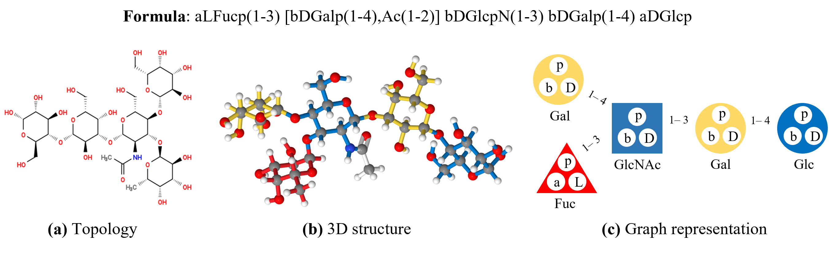
Carbohydrates (examples in Figure 1), also called saccharides, are one of the major biomolecule classes aside from proteins, lipids, and nucleic acids on earth. At the macroscale, carbohydrates are common in sugars and digestive fibers in our diets, and at the microscale, they are widespread on cell membranes and in metabolic pathways. Monosaccharides (introduced in Figure 1), also known as simple sugars (Chaplin and Kennedy, 1986), are the base units of carbohydrates and are typically composed of carbon, hydrogen, and oxygen atoms in specific ratios. Glycosidic bonds, which link monosaccharides into chains or trees, are formed via condensation reactions between the connected monosaccharides. Long chains of monosaccharides are also called polysaccharides. Structure patterns closely relate to the NMR chemical shift spectra and functions (Blanco and Blanco, 2022) of carbohydrates. Importantly, five attributes are the minimum structural information necessary to describe a monosaccharide in a given carbohydrate: (1) Fischer configuration, (2) stem type, (3) ring size, (4) anomeric state, and (5) type and location of modifications (Herget et al., 2009), additional features may be helpful, as discussed in Table 2 and Appendix B. Furthermore, the central ring carbon atoms and their corresponding hydrogen atoms are labeled in a universal and formulaic way in carbohydrates, which aids in building carbohydrate-specific features in the ML pipeline.
Nuclear Magnetic Resonance (NMR):
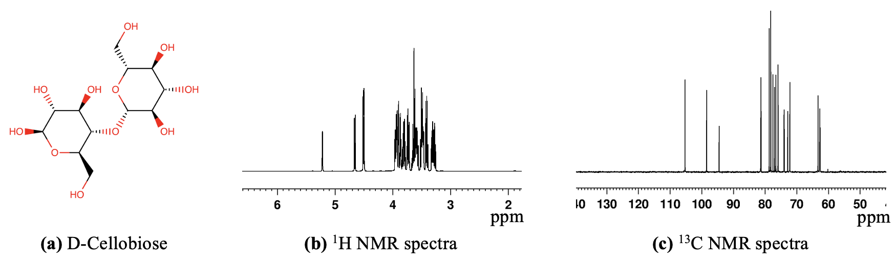
NMR spectra provide key structural features of carbohydrates, including the stereochemistry of monosaccharides, glycosidic linkage types, and conformational preferences. Its non-destructive nature, high sensitivity, and ability to analyze samples in solution make NMR an indispensable tool for carbohydrate research. Arguably, the most accessible and complete NMR parameter for computational structural studies is the 1D chemical shifts (Jonas et al., 2022), where in carbohydrates, usually only the hydrogen 1H and carbon 13C nuclei shifts are measurable (Toukach and Ananikov, 2013). Figure 2 shows a simple carbohydrate and its 1H and 13C NMR spectra. As another challenge specific to carbohydrates, carbohydrate NMR peaks are constrained to a much narrower region of spectra range than proteins, making them harder to separate and leading to an over-reliance on manual interpretation (Toukach and Ananikov, 2013). Thus, the development of theoretical and computational tools that can more automatically and accurately relate a carbohydrate structure and its NMR parameters is a high priority for the field (Hart and Copeland, 2010; Herget et al., 2009; Toukach and Ananikov, 2013; Jonas et al., 2022).
Structure-related Chemical Shift Prediction Methods:
The primary computational methods for chemical shift prediction of carbohydrates can be grouped into four categories: ab initio methods, rules-based and additive increment-based methods, substructure codes, and data-driven ML approaches (Jonas et al., 2022). Ab initio methods, such as density functional theory (DFT) methods, are so far the most accurate as they are based on foundational physics and chemistry theory, but have the lowest throughput and often require considerable expert parameter sweeping and tuning (Tantillo, 2018; Kevin et al., 2019). Additive increment-based approaches, such as CASPER (Jansson et al., 2006; Lundborg and Widmalm, 2011), rely on carefully designed rules, which have limited generalization power and have yet to be extensively validated against experimental data (Jonas et al., 2022). Substructure codes, such as HOSE codes (Kuhn et al., 2008), are the oldest prediction method and still can provide competitive performance in some cases (Jonas et al., 2022). However, substructure code methods have limitations in encoding stereochemical information and distinguishing conformers (though improvements were made recently in this area (Kuhn and Johnson, 2019)). Most HOSE code methods are based on neighborhood search that requires closely similar examples to reach adequate prediction quality (Kuhn et al., 2008). HOSE codes were tested with several large experimental datasets containing assorted biomolecules, with accuracy ranging from approximately 1-3.5 ppm for 13\ceC and 0.15-0.30 ppm for 1\ceH (mean absolute error) (Jonas et al., 2022) (performance of NMR chemical shift prediction on carbohydrates has not been reported to our knowledge). GODESS is a high-quality carbohydrate-specific hybrid of HOSE-like methods and rules-based methods for NMR prediction (Kapaev and Toukach, 2015, 2018). GODESS is capable of generating both structure files in standard carbohydrate format, and atomic-level NMR chemical shift predictions for central ring carbon and hydrogen atoms as well as for some modification group atoms. In this study, we used GODESS to produce the experimentally informed simulated data in GlycoNMR.Sim.
Lastly, ML methods, especially graph neural networks (GNNs) (Battaglia et al., 2018; Zhou et al., 2020), have shown great potential for predicting NMR spectra for biomolecules (Jonas and Kuhn, 2019; Kang et al., 2020; Yang et al., 2021b; McGill et al., 2021; Jonas et al., 2022). Nevertheless, they are relatively unexplored in carbohydrates. To help fill in this gap, here we present, to our knowledge, the first large, ML-friendly dataset for prediction of NMR chemical shifts for carbohydrates.
Relation to ML in other biomolecules:
A large range of papers exists for tackling problems in NMR with ML broadly. Comprehensive general reviews of ML applications in NMR in recent years include (Chen et al., 2020; Bratholm et al., 2021; Yokoyama et al., 2022; Kuhn, 2022; Li et al., 2022; Cortés et al., 2023). The most extensive review of ML to predict NMR spectra of biomolecules is (Jonas et al., 2022) (especially Table 1 in the review). CNNs, MPNNs, and machine were found to have the best performance as a general recent trend in NMR for diverse biomolecules (Jonas and Kuhn, 2019; Dračínskỳ et al., 2019; Kwon et al., 2020; Li et al., 2021), though large differences in sample size and dataset composition make firm conclusions hard to draw (low statistics plague the NMR field due to issues in public datasets). GNNs are by far the most commonly recently used ML tools in this area, though, while feedforward networks dominated earlier work (Jonas et al., 2022).
Relation to Graph-based MRL:
Graph-based MRL has gained accelerating amounts of attention due to its ability to capture local connectivity and topological information of biomolecules (Gilmer et al., 2017; Kipf and Welling, 2017; Hamilton et al., 2017; Xu et al., 2019; Veličković et al., 2018). In graph-based MRL, molecules can be encoded in either 2D or 3D graphs with atoms seen as nodes. In a 2D molecular graph, edges can be pre-determined chemical bonds. In a 3D molecular graph, edges are determined based on the 3D coordinates of atoms, to capture their atomic interactions. Several message-passing schemes have been developed for GNNs to use spatial information such as atom interactions (Schütt et al., 2017), bond rotations and angles between bonds (Gasteiger et al., 2020a, b; Wang et al., 2022), spherical coordinate systems (Liu et al., 2022) and topological geometries (Fang et al., 2022). We chose GNN-based MRL models as the baseline due to their strong expressive power and promising performance on other kinds of biomolecules. For example, encoding bond distance as continuous numbers is expensive computationally, so GNNs encode simplified bond information to avoid the full computational cost (Yang et al., 2021b). Chemists also know that atoms in carbohydrates interact non-negligibly up to 3-4 atoms away, and GNN node-edge structures are well-posed to account for these interactions (Kapaev and Toukach, 2015). We thus formulated the structure-related chemical shift prediction problem as a node regression task, and trained GNNs to predict the chemical shifts of primary monosaccharide ring carbons and hydrogen atoms. We used the Root-Mean-Square Error (RMSE) to evaluate the predicted and the ground truth chemical shifts.
3 Datasets and Baseline Model
We designed a pipeline specifically for annotating the NMR data that we gathered to facilitate the development of ML techniques for predicting atomic NMR shifts of carbohydrates. Two ML-friendly NMR chemical shift datasets GlycoNMR.Exp and GlycoNMR.Sim were constructed. They contain both the 3D structures and the curated 1H and 13C NMR chemical shifts of the carbohydrates. The data statistics are summarized in Table 1. In addition, Table 7 and Figure 5 (in Appendix) show the list of covered monosaccharides and a histogram of carbohydrate sizes (number of monosaccharides) in our datasets. A set of carbohydrate-specific features was engineered to describe the atom structure dynamics in a carbohydrate. We incorporated the features to build a baseline model (see Section 3.2).
GlycoNMR.Exp:
This dataset mainly contains the experimental NMR data obtained from Glycosciences.DB (Böhm et al., 2019). Glycosciences.DB inherited data from the discontinued Complex Carbohydrate Structure Database (CCSD/CarbBank) (Doubet and Albersheim, 1992). It was semi-automatically populated (with moderator oversight) by the carbohydrate entries in the worldwide Protein Data Bank (wwPDB) (Böhm et al., 2019). The NMR data was supplied by SugaBase (Vliegenthart et al., 1992) or manually uploaded by researchers. Glycosciences.DB contains around 3400 carbohydrate entries associated with NMR shifts (however, most only with partial annotation on 1H or 13C). We found 299 carbohydrates had both structures and complete (or near complete) 1H and 13C shifts, and included them in GlycoNMR.Exp for further annotation and processing to make the dataset ML-friendly (details in Section 3.1). This requires substantial domain expertise and efforts on our part, due to the inconsistent and sometimes ambiguous labeling and organization of the diversely-sourced data from this repository. For better illustration, we present a medium-sized carbohydrate data, including both its raw data file 1, 2 and annotated and processed file. Notice that raw data file 1, 2 records the carbohydrate structure and NMR shift separately.
GlycoNMR.Sim:
This dataset contains the simulated NMR chemical shifts produced by using GODESS (http://csdb.glycoscience.ru) (Toukach and Egorova, 2016). GODESS combines incremental rule-based methods (called “empirical” simulation in GODESS) and/or HOSE-like “statistical” methods, and is informed by the CSDB experimental data (Kapaev et al., 2014; Kapaev and Toukach, 2016, 2018; Toukach and Egorova, 2022). GODESS recently demonstrated superior performance in simulating certain carbohydrate NMR shifts and could sometimes perform comparably to DFT (Kapaev et al., 2014; Kapaev and Toukach, 2016, 2018). Hence, we chose it to produce a simulation dataset to amend the lack of publicly available experimental NMR data for carbohydrates. GODESS requires the formula of carbohydrates to be written in the correct CSDB format (Toukach and Egorova, 2019), and does not produce results for those formulas that it deems chemically impossible or incorrect. Helpfully, GODESS scores the trustworthiness of each simulation result. We excluded simulation results with low trustworthiness (error > 2 ppm). We were able to simulate and curate NMR chemical shifts for 200,000 atoms in 2,310 carbohydrates. For a demonstration, we present a large-sized carbohydrate, including both its simulated raw data files 1, 2, 3 and the annotated and processed file. Raw data file 1 contains the structural information of the carbohydrate, while the 13C and 1H NMR chemical shifts are stored separately in raw data file 2 and 3, respectively.
The GlycoNMR datasets are much larger than those used in most carbohydrate-specific studies, which typically have < 100 molecules (Furevi et al., 2022). The size of our data is also comparable to those of the protein or biomolecule NMR datasets, which usually number in the hundreds to low thousands of molecules at best (see Table 1 in (Jonas et al., 2022), or (Yang et al., 2021b)).
| Data source | # Carbohydrate | # Monosaccharide | # Atom | # labeled NMR shifts |
|---|---|---|---|---|
| GlycoNMR.Exp | 299 | 1,130 | 27,267 | 11,848 |
| GlycoNMR.Sim | 2,310 | 16,030 | 372,958 | 199,695 |
3.1 Data Annotation
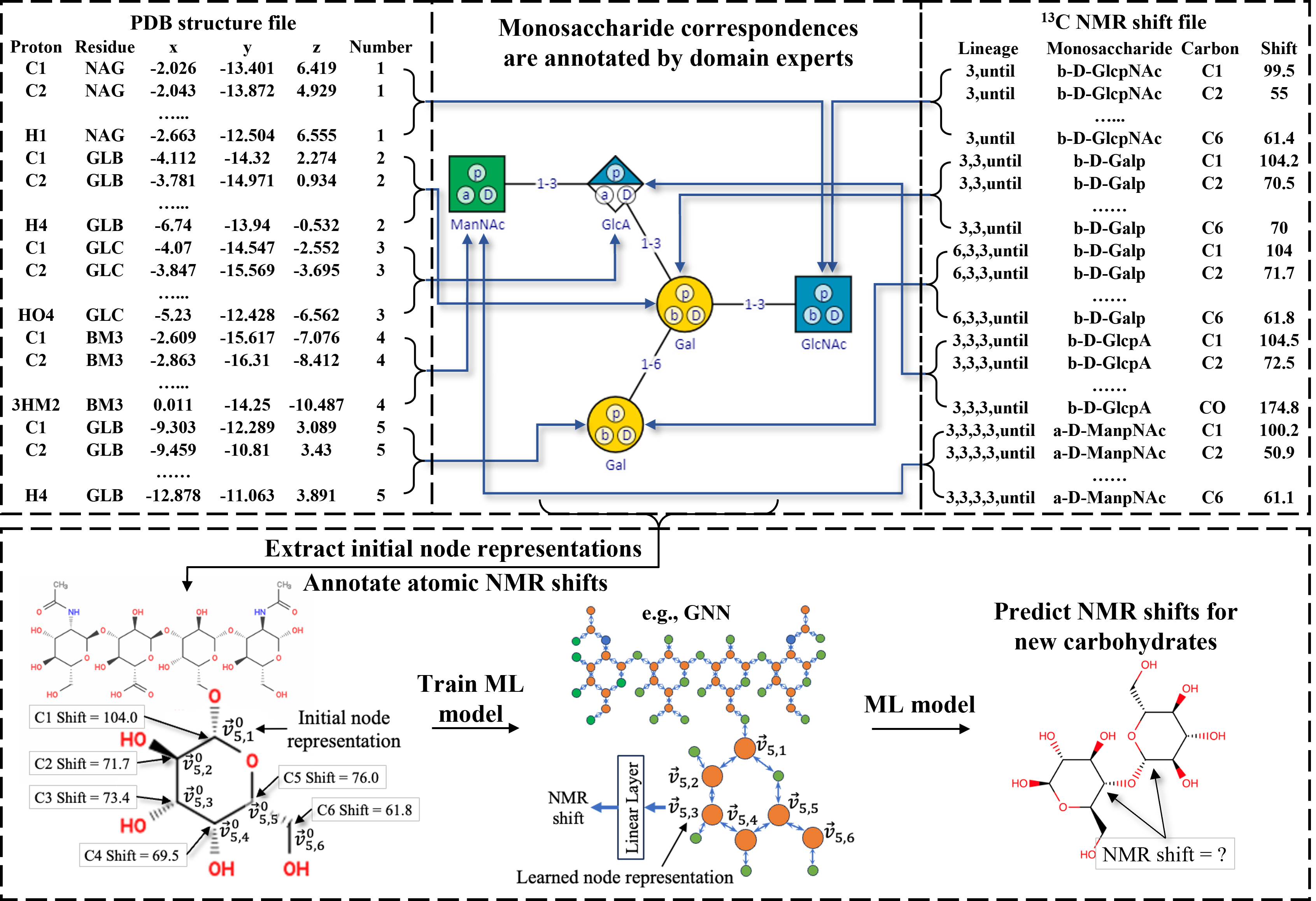
We performed extensive data annotation to associate the 3D structure of each carbohydrate with its NMR chemical shifts. GlycoNMR.Exp and GlycoNMR.Sim stores the structure data for each carbohydrate in the Protein Data Bank (PDB) format amended for carbohydrates, which contains comprehensive 3D structural information, including atom types, ring positions, 3D atomic coordinates, connectivity of the atom, and three-letter abbreviations for monosaccharides. The corresponding NMR data for a given carbohydrate is stored in a separate file, which contains: (1) the hydrogen and carbohydrate chemical shifts per atom per monosaccharide unit and (2) the lineage information of each monosaccharide to its root. We first matched the monosaccharides in the PDB file with those in the NMR file and then used the ring position information to match atoms across the PDB and NMR files. Unfortunately, the order of the monosaccharides in the PDB file often does not match that in the NMR file. The three-letter abbreviations of monosaccharides in PDB files increase the matching difficulty due to the inherent ambiguity and inconsistency in carbohydrate naming convention across time and labs (Toukach and Egorova, 2019). In addition, one carbohydrate can contain multiple monosaccharide units of the same type. For example, in the Glycosciences.DB-sourced PDB files, a single type of monosaccharide can manifest as multiple residues with identical three-letter coding names or vice versa. Hence, we had to utilize domain knowledge to reduce such ambiguities as much as possible when handling the Glycosciences.DB data. Furthermore, we validated topological connections between monosaccharides using the PDB structure data (see Figure 3). We matched the topological connections between monosaccharide units in the PDB files from Glycosciences.DB and GODESS with the lineage information used by their corresponding NMR data files. This allowed us to match the monosaccharides in the PDB files with those in the NMR files, and then use ring positions to associate atoms in a PDB file with atoms in the corresponding NMR data file. Detailed annotation process recorded in Appendix J, including GitHub repos and examples for data annotation.
3.2 Glycoscience-informed Feature Engineering
We derived a set of structural features for the atoms in carbohydrates, which are categorized into monosaccharide-level and atom-level (see Table 2). The monosaccharide-level features describe the monosaccharide context of an atom, including the stem type, configuration, ring size, anomeric status of the monosaccharide, and modifications to the monosaccharide. These features encode the stereochemistry properties and structural dynamics of a monosaccharide and provide information about the overall electronic environment of each atom. The atom-level features include the ring position (wherein the ring the atom is located or is attached) and atom type. To briefly introduce the ring position of an atom, monosaccharide units are classified as either aldoses or ketoses. The aldehyde carbon in aldoses is always numbered as C1 and the ketone carbon in ketoses is labeled as the lowest possible number (Fontana and Widmalm, 2023). The ring position provides information about what other atoms and/or functional groups the atom interacts with. Both categories of features play a significant role in determining the NMR chemical shift value of the atom. We enhanced the carbohydrate PDB structure files by adding the above features and subsequently converted these enriched files into a tabular format where each row describes an atom along with its 3D coordinate and its features and the features associated with the monosaccharide it belongs to. Table 8 in the Appendix describes the processed PDB file to illustrate the above feature engineering effort.
| Feature | Explanation | Example values |
| Monosaccharide | ||
| Configuration | Fischer convention | D, L |
| Stem type | Basic monosaccharide unit | Gal, Glc, Man, .. |
| Ring size | Number of ring carbons | Pyranose , Furanose |
| Anomer | Anomeric orientation | |
| hydroxyl group | ||
| Modifications | Modification groups | Ac, Sulfate, Me, Deoxygenation |
| Atom | ||
| Ring position | Atom position in carbon ring | C1, C2, C3, C4, … |
| Atom type | Chemical elements | C, H, N, O, … |
3.3 Evaluation metric
To evaluate the performance of several MRL models in NMR shift prediction, we calculate the Root-Mean-Square Error (RMSE) between the predicted NMR chemical shifts and the ground truth NMR shifts. The formula for calculating the RMSE is provided in Appendix G. In addition, RMSE is sensitive to outliers, and it can also help us find mismatches caused by humans, especially for the experimental dataset that requires extensive data annotation. We repeatedly apply an outlier check by comparing the ground truth NMR shift and the predicted NMR shift generated by the baseline model.
3.4 Baseline Model
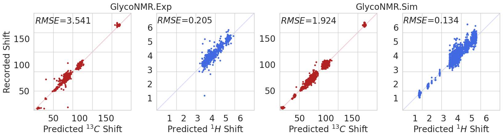
We adopted a 2D graph convolutional neural network (Kipf and Welling, 2017) as our baseline model, a standard architecture that is easy to build off of. We added two linear layers (one for input and the other for output). Each carbohydrate is represented as a graph, with nodes representing atoms and edges representing bonds between atoms. Each node is associated with the features described above. If a carbohydrate structure file does not provide information about the bond connectivity between atoms, we added edges between atom nodes based on their distances. The distance thresholds are for \ceC-\ceC (Liu et al., 2021), for \ceH-\ceX (Guzmán-Afonso et al., 2019), and for \ceX-\ceX (Gunbas et al., 2012), where X stands for other atoms. For each of the GlycoNMR.Exp and GlycoNMR.Sim datasets, we randomly split the carbohydrates into the 80/20 train/validation subsets. Using the training subset, a model was trained to predict 13C or 1H NMR chemical shifts and was evaluated on the corresponding validation subset. The validation results are presented in Figure 4, showing that the baseline model performs reasonably well despite its relatively simple model architecture.
We investigated the impact of individual features (described in Section 3.2) on the baseline model by examining the change in the model performance after removing one single feature. We reported the results in Table 3. We observed that the model performance dropped drastically after removing the ring position feature. Furthermore, when either the stem type feature or the anomer feature is removed, the model performance on carbon drops by a relatively large margin. Other features also had impacts, although their effects are relatively mild. This observation resonates with our expectation when designing the features that the ring position should encode important information about the local context of an atom. Note that removing the 3D-related feature "configuration" improves the performance of the 2D model slightly. The results here provide a direction for improvements, for example, designing new structural features and increasing the interpretability of the model.
| GlycoNMR.Exp | Ring position | Modification | Stem type | Anomer | Configuration | Ring size | None |
|---|---|---|---|---|---|---|---|
| 1H | 0.376 | N/A | 0.271 | 0.240 | 0.206 | 0.220 | 0.205 |
| 13C | 20.218 | N/A | 4.475 | 3.749 | 3.461 | 3.575 | 3.541 |
| GlycoNMR.Sim | Ring position | Modification | Stem type | Anomer | Configuration | Ring size | None |
| 1H | 0.507 | 0.137 | 0.185 | 0.187 | 0.136 | 0.135 | 0.134 |
| 13C | 20.5529 | 1.991 | 2.827 | 2.258 | 1.910 | 1.977 | 1.924 |
4 Benchmark Study on GlycoNMR
To investigate the benefits of encoding 3D structural information, we adapted and benchmarked four state-of-the-art 3D graph-based MRL methods on our datasets: SchNet (Schütt et al., 2017), DimeNet++ (Gasteiger et al., 2020b), ComENet (Wang et al., 2022) and SphereNet (Liu et al., 2022). These models were originally designed to predict the graph-level quantum properties of small molecules from their structures. To apply them to our tasks, we replaced their global pooling layer, which is needed for predicting the properties of whole molecules, and added a layer that maps the learned embedding of each atom to its NMR chemical shift. We fixed the hidden embedding size across all models for a fair comparison. We trained two models for each dataset: one for predicting the 13C NMR shifts and the other for predicting the 1H NMR shifts. For both datasets, we randomly partitioned the carbohydrates into an 80/10/10 split for training, validation, and testing. Additionally, for the GlycoNMR.Exp dataset, given its smaller sample size, to prevent overfitting, we split the training subset into 60% and 20%. We trained the model on the 60% and fine-tuned its hyperparameters on the 20% of the training data. Then, the whole training subset is used to retrain the model using the fine-tuned hyperparameters. Early stopping based on the performance of the validation subset was used during training in both cases, and the RMSE on the test subset is reported.
4.1 Benchmark of 3D-GNN models
We evaluate the adapted 3D-based MRL methods in carbohydrate NMR shift prediction under two settings. In the first setting, the representation of each atom is initialized with only the atomic-level features. This adheres to the initialization method outlined in numerous existing publications on MRL, including (Zhou et al., 2023; Liu et al., 2022). In the second setting, we incorporated monosaccharide-level features into the initial atom representation, which we introduced in Table 2, detailed in and further discussed in Table 3. The test results are reported in Table 4 (rows - show the results of the first setting, and rows - for the second setting). The running time comparison table is provided in Section 4.3. We also provided additional multi-task learning benchmarks in Section 4.2, where we trained one MRL model for each dataset to predict the 13C and 1H shifts jointly.
In general, under the first setting, the 3D-based MRL methods perform better than the 2D-baseline model (see Figure 4). SphereNet achieves the lowest RMSE on the GlycoNMR.Sim dataset, while SchNet has the lowest RMSE on the GlycoNMR.Exp dataset for both 13C and 1H NMR chemical shift prediction. Direct incorporation of 3D structural information into GNNs yields promising results in predicting NMR chemical shifts. In addition, under the second setting, we notice a marginal overall improvement in model performance with additional incorporation of monosaccharide-level features.
| GlycoNMR.Sim | GlycoNMR.Exp | |||
| 13C | 1H | 13C | 1H | |
| ComENet (Wang et al., 2022) + atom feat. | 1.749 | 0.130 | 3.316 | 0.162 |
| DimeNet++ (Gasteiger et al., 2020b) + atom feat. | 2.114 | 0.132 | 4.324 | 0.160 |
| SchNet (Schütt et al., 2017) + atom feat. | 1.633 | 0.136 | 3.217 | 0.170 |
| SphereNet (Liu et al., 2022) + atom feat. | 2.082 | 0.129 | 2.993 | 0.213 |
| ComENet (Wang et al., 2022) + extra feat. | 1.431 | 0.116 | 2.938 | 0.168 |
| DimeNet++ (Gasteiger et al., 2020b) + extra feat. | 1.449 | 0.113 | 2.550 | 0.145 |
| SchNet (Schütt et al., 2017) + extra feat. | 1.487 | 0.118 | 2.492 | 0.140 |
| SphereNet (Liu et al., 2022) + extra feat. | 1.353 | 0.110 | 3.044 | 0.146 |
In the recent protein and small biomolecule computational studies, the MAEs of NMR chemical shift prediction tasks range from 0.1-0.3 ppm for 1H and 0.7-4 ppm for 13C (Jonas et al., 2022), which depend on specifics of models and datasets (e.g., molecular characteristics, sample size and diversity, simulated or experimental data). SphereNet results from our carbohydrate-specific data are comparable to the reported results on other classes of biomolecules. This computational error range can be compared to an experimental error reported for NMR data collection, which was estimated to be 0.51 ppm for 13C and 0.09 ppm for 1H, according to the mean absolute error across 50,000 shifts in the nmrshiftdb2 (which contains a wide variety of biomolecules) (Jonas and Kuhn, 2019). Although it is difficult to compare errors across studies on different molecule classes, and across laboratory conditions and NMR instruments (Stavarache et al., 2022), these observations suggest that the results reported in Table 4 could serve as a useful reference for future efforts in MRL on carbohydrates.
4.2 Benchmark of multi-task NMR shift prediction
We trained 3D GNN models to perform multi-task learning on both GlycoNMR.Sim and GlycoNMR.Exp. Each 3D-based model is trained to predict the carbon NMR shift and the hydrogen shift jointly. The results are summarized in Table 5. We notice that there is an overall significant drop in performance across all 3D GNN models.
4.3 Running time comparison
| Dataset | ComeNet | DimeNet++ | SchNet | SphereNet |
|---|---|---|---|---|
| GlycoNMR.Sim | 7.564 | 20.581 | 3.615 | 31.831 |
| GlycoNMR.Exp | 1.257 | 2.312 | 0.754 | 2.032 |
Running time comparison of 3D GNN models, the duration in seconds for each training epoch is reported. For a fair comparison across the 3D-based GNN models, in GlycoNMR.Sim dataset, we set the batch size to 4, the number of hidden channels to 128, and the number of layers to 4, in GlycoNMR.Exp, we set the batch size to 2, the number of hidden channels to 64, and the number of layers to 2.
5 Discussion and Conclusions
Carbohydrate research has historically lagged behind other major molecular classes (e.g. proteins, small molecules, DNA, etc.) due to theoretical bottlenecks and data quality issues. To help improve this situation, we introduced the first ML-friendly, carbohydrate-specific NMR dataset (GlycoNMR) and pipelines for encoding carbohydrates in predictively powerful GNNs. We hope this study immediately provides useful resources for ML researchers to engage in this new frontier and form a new force to make glyco-related sciences one of the main applications that drive ML research.
As a limitation of the current dataset, most experimentalists only upload NMR spectra peak positions (which is what we predicted), and raw spectral files are rarely openly provided in carbohydrate research. However, the full peak shapes (e.g., widths, heights) and broader spectral patterns also encode rich structural information, but major changes in open data norms must occur in glycoscience to make such data available to ML researchers. Additionally, many properties (e.g., functional, immunological, solvent-related, etc.) or multimodal datasets can be incorporated into future data and models to expand ML applications in this realm (Burkholz et al., 2021). Solvent-carbohydrate interactions, for example, remain poorly understood and theoretically important for understanding NMR data, but most public data remains in water (Klepach et al., 2015; Kirschner and Woods, 2001; Hassan et al., 2015). Future work should also explore more hierarchical structure graphs specifically tailored towards carbohydrates (Mohapatra et al., 2022). Overall, increasingly strong collaboration between glyco-focused scientists and ML-focused researchers is essential over the next decade in the field of glycoscience, as the quality and scope of structural and functional carbohydrate-specific databases must continue to improve and grow in parallel with the power of ML tools that utilize them.
Acknowledgments
This work was supported by GlycoMIP, a National Science Foundation Materials Innovation Platform funded through Cooperative Agreement DMR-1933525.
Data Availability
GlycoNMR is freely available for academic purposes. The full datasets will be made public upon completion of the peer review and can be downloaded here by early access.
References
- Wu et al. [2018] Zhenqin Wu, Bharath Ramsundar, Evan N Feinberg, Joseph Gomes, Caleb Geniesse, Aneesh S Pappu, Karl Leswing, and Vijay Pande. Moleculenet: a benchmark for molecular machine learning. Chemical Science, 2018.
- Rong et al. [2020] Yu Rong, Yatao Bian, Tingyang Xu, Weiyang Xie, Ying Wei, Wenbing Huang, and Junzhou Huang. Self-supervised graph transformer on large-scale molecular data. In Advances in Neural Information Processing Systems, 2020.
- Méndez-Lucio et al. [2021] Oscar Méndez-Lucio, Mazen Ahmad, Ehecatl Antonio del Rio-Chanona, and Jörg Kurt Wegner. A geometric deep learning approach to predict binding conformations of bioactive molecules. Nature Machine Intelligence, 2021.
- Jumper et al. [2021] John Jumper, Richard Evans, Alexander Pritzel, Tim Green, Michael Figurnov, Olaf Ronneberger, Kathryn Tunyasuvunakool, Russ Bates, Augustin Žídek, Anna Potapenko, et al. Highly accurate protein structure prediction with AlphaFold. Nature, 2021.
- Wengert et al. [2021] Simon Wengert, Gábor Csányi, Karsten Reuter, and Johannes T Margraf. Data-efficient machine learning for molecular crystal structure prediction. Chemical Science, 2021.
- Yan et al. [2022] Keqiang Yan, Yi Liu, Yuchao Lin, and Shuiwang Ji. Periodic graph transformers for crystal material property prediction. In Advances in Neural Information Processing Systems, 2022.
- Zhou et al. [2023] Gengmo Zhou, Zhifeng Gao, Qiankun Ding, Hang Zheng, Hongteng Xu, Zhewei Wei, Linfeng Zhang, and Guolin Ke. Uni-mol: A universal 3D molecular representation learning framework. In International Conference on Learning Representations, 2023.
- Guo et al. [2023] Zhichun Guo, Bozhao Nan, Yijun Tian, Olaf Wiest, Chuxu Zhang, and Nitesh V. Chawla. Graph-based molecular representation learning. In International Joint Conference on Artificial Intelligence, 2023.
- Oldenkamp et al. [2019] Heidi F Oldenkamp, Julia E Vela Ramirez, and Nicholas A Peppas. Re-evaluating the importance of carbohydrates as regenerative biomaterials. Regenerative Biomaterials, 2019.
- Apweiler et al. [1999] Rolf Apweiler, Henning Hermjakob, and Nathan Sharon. On the frequency of protein glycosylation, as deduced from analysis of the SWISS-PROT database. Biochimica et Biophysica Acta-General Subjects, 1999.
- Hart and Copeland [2010] Gerald W Hart and Ronald J Copeland. Glycomics hits the big time. Cell, 2010.
- Varki [2017] Ajit Varki. Biological roles of glycans. Glycobiology, 2017.
- Paszek et al. [2014] Matthew J Paszek, Christopher C DuFort, Olivier Rossier, Russell Bainer, Janna K Mouw, Kamil Godula, Jason E Hudak, Jonathon N Lakins, Amanda C Wijekoon, Luke Cassereau, et al. The cancer glycocalyx mechanically primes integrin-mediated growth and survival. Nature, 2014.
- Tondepu and Karumbaiah [2022] Chaitanya Tondepu and Lohitash Karumbaiah. Glycomaterials to investigate the functional role of aberrant glycosylation in glioblastoma. Advanced Healthcare Materials, 2022.
- Coullerez et al. [2006] Géraldine Coullerez, Peter H Seeberger, and Marcus Textor. Merging organic and polymer chemistries to create glycomaterials for glycomics applications. Macromolecular Bioscience, 2006.
- Reichardt et al. [2013] Niels C Reichardt, Manuel Martín-Lomas, and Soledad Penadés. Glyconanotechnology. Chemical Society Reviews, 2013.
- Huang et al. [2017] Mia L Huang, Sean C Purcell, Stephen Verespy III, Yinan Wang, and Kamil Godula. Glycocalyx scaffolding with synthetic nanoscale glycomaterials. Biomaterials Science, 2017.
- Pignatelli et al. [2020] C Pignatelli, F Cadamuro, S Magli, L Rossi, L Russo, and F Nicotra. Glycans and hybrid glyco-materials for artificial cell microenvironment fabrication. In Carbohydrate Chemistry. Royal Society of Chemistry, 2020.
- Richards and Gibson [2021] Sarah-Jane Richards and Matthew I Gibson. Toward glycomaterials with selectivity as well as affinity. JACS Au, 2021.
- Cao et al. [2022] Xuefeng Cao, Shuaishuai Wang, Madhusudhan Reddy Gadi, Ding Liu, Peng G Wang, Xiu-Feng Wan, Jian Zhang, Xi Chen, Lauren E Pepi, Parastoo Azadi, et al. Systematic synthesis of bisected N-glycans and unique recognitions by glycan-binding proteins. Chemical Science, 2022.
- Pakulski and Benner [1994] J Dean Pakulski and Ronald Benner. Abundance and distribution of carbohydrates in the ocean. Limnology and Oceanography, 1994.
- Gullström et al. [2018] Martin Gullström, Liberatus D Lyimo, Martin Dahl, Göran S Samuelsson, Maria Eggertsen, Elisabeth Anderberg, Lina M Rasmusson, Hans W Linderholm, Anders Knudby, Salomão Bandeira, et al. Blue carbon storage in tropical seagrass meadows relates to carbonate stock dynamics, plant–sediment processes, and landscape context: insights from the Western Indian Ocean. Ecosystems, 2018.
- Ratner et al. [2004] Daniel M Ratner, Eddie W Adams, Matthew D Disney, and Peter H Seeberger. Tools for glycomics: mapping interactions of carbohydrates in biological systems. ChemBioChem, 2004.
- Duus et al. [2000] Jens Ø Duus, Charlotte H Gotfredsen, and Klaus Bock. Carbohydrate structural determination by NMR spectroscopy: modern methods and limitations. Chemical Reviews, 2000.
- Brown et al. [2018] Geoffrey D Brown, Julia Bauer, Helen MI Osborn, and Rainer Kuemmerle. A solution NMR approach to determine the chemical structures of carbohydrates using the hydroxyl groups as starting points. ACS Omega, 2018.
- Lundborg and Widmalm [2011] Magnus Lundborg and Goran Widmalm. Structural analysis of glycans by NMR chemical shift prediction. Analytical Chemistry, 2011.
- Toukach and Ananikov [2013] Filip V Toukach and Valentine P Ananikov. Recent advances in computational predictions of NMR parameters for the structure elucidation of carbohydrates: methods and limitations. Chemical Society Reviews, 2013.
- Cobas [2020] Carlos Cobas. NMR signal processing, prediction, and structure verification with machine learning techniques. Magnetic Resonance in Chemistry, 2020.
- Jonas et al. [2022] Eric Jonas, Stefan Kuhn, and Nils Schlörer. Prediction of chemical shift in NMR: A review. Magnetic Resonance in Chemistry, 2022.
- David et al. [2020] Laurianne David, Amol Thakkar, Rocío Mercado, and Ola Engkvist. Molecular representations in AI-driven drug discovery: a review and practical guide. Journal of Cheminformatics, 2020.
- Shi et al. [2021] Chence Shi, Shitong Luo, Minkai Xu, and Jian Tang. Learning gradient fields for molecular conformation generation. In International Conference on Machine Learning, 2021.
- Yang et al. [2021a] Shuwen Yang, Ziyao Li, Guojie Song, and Lingsheng Cai. Deep molecular representation learning via fusing physical and chemical information. In Advances in Neural Information Processing Systems, 2021a.
- Zhang et al. [2021] Zaixi Zhang, Qi Liu, Hao Wang, Chengqiang Lu, and Chee-Kong Lee. Motif-based graph self-supervised learning for molecular property prediction. In Advances in Neural Information Processing Systems, 2021.
- Shi et al. [2020] Chence Shi, Minkai Xu, Zhaocheng Zhu, Weinan Zhang, Ming Zhang, and Jian Tang. Graphaf: a flow-based autoregressive model for molecular graph generation. In International Conference on Learning Representations, 2020.
- Zhu et al. [2022] Jinhua Zhu, Yingce Xia, Lijun Wu, Shufang Xie, Tao Qin, Wengang Zhou, Houqiang Li, and Tie-Yan Liu. Unified 2D and 3D pre-training of molecular representations. In ACM SIGKDD Conference on Knowledge Discovery and Data Mining, 2022.
- Chen et al. [2019] Xin Chen, Xien Liu, and Ji Wu. Drug-drug interaction prediction with graph representation learning. In IEEE International Conference on Bioinformatics and Biomedicine, 2019.
- Lyu et al. [2021] Tengfei Lyu, Jianliang Gao, Ling Tian, Zhao Li, Peng Zhang, and Ji Zhang. MDNN: A multimodal deep neural network for predicting drug-drug interaction events. In International Joint Conference on Artificial Intelligence, 2021.
- Toukach and Egorova [2019] Philip V Toukach and Ksenia S Egorova. New features of carbohydrate structure database notation (CSDB linear), as compared to other carbohydrate notations. Journal of Chemical Information and Modeling, 2019.
- Ranzinger et al. [2015] Rene Ranzinger, Kiyoko F Aoki-Kinoshita, Matthew P Campbell, Shin Kawano, Thomas Lütteke, Shujiro Okuda, Daisuke Shinmachi, Toshihide Shikanai, Hiromichi Sawaki, Philip Toukach, et al. GlycoRDF: an ontology to standardize glycomics data in RDF. Bioinformatics, 2015.
- Toukach and Egorova [2022] Philip V Toukach and Ksenia S Egorova. Source files of the carbohydrate structure database: the way to sophisticated analysis of natural glycans. Scientific Data, 2022.
- Böhm et al. [2019] Michael Böhm, Andreas Bohne-Lang, Martin Frank, Alexander Loss, Miguel A Rojas-Macias, and Thomas Lütteke. Glycosciences.DB: an annotated data collection linking glycomics and proteomics data. Nucleic Acids Research, 2019.
- Herget et al. [2009] Stephan Herget, René Ranzinger, Robin Thomson, Martin Frank, and Claus-Wilhelm von der Lieth. Introduction to carbohydrate structure and diversity. Bioinformatics for Glycobiology and Glycomics: An Introduction, 2009.
- Paruzzo et al. [2018] Federico M Paruzzo, Albert Hofstetter, Félix Musil, Sandip De, Michele Ceriotti, and Lyndon Emsley. Chemical shifts in molecular solids by machine learning. Nature Communications, 2018.
- Loß et al. [2002] Alexander Loß, Peter Bunsmann, Andreas Bohne, Annika Loß, Eberhard Schwarzer, Elke Lang, and Claus-W Von der Lieth. SWEET-DB: an attempt to create annotated data collections for carbohydrates. Nucleic Acids Research, 2002.
- Kapaev and Toukach [2015] Roman R Kapaev and Philip V Toukach. Improved carbohydrate structure generalization scheme for 1H and 13C nmr simulations. Analytical Chemistry, 2015.
- Kapaev and Toukach [2018] Roman R Kapaev and Philip V Toukach. GRASS: semi-automated NMR-based structure elucidation of saccharides. Bioinformatics, 2018.
- Chaplin and Kennedy [1986] M Chaplin and JJMS Kennedy. Monosaccharides. Mass Spectrom, 1986.
- Blanco and Blanco [2022] Antonio Blanco and Gustavo Blanco. Chapter 4 - Carbohydrates. In Antonio Blanco and Gustavo Blanco, editors, Medical Biochemistry. Academic Press, 2022.
- Tantillo [2018] D.J. Tantillo. Applied Theoretical Organic Chemistry. World Scientific, 2018.
- Kevin et al. [2019] EB Kevin, M Jonathan, et al. The optimal DFT approach in DP4 NMR structure analysis–pushing the limits of relative configuration elucidation. Organic & Biomolecular Chemistry, 2019.
- Jansson et al. [2006] Per-Erik Jansson, Roland Stenutz, and Göran Widmalm. Sequence determination of oligosaccharides and regular polysaccharides using NMR spectroscopy and a novel web-based version of the computer program CASPER. Carbohydrate Research, 2006.
- Kuhn et al. [2008] Stefan Kuhn, Björn Egert, Steffen Neumann, and Christoph Steinbeck. Building blocks for automated elucidation of metabolites: Machine learning methods for NMR prediction. BMC Bioinformatics, 2008.
- Kuhn and Johnson [2019] Stefan Kuhn and Sean R Johnson. Stereo-aware extension of HOSE codes. ACS Omega, 2019.
- Battaglia et al. [2018] Peter W Battaglia, Jessica B Hamrick, Victor Bapst, Alvaro Sanchez-Gonzalez, Vinicius Zambaldi, Mateusz Malinowski, Andrea Tacchetti, David Raposo, Adam Santoro, Ryan Faulkner, et al. Relational inductive biases, deep learning, and graph networks. arXiv preprint arXiv:1806.01261, 2018.
- Zhou et al. [2020] Jie Zhou, Ganqu Cui, Shengding Hu, Zhengyan Zhang, Cheng Yang, Zhiyuan Liu, Lifeng Wang, Changcheng Li, and Maosong Sun. Graph neural networks: A review of methods and applications. AI Open, 2020.
- Jonas and Kuhn [2019] Eric Jonas and Stefan Kuhn. Rapid prediction of NMR spectral properties with quantified uncertainty. Journal of Cheminformatics, 2019.
- Kang et al. [2020] Seokho Kang, Youngchun Kwon, Dongseon Lee, and Youn-Suk Choi. Predictive modeling of NMR chemical shifts without using atomic-level annotations. Journal of Chemical Information and Modeling, 2020.
- Yang et al. [2021b] Ziyue Yang, Maghesree Chakraborty, and Andrew D White. Predicting chemical shifts with graph neural networks. Chemical Science, 2021b.
- McGill et al. [2021] Charles McGill, Michael Forsuelo, Yanfei Guan, and William H Green. Predicting infrared spectra with message passing neural networks. Journal of Chemical Information and Modeling, 2021.
- Chen et al. [2020] Dicheng Chen, Zi Wang, Di Guo, Vladislav Orekhov, and Xiaobo Qu. Review and prospect: deep learning in nuclear magnetic resonance spectroscopy. Chemistry–A European Journal, 2020.
- Bratholm et al. [2021] Lars A Bratholm, Will Gerrard, Brandon Anderson, Shaojie Bai, Sunghwan Choi, Lam Dang, Pavel Hanchar, Addison Howard, Sanghoon Kim, Zico Kolter, et al. A community-powered search of machine learning strategy space to find NMR property prediction models. Plos ONE, 2021.
- Yokoyama et al. [2022] Daiki Yokoyama, Sosei Suzuki, Taiga Asakura, and Jun Kikuchi. Chemometric analysis of NMR spectra and machine learning to investigate membrane fouling. ACS Omega, 2022.
- Kuhn [2022] Stefan Kuhn. Applications of machine learning and artificial intelligence in NMR. Magnetic Resonance in Chemistry, 2022.
- Li et al. [2022] Da-Wei Li, Alexandar L Hansen, Lei Bruschweiler-Li, Chunhua Yuan, and Rafael Brüschweiler. Fundamental and practical aspects of machine learning for the peak picking of biomolecular NMR spectra. Journal of Biomolecular NMR, 2022.
- Cortés et al. [2023] Iván Cortés, Cristina Cuadrado, Antonio Hernández Daranas, and Ariel M Sarotti. Machine learning in computational NMR-aided structural elucidation. Frontiers in Natural Products, 2023.
- Dračínskỳ et al. [2019] Martin Dračínskỳ, Pablo Unzueta, and Gregory JO Beran. Improving the accuracy of solid-state nuclear magnetic resonance chemical shift prediction with a simple molecular correction. Physical Chemistry Chemical Physics, 2019.
- Kwon et al. [2020] Youngchun Kwon, Dongseon Lee, Youn-Suk Choi, Myeonginn Kang, and Seokho Kang. Neural message passing for NMR chemical shift prediction. Journal of Chemical Information and Modeling, 2020.
- Li et al. [2021] Da-Wei Li, Alexandar L Hansen, Chunhua Yuan, Lei Bruschweiler-Li, and Rafael Brüschweiler. Deep picker is a deep neural network for accurate deconvolution of complex two-dimensional NMR spectra. Nature Communications, 2021.
- Gilmer et al. [2017] Justin Gilmer, Samuel S Schoenholz, Patrick F Riley, Oriol Vinyals, and George E Dahl. Neural message passing for quantum chemistry. In International Conference on Machine Learning. PMLR, 2017.
- Kipf and Welling [2017] Thomas N. Kipf and Max Welling. Semi-supervised classification with graph convolutional networks. In International Conference on Learning Representations, 2017.
- Hamilton et al. [2017] Will Hamilton, Zhitao Ying, and Jure Leskovec. Inductive representation learning on large graphs. In Advances in Neural Information Processing Systems, 2017.
- Xu et al. [2019] Keyulu Xu, Weihua Hu, Jure Leskovec, and Stefanie Jegelka. How powerful are graph neural networks? In International Conference on Learning Representations, 2019.
- Veličković et al. [2018] Petar Veličković, Guillem Cucurull, Arantxa Casanova, Adriana Romero, Pietro Liò, and Yoshua Bengio. Graph attention networks. In International Conference on Learning Representations, 2018.
- Schütt et al. [2017] Kristof Schütt, Pieter-Jan Kindermans, Huziel Enoc Sauceda Felix, Stefan Chmiela, Alexandre Tkatchenko, and Klaus-Robert Müller. Schnet: A continuous-filter convolutional neural network for modeling quantum interactions. In Advances in Neural Information Processing Systems, 2017.
- Gasteiger et al. [2020a] Johannes Gasteiger, Janek Groß, and Stephan Günnemann. Directional message passing for molecular graphs. In International Conference on Learning Representations, 2020a.
- Gasteiger et al. [2020b] Johannes Gasteiger, Shankari Giri, Johannes T. Margraf, and Stephan Günnemann. Fast and uncertainty-aware directional message passing for non-equilibrium molecules. In Machine Learning for Molecules Workshop, NeurIPS, 2020b.
- Wang et al. [2022] Limei Wang, Yi Liu, Yuchao Lin, Haoran Liu, and Shuiwang Ji. ComENet: Towards complete and efficient message passing for 3D molecular graphs. In Advances in Neural Information Processing Systems, 2022.
- Liu et al. [2022] Yi Liu, Limei Wang, Meng Liu, Yuchao Lin, Xuan Zhang, Bora Oztekin, and Shuiwang Ji. Spherical message passing for 3D molecular graphs. In International Conference on Learning Representations, 2022.
- Fang et al. [2022] Xiaomin Fang, Lihang Liu, Jieqiong Lei, Donglong He, Shanzhuo Zhang, Jingbo Zhou, Fan Wang, Hua Wu, and Haifeng Wang. Geometry-enhanced molecular representation learning for property prediction. Nature Machine Intelligence, 2022.
- Doubet and Albersheim [1992] Scott Doubet and Peter Albersheim. Letter to the glyco-forum: Carbbank. Glycobiology, 1992.
- Vliegenthart et al. [1992] JFG Vliegenthart, JA van Kuik, and K Hård. A 1H NMR database computer program for the analysis of the primary structure of complex carbohydrates. Carbohydrate Research, 1992.
- Toukach and Egorova [2016] Philip V Toukach and Ksenia S Egorova. Carbohydrate structure database merged from bacterial, archaeal, plant and fungal parts. Nucleic Acids Research, 2016.
- Kapaev et al. [2014] Roman R Kapaev, Ksenia S Egorova, and Philip V Toukach. Carbohydrate structure generalization scheme for database-driven simulation of experimental observables, such as NMR chemical shifts. Journal of Chemical Information and Modeling, 2014.
- Kapaev and Toukach [2016] Roman R Kapaev and Philip V Toukach. Simulation of 2D NMR spectra of carbohydrates using GODESS software. Journal of Chemical Information and Modeling, 2016.
- Furevi et al. [2022] Axel Furevi, Alessandro Ruda, Thibault Angles d’Ortoli, Hani Mobarak, Jonas Ståhle, Christoffer Hamark, Carolina Fontana, Olof Engström, Patricia Apostolica, and Göran Widmalm. Complete 1H and 13C NMR chemical shift assignments of mono-to tetrasaccharides as basis for NMR chemical shift predictions of oligo-and polysaccharides using the computer program CASPER. Carbohydrate Research, 2022.
- Fontana and Widmalm [2023] Carolina Fontana and Goran Widmalm. Primary structure of glycans by NMR spectroscopy. Chemical Reviews, 2023.
- Liu et al. [2021] Sai Liu, Yihua Lu, Xi Zhu, and Min Wang. Theoretical predictions of two new chiral solid carbon oxides. Physics Letters A, 385:126941, 2021.
- Guzmán-Afonso et al. [2019] Candelaria Guzmán-Afonso, You-lee Hong, Henri Colaux, Hirofumi Iijima, Akihiro Saitow, Takuma Fukumura, Yoshitaka Aoyama, Souhei Motoki, Tetsuo Oikawa, Toshio Yamazaki, et al. Understanding hydrogen-bonding structures of molecular crystals via electron and nmr nanocrystallography. Nature communications, 10(1):3537, 2019.
- Gunbas et al. [2012] Gorkem Gunbas, Nema Hafezi, William L Sheppard, Marilyn M Olmstead, Irini V Stoyanova, Fook S Tham, Matthew P Meyer, and Mark Mascal. Extreme oxatriquinanes and a record c–o bond length. Nature chemistry, 4(12):1018–1023, 2012.
- Stavarache et al. [2022] Cristina Stavarache, Alina Nicolescu, Cătălin Duduianu, Gabriela Liliana Ailiesei, Mihaela Balan-Porcăraşu, Mihaela Cristea, Ana-Maria Macsim, Oana Popa, Carmen Stavarache, Anca Hîrtopeanu, et al. A real-life reproducibility assessment for NMR metabolomics. Diagnostics, 2022.
- Burkholz et al. [2021] Rebekka Burkholz, John Quackenbush, and Daniel Bojar. Using graph convolutional neural networks to learn a representation for glycans. Cell Reports, 2021.
- Klepach et al. [2015] Thomas Klepach, Hongqiu Zhao, Xiaosong Hu, Wenhui Zhang, Roland Stenutz, Matthew J Hadad, Ian Carmichael, and Anthony S Serianni. Informing saccharide structural NMR studies with density functional theory calculations. Glycoinformatics, 2015.
- Kirschner and Woods [2001] Karl N Kirschner and Robert J Woods. Solvent interactions determine carbohydrate conformation. Proceedings of the National Academy of Sciences, 2001.
- Hassan et al. [2015] El-Sayed RE Hassan, Fabrice Mutelet, and Mohammed Bouroukba. Experimental and theoretical study of carbohydrate–ionic liquid interactions. Carbohydrate polymers, 2015.
- Mohapatra et al. [2022] Somesh Mohapatra, Joyce An, and Rafael Gómez-Bombarelli. Chemistry-informed macromolecule graph representation for similarity computation, unsupervised and supervised learning. Machine Learning: Science and Technology, 2022.
- Štrumbelj and Kononenko [2014] Erik Štrumbelj and Igor Kononenko. Explaining prediction models and individual predictions with feature contributions. Knowledge and information systems, 41:647–665, 2014.
- Toukach et al. [2007] Philip Toukach, Hiren J Joshi, Rene Ranzinger, Yuri Knirel, and Claus-W von der Lieth. Sharing of worldwide distributed carbohydrate-related digital resources: online connection of the bacterial carbohydrate structure database and glycosciences. de. Nucleic acids research, 35(suppl_1):D280–D286, 2007.
Appendix A Further Descriptive Analyses of Each Dataset
In this section, we provide a detailed data analysis of GlycoNMR, focusing on both the quantity and variety of monosaccharides within our dataset.
A.1 Histogram distribution of carbohydrate lengths in both datasets
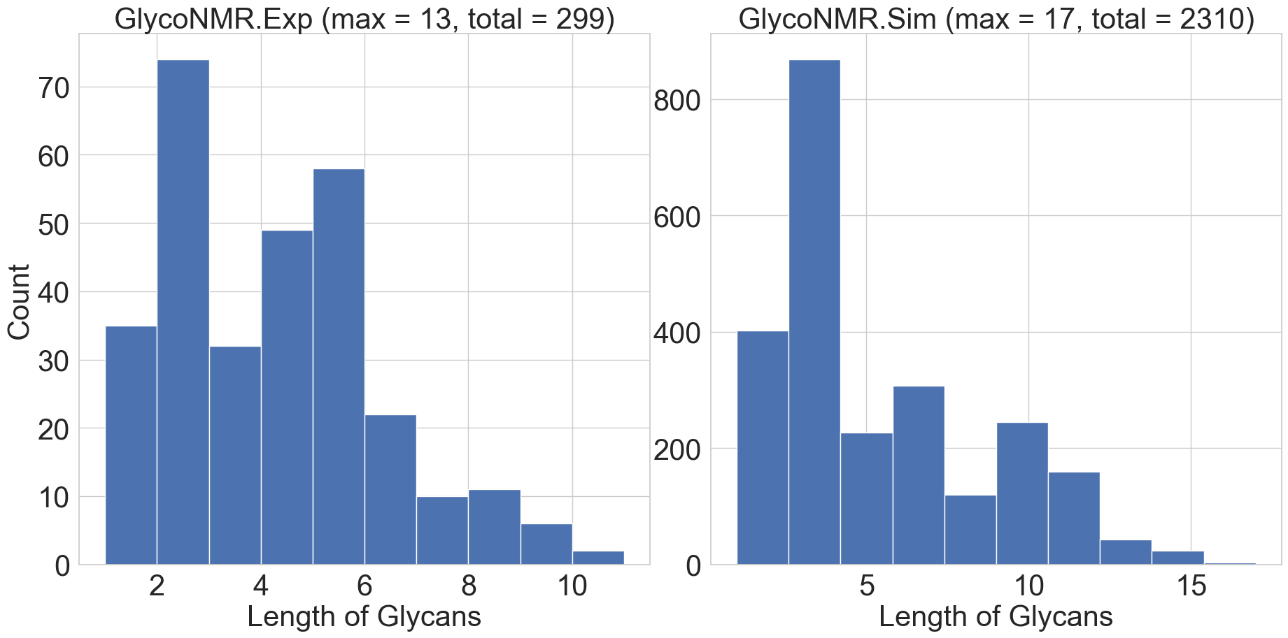
We further analyze the data volume of GlycoNMR. We plot the distributions of the number of monosaccharides that every carbohydrate contains in both GlycoNMR.Exp and GlycoNMR.Sim. In Figure 5, we use ’length of glycan’ to denote the number of monosaccharides that the carbohydrate contains. We observe both histograms exhibit a right-skewed distribution in the length of the glycan. This indicates that GlycoNMR.Exp contains a greater proportion of small and middle-sized carbohydrates than large-sized carbohydrates. Therefore, existing MRL methods may be biased towards smaller carbohydrates.
A.2 Percentage of monosaccharide types in both datasets
We investigate the diversity of monosaccharide types in GlycoNMR. For each dataset, we count the occurrence of all monosaccharides and present the percentage of the top eight most frequently appearing monosaccharides in Table 7. The entry "Others" represents the category of relatively infrequently appeared monosaccharides, including stem type: ManA, Neu, GalN, Ara, etc. We demonstrate that GlycoNMR covers the most commonly occurring stems of monosaccharides as introduced in [Chaplin and Kennedy, 1986] for example.
| GlycoNMR.Sim | GlycoNMR.Exp | ||
|---|---|---|---|
| Monosaccharide | Percentage | Monosaccharide | Percentage |
| Glc | 18.86% | Gal | 19.73% |
| Gal | 17.5% | Glc | 17.7% |
| GlcNAc | 12.18% | GlcNAc | 12.21% |
| Fuc | 12.1% | Rha | 11.06% |
| Xyl | 8.51% | Man | 6.81% |
| Man | 6.23% | Fuc | 4.87% |
| GlcA | 6.19% | Kdo | 4.78% |
| GalA | 5.49% | GlcA | 4.42% |
| Others | 12.94% | Others | 18.42% |
A.3 Feature statistics
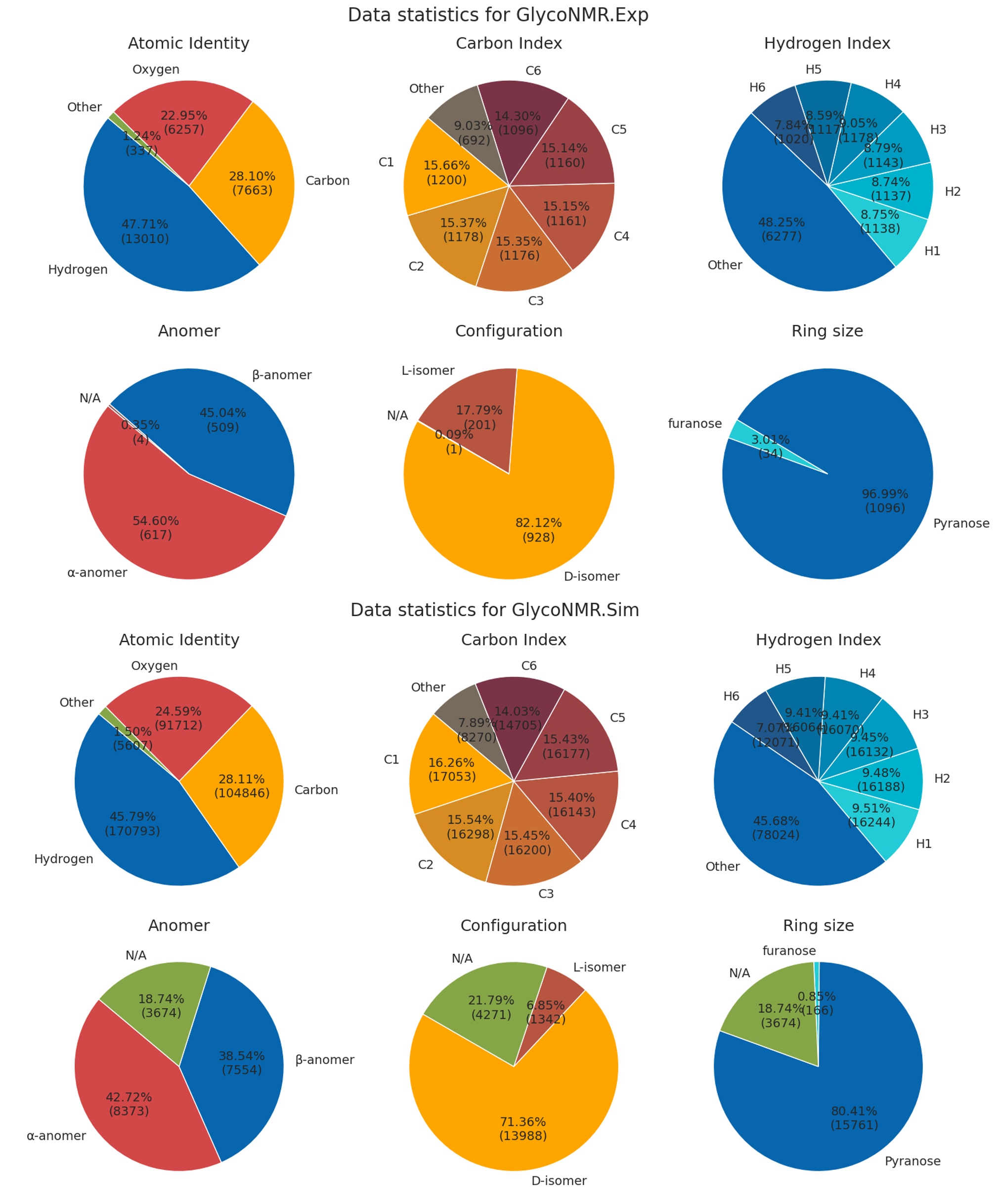
In this section, we present detailed feature statistics for both GlycoNMR.Exp and GlycoNMR.Sim. Specifically, we show the percentage of atom-level features and monosaccharide-level features. For the atom level feature (first-row and the third-row of Figure 6), we present the proportional distribution of values for atom type, carbon atom position, and hydrogen atom position. For the description of atom identity (top left), ’other’ indicates other types of atoms, including nitrogen, phosphorus, and sulfur. For the description of carbon atom position (top middle), ‘Other’ indicates the off-ring carbons. Similarly, for the description of the hydrogen atom position (top right), ‘Other’ indicates off-ring hydrogens. For the monosaccharide level feature (the second row and the fourth row of Figure 6), we included Anomer (bottom left, indicates the hydroxyl group), Configuration (bottom middle, indicates Fischer project information), and Ring Size (bottom right, number of in-ring carbons) as introduced in Table 2. The ‘N/A’ of each pie chart indicates that the information is not contained in the PDB file.
A.4 Ring Position vs. Shift Relationship
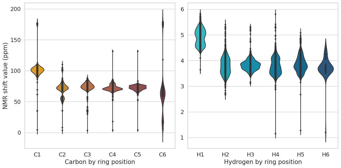
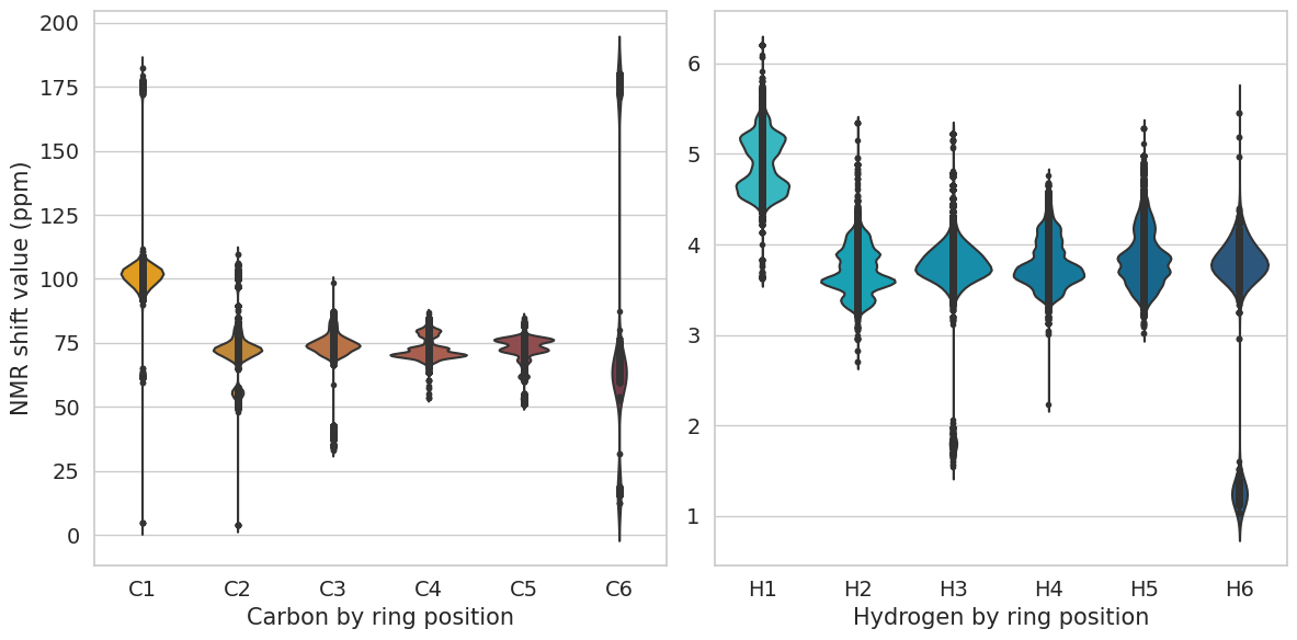
We investigate the relationship between the ring position and the NMR shift values. We plot the distribution of the NMR shift values by carbon and hydrogen ring positions. For both Figure 7 and Figure 8, the x-axis indicates the ring position of the atom (Carbon / Hydrogen), and the y-axis indicates the NMR shift values of the corresponding atoms. We notice that the distribution of NMR shift values for the ring positions C1 and C6 significantly vary from those of C2, C3, C4, and C5, similarly of H1 and H6 to H2, H3, H4, and H5. A fundamental factor that determines the NMR shift value is the atom’s electronic environment, especially bonded or non-bonded interactions within 1-3 atom distances away from the atom of interest.
Appendix B Details on Features Tables
In this section, we present a comprehensive description of the processed PDB file, including the curated features mentioned in Section 2 and Section 3.2. For each feature, we provide its data type along with a detailed explanation. Lines 1-8 in Table 6 record attributes presented in the original PDB file. We incorporate the Atom_name and Atom_type as components of the node features. Coordinate x, y, and z is used as spatial information to construct the MRL models. Lines 9-15 record the processed node features as introduced Table 2. Lines 15-25 describe the feature: Modifications, that are used in GlycoNMR.Sim. On curating the feature Modification, we first identify the modification group using Lineage, Atom_num, Residue_name, and atom connectivity. Then, we calculate each atom’s distance(atom path) to the identified modification group, set up several distance thresholds to convert them into categorical values and incorporate them as node features. Notice that the atom connectivity information is generally missing in GlycoNMR.Exp, thus it can be ambiguous to match the atoms to their corresponding modification groups, and we omitted this feature for now in the smaller Glycosciences.DB-sourced dataset only (in contrast, Modification was included in the GODESS-sourced dataset). Future databases of new experimental results in carbohydrate NMR spectra should seek to improve the clarity in this area, such as with more uniform standards in data annotation by the original uploaders. Last, we use the labeled in-ring atoms’ NMR shift as ground truth values.
| Value | Datatype | Descriptions |
| Atom_num | Numerical | Atom index number in the carbohydrate |
| Atom_name | Categorical | Atom name that also indicates its within-monosaccharide position index |
| Residual_name | Categorical | Three letters abbreviation of monosaccharide name |
| Residual_num | Numerical | Monosaccharide order number assigned |
| x | Numerical | X coordinate of the atom |
| y | Numerical | Y coordinate of the atom |
| z | Numerical | Z coordinate of the atom |
| Atom_type | Categorical | Chemical element type of the atom |
| Residual_accurate_name | Categorical | Full name of monosaccharide or modification group that atom belongs to |
| Lineage | String | Lineage (linkage) information of the current residue |
| Ac_component | Categorical | Whether atom is in an Ac modification |
| bound_AB | Categorical | Anomeric orientation of hydroxyl group |
| fischer_projection_DL | Categorical | Fischer convention |
| reformulated_standard_mono | Categorical | Monosaccharide stem name |
| carbon_number_PF | Categorical | Number of ring carbons (ring size) |
| Me_min_atom_distance | Numerical | Distance of the shortest atom path to Me modification group |
| Me_min_atom_path | Categorical list | The shortest atom path to Me modification |
| Ser_atom_distance | Numerical | Distance of the shortest atom path to Ser modification group |
| Ser_atom_path | Categorical list | The shortest atom path to Ser modification |
| Ac_min_atom_distance | Numerical | Distance of the shortest atom path to Ac modification group |
| Ac_min_atom_path | Categorical list | The shortest atom path to Ac modification |
| S_min_atom_distance | Numerical | Distance of the shortest atom path to S-related modification group |
| S_min_atom_path | Categorical list | The shortest atom path to S-related modification |
| Gc_min_atom_distance | Numerical | Distance of the shortest atom path to Gc modification group |
| Gc_min_atom_path | Categorical list | The shortest atom path to Gc modification |
| main_ring_shift | Numerical | Chemical shift values of all labeled main ring atoms |
| shift | Numerical | Chemical shift values of all labeled atoms |
Appendix C Shapley analysis of Feature contributions
We calculate the Shapley values in Table 9 for the atomic-level and monosaccharide-level features as we introduced in Table 2, following the implementation method if [Štrumbelj and Kononenko, 2014]. We noticed that, in general, all Shapley values are positive. Among all the features, the ring position of both carbon and hydrogen atoms plays a significant role in the NMR shift prediction. In addition, incorporating the stem type of the monosaccharides in the 2D GNN can marginally decrease the prediction error. The remaining features, such as modification group, anomer, configuration, and ring size, have a relatively minor impact on overall model performance.
| GlycoNMR.Exp | Ring position | Modification | Stem type | Anomer | Configuration | Ring size |
|---|---|---|---|---|---|---|
| 1H | 0.457 | N/A | 0.088 | 0.061 | 0.009 | 0.008 |
| 13C | 16.852 | N/A | 2.640 | 0.515 | 0.257 | 0.085 |
| GlycoNMR.Sim | Ring position | Modification | Stem type | Anomer | Configuration | Ring size |
| 1H | 0.387 | 0.014 | 0.112 | 0.187 | 0.051 | 0.014 |
| 13C | 13.007 | 0.321 | 3.619 | 0.465 | 0.199 | 0.055 |
Appendix D Possible Future Research Topics
In this section, we provide several unexplored glycoscience-related research topics that GlycoNMR can be used for. We believe these topics can potentially benefit the overall ML and glycoscience community.
Overview
A common problem in glycosciences is matching structure to NMR spectra. For example, a scientist may want to verify they have generated the correct structure in the laboratory, by examining a compound’s spectra after synthesis. NMR spectral peak positions provide key features for carbohydrate structure identification, including the stereochemistry of monosaccharides, glycosidic linkage types, atomic interactions and couplings, and conformational preferences. Individual atoms (with net spin) in a carbohydrate generate the key spectral peaks for structure interpretation, which in practice in carbohydrates is typically the central ring carbon and hydrogen atoms, plus certain modification groups. Chemical shift values reported in ppm units are also independent of spectrometer frequency and thus comparable across labs and equipment settings. In carbohydrates, usually, only the hydrogen 1H and carbon 13C nuclei shifts are measurable, making spectra harder to interpret than protein spectra where nitrogen and phosphorus shifts are also accessible [Toukach and Ananikov, 2013]. As another challenge specific to carbohydrates, carbohydrate NMR peaks are constrained to a much narrower region of spectra range than proteins, making them harder to separate and leading to an over-reliance on manual interpretation [Toukach and Ananikov, 2013]. The development of theoretical and computational ML-based tools that can utilize large datasets to find and predict relationships between carbohydrate structure and its NMR parameters is a high priority for the field.
Customized models for carbohydrate data
Models specifically designed to accommodate the unique characteristics and structure of the carbohydrate data are important to develop. As introduced in Section 2, carbohydrates are a special type of biomolecule that is formed via the condensation reactions of monosaccharides. We conduct heavy feature engineering to extract the monosaccharide-related features, and our experimental results in Table 3 have already demonstrated the usefulness of monosaccharide information (stem type) in NMR shift prediction. However, we incorporate them as atom-level features in our baseline and the 3D-based MRL models. In this case, the existing models may fail to capture the spatial information between monosaccharides, and more neural network layers corresponding to the structural hierarchies inherent to carbohydrates could improve prediction quality in future work. On the other hand, a carbohydrate’s unique atoms-to-monosaccharides-to-carbohydrate characteristic inherently satisfies a hierarchical graph structure, so the information is partly captured in the current implementation. We believe that developing a customized MRL model (e.g., learning representations for both atoms and monosaccharides) can help learn a better node representation for accurate NMR shift predictions in future work.
Theoretically advancing NMR-based structural analysis approaches in ML directions requires having a comprehensive database where the same base monosaccharide units have various neighboring units or modification groups swapped out or removed across data entries, in order to see how the spectra changes as various components are combined or removed to better train models. Such comprehensive databases have been established and well-studied in protein ML research, but a lack of ML-friendly databases and poor open access data norms have hindered parallel progress in carbohydrates. While our database is certainly not comprehensive and complete, with carbohydrates being more diverse and varied than any other class of biomolecule, our approximately 2600 NMR spectra and structure files tailored for ease of use in ML pipeline is the first of its size for ML studies.
For additional ideas for boosting the data size and quality in future work: by our assessment, GODESS provides the best balance of accuracy, efficiency, and accessibility for the simulation of 1D NMR of carbohydrates. However, as with any simulation method, it likely has some biases and simplifications not seen in experimental data which are difficult to reveal without a large experimental dataset for comparison. Thus, it is important for future work to expand this dataset to include simulation datasets from other sources (e.g. CASPER [Furevi et al., 2022]), as well as to expand the experimental dataset for comparison to the theoretical predictions. The experimental dataset expansion will necessitate a serious and concentrated effort on the part of glycoscience researchers to improve the open data norms of their field.
Predicting NMR spectra:
As presented in Section 3.1, extensive data annotation is required for preparing the atom-level carbohydrate NMR chemical shift data. Notably, for annotating each carbohydrate, the key step is to match the monosaccharides present in the PDB (Protein Data Bank) structure file to the monosaccharides present in the NMR (Nuclear Magnetic Resonance) chemical shift file. This step not only demands significant effort but also necessitates domain expertise, but will continue to do so at least until the experimental glycoscience field adopts more uniform standards in data files.
In the field of glycosciences, the ideal scenario is to predict the full continuous spectrum (peak widths and noise included) depicted in Figure 2 (b) and (c) directly from the carbohydrate structure. In our case, the NMR chemical shift prediction problem of just peaks is reformulated as graph-regression tasks with promising initial performance. The biggest improvements in this direction will necessitate both increasingly larger and more diverse experimental datasets, as well as model innovations.
Appendix E Model Setup and Computation Resources
To ensure a fair comparison, the hidden embedding size for all 3D GNN models is set to 128, and the number of hidden layers is set to 4 in the GlycoNMR.Sim dataset. In the GlycoNMR.Exp dataset, due to the limitations in data size and to prevent over-fitting, the number of hidden layers is set to 2. It takes around 5-34 seconds to train a single epoch with a batch size of 4, depending on different models. All data processing and model training is performed on a Linux workstation with an Intel Core i7 CPU, 32GB memory, and two GeForce RTX 3090 GPUs. Our entire training time for all models in aggregate was on the scale of several hours. Loading codes for the dataset will also be provided in the linked anonymous GitHub after the completion of the peer review. We also provided more detailed run-time information and epoch numbers in the anonymous GitHub repository.
Appendix F GlycoNMR licensing
Disclaimer on GlycoNMR.Exp
GlycoNMR.Exp is freely available under CC BY 4.0 license and can be downloaded within this link. GlycoNMR.Exp is laboriously curated from Glycoscience.DB to facilitate machine learning research on NMR shift predictions of carbohydrates. Glycosciences.DB experimental data uploaded from various labs can be downloaded within this link. Glycosciences.DB [Böhm et al., 2019], as part of the Glycosciences.de [Toukach et al., 2007] portal, is provided for the glycoscience community with unrestricted open access intent. According to [Toukach et al., 2007]: "All glycan-related scientific data of the GLYCOSCIENCES.de portal are freely accessible via the Internet following the open access philosophy: ‘free availability and unrestricted use’."
Disclaimer on GlycoNMR.Sim
GlycoNMR.Sim is freely available under CC BY 4.0 license and can be downloaded within this link. GlycoNMR.Sim is extensively curated from the simulation software GODESS. The GODESS experimentally-informed simulation data without preprocessing can be downloaded within this link. GODESS simulation output is free to use and does not have a license (see https://glic.glycoinfo.org/software/), if proper attribution to the references is done.
Appendix G RMSE formula for Benchmarks
The RMSE was calculated according to the usual equation in all results presented throughout the manuscript:
Where is the recorded NMR chemical shift, is the prediction from our GNN model on the atom from the test set, and N is the number of the test data points.
Appendix H Random Forest Baseline
| GlycoNMR.Sim | GlycoNMR.Exp | |||
|---|---|---|---|---|
| 13C | 1H | 13C | 1H | |
| Random Forest | 2.446 | 0.132 | 4.117 | 0.178 |
We conducted a traditional ML baseline experiment using random forest to predict atomic NMR shifts. The features of each atom (represented as a node in its carbohydrate graph) follow the same initializing method as used for training the 2D GNN model. In addition, we follow the same splitting method as we did in Section 3.4. In general, the baseline model slightly underperforms relative to the 2D GNN model. This demonstrates the effectivenss of our feature engineering step in Section 3.2.
Appendix I Hyperparameter selection for GlycoNMR.Exp
We fine-tune the 3D-based GNN models on GlycoNMR.Exp to prevent overfitting, The hyperparameter is selected from the following ranges: learning rate [0.001, 0.01], batch size: [2, 4, 8], number of layers: [2, 3, 4], hidden channel size: [32, 64, 128, 256], and the cut-off distance for deciding the interactions between atoms: [4.0, 5.0]. We unfortunately did not have time to do more substantial hyperparameter tuning. We believe users of our dataset will be in better positions to provide better results than us with the innovative design of the 3D-based MRL model and substantial hyperparameter selection.
Appendix J Data annotation supplements
In this section, we provide two supplemental repositories to help illustrate our data preprocessing pipeline. One of our major contributions is to extensively curate the raw files from the Glycosciences.DB- and GODESS-sourced datasets to make the GlycoNMR dataset friendly to machine learning researchers. To achieve this, we have made significant efforts in data preprocessing and provided a reproducible protocol for use in curating future carbohydrate-related NMR/structure databases.
J.1 Overview
We summarize the data preprocessing pipelines on Glycosciences.DB in the following five steps. 1, We manually and semi-automatically checked the carbohydrate data scraped from Glycosciences.DB, and we applied exclusion criterion of only maintaining carbohydrates with complete or nearly complete NMR peak shift lists. 2, We reformulated all the PDB files (as well as the NMR label files) into an interpretable and consistent format, as they are uploaded from various labs. 3, We examined the carbohydrates with branched monosaccharide chains, and manually matched the monosaccharide IDs from the PDB file and the NMR label file. 4, We trained a simple 2D GNN model and predicted the NMR chemical shifts for each annotated atom. 5, We examined the carbohydrates with the highest ranked errors and applied an outlier check using domain knowledge over many iterations of annotation debugging and validation, until we had a complete semi-automated pipeline that could correct the most common reasons for annotation mismatch between the NMR and structure file. While developing the annotation pipeline, if the error resulted from mismatches in monosaccharide IDs in Step 4, we then go back to the previous steps 2, 3 and 4. The data preprocessing pipeline in GODESS is relatively similar to the Glycoscience.DB. We constructed a more streamlined semi-automatic pipeline to annotate the GODESS dataset since the dataset is generated from a single simulation software with more consistent formatting. We introduced this pipeline in our released repository provided below.
To further demonstrate our efforts, we released two repositories for reference on data cleaning, processing, and annotating:
Creating GlycoNMR.Sim from the GODESS (https://anonymous.4open.science/r/GODESS_preprocess-F9CD/README.md)
Creating GlycoNMR.Exp from the Glycosciences.DB (https://anonymous.4open.science/r/GlycoscienceDB_preprocess-B678/README.md).
The data preprocessing steps are provided in detail in the README.md file.
J.2 An annotation example from GlycoNMR.Exp
For carbohydrate file DB26380, we need to manually annotate the PDB file by assigning each central ring carbon and hydrogen atoms with their corresponding shift values, which is stored in the NMR label file. To achieve this, we need to associate the atoms’ parent monosaccharide IDs between the two files. We first draw a sketch of the carbohydrate structure consisting of the basic monosaccharide components from the CSV file using the linkage information. Atoms with the same linkages are from the same monosaccharides. For example, atoms from lines 13-19 belong to monosaccharide B-D-GLCPN. We utilize linkage information to identify monosaccharide components but not monosaccharide names such as ‘B-D-GLCPN’ because, in some scenarios, the same monosaccharide name may indicate different monosaccharide components (i.e., there can be multiple monosaccharide units with the same name in a carbohydrate, but the linkage information can be used to tell them apart for NMR shift matching purposes). For example, lines 62-67, 68-73, and 74-79 of the NMR label file refer to three separate monosaccharide unit components, that are parents of different sets of atoms and appear in different locations of the carbohydrate chain, but still have the same monosaccharide chemical name. DB26380’s sketch plot can be found on the 8th page (plot number 23) of our annotation document for branched carbohydrates. Second, we again inspect the PDB file and match the monosaccharide components with the help of the SWECON information which provides additional secondary linkage information at the bottom of Glycosciences.DB PDB file (lines 306-315) and our domain expertise. Then, for another example of a common issue causing mismatches between monosaccharide shift and structure, in DB26380, we noticed that the Phosphoryl group ‘PO3’ (lines 39-42, 64-67) is treated as a monosaccharide component in the PDB file despite not being a monosaccharide, therefore the monosaccharide shift file ID ordering 3 and 13 should be disregarded when comparing to the PDB structure file, and the 4th monosaccharide residue in the PDB file should instead be matched with the 3rd monosaccharide parent and its atom components in the NMR label file. A detailed match is presented in our PDF document mentioned above. Then last, when all parent monosaccharides are correctly matched between structure and shift files, we assign the corresponding monosaccharide atoms’ shift from the label file to the PDB file by their atom names.
J.3 An annotation example from GlycoNMR.Sim
For glycan with name: ’aDXylp(1-6)bDGlcp(1-4)[aLFucp(1-2)bDGalp(1-2)aDXylp(1-6)]bDGlcp(1-4)[aLFucp(1-2)bDGalp(1-2)aDXylp(1-6)]bDGlcp(1-4)xDGlca’ and its corresponding PDB file, monosaccharide bond linkage ’(1-4)’ indicates the carbon with position number 1 is connected to the carbon with position number 4 via a dehydration synthesis reaction, where ‘xDGlca’ is the precursor monosaccharides (in other words ‘root’). From line 223 of the PDB file, we notice that atom 1 is connected to atoms 28 and 2, this indicates that the monosaccharide with ID 2 is connected to a monosaccharide with ID in the following bounds (C1 - O4 - C4), where C indicates the carbon and O indicate the oxygen and the following number indicates the ring position. In this case, from the 3rd line of the label file, we can match the monosaccharides residue ‘b-D-Glcp’ from the label file to the monosaccharides ID 2 in the PDB file using the linkage information ‘, 4’ which indicates the following bounds (C1 - O4 - C4). Then, again, we assign the corresponding monosaccharide atom’s shift from the NMR label file to the PDB file by its atom name.
Appendix K Example codes and demos
We provide four Jupyter Notebook demos in the anonymous GitHub repo for detailed instructions. They introduce step by step on how to utilize the GlycoNMR.Sim and GlycoNMR.Exp datasets to train a 3D or 2D GNN model.
Train a 2D-based GNN model on GlycoNMR.Sim: https://anonymous.4open.science/r/GlycoNMR-D381/2D_example_Sim_GlycoNMR.ipynb.
Train a 2D-based GNN model on GlycoNMR.Exp: https://anonymous.4open.science/r/GlycoNMR-D381/2D_example_Exp_GlycoNMR.ipynb.
Train a 3D-based GNN model on GlycoNMR.Sim: https://anonymous.4open.science/r/GlycoNMR-D381/3D_example_Exp_GlycoNMR.ipynb.
Train a 3D-based GNN model on GlycoNMR.Exp: https://anonymous.4open.science/r/GlycoNMR-D381/3D_example_Sim_GlycoNMR.ipynb.