Fluorescent single-molecule STM probe
Abstract
The plasmonic tip of a scanning tunnelling microscope (STM) is functionalized with a single fluorescent molecule and is scanned on a plasmonic substrate. The tunneling current flowing through the tip-molecule-substrate junction generates a narrow-line emission of light corresponding to the fluorescence of the negatively charged molecule suspended at the apex of the tip, i.e., the emission of the excited molecular anion (trion). The fluorescence of this molecular probe is recorded for tip-substrate nanocavities featuring different plasmonic resonances, for different tip-substrate distances and applied bias voltages, and on different substrates. We demonstrate that the width of the emission peak can be used as a probe of the trion-plasmon coupling strength and that the energy of the emitted photons is governed by the molecule interactions with its electrostatic environment. Additionally, we theoretically elucidate why the direct contact of the suspended molecule with the metallic tip does not totally quench the radiative emission of the molecule.
pacs:
78.67.-n,78.60.Fi,68.37.EfIntroduction
Scanning probe microscopes (SPM) have revolutionized our perception of the atomic-scale world, providing topographic, electronic, magnetic, optical or mechanical information of a surface with ultimate spatial resolution. To address some specific properties of the probed sample, it proved advantageous to modify the chemical nature of the extremity of the scanning probe tip. Molecule-functionalized tips have played here a key role, enabling unprecedented resolution over the skeleton of molecules [1, 2] or addressing electrostatic [3, 4, 5, 6], magnetic [7] and transport properties [8, 9, 10]. In parallel, tip-induced electroluminescence [11, 12, 13, 14, 15, 16], photoluminescence [17, 18, 19] or Raman spectroscopies [20, 21] have reached sub-nanometer spatial resolution by making use of the extreme confinement of electromagnetic fields at the end of scanning tunneling microscope (STM) tips made of plasmonic materials. These techniques have made it possible to measure the fluorescence and scattering properties of individual molecules lying flat on thin decoupling layers with sub-molecular precision. Transferring a fluorescent molecule to the tip of an SPM would allow sensing and mapping the local electromagnetic field of a sample with the same spatial resolution. This has been attempted with terylene molecules embedded in micron-size nanocrystals [22], semiconductor quantum dots (SCQD) fixed on the tip of an optical fiber [23], and NV centers in diamond nanocrystals attached to atomic force microscope tips [24]. Whereas these nearfield probes have been successfully used to sense electrostatic, electromagnetic and magnetic fields, the spatial resolution they provide remains limited by the (large) size of the emitter (SCQD) or of the nanocrystal into which the emitter is embedded. Hence, SPM tips preserving the fluorescence properties of a single-molecule directly attached to the tip apex are highly desirable, but have not been reported so far. This is presumably because direct contact between the molecule and the metal generally causes total quenching of the molecule fluorescence, due to ultrafast electron and energy transfer from the excited molecule to the metal [25].
Here we build on recent works demonstrating that a single 3,4,9,10-perylenetetracarboxylic-dianhydride (PTCDA) molecule can be suspended at the apex of a metallic STM tip [26, 27], and report on the electrically-induced fluorescence of this functionalized PTCDA tip. By performing a theoretical analysis, we show that in this configuration the spatial overlap between the molecular and tip electronic orbitals is much smaller than in geometries where the molecule would lie flat on the substrate. This leads to a reduced charge-transfer rate between the molecule and the metal, thus preserving the molecular fluorescence properties. As the PTCDA molecule is hanging approximately aligned with the tip axis in the STM junction, the molecular transition dipole is close to collinear with the plasmonic electric field in the gap, a situation that strongly contrasts with the on-surface flat-lying configuration of the molecules studied in usual tip-induced fluorescence measurements [18]. This collinear geometry leads to a large increase of the coupling strength between the molecular exciton and the tip-sample plasmonic cavity by up to two orders of magnitude compared to the perpendicular (flat-lying) configuration. Eventually, the spectral characteristics of the PTCDA tips are investigated as a function of the plasmonic response of the tip-sample cavity, the bias voltage and the tip-sample distance, revealing that the emission of PTCDA tips can be used as a probe of the local electromagnetic and electrostatic fields.
Results
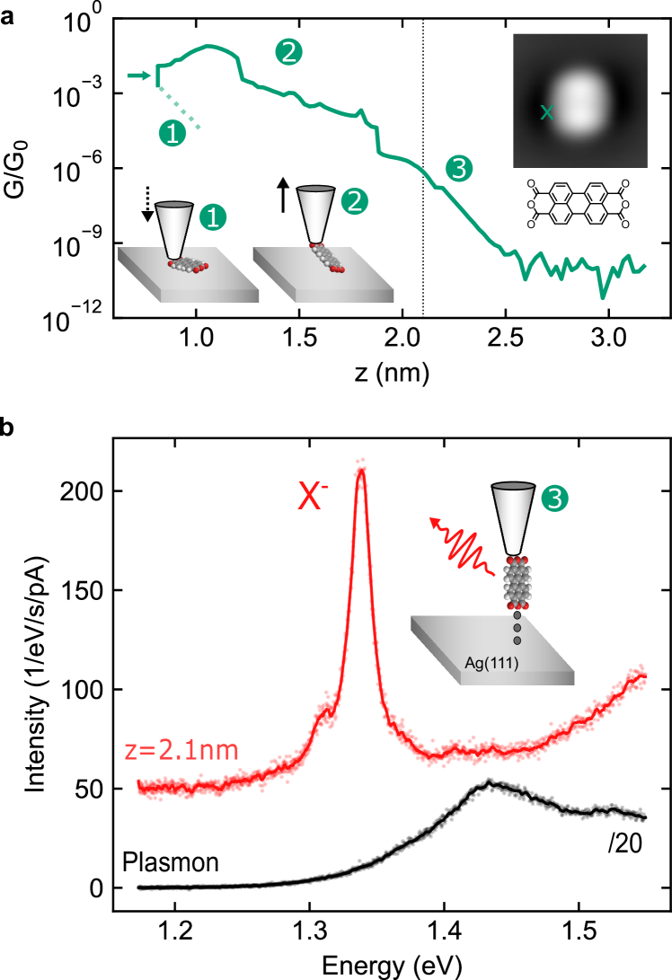
The procedure for making fluorescent PTCDA tips in a low-temperature, ultra high vacuum STM is schematized in Figure 1 (see Methods for details of the sample preparation). A silver-covered tungsten tip is approached (dotted line in Fig. 1a) to the oxygen atoms located at the extremity of a PTCDA molecule deposited on Ag(111) until a contact is reached (current jump indicated by the green arrow in Fig. 1a). At this contact point, the last tip atom is expected to be at from the metallic surface [28]. The tip is then retracted together with the attached molecule at its extremity. During this procedure the electrical current traversing the junction is monitored (solid green line Fig. 1a). The presence of the molecule at the tip is confirmed by the much larger conductance values recorded during retraction ((2) Fig. 1a).
Eventually, the complete detachment of the molecule from the surface is identified ((3) Fig. 1a) at by a strong slope change of the exponentially decaying conductance with distance. As discussed in Ref. [29] and [30], the PTCDA molecule adopts an unexpected up-standing configuration at the tip apex thanks to stabilizing electrostatic dipole forces that prevent the molecule from toppling over onto the tip shaft. Besides, it has been demonstrated that for low positive voltage tunneling conditions (), the attached molecule is negatively charged (PTCDA-) [31, 26, 27, 32, 33], a state that reflects the electron acceptor character of the molecule. In Fig. 1b we report STM-induced luminescence (STML) spectra acquired at , first with a clean metal tip, revealing a broad feature characteristic of plasmonic emission (black spectrum), and then with a fully detached () PTCDA tip on top of the bare silver surface (red spectrum). In contrast to the spectrally broad plasmonic emission observed when the clean tip is used, the STML spectrum measured using the PTCDA tip exhibits a much sharper and more intense emission peak, which is centered at a photon energy of . In agreement with a recent report [34], we assign this peak to the luminescence of negatively charged PTCDA (later referred to as negative trion X-). This result provides experimental evidence that the luminescence properties of the molecule suspended at the tip are preserved, despite the direct molecule-metal contact. The PTCDA tip can therefore be considered as a fluorescent molecular probe, whose spectral emission properties measured in the far field can be used to probe its local environment.
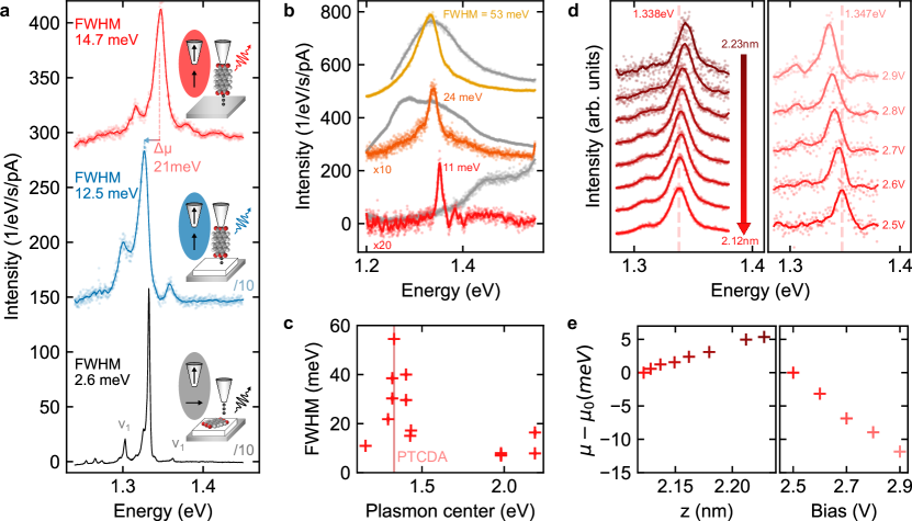
As a first step to characterize this fluorescent probe, we compare spectra (Fig. 2a) successively obtained with a PTCDA tip located above the bare Ag(111) (red spectrum), above a two atomic layer-thick (2 ML) NaCl island grown on Ag(111) (blue spectrum) and with the exact same molecule adsorbed flat on 3ML NaCl but excited using a bare metal tip (black spectrum). For this last spectrum, the molecule was released from the tip by applying a low voltage pulse on top of NaCl. All spectra exhibit an intense X- emission line together with two vibronic satellites (), located at on each side of the X- line, that occur from an in-plane breathing mode of the molecule [36]. When the PTCDA tip is used, we observe that the STML spectrum measured on top of 2 ML NaCl-covered Ag(111) is rigidly redshifted by relative to the spectrum measured on bare Ag(111). This shift suggests that the emission properties of the tip-suspended molecule are dependent on its local dielectric environment, which is modified by the presence of the NaCl island.
Moreover, the full width at half maximum (FWHM) of the emission peak is one order of magnitude larger in the spectra measured using the PTCDA tip () than in the spectra measured using the clean tip on the PTCDA molecule lying flat on the NaCl (). The width of molecular emission lines in STML experiments may have different origins and may be due to dephasing induced by the coupling of the trion with its electrostatic and/or phononic environments, non-radiative decay paths or a strongly shortened trion fluorescence lifetime compared to the free molecule because of the particular electromagnetic environment of the STM junction (Purcell effect). When the latter dominates, the FWHM is determined by the trion-plasmon coupling strength.
To identify the respective role of dephasing and lifetime-shortening on the emission linewidth of the PTCDA tip, we investigate the FWHM of the X- line as a function of the energy of the gap plasmon of the tip-sample junction. It is well known that the plasmonic response of the junction strongly depends on the nanoscale structure of the tip apex, a parameter that can be tuned by controlled indentations of the tip in the bare metal surface [37]. In Fig. 2b, we show the STML spectra of three different PTCDA tips above bare Ag(111) together with the plasmonic response of the tip-sample junction recorded before functionalization with the PTCDA molecule. The plasmon peak is either strongly blue-detuned (bottom spectrum), weakly red-detuned (middle spectrum), or in-resonance (top spectrum) with the X- line. Here, the smaller the frequency mismatch between the plasmonic resonance and the trion emission, the broader the FWHM of the X- emission. This correlation is better evidenced in Fig. 2c where the FWHM of the X- line is plotted as a function of the energy of the plasmon resonance maximum for 13 different tips. This plot also reveals that the FWHM does not reach values lower than , even for extremely non-resonant conditions. This suggests that the line width is in this case limited by dephasing or non-radiative decay paths that do not involve coupling to plasmons. In contrast, the FWHM increase observed at resonance indicates a reduction of the excited state lifetime due to the stronger coupling of the trion with the gap plasmons. The FWHM then reaches values as high as , corresponding to a fluorescence lifetime of . The trion-plasmon coupling is here still below the threshold of strong coupling [38]. As a figure of merit to quantify the regime of plasmon-trion coupling, we use the ratio between the experimental plasmon-trion coupling (i.e., the Jaynes-Cummings coupling strength as defined in Ref. [15]) and the experimental width of the plasmon . Assuming that the trion couples to a single plasmon mode as described in Ref. [15], we estimate for the resonant case (top spectrum Fig. 2b) from the plasmon-induced width and ( is the reduced Planck constant) as . Since is smaller than we conclude that the plasmonic losses still dominate over the plasmon-trion coupling and we therefore observe a weak trion-plasmon coupling. For flat PTCDA adsorbed on 4 ML NaCl, a FWHM as low as could be measured (see Supporting Information (SI) [35]), setting a lower limit for the trion lifetime of when the tip and molecular dipoles are orthogonal to each other (compare sketches in Fig.2a).
Overall, our results show that the excited state lifetime of the PTCDA trion is two orders of magnitude shorter when the transition dipole of the molecule is parallel to the electric field of the picocavity plasmons than when it is orthogonal to it, i.e., than in the configuration used in previous STML studies. Furthermore, the coupling strength is maximum when the plasmon is tuned in resonance with the X- line. This also indicates that the FWHM of X- can be used to probe variations of the local density of photonic states at metallic surfaces, e.g., at the surface of plasmonic nanostructures [22, 39].
In principle, not only the FWHM, but also the energy position of the X- line can be used for probing the environment of the molecular-tip. In previous reports, emission peak positions have been shown to vary as a function of both the local electromagnetic field, an effect known as photonic Lamb shift [40], and the electrostatic field where the spectral shift is described in terms of a Stark effect [15]. In Fig. 2d, we monitor the X- line as a function of the tip-sample distance (at constant voltage) and as a function of the voltage (at constant tip-sample distance). The FWHM of the line () remains constant for the two sets, indicating that the trion-plasmon coupling does not significantly vary over this range of distances or voltages. In contrast, the X- line experiences a shift to lower energies with tip approach and with increasing voltages. Fig. 2e reveals that this line-shift evolves essentially linearly with distance, at a rate of , and with voltage, at a rate of . These values are close to the rates reported for phthalocyanine molecules lying flat on NaCl and addressed by a metal tip [15]. In this case, the line-shifts caused by changes in the tip-sample distance and voltage were dominated by the Stark effect due to the electrostatic field in the STM junction. This effect is likely at play in Fig. 2(d, e), suggesting that one can use the spectral shift of the PTCDA tip emission line as a probe of the local electrostatic field.
Discussion
To explain the unexpected observation of a bright fluorescence from a molecule that is in direct contact with a metallic electrode, we perform a series of quantum chemical and quasi-electrostatic calculations. We develop a theoretical description that compares (i) the interaction between the molecular exciton and the tip/substrate plasmons as discussed in detail in Ref. [15], and (ii) the charge transfer between the molecule and the tip, which is expected to strongly contribute to the quenching of the molecule’s photon emission. Our model is thus able to capture the most relevant physical processes providing order-of-magnitude estimates of the photon emission efficiency.
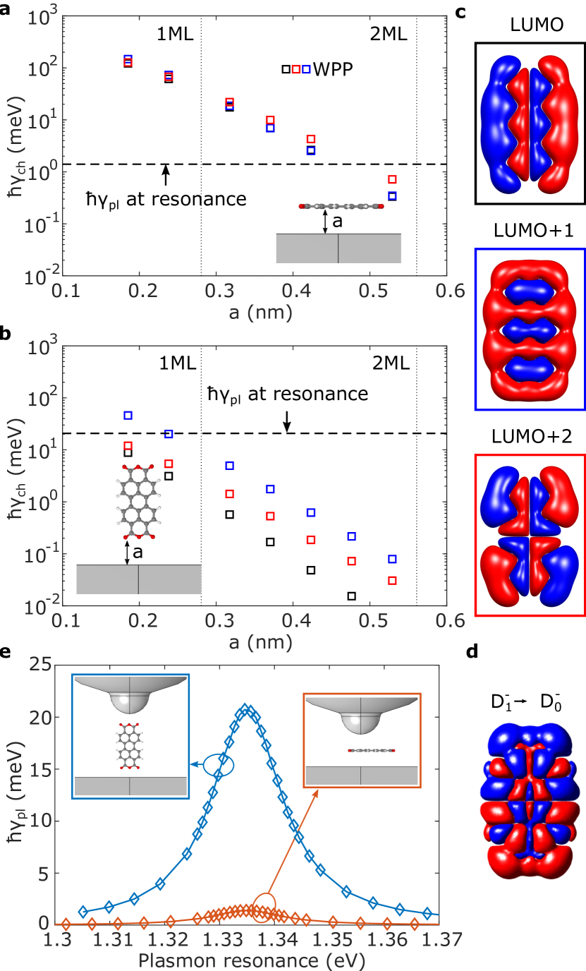
We describe the charge transfer in an effective single-particle picture in which we estimate the charge transfer rate of several molecular orbitals into the metal surface using the real-time wave-packet propagation (WPP) technique [41] (see SI [35] for details and additional estimates using a perturbation approach). The molecule is assumed to be positioned above a planar metal surface and the distance between the molecule and the image plane of the surface is varied. We consider two different configurations: the molecule lying flat on the surface (Fig. 3a) and the molecule in the lifted configuration with its plane perpendicular to the surface (Fig. 3b). In the latter case, describes the distance between image plane and the bottom oxygen atoms of the molecule. The decay rate induced by the charge transfer is shown in Fig. 3a (Fig. 3b) for the flat-lying (lifted) configuration for the LUMO (lowest unoccupied molecular orbital) (black), LUMO+1 (blue), and LUMO+2 (red) as squares calculated using the full real-time WPP. The orbital labeling is derived from the electronic configuration of the neutral molecule, i.e., the LUMO is split into the singly occupied and unoccupied orbitals in the negative ground state D. We note that both LUMO and LUMO+2 have a significant contribution to the electronic configuration of (see SI [35] for details). The corresponding orbitals are shown in Fig. 3c. Fig. 3b (Fig. 3a) demonstrates that at a distance of about - roughly corresponding to the distance between the atoms binding the tip and the molecule [29, 30] - the electronic decay rate of the orbitals reaches up to () for the lifted (flat-lying) configuration. In other words, the lifted configuration leads to a ten-times weaker metal-molecule electronic coupling than for a flat-lying molecule.
To know if the charge transfer rate is fast enough to quench the trion emission, one should compare it with the radiative decay rate expressed by . To calculate , we first describe the electronic excitations in the negative PTCDA molecule using time-dependent density-functional theory (TDDFT) (see SI [35] for details). We relax the molecular geometry in its first excited state D and extract the molecule’s transition charge density (Fig. 3d) - generalizing the concept of the transition dipole moment. We next calculate a quasi-static electric potential generated by the source charge density placed in plasmonic environment formed by the tip and the substrate. We obtain the plasmon-induced broadening of the trion as [15] and show the result in Fig. 3e as a function of the plasmon resonance energy, which is artificially tuned by changing the tip geometry (see SI [35]). The molecule is considered in two geometrical configurations in the center of the tip-substrate gap, once parallel to the surface, and once in the upright geometry. As demonstrated in Fig. 3e, the resonance between the trionic state and the plasmonic mode is necessary to reach large trion broadening of up to for the upright molecule, on the order of the experimental value reported in Fig. 2c (see SI [35] for a detailed discussion). Our calculation thus supports the conclusion that the plasmon resonance is responsible for the observed broadening of the trion line. Besides, in the lifted configuration, the broadening is times larger than the maximal plasmon-exciton coupling obtained for the flat-lying molecule (Fig. 3e). In other words, the fluorescence decay is times faster for molecules oriented aligned with the tip axis than for flat-lying molecules. The plasmon-induced trion decay rate in both configuration is represented by horizontal dashed line in Fig. 3a, b. For the flat-lying case (Fig. 3a), we see that the plasmon-induced trionic decay rate dominates over the charge-transfer rate only for , a molecule-substrate distance that is close to the one corresponding to 2 ML NaCl where STM-induced fluorescence signal starts to be observed. In contrast, the plasmon-induced trion decay rate already dominates for for the lifted configuration (Fig. 3b). Overall, these data show that the observation of fluorescence in the lifted configuration results from the combination of a reduced electronic coupling between molecule and electrode states and from the enhanced electromagnetic coupling between the vertically polarized tip-plasmons and the molecular excited states.
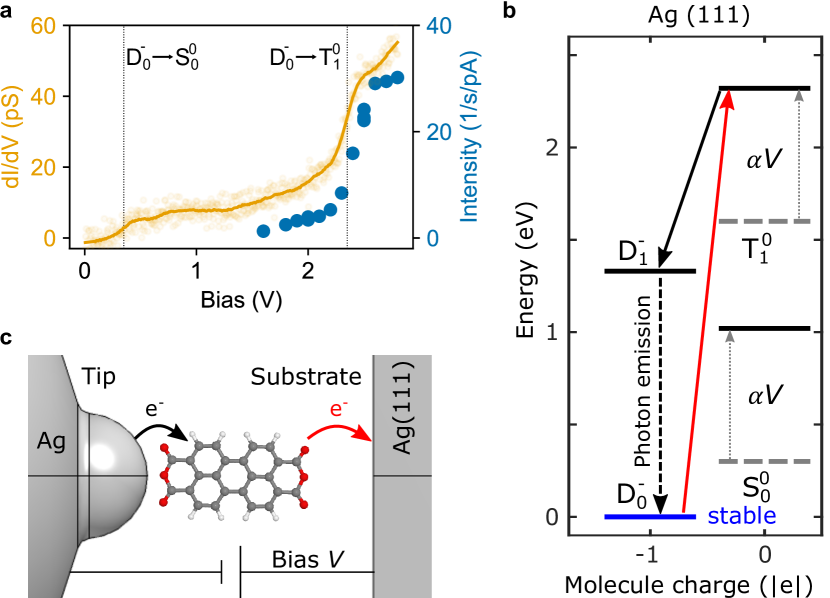
Next, we briefly discuss the mechanisms that are likely involved in the STM-induced excitation of the PTCDA molecule suspended on the tip. In STML the mechanism bringing the molecule to the excited state usually depends on a sequence of charge transfer events that are specific to the considered system [42, 43]. Figure 4a shows a spectrum simultaneously recorded with the total intensity of the X- line emitted by the molecule. The curve shows a smooth step-like increase of the bias voltage at and . The step at is accompanied by an equally smooth onset of photon emission. To explain this voltage-dependent behaviour we propose a model of charge transfer events between the many-body electronic states of the molecule (see Fig. 4b). In the model we include electronic states of the neutral molecule (singlet ground state S0 and a triplet state T), two doublet states (D and D) of the negatively charged molecule. The energies of the states displayed in the diagram are aligned with respect to the work function of the Ag tip which we estimate at [44, 45]. When a voltage is applied, the relative energy of the different charge states shifts by as indicated by the grey dotted arrows. Here indicates the fractional voltage drop experienced by the molecule with respect to the tip electrode. We envision the mechanism as follows. First, by applying a small bias of about the singly negatively charged molecule can be transiently neutralized by tunneling to the substrate and brought into the S state. For a voltage of the transition into the neutral triplet T state is enabled (red arrows in Fig. 4a, c). Once in the neutral triplet state, the molecule can be rapidly charged from the tip (black arrows) and can end up in the excited doublet configuration (D). D readily decays into the ground state either by non-radiatively exchanging an electron with the tip or by photon emission. We note that when the lifted molecule is brought above the NaCl/Ag(111) surface, the mechanism leading to light emission can differ from the present discussion (see SI [35]).
In conclusion, we demonstrated that a PTCDA molecule preserves its intrinsic emission properties even when it is directly attached to a plasmonic scanning probe tip. This is due to the relatively low spatial overlap between the tip and molecule electronic orbitals — which in turn leads to weak luminescence quenching by charge transfer — and by the strongly increased radiative decay probability (by up to 2 orders of magnitude compared to flat-lying PTCDA) of the molecular emitter fixed vertically at the apex of the plasmonic tip. This increased trion-plasmon interaction, however, remains insufficient to reach the strong coupling regime. Our data also demonstrate that the fluorescent properties of the molecular probe are sensitive to their electromagnetic and electrostatic environment. Eventually, the excitation mechanism by tunneling electrons has been discussed. Further work will be devoted to characterizing the spatial resolution that is achievable with this atomic-scale sensor that could be used, in the close future, in resonant energy transfer microscopy experiments having ultimate lateral and axial precision.
Methods
The STM data was acquired with a low temperature () Unisoku setup operating in ultrahigh vacuum and adapted to detect the light emitted at the tip-sample junction. The optical detection setup is composed of a spectrograph coupled to a CCD camera; a grating having a groove density of 300 lines/mm was used and provided a spectral resolution of for all the data presented in the paper with the exception of the data of Fig. S3 where a grating with 1200 lines/mm was used. Tungsten STM-tips were introduced in the sample to cover them with silver so as to tune their plasmonic response. The Ag(111) substrate was cleaned with successive Ar+-ion sputtering and annealing cycles.
Approximately 0.5 monolayers of NaCl were sublimated on Ag(111) kept at room temperature. The sample was then flash-annealed up to to obtain square domains of bi- and tri-layers of NaCl. PTCDA was evaporated in situ on the sample held at using a molecular beam evaporator (), resulting in a sparse distribution of individual molecules.
References
References
- Temirov et al. [2008a] R. Temirov, S. Soubatch, O. Neucheva, A. C. Lassise, and F. S. Tautz, A novel method achieving ultra-high geometrical resolution in scanning tunnelling microscopy, New J. Phys. 10, 053012 (2008a).
- Gross et al. [2009] L. Gross, Z. L. Wang, D. Ugarte, F. Mohn, N. Moll, W. a. Heer, P. Vincent, P. Liljeroth, C. Journet, G. Meyer, V. T. Binh, M. Poot, H. S. J. V. D. Zant, A. Aguasca, A. Bachtold, K. Kim, A. Zettl, P. Hung, H. W. C. Postma, M. Bockrath, X. Blase, and S. Roche, The chemical structure of a molecule resolved by atomic force microscopy, Science 325, 1110 (2009).
- Wagner et al. [2019] C. Wagner, M. F. B. Green, M. Maiworm, P. Leinen, T. Esat, N. Ferri, N. Friedrich, R. Findeisen, A. Tkatchenko, R. Temirov, and F. S. Tautz, Quantitative imaging of electric surface potentials with single-atom sensitivity, Nat. Mater. 18, 853 (2019).
- Mallada et al. [2021] B. Mallada, A. Gallardo, M. Lamanec, B. De La Torre, V. Špirko, P. Hobza, and P. Jelinek, Real-space imaging of anisotropic charge of -hole by means of kelvin probe force microscopy, Science 374, 863 (2021).
- Tallarida et al. [2017] N. Tallarida, J. Lee, and V. A. Apkarian, Tip-enhanced raman spectromicroscopy on the angstrom scale: bare and co-terminated ag tips, ACS Nano 11, 11393 (2017).
- Lee et al. [2018] J. Lee, N. Tallarida, X. Chen, L. Jensen, and V. A. Apkarian, Microscopy with a single-molecule scanning electrometer, Sci. Adv. 4, eaat5472 (2018).
- Verlhac et al. [2019] B. Verlhac, N. Bachellier, L. Garnier, M. Ormaza, P. Abufager, R. Robles, M.-L. Bocquet, M. Ternes, N. Lorente, and L. Limot, Atomic-scale spin sensing with a single molecule at the apex of a scanning tunneling microscope, Science 366, 623 (2019).
- Schull et al. [2009] G. Schull, T. Frederiksen, M. Brandbyge, and R. Berndt, Passing current through touching molecules, Phys. Rev. Lett. 103, 206803 (2009).
- Lafferentz et al. [2009] L. Lafferentz, F. Ample, H. Yu, S. Hecht, C. Joachim, and L. Grill, Conductance of a single conjugated polymer as a continuous function of its length, Science 323, 1193 (2009).
- Schull et al. [2011] G. Schull, T. Frederiksen, A. Arnau, D. Sánchez-Portal, and R. Berndt, Atomic-scale engineering of electrodes for single-molecule contacts, Nature Nanotechnology 6, 23 (2011).
- Qiu et al. [2003] X. H. Qiu, G. V. Nazin, and W. Ho, Vibrationally resolved fluorescence excited with submolecular precision, Science 299, 542 (2003).
- Zhang et al. [2016] Y. Zhang, Y. Luo, Y. Zhang, Y.-J. Yu, Y.-M. Kuang, L. Zhang, Q.-S. Meng, Y. Luo, J.-L. Yang, Z.-C. Dong, et al., Visualizing coherent intermolecular dipole–dipole coupling in real space, Nature 531, 623 (2016).
- Imada et al. [2016] H. Imada, K. Miwa, M. Imai-Imada, S. Kawahara, K. Kimura, and Y. Kim, Real-space investigation of energy transfer in heterogeneous molecular dimers, Nature 538, 364 (2016).
- Doppagne et al. [2020] B. Doppagne, T. Neuman, R. Soria-Martinez, L. E. P. López, H. Bulou, M. Romeo, S. Berciaud, F. Scheurer, J. Aizpurua, and G. Schull, Single-molecule tautomerization tracking through space-and time-resolved fluorescence spectroscopy, Nat. Nanotechnol. 15, 207 (2020).
- Rosławska et al. [2022] A. Rosławska, T. Neuman, B. Doppagne, A. G. Borisov, M. Romeo, F. Scheurer, J. Aizpurua, and G. Schull, Mapping lamb, stark, and purcell effects at a chromophore-picocavity junction with hyper-resolved fluorescence microscopy, Phys. Rev. X 12, 011012 (2022).
- Doležal et al. [2022a] J. Doležal, S. Canola, P. Hapala, R. C. de Campos Ferreira, P. Merino, and M. Švec, Real space visualization of entangled excitonic states in charged molecular assemblies, ACS Nano 16, 1082 (2022a).
- Yang et al. [2020] B. Yang, G. Chen, A. Ghafoor, Y. Zhang, Y. Zhang, Y. Zhang, Y. Luo, J. Yang, V. Sandoghdar, J. Aizpurua, Z. Dong, and J. G. Hou, Sub-nanometre resolution in single-molecule photoluminescence imaging, Nat. Photonics 14, 693 (2020).
- Imada et al. [2021] H. Imada, M. Imai-Imada, K. Miwa, H. Yamane, T. Iwasa, Y. Tanaka, N. Toriumi, K. Kimura, N. Yokoshi, A. Muranaka, M. Uchiyama, T. Taketsugu, Y. K. Kato, H. Ishihara, and Y. Kim, Single-molecule laser nanospectroscopy with micro–electron volt energy resolution, Science 373, 95 (2021).
- Rosławska et al. [2023] A. Rosławska, K. Kaiser, M. Romeo, E. Devaux, F. Scheurer, S. Berciaud, T. Neuman, and G. Schull, Submolecular-scale control of phototautomerization, , arXiv 2305.13157, https://doi.org/10.48550/arXiv.2305.13157 (2023).
- Zhang et al. [2013] R. Zhang, Y. Zhang, Z. C. Dong, S. Jiang, C. Zhang, L. G. Chen, L. Zhang, Y. Liao, J. Aizpurua, Y. Luo, J. L. Yang, and J. G. Hou, Chemical mapping of a single molecule by plasmon-enhanced raman scattering, Nature 498, 82 (2013).
- Lee et al. [2019] J. Lee, K. T. Crampton, N. Tallarida, and V. A. Apkarian, Visualizing vibrational normal modes of a single molecule with atomically confined light, Nature 568, 78 (2019).
- Michaelis et al. [2000] J. Michaelis, C. Hettich, J. Mlynek, and V. Sandoghdar, Optical microscopy using a single-molecule light source, Nature 405, 325 (2000).
- Cadeddu et al. [2016] D. Cadeddu, J. Teissier, F. R. Braakman, N. Gregersen, P. Stepanov, J.-M. Gérard, J. Claudon, R. J. Warburton, M. Poggio, and M. Munsch, A fiber-coupled quantum-dot on a photonic tip, Appl. Phys. Lett. 108, 011112 (2016).
- Rondin et al. [2014] L. Rondin, J.-P. Tetienne, T. Hingant, J.-F. Roch, P. Maletinsky, and V. Jacques, Magnetometry with nitrogen-vacancy defects in diamond, Rep. Prog. Phys. 77, 056503 (2014).
- Doppagne et al. [2018] B. Doppagne, M. C. Chong, H. Bulou, A. Boeglin, F. Scheurer, and G. Schull, Electrofluorochromism at the single-molecule level, Science 361, 251 (2018).
- Wagner et al. [2015] C. Wagner, M. F. Green, P. Leinen, T. Deilmann, P. Krüger, M. Rohlfing, R. Temirov, and F. S. Tautz, Scanning quantum dot microscopy, Phys. Rev. Lett. 115, 026101 (2015).
- Temirov et al. [2018] R. Temirov, M. F. B. Green, N. Friedrich, P. Leinen, T. Esat, P. Chmielniak, S. Sarwar, J. Rawson, P. Kögerler, C. Wagner, M. Rohlfing, and F. S. Tautz, Molecular model of a quantum dot beyond the constant interaction approximation, Phys. Rev. Lett. 120, 206801 (2018).
- Fournier et al. [2011] N. Fournier, C. Wagner, C. Weiss, R. Temirov, and F. S. Tautz, Force-controlled lifting of molecular wires, Phys. Rev. B: Condens. Matter 84, 16 (2011).
- Knol et al. [2021] M. Knol, H. H. Arefi, D. Corken, J. Gardner, F. S. Tautz, R. J. Maurer, and C. Wagner, The stabilization potential of a standing molecule, Sci. Adv. 7, eabj9751 (2021).
- Arefi et al. [2022] H. H. Arefi, D. Corken, F. S. Tautz, R. J. Maurer, and C. Wagner, Design principles for metastable standing molecules, J. Phys. Chem. C 126, 6880 (2022).
- Temirov et al. [2008b] R. Temirov, A. Lassise, F. B. Anders, and F. S. Tautz, Kondo effect by controlled cleavage of a single-molecule contact, Nanotechnology 19, 065401 (2008b).
- Esat et al. [2018] T. Esat, N. Friedrich, F. S. Tautz, and R. Temirov, A standing molecule as a single-electron field emitter, Nature 558, 573 (2018).
- Esat et al. [2023] T. Esat, M. Ternes, R. Temirov, and F. S. Tautz, Electron spin secluded inside a bottom-up assembled standing metal-molecule nanostructure, arXiv:2301.11762 https://doi.org/10.48550/arXiv.2301.11762 (2023).
- Doležal et al. [2022b] J. Doležal, S. Canola, P. Hapala, R. C. de Campos Ferreira, P. Merino, and M. Švec, Real space visualization of entangled excitonic states in charged molecular assemblies, ACS Nano 16, 1082 (2022b).
- [35] See Supporting Information for details.
- Paulheim et al. [2016] A. Paulheim, C. Marquardt, M. Sokolowski, M. Hochheim, T. Bredow, H. Aldahhak, E. Rauls, and W. G. Schmidt, Surface induced vibrational modes in the fluorescence spectra of PTCDA adsorbed on the KCl(100) and NaCl(100) surfaces, PCCP 18, 32891 (2016).
- Imada et al. [2017] H. Imada, K. Miwa, M. Imai-Imada, S. Kawahara, K. Kimura, and Y. Kim, Single-molecule investigation of energy dynamics in a coupled plasmon-exciton system, Phys. Rev. Lett. 119, 013901 (2017).
- Chikkaraddy et al. [2016] R. Chikkaraddy, B. De Nijs, F. Benz, S. J. Barrow, O. A. Scherman, E. Rosta, A. Demetriadou, P. Fox, O. Hess, and J. J. Baumberg, Single-molecule strong coupling at room temperature in plasmonic nanocavities, Nature 535, 127 (2016).
- Schell et al. [2014] A. W. Schell, P. Engel, J. F. M. Werra, C. Wolff, K. Busch, and O. Benson, Scanning single quantum emitter fluorescence lifetime imaging: quantitative analysis of the local density of photonic states, Nano Lett. 14, 2623 (2014).
- Zhang et al. [2017] Y. Zhang, Q.-S. Meng, L. Zhang, Y. Luo, Y.-J. Yu, B. Yang, Y. Zhang, R. Esteban, J. Aizpurua, Y. Luo, et al., Sub-nanometre control of the coherent interaction between a single molecule and a plasmonic nanocavity, Nat. Commun. 8, 15225 (2017).
- Aguilar-Galindo et al. [2021] F. Aguilar-Galindo, M. Zapata-Herrera, S. Díaz-Tendero, J. Aizpurua, and A. G. Borisov, Effect of a dielectric spacer on electronic and electromagnetic interactions at play in molecular exciton decay at surfaces and in plasmonic gaps, ACS Photonics 8, 3495 (2021).
- Miwa et al. [2019] K. Miwa, H. Imada, M. Imai-Imada, K. Kimura, M. Galperin, and Y. Kim, Many-body state description of single-molecule electroluminescence driven by a scanning tunneling microscope, Nano Lett. 19, 2803 (2019).
- Jiang et al. [2023] S. Jiang, T. Neuman, R. Bretel, A. Boeglin, F. Scheurer, E. Le Moal, and G. Schull, Many-body description of stm-induced fluorescence of charged molecules, Phys. Rev. Lett. 130, 126202 (2023).
- Dweydari and Mee [1975] A. W. Dweydari and C. H. B. Mee, Work function measurements on (100) and (110) surfaces of silver, Phys. Status Solidi A 27, 223 (1975).
- Chulkov et al. [1999] E. Chulkov, V. Silkin, and P. Echenique, Image potential states on metal surfaces: binding energies and wave functions, Surf. Sci. 437, 330 (1999).
- Newville et al. [2021] M. Newville, R. Otten, A. Nelson, A. Ingargiola, T. Stensitzki, D. Allan, A. Fox, F. Carter, Michał, R. Osborn, D. Pustakhod, lneuhaus, S. Weigand, Glenn, C. Deil, Mark, A. L. R. Hansen, G. Pasquevich, L. Foks, N. Zobrist, O. Frost, A. Beelen, Stuermer, azelcer, A. Hannum, A. Polloreno, J. H. Nielsen, S. Caldwell, A. Almarza, and A. Persaud, Lmfit/lmfit-py: 1.0.3 (2021).
- Horcas et al. [2007] I. Horcas, R. Fernández, J. Gomez-Rodriguez, J. Colchero, J. Gómez-Herrero, and A. Baro, WSXM: A software for scanning probe microscopy and a tool for nanotechnology, Rev. Sci. Instrum. 78, https://doi.org/10.1063/1.2432410 (2007).
Acknowledgements
JA and XA would like acknowledge the Spanish Ministry of Science and Innovation, Project Nr. PID2022-139579NB-I00, and XA acknowledges his grant Nr. FPU21/02963. NF acknowledges funding from the Spanish government MCIN/AEI/10.13039/501100011033 through grants MAT2016-78293-C61 and PID2019-107338RB-C61, and by the European Union (EU) H2020 program through the FET Open project SPRING (grant agreement No. 863098). This work is supported by “Investissements d’Avenir” LabEx PALM (ANR-10-LABX-0039-PALM). TN acknowledges the Lumina Quaeruntur fellowship of the Czech Academy of Sciences. Computational resources were supplied by the project ”e-Infrastruktura CZ” (e-INFRA CZ LM2018140) supported by the Ministry of Education, Youth and Sports of the Czech Republic. This project has received funding from the European Research Council (ERC) under the European Union’s Horizon 2020 research and innovation programme (grant agreement no. 771850). This work of the interdisciplinary Thematic Institute QMat, as part of the ITI 2021 2028 program of the University of Strasbourg, CNRS and Inserm, was supported by IdEx Unistra (ANR 10 IDEX 0002), as well as by SFRI STRAT’US project (ANR 20 SFRI 0012) and EUR QMAT ANR-17-EURE-0024 under the framework of the French investments for the Future Program. This work is supported by the Agence Nationale de la Recherche (ANR) under Contract No. ANR-22-CE09-0008.
Author information
Contributions
GS conceived and designed the experiment. MR developed specialized software for the data acquisition. NF, AR, EL performed the STML experiments. NF analyzed the data. XA, JA, AB and TN developed the theoretical models and performed the numerical simulations. NF, TN and GS wrote the manuscript with input from all authors.
Ethics Information
Competing interests
The authors declare no competing interests.