MI-Gen: Multiple Instance Generation of Pathology Reports for Gigapixel Whole-Slide Images
Abstract
Whole slide images are the foundation of digital pathology for the diagnosis and treatment of carcinomas. Writing pathology reports is laborious and error-prone for inexperienced pathologists. To reduce the workload and improve clinical automation, we investigate how to generate pathology reports given whole slide images. On the data end, we curated the largest WSI-text dataset (TCGA-PathoText). In specific, we collected nearly 10000 high-quality WSI-text pairs for visual-language models by recognizing and cleaning pathology reports which narrate diagnostic slides in TCGA. On the model end, we propose the multiple instance generative model (MI-Gen) which can produce pathology reports for gigapixel WSIs. We benchmark our model on the largest subset of TCGA-PathoText. Experimental results show our model can generate pathology reports which contain multiple clinical clues. Furthermore, WSI-text prediction can be seen as an approach of visual-language pre-training, which enables our model to be transferred to downstream diagnostic tasks like carcinoma grading and phenotyping. We observe that simple semantic extraction from the pathology reports can achieve the best performance (0.838 of F1 score) on BRCA subtyping without adding extra parameters or tricky fine-tuning. Our collected dataset and related code will all be publicly available.
1 Introduction
Whole-slide images (WSI) based diagnostic pathology is the foundation and gold standard for early tumor discovery and developing therapies. Due to the enormous size and large amount of heterogeneous information that exists in WSIs, the reading and interpretation of the slide usually necessitates specialized pathologists to complete a relatively reliable diagnosis. Recently, with the advancement of the automation of WSIs, computational pathology has achieved remarkable success and, assisted with deep learning, some can even outperform experienced pathologists in certain tasks like tissue phenotyping [47, 62, 29, 39, 35], histological semantic segmentation [34, 8, 52, 50], and nuclei detection [49, 9, 1, 44]. These advanced methods have largely improved the automation of the pathological reading workflow, especially for those less-experienced pathologists in rural areas [63].
In spite of the ”clinical-grade” performance of these computational pathology approaches, pathologists still need to organize the findings and write textual reports for each slide. Hundreds to thousands of WSIs need to be summarized in text in the pathology departments every day [17]. The automation of diagnostic reports can largely reduce the workload of pathologists. Furthermore, the content of pathology reports usually includes the diagnostic results previous pathological models can provide [7]. Therefore, it motivates us to take a step forward to achieve the automatic generation of pathology reports. On the data end, the great advancement of computational pathology in the past years owes very much to the emergence of publicly available pathology datasets. For example, The Cancer Genome Atlas (TCGA), as the largest cancer gene information database, contains at least 10,000 slides with associated clinical information from different centers. Many works of computational pathology rely on the subsets of TCGA to verify the effectiveness of their morels [31, 21, 5, 27, 11]. However, there are few paired WSI-text datasets with public access due to privacy and some other reasons. And asking pathologists to annotate the existing public whole slide images is expensive and error-prone. To deal with the data shortage, some researchers resort to books, articles, and webs [18, 40, 23] to obtain large-scale image-text pairs. However, their collected texts are limited to patch-level descriptions. Furthermore, the correspondence between image and text is sometimes noisy and not well-aligned. Therefore, collecting high-quality WSI-text pairs is worth exploring and can boost the development of visual-language models in computational pathology.
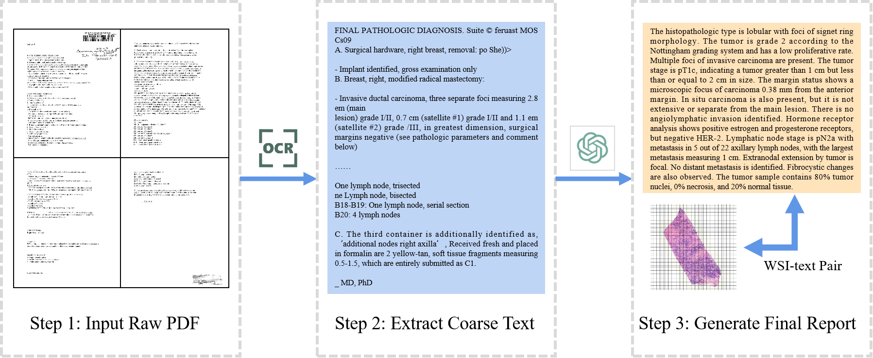
We notice that TCGA includes scanning copies of pathology reports in the format of PDF111https://portal.gdc.cancer.gov/. But they are too long with redundant information and present in a complex structure. Therefore, we propose a pipeline to extract and clean pathological texts from TCGA, which can convert complex PDF files to concise WSI-text pairs with the assistance of large language models (LLM), as shown in Fig. 1. The pairs we collected are also high-quality since they contain specialized knowledge from professional pathologists and have abundant clinical clues at slide-level, thus benefiting the construction of a more advanced visual-language model. To be specific, we first recognize WSI-PDF pairs from the TCGA dataset. Each pair contains a diagnostic whole slide image and its associated pathology report. We apply Optical Character Recognition (OCR) models [51] to extract text from the PDF files and obtain coarse reports. It is worth noting that OCR will transform all the characters in the PDF which inevitably results in text noise like page ID, signature, etc. Finally, we resort to LLMs like GPT-3.5-Turbo [6] to filter and summarize the coarse texts with prompts [37]. The proposed dataset is named as TCGA-PathoText containing total 9009 WSI-text pairs.
There are still challenges to achieving slide-level generation on the model end. Recent years have witnessed the boom of visual-language models in image captioning [55, 59, 46, 60]. And in the medical area, the generation of radiology reports has been explored by several works [25, 12, 36, 56]. However, the huge size of WSIs precludes existing machine learning techniques in image captioning from being transferred to the applications on WSIs. Existing image captioning methods for natural images and medical images all follow an end-to-end diagram where the entire image is encoded as the hidden state. It is unaffordable to directly process the WSIs with more than 10 gigapixels unless WSIs are resized or disentangled sacrificing much fine-grained information. In terms of the pathological approaches, PLIP [23] and MI-Zero [40] both adopt a CLIP-like [45] structure which largely improves the zero-shot ability but is still unable to directly generate pathology reports. To deal with this problem, we introduce a Multiple Instance Generation (MI-Gen) framework to achieve WSI-based report generation. It contains a visual extractor which encodes the non-overlap patches with the sliding window and a sequence-to-sequence generator to produce pathology texts.
In this work, our contributions can be concluded as follows:
-
1.
We propose a pipeline to curate high-quality WSI-text pairs from TCGA. The dataset (TCGA-PathoText) contains about ten thousand pairs which will be publicly accessible. It can potentially promote the development of visual-language models in pathology.
-
2.
We design a multiple instance generation framework (MI-Gen). By incorporating the position-aware module, our model is more sensitive to the spatial information in WSIs.
-
3.
We benchmark diverse sequence-to-sequence models on the subset of TCGA-PathoText. Downstream tasks are also included. As a generative model, our method is able to provide more abundant and accurate clinical results compared to previous MIL methods.
2 Related Works
2.1 Multiple Instance Learning for WSIs
Under multiple instance learning (MIL) frameworks, the slide is tiled where each small patch acts as an instance and the whole slide acts as a bag. Given only the bag label, the features of each instance are extracted and aggregated to make the prediction. MIL is essentially proposed to recognize the most relevant instance and thus previous works focus on the aggregation module. Wang et al. [57] adopted pooling operations to process instance features. They simply discard the less activated instance and keep the most discriminative one, which results in loss-of-details inevitably. Ilse et al. [24] proposed to use attention-weighted averaging as the aggregation operator. Chikontwe et al. [13] proposed a center loss to map the instance embeddings and reduce the intra-class variations. Some other works [62, 48, 58] specified the feature distances as attention scores and build convolutional neural networks to extract the instance-level correlation. Hou et al. [21] explicitly built the mutual-instance relationship of multi-resolution patches using a heterogeneous graph. Shao et al. [47] introduced the transformer-based model considering both the local context and the global correlation among the instances in the process of diagnosis. Our work also extracts features from non-overlap patches as individual instances from the whole bag.
2.2 Image Captioning
The most related task to ours is image captioning where textual descriptions are generated automatically given images. Most image captioning models are characterized by the encoder-decoder structure. The encoder is used to extract features from the image followed by the decoder which generates captions in sequence. Many works adopt a CNN-RNN framework [55, 16, 28, 41]. Some other researchers have managed to incorporate attention mechanisms into the encoding of images [59, 2, 60, 38]. Recently, transformer-based models [14, 20, 30, 22] have emerged for their powerful representation power and parallel nature. Typically, they consist of an encoder with a stack of self-attention and a decoder that learns to allocate attention to image and word embeddings.
Medical visual-language models have also boomed for generating radiology reports. Different from the captions that narrate natural images, radiology reports are longer and contain many terminologies in certain patterns. Jing et al. [25] first investigated the generation of radiology reports in a multi-task learning framework. Yuan et al. [61] extended the generation pipeline by incorporating multi-view image fusion. To fully exploit the intrinsic characteristics of radiology reports, Li et al. [32] incorporated retrieval modules and Jing et al. [26] explored the structure information. Also, transformer models have been investigated. Chen et al. [12] proposed a memory-drive transformer to generate radiology reports. Miura et al. [42] proposed new rewards to generate factually complete and consistent reports. Tsuneki et al. [53] tried to caption the whole slide image. Their captions are somewhat short and the model they use is not so advanced. In our work, the captions we collected are longer and more like a report which tends to give a gross description of the slide.
3 Multiple Instance Generation: Problem Formulation
In the multiple instance generation framework, the huge image can be seen as a bag which contains a group of instances which are non-overlap patches with a much smaller resolution. Denote the cohort of instances as where is the -th sample in the dataset and is the sequence length which is determined by the patch size. Usually, when the patch size is 256, the sequence length can be larger than ten thousand. A visual extractor is adopted to extract image embeddings from the patches, denoted as where is the embedding size.
A sequence-to-sequence model is incorporated to generate the target sequence where is the ground truth sequence length. Like previous text generation tasks, the encoder-decoder parameterized by is trained using language model objective which maximizes the sum of conditional possibilities of individual words in the sequence. Therefore, the negative log-likelihood (NLL) loss can be calculated as:
| (1) |
The probability of -th word is calculated based on the patch embeddings and the previous sequence .
4 Method
4.1 TCGA-PathoText Construction
The first step in constructing TCGA-PathoText is to find out the diagnostic slides and their corresponding pathology reports in the TCGA. The diagnostic slides from TCGA cover diverse disease types originating at different primary sites. The pathology reports in the format of PDF usually contain multiple pages. We use OCR methods to transform the files into text. However, the text is still noisy because the report itself contains redundant information, and OCR sometimes generates garbled code. Therefore, we apply LLMs to filter and summarize the report. Given the prompt of ”Please summarize the following pathology report:”, the text is filtered and shortened for training. This pipeline is completely automatic without the need for labeling with experts.
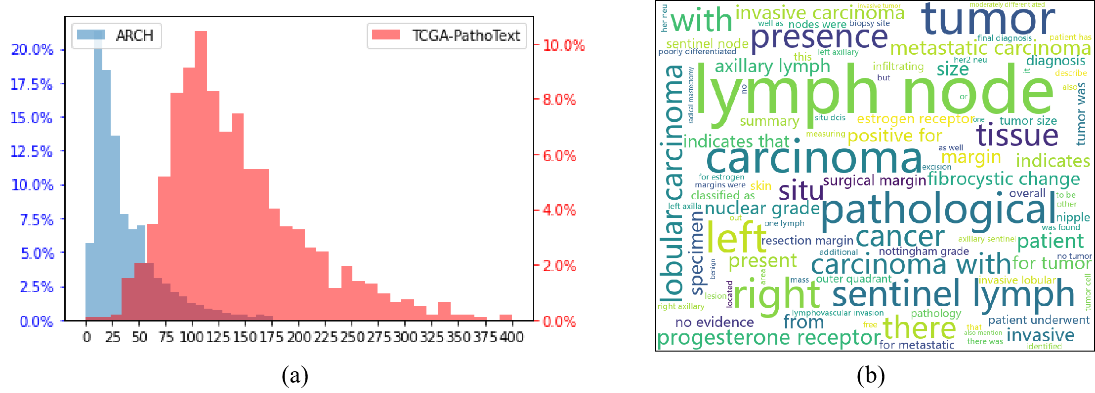
TCGA-PathoText contains 9009 WSI-text pairs in total. Our collected text is distilled from clinical pathology reports with well-aligned correspondence and abundant pathological content. Therefore, our text is much longer than ARCH [18] (patch-level descriptions collected from books and articles) by the histogram of text lengths, as illustrated in Fig. 2. TCGA-PathoText is broken down into different subsets according to the disease type. For instance, TCGA-BRCA, which is the largest subset in TCGA, contains 1098 cases of breast cancer. The corresponding WSI-text subset in our TCGA-PathoText is named as TCGA-PathoText(BRCA). More details about TCGA-PathoText are demonstrated in the Supplementary Materials.
4.2 WSI-text Generation
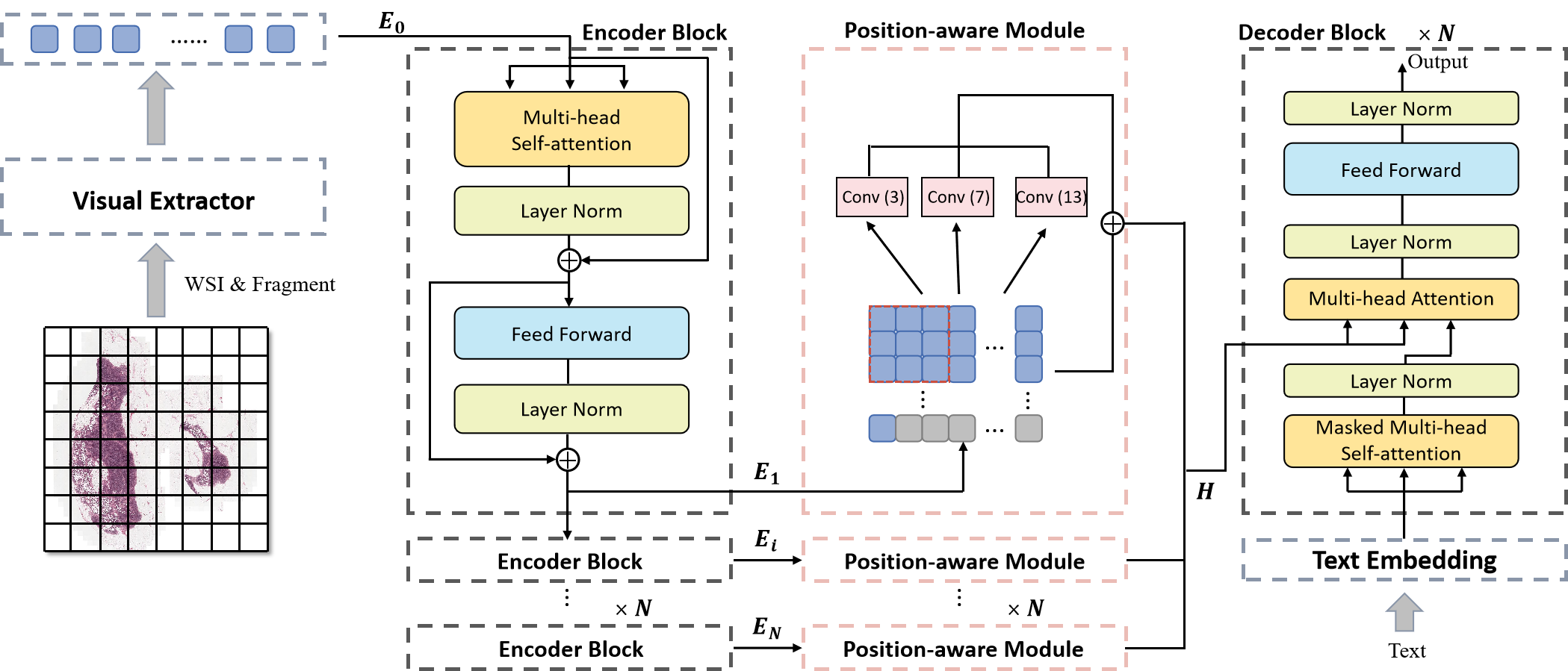
As illustrated in Fig. 3, our proposed generative model consists of two parts: the visual extractor, and the encoder-decoder. We incorporate the hierarchical position-aware module into Transformer encoder layers so as to strengthen the model awareness of spatial information in WSIs. The details are described below.
4.2.1 Visual Extractor
Given a whole-slide image with huge resolution, it is comprised of small patches . It is unable to accumulate gradients in the visual extractor due to the large size of WSIs. Therefore, we apply pre-trained neural networks to extract features from these patches. The visual extractor is non-trainable during training. The initial image embeddings are denoted as which are fed to the subsequent modules.
4.2.2 Encoder-Dcoder
Transformer has shown remarkable success in the past few years due to its strong capability of modeling the long sequence and interaction among individual tokens with self-attention. Therefore, we adopt Transformer as our backbone. Denote the Transformer encoder consists transformer layers. The embeddings are processed by the transformer layers iteratively as:
| (2) |
where refers to the -th transformer layer. In standard Transformer, the hidden state of the last encoder layer is directly fed to the cross-attention module with text embeddings for decoding. Whereas, we propose hierarchical position-aware modules to aggregate the embeddings from different encoder layers. The position-aware modules are inserted after each encoder layer so that more abundant context information is captured. Therefore, the hidden state H for decoding can be formulated as:
| (3) |
In terms of the decoder, we adopt the standard Transformer decoder where the inputs are the hidden state and text embeddings. In the training stage, a batch of samples is decoded in parallel with a subsequent mask. While in the inferring stage, the report is generated word by word. Therefore, the decoding will become more time-consuming if the target report is longer.
| Visual Extractor & Pre-train | Encoder-Decoder | BLEU-1 | BLEU-2 | BLEU-3 | BLEU-4 | METEOR | ROGUE |
|---|---|---|---|---|---|---|---|
| ResNet&ImageNet | CNN-RNN[55] | 0.334 | 0.209 | 0.122 | 0.074 | 0.137 | 0.248 |
| att-LSTM[59] | 0.367 | 0.234 | 0.128 | 0.085 | 0.151 | 0.262 | |
| vanilla Transformer [54] | 0.395 | 0.230 | 0.135 | 0.089 | 0.145 | 0.254 | |
| Mem-Transformer [12] | 0.317 | 0.207 | 0.136 | 0.091 | 0.129 | 0.270 | |
| Ours | 0.403 | 0.254 | 0.168 | 0.117 | 0.163 | 0.280 | |
| ViT&ImageNet | CNN-RNN[55] | 0.328 | 0.201 | 0.127 | 0.082 | 0.142 | 0.253 |
| att-LSTM[59] | 0.341 | 0.211 | 0.132 | 0.083 | 0.145 | 0.265 | |
| vanilla Transformer[54] | 0.346 | 0.216 | 0.137 | 0.091 | 0.149 | 0.273 | |
| Mem-Transformer[12] | 0.332 | 0.216 | 0.144 | 0.100 | 0.147 | 0.268 | |
| Ours | 0.380 | 0.252 | 0.169 | 0.110 | 0.157 | 0.279 | |
| ViT&HIPT | CNN-RNN[55] | 0.342 | 0.215 | 0.141 | 0.084 | 0.148 | 0.260 |
| att-LSTM[59] | 0.372 | 0.230 | 0.135 | 0.090 | 0.150 | 0.269 | |
| vanilla Transformer[54] | 0.383 | 0.237 | 0.151 | 0.096 | 0.152 | 0.264 | |
| Mem-Transformer[12] | 0.344 | 0.218 | 0.150 | 0.103 | 0.151 | 0.268 | |
| Ours | 0.446 | 0.286 | 0.183 | 0.120 | 0.171 | 0.271 |
Position-aware Module. Morphological and spatial information plays a key role when pathologists are diagnosing WSIs. It has been confirmed in [47] that convolutional layers improve position information awareness. Inspired by this, we incorporate a hierarchical position-aware module (PAM) into the encoding of image embeddings. Considering the varying sizes of tokens in WSIs, we also conduct padding so that the feature map can be reshaped for fitting convolutional layers. Convolution kernels of various sizes are adopted to capture heterogeneous spatial information.
In specific, denote the sequence of image embeddings as . Firstly, we pad the sequence until its length becomes a square number . Then, the 1-D sequence can be reshaped into 2-D space . We adopt several CNNs to process and aggregate the 2-D embeddings:
| (4) |
Heterogeneous spatial information from the CNNs with varying kernel sizes is summed together. Finally, the hidden state returns to 1-D space as a sequence for decoding. Our PAMs are inserted hierarchically after each encoder block, encoding abundant spatial information at different depth, which improves the awareness of spatial descriptions in the generated reports.
5 Experiments and Results
5.1 Implementation Details
Datasets. We train and validate our generative model on TCGA-PathoText(BRCA) which includes 845 pairs for training, 98 pairs for validating, and 98 pairs for testing. After training with WSI-text pairs from TCGA-PathoText, we evaluate the transfer performance on two WSI classification datasets: Camelyon-16 [4] for positive/negative binary classification and TCGA-BRCA for tumor subtyping. Camelyon-16 contains 400 hematoxylins and eosin (HE) stained whole slide images for breast cancer. Our model is finetuned on the official training set of which 10 is randomly selected to constitute the validation set. TCGA-BRCA contains 1041 whole slide images with the label of invasive ductal (IDC) or invasive lobular carcinoma (ILC). 10 of TCGA-BRCA is randomly selected for validation and inference.
Model Setting. The number of encoder layers and decoder layers are both 3. For self-attention modules, the number of heads is 4 and the size of embeddings is 512. Three CNNs are adopted in the PAM with the kernel size of 3, 7, and 13 respectively. We use Adam with the learning rate of 1e-4 to optimize the model. The weight decay is 5e-5. We adopt beam search with the size of 3 as the sampling method. Our model is trained on four A100-80G GPUs.
5.2 Baselines
Considering the huge size of WSIs, the visual extractors are frozen during training. Therefore, the patch embeddings for text generation are fixed. To explore the effect of patch embeddings on the quality of generated text. We explore different pre-trained visual extractors. Two kinds of visual models are chosen as our visual extractor: ResNet [19] which is composed of convolutional layers and ViT [15] which is based on Transformer. The output of the visual extractor is the image features with the length of 1024 and 384 for ResNet and ViT respectively. For the pre-training of visual extractors, we explore two strategies: 1) ImageNet (out-of-domain) pre-training on extensive natural images, and 2) hierarchical self-supervised learning with a pyramid transformer (HIPT) [10] on TCGA (in-domain).
We compare our encoder-decoder with several popular image captioning methods. There are two models which decode by LSTMs: CNN-RNN [55] and att-LSTM [59] . We also re-implemented two state-of-the-art Transformer-based models. The first is Vanilla Transformer which can be seen as the ablated version of our method. The other is Mem-Transformer [12] which is specially designed for medical report generation by incorporating a memory mechanism in the decoder.
5.3 Quantitative Results
As illustrated in Table 1, we adopt standard image captioning evaluation metrics to evaluate the generation performance: BLEU [43], METEOR [3], and ROUGE [33].
CNN-RNN adopts a standard encoder-decoder structure where images are first processed by convolutional layers and fed to an LSTM decoder for text generation. Att-LSTM adds an attention module between CNN and LSTM. We can observe that LSTM decoders perform relatively worse than transformer-based models. It shows the superiority of Transformer for modeling long sequences. Mem-Transformer incorporates the relational memory to implicitly model similar patterns in different reports. However, it does not show a better performance than Vanilla Transformer. The potential reason might be that pathology report in TCGA-PathoText is much longer than other medical image captioning datasets, thus having too heterogeneous structures to be memorized. In our model, we adopt hierarchical position-aware modules to capture spatial semantics in the WSI. This mechanism facilitates our model to achieve the best performance no matter what the visual extractor is.
In terms of visual extractors, ResNet and ViT do not show a large difference when they are pre-trained on ImageNet. But ViT pre-trained with HIPT demonstrates a significant improvement. It is not surprising because domain-aligned pre-training usually outperforms out-of-domain ImageNet pre-training in pathology [27].
5.4 Qualitative Results
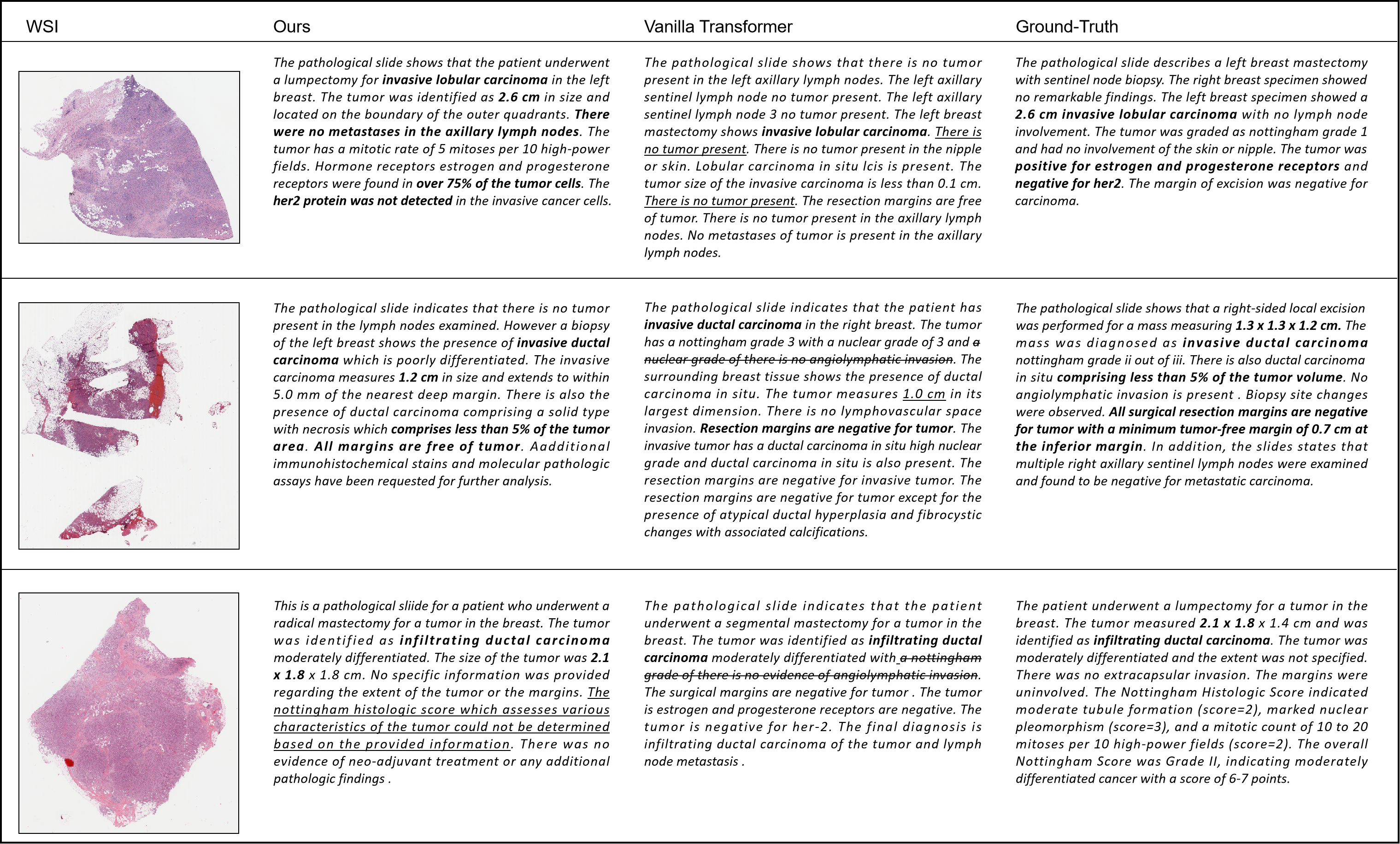
Three samples of WSIs with their corresponding pathology report from different models are illustrated in Figure 4. The first sentence is usually a general description of the WSI including the diagnosis of tumor subtype: ”invasive lobular carcinoma” or ”invasive lobular carcinoma”. We can find that our model and Vanilla Transformer both give accurate subtyping results for these three WSIs. To further measure their ability of subtyping quantitatively, we go through all the generated reports and related results will be presented in the following section 5.5.
The pathology reports from ground-truth contain spatial information like tumor size (”2.6 cm”, ”1.31.31.2 cm”). Vanilla Transformer fails to give a tumor spatial description (the first and third WSI) or generate not precise results (the second WSI), which reflects its disability to capture spatial features. Our model provides the perfectly right descriptions in the first and second row (”2.6 cm”, ”less than 5 of the tumor area”) and partially correct results for the others (”1.2 cm”, ”2.11.81.8 cm”). The potential reason why 3-D size can not be accurately predicted may be that the WSI can only provide 2-D spatial information. For the first WSI, though ground-truth only presents positive/negative results for hormone receptors, our model still has the tendency to generate spatial descriptions (”75 of the tumor cells”, ”not detected”). Although it is impossible to check their correctness, the corresponding binary classification of these spatial descriptions is exactly right. This comparison demonstrates that position-aware modules largely improve the spatial awareness of our model.
Our model also generates more medical terms that are consistent with the ground-truth. Vanilla Transformer sometimes produces results that are not only inconsistent with the ground-truth but also self-contradictory, as shown in the first row (”There is no tumor present” following the claim of ”invasive lobular carcinoma”). There are also repetitive sentences in reports from Vanilla Transformer. In terms of the Nottingham score of the third WSI, our model admits to being unable to make judgements. However, Vanilla Transformer generates text that is grammatically incorrect.
5.5 Transfer for WSI classification
Recent years have witnessed that large-scale visual-language pre-training benefits the performance on downstream tasks such as image classification [40, 45]. After training on the WSI-text pairs, we evaluate the transfer ability of our model on two WSI-level classification tasks: Camelyon-16 classification [4] and BRCA subtyping. We also re-produced several MIL approaches which are specially designed for WSI classification for comparison: AB-MIL [24], DS-MIL [29], CLAM-SB [39], and TransMIL [47]. They also segment the foreground regions and obtained pathological patches with a resolution of 256. These patches are transformed into embeddings by a visual extractor. For a fair comparison, we adopted the same ImageNet pre-trained ResNet-50 to extract image embeddings.
We adopt the encoder of our model with a trainable token attached at the head of the visual tokens. After encoding, this token is fed to a classifier to generate the classification result. Due to the existence of subtyping diagnosis in the text, we have also gone through all the reports and directly extracted semantic information for BRCA subtyping. Related results are demonstrated in Table 2. The first row is the result of the fully-supervised model which is trained under patch-level supervision so as to show the upper bound for the task.
For the unseen domain Camelyon-16, our model can achieve a comparable performance. In terms of TCGA-BRCA subtyping, our model can achieve the best AUC performance by fine-tuning compared to other MIL baselines. It is worth noting that we can also obtain state-of-the-art classification results by simply extracting semantic information from the generated report without any additional parameters or training, which shows the potential of WSI-text generation in computational pathology.
| Camelyon-16 | TCGA-BRCA | |||
| F1 | AUC | F1 | AUC | |
| Fully-supervised | 0.967 | 0.992 | - | - |
| Max-pooling | 0.805 | 0.824 | 0.644 | 0.826 |
| AB-MIL [24] | 0.828 | 0.851 | 0.771 | 0.869 |
| DS-MIL [29] | 0.857 | 0.892 | 0.775 | 0.875 |
| CLAM-SB [39] | 0.839 | 0.875 | 0.797 | 0.879 |
| TransMIL [47] | 0.846 | 0.883 | 0.806 | 0.889 |
| Fine-tuning | 0.841 | 0.855 | 0.780 | 0.897 |
| Semantic Extraction | - | - | 0.838 | - |
5.6 Effect of Spatial Information
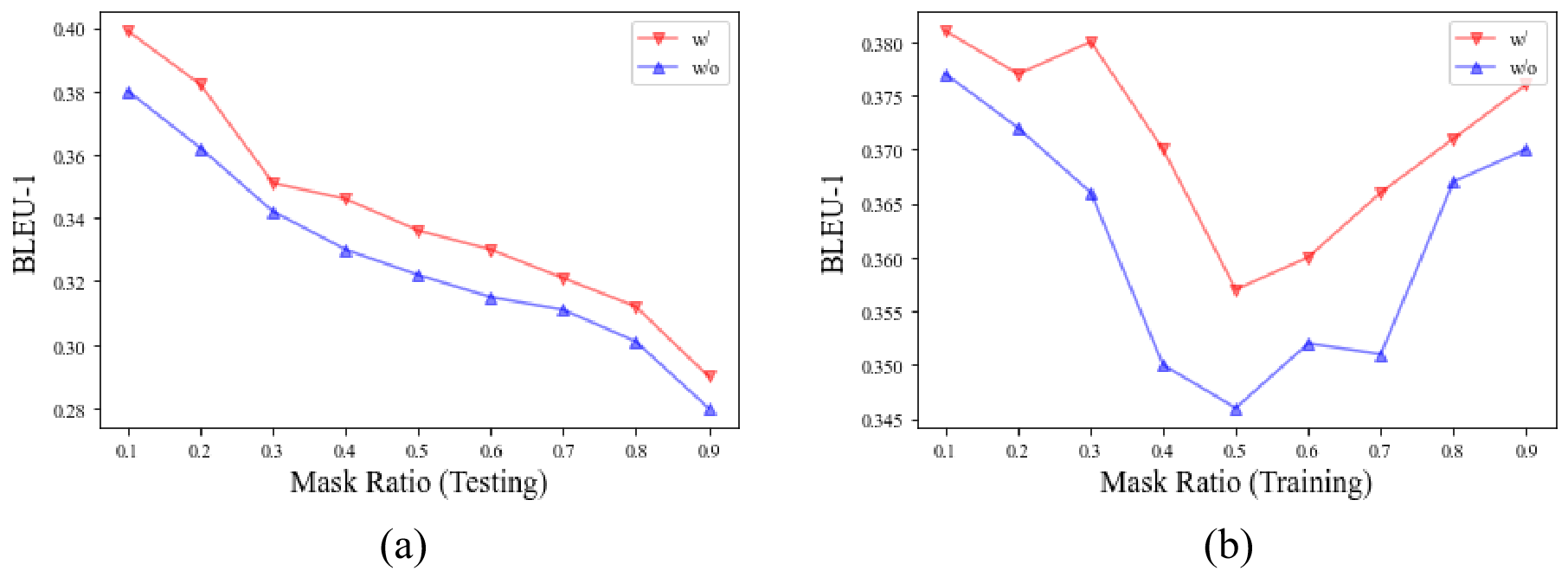
In the clinical scenario, an experienced pathologist allocates more attention to certain regions of interest instead of carefully checking all the patches in the WSIs. Therefore, to further analyze the robustness of our model and the proposed position-aware module, we add noise to the input by randomly masking some patches in the WSI. The quantitative results are demonstrated in Fig. 5: ’w/’ represents our proposed model with position-aware modules while ’w/o’ refers to the version without the position-aware module.
As shown in Fig. 5 (a), with the growth of mask ratio in test data, the performance drops continually. However, the performance of our model shows a consistent superiority over the model without the position-aware module. The position information contributes to an improvement of about on BLEU-1 over text generation. When we conduct masking in the training stage, the overall performance does not show a consistent loss. Our model is more stable and still demonstrates its robustness. The spatial information enhances the model performance by around of BLEU-1.
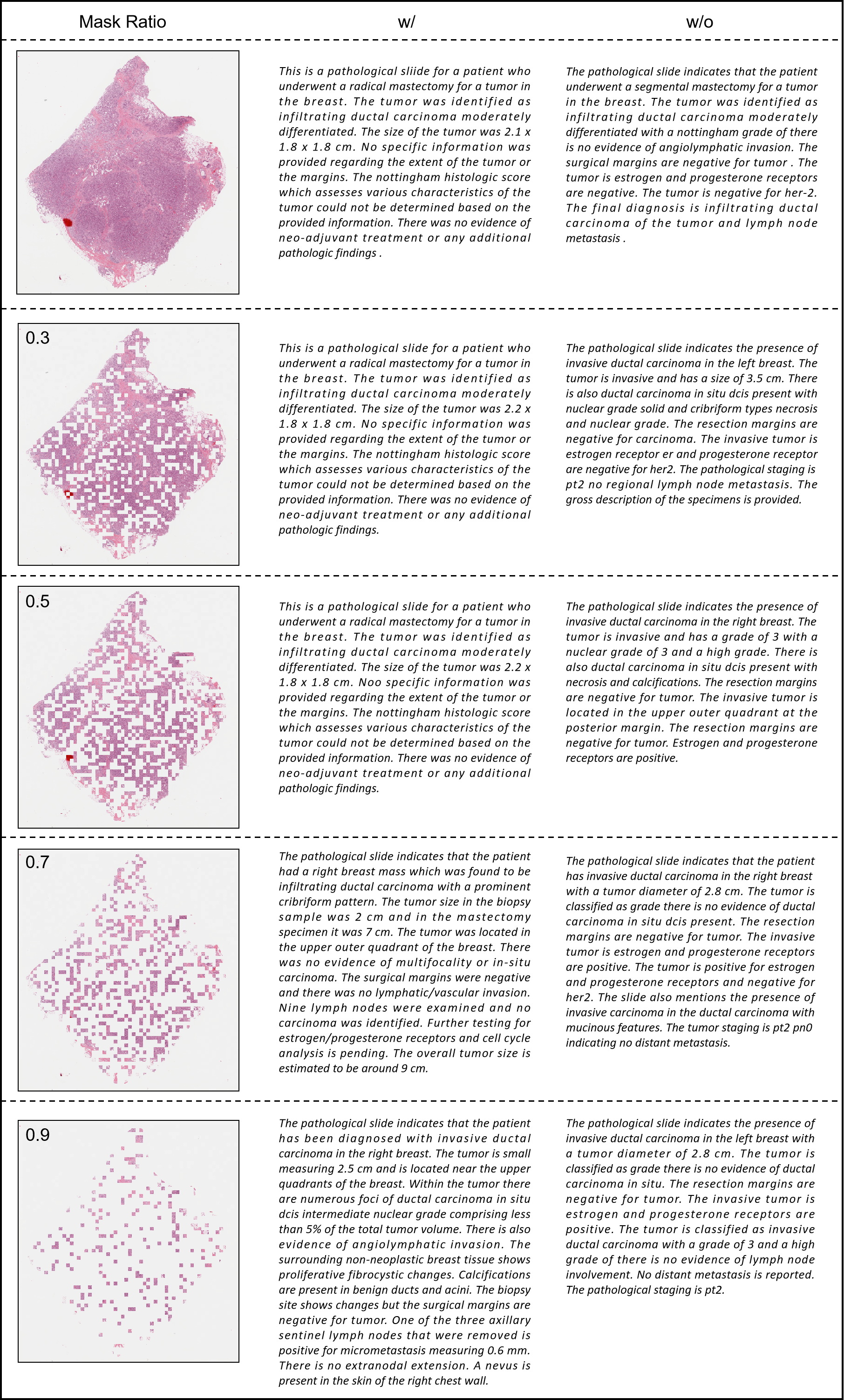
We also visualize the results, as illustrated in Fig. 6. The first column presents the masked WSIs with a higher mask ratio from top to down. We can observe that our model can maintain the report structure when the mask rate is low (0.3 and 0.5) sacrificing a little accuracy in tumor size identification (”2.21.81.8 cm” versus ”2.11.81.8 cm”). At the same time, the version without position information fails to describe the size of the carcinoma and even gramma error appears. when the mask rate is very high (0.7 and 0.9), our model also fails to give a relatively accurate spatial description. And the model without position information generates more language-related errors. More ablation studies can be observed in Supplementary Materials.
6 Limitations and Future Work
Although it is a bit regretful that we are not able to train a universal model on the whole TCGA-PathoText due to the GPU limitation, we are still confident that our pipeline can be scaled to a larger domain. By bridging the gap among diverse disease types, we can obtain a more powerful pathological model in the future. In terms of the limitations of our model, we notice that the decoding time is long because the target report has many more tokens than patch-level descriptions. The average decoding time is nearly one hour for our validation set. Although it is clinically acceptable to wait for some time to receive the diagnosis results, we still hope to make decoding faster which at least helps to save costs when we are training at a very large-scale dataset. In addition, since the visual extractor is frozen during training, image embeddings and text embeddings may not be well-aligned. Visual features are either obtained from well-constructed backbones pre-trained on natural images or from self-supervised models pre-trained in pathology datasets. Since they are not adapted to the generative task, these task-agnostic features may hinder the performance.
7 Conclusion
In this paper, we have shown the feasibility of pathology report generation. On the data end, we collected nearly 10000 WSI-text pairs by transforming PDF files in TCGA into concise and comprehensive pathology reports with the aid of LLMs. Our proposed TCGA-PathoText is the largest WSI-text dataset so far to the best of our knowledge and is going to be available to the public. On the model end, we introduce MI-Gen as a generative model for WSI-level descriptions. By benchmarking different baselines on the subset of TCGA-PathoText, we also reveal the superiority of our model in being aware of the spatial information among the WSIs. Furthermore, the transfer performance on BRCA subtyping reflects the advancement of WSI-text generation since the pathology reports contain diverse clinical clues which have covered previous discriminative tasks. Many directions deserve exploring in the future including considering more WSI-text pairs, improving the alignment of visual and textual embeddings, and boosting the decoding efficiency in text generation. In addition, other fields that utilize high-resolution images like remote sensing can be inspired by our work.
References
- Abousamra et al. [2021] Shahira Abousamra, David Belinsky, John Van Arnam, Felicia Allard, Eric Yee, Rajarsi Gupta, Tahsin Kurc, Dimitris Samaras, Joel Saltz, and Chao Chen. Multi-class cell detection using spatial context representation. In Proceedings of the IEEE/CVF International Conference on Computer Vision, pages 4005–4014, 2021.
- Anderson et al. [2018] Peter Anderson, Xiaodong He, Chris Buehler, Damien Teney, Mark Johnson, Stephen Gould, and Lei Zhang. Bottom-up and top-down attention for image captioning and visual question answering. In 2018 IEEE/CVF Conference on Computer Vision and Pattern Recognition, pages 6077–6086, 2018.
- Banerjee and Lavie [2005] Satanjeev Banerjee and Alon Lavie. Meteor: An automatic metric for mt evaluation with improved correlation with human judgments. In Proceedings of the acl workshop on intrinsic and extrinsic evaluation measures for machine translation and/or summarization, pages 65–72, 2005.
- Bejnordi et al. [2017] Babak Ehteshami Bejnordi, Mitko Veta, Paul Johannes Van Diest, Bram Van Ginneken, Nico Karssemeijer, Geert Litjens, Jeroen AWM Van Der Laak, Meyke Hermsen, Quirine F Manson, Maschenka Balkenhol, et al. Diagnostic assessment of deep learning algorithms for detection of lymph node metastases in women with breast cancer. Jama, 318(22):2199–2210, 2017.
- Bergner et al. [2022] Benjamin Bergner, Christoph Lippert, and Aravindh Mahendran. Iterative patch selection for high-resolution image recognition. arXiv preprint arXiv:2210.13007, 2022.
- Brown et al. [2020] Tom Brown, Benjamin Mann, Nick Ryder, Melanie Subbiah, Jared D Kaplan, Prafulla Dhariwal, Arvind Neelakantan, Pranav Shyam, Girish Sastry, Amanda Askell, et al. Language models are few-shot learners. Advances in neural information processing systems, 33:1877–1901, 2020.
- Buckley et al. [2012] Julliette M Buckley, Suzanne B Coopey, John Sharko, Fernanda Polubriaginof, Brian Drohan, Ahmet K Belli, Elizabeth MH Kim, Judy E Garber, Barbara L Smith, Michele A Gadd, et al. The feasibility of using natural language processing to extract clinical information from breast pathology reports. Journal of pathology informatics, 3(1):23, 2012.
- Chan et al. [2019] Lyndon Chan, Mahdi S Hosseini, Corwyn Rowsell, Konstantinos N Plataniotis, and Savvas Damaskinos. Histosegnet: Semantic segmentation of histological tissue type in whole slide images. In Proceedings of the IEEE/CVF International Conference on Computer Vision, pages 10662–10671, 2019.
- Chen et al. [2023] Pingyi Chen, Chenglu Zhu, Zhongyi Shui, Jiatong Cai, Sunyi Zheng, Shichuan Zhang, and Lin Yang. Exploring unsupervised cell recognition with prior self-activation maps. In Medical Image Computing and Computer Assisted Intervention – MICCAI 2023, pages 559–568, Cham, 2023. Springer Nature Switzerland.
- Chen et al. [2022] Richard J Chen, Chengkuan Chen, Yicong Li, Tiffany Y Chen, Andrew D Trister, Rahul G Krishnan, and Faisal Mahmood. Scaling vision transformers to gigapixel images via hierarchical self-supervised learning. In Proceedings of the IEEE/CVF Conference on Computer Vision and Pattern Recognition, pages 16144–16155, 2022.
- Chen and Lu [2023] Yuan-Chih Chen and Chun-Shien Lu. Rankmix: Data augmentation for weakly supervised learning of classifying whole slide images with diverse sizes and imbalanced categories. In Proceedings of the IEEE/CVF Conference on Computer Vision and Pattern Recognition, pages 23936–23945, 2023.
- Chen et al. [2020] Zhihong Chen, Yan Song, Tsung-Hui Chang, and Xiang Wan. Generating radiology reports via memory-driven transformer. In Proceedings of the 2020 Conference on Empirical Methods in Natural Language Processing, 2020.
- Chikontwe et al. [2020] Philip Chikontwe, Meejeong Kim, Soo Jeong Nam, Heounjeong Go, and Sang Hyun Park. Multiple instance learning with center embeddings for histopathology classification. In International Conference on Medical Image Computing and Computer-Assisted Intervention, pages 519–528. Springer, 2020.
- Cornia et al. [2020] Marcella Cornia, Matteo Stefanini, Lorenzo Baraldi, and Rita Cucchiara. Meshed-Memory Transformer for Image Captioning. In Proceedings of the IEEE/CVF Conference on Computer Vision and Pattern Recognition, 2020.
- Dosovitskiy et al. [2020] Alexey Dosovitskiy, Lucas Beyer, Alexander Kolesnikov, Dirk Weissenborn, Xiaohua Zhai, Thomas Unterthiner, Mostafa Dehghani, Matthias Minderer, Georg Heigold, Sylvain Gelly, et al. An image is worth 16x16 words: Transformers for image recognition at scale. arXiv preprint arXiv:2010.11929, 2020.
- Fang et al. [2015] Hao Fang, Saurabh Gupta, Forrest Iandola, Rupesh K Srivastava, Li Deng, Piotr Dollár, Jianfeng Gao, Xiaodong He, Margaret Mitchell, John C Platt, et al. From captions to visual concepts and back. In Proceedings of the IEEE conference on computer vision and pattern recognition, pages 1473–1482, 2015.
- Farahani et al. [2015] Navid Farahani, Anil V Parwani, and Liron Pantanowitz. Whole slide imaging in pathology: advantages, limitations, and emerging perspectives. Pathology and Laboratory Medicine International, pages 23–33, 2015.
- Gamper and Rajpoot [2021] Jevgenij Gamper and Nasir Rajpoot. Multiple instance captioning: Learning representations from histopathology textbooks and articles. In Proceedings of the IEEE/CVF conference on computer vision and pattern recognition, pages 16549–16559, 2021.
- He et al. [2016] Kaiming He, Xiangyu Zhang, Shaoqing Ren, and Jian Sun. Deep residual learning for image recognition. In Proceedings of the IEEE conference on computer vision and pattern recognition, pages 770–778, 2016.
- Herdade et al. [2019] Simao Herdade, Armin Kappeler, Kofi Boakye, and Joao Soares. Image captioning: Transforming objects into words. In Advances in Neural Information Processing Systems. Curran Associates, Inc., 2019.
- Hou et al. [2022] Wentai Hou, Lequan Yu, Chengxuan Lin, Helong Huang, Rongshan Yu, Jing Qin, and Liansheng Wang. H^ 2-mil: Exploring hierarchical representation with heterogeneous multiple instance learning for whole slide image analysis. In Proceedings of the AAAI conference on artificial intelligence, pages 933–941, 2022.
- Huang et al. [2019] L. Huang, W. Wang, J. Chen, and X. Wei. Attention on attention for image captioning. In 2019 IEEE/CVF International Conference on Computer Vision (ICCV), pages 4633–4642, Los Alamitos, CA, USA, 2019. IEEE Computer Society.
- Huang et al. [2023] Zhi Huang, Federico Bianchi, Mert Yuksekgonul, Thomas J Montine, and James Zou. A visual–language foundation model for pathology image analysis using medical twitter. Nature medicine, 29(9):2307–2316, 2023.
- Ilse et al. [2018] Maximilian Ilse, Jakub Tomczak, and Max Welling. Attention-based deep multiple instance learning. In International conference on machine learning, pages 2127–2136. PMLR, 2018.
- Jing et al. [2018] Baoyu Jing, Pengtao Xie, and Eric Xing. On the automatic generation of medical imaging reports. In Proceedings of the 56th Annual Meeting of the Association for Computational Linguistics (Volume 1: Long Papers), pages 2577–2586, Melbourne, Australia, 2018. Association for Computational Linguistics.
- Jing et al. [2019] Baoyu Jing, Zeya Wang, and Eric Xing. Show, describe and conclude: On exploiting the structure information of chest X-ray reports. In Proceedings of the 57th Annual Meeting of the Association for Computational Linguistics, pages 6570–6580. Association for Computational Linguistics, 2019.
- Kang et al. [2023] Mingu Kang, Heon Song, Seonwook Park, Donggeun Yoo, and Sérgio Pereira. Benchmarking self-supervised learning on diverse pathology datasets. In Proceedings of the IEEE/CVF Conference on Computer Vision and Pattern Recognition, pages 3344–3354, 2023.
- Karpathy and Fei-Fei [2015] Andrej Karpathy and Li Fei-Fei. Deep visual-semantic alignments for generating image descriptions. In Proceedings of the IEEE conference on computer vision and pattern recognition, pages 3128–3137, 2015.
- Li et al. [2021] Bin Li, Yin Li, and Kevin W Eliceiri. Dual-stream multiple instance learning network for whole slide image classification with self-supervised contrastive learning. In Proceedings of the IEEE/CVF conference on computer vision and pattern recognition, pages 14318–14328, 2021.
- Li et al. [2019] Guang Li, Linchao Zhu, Ping Liu, and Yi Yang. Entangled transformer for image captioning. 2019 IEEE/CVF International Conference on Computer Vision (ICCV), pages 8927–8936, 2019.
- Li et al. [2023] Honglin Li, Chenglu Zhu, Yunlong Zhang, Yuxuan Sun, Zhongyi Shui, Wenwei Kuang, Sunyi Zheng, and Lin Yang. Task-specific fine-tuning via variational information bottleneck for weakly-supervised pathology whole slide image classification. In Proceedings of the IEEE/CVF Conference on Computer Vision and Pattern Recognition, pages 7454–7463, 2023.
- Li et al. [2018] Yuan Li, Xiaodan Liang, Zhiting Hu, and Eric P Xing. Hybrid retrieval-generation reinforced agent for medical image report generation. In Advances in Neural Information Processing Systems. Curran Associates, Inc., 2018.
- Lin [2004] Chin-Yew Lin. Rouge: A package for automatic evaluation of summaries. In Text summarization branches out, pages 74–81, 2004.
- Lin et al. [2019] Huangjing Lin, Hao Chen, Simon Graham, Qi Dou, Nasir Rajpoot, and Pheng-Ann Heng. Fast scannet: Fast and dense analysis of multi-gigapixel whole-slide images for cancer metastasis detection. IEEE transactions on medical imaging, 38(8):1948–1958, 2019.
- Lin et al. [2023] Tiancheng Lin, Zhimiao Yu, Hongyu Hu, Yi Xu, and Chang-Wen Chen. Interventional bag multi-instance learning on whole-slide pathological images. In Proceedings of the IEEE/CVF Conference on Computer Vision and Pattern Recognition, pages 19830–19839, 2023.
- Liu et al. [2019] Guanxiong Liu, Tzu-Ming Harry Hsu, Matthew McDermott, Willie Boag, Wei-Hung Weng, Peter Szolovits, and Marzyeh Ghassemi. Clinically accurate chest x-ray report generation. In Machine Learning for Healthcare Conference, pages 249–269. PMLR, 2019.
- Liu et al. [2023] Pengfei Liu, Weizhe Yuan, Jinlan Fu, Zhengbao Jiang, Hiroaki Hayashi, and Graham Neubig. Pre-train, prompt, and predict: A systematic survey of prompting methods in natural language processing. ACM Computing Surveys, 55(9):1–35, 2023.
- Lu et al. [2017] Jiasen Lu, Caiming Xiong, Devi Parikh, and Richard Socher. Knowing when to look: Adaptive attention via a visual sentinel for image captioning. In 2017 IEEE Conference on Computer Vision and Pattern Recognition (CVPR), pages 3242–3250, 2017.
- Lu et al. [2021] Ming Y Lu, Drew FK Williamson, Tiffany Y Chen, Richard J Chen, Matteo Barbieri, and Faisal Mahmood. Data-efficient and weakly supervised computational pathology on whole-slide images. Nature biomedical engineering, 5(6):555–570, 2021.
- Lu et al. [2023] Ming Y Lu, Bowen Chen, Andrew Zhang, Drew FK Williamson, Richard J Chen, Tong Ding, Long Phi Le, Yung-Sung Chuang, and Faisal Mahmood. Visual language pretrained multiple instance zero-shot transfer for histopathology images. In Proceedings of the IEEE/CVF Conference on Computer Vision and Pattern Recognition, pages 19764–19775, 2023.
- Mao et al. [2014] Junhua Mao, Wei Xu, Yi Yang, Jiang Wang, Zhiheng Huang, and Alan Yuille. Deep captioning with multimodal recurrent neural networks (m-rnn). arXiv preprint arXiv:1412.6632, 2014.
- Miura et al. [2021] Yasuhide Miura, Yuhao Zhang, Emily Tsai, Curtis Langlotz, and Dan Jurafsky. Improving factual completeness and consistency of image-to-text radiology report generation. In Proceedings of the 2021 Conference of the North American Chapter of the Association for Computational Linguistics: Human Language Technologies, Online, 2021. Association for Computational Linguistics.
- Papineni et al. [2002] Kishore Papineni, Salim Roukos, Todd Ward, and Wei-Jing Zhu. Bleu: a method for automatic evaluation of machine translation. In Proceedings of the 40th annual meeting of the Association for Computational Linguistics, pages 311–318, 2002.
- Qu et al. [2020] Hui Qu, Pengxiang Wu, Qiaoying Huang, Jingru Yi, Zhennan Yan, Kang Li, Gregory M. Riedlinger, Subhajyoti De, Shaoting Zhang, and Dimitris N. Metaxas. Weakly Supervised Deep Nuclei Segmentation Using Partial Points Annotation in Histopathology Images. IEEE transactions on medical imaging, 39(11):3655–3666, 2020.
- Radford et al. [2021] Alec Radford, Jong Wook Kim, Chris Hallacy, Aditya Ramesh, Gabriel Goh, Sandhini Agarwal, Girish Sastry, Amanda Askell, Pamela Mishkin, Jack Clark, et al. Learning transferable visual models from natural language supervision. In International conference on machine learning, pages 8748–8763. PMLR, 2021.
- Rennie et al. [2017] Steven J Rennie, Etienne Marcheret, Youssef Mroueh, Jerret Ross, and Vaibhava Goel. Self-critical sequence training for image captioning. In Proceedings of the IEEE conference on computer vision and pattern recognition, pages 7008–7024, 2017.
- Shao et al. [2021] Zhuchen Shao, Hao Bian, Yang Chen, Yifeng Wang, Jian Zhang, Xiangyang Ji, et al. Transmil: Transformer based correlated multiple instance learning for whole slide image classification. Advances in Neural Information Processing Systems, 34:2136–2147, 2021.
- Sharma et al. [2021] Yash Sharma, Aman Shrivastava, Lubaina Ehsan, Christopher A Moskaluk, Sana Syed, and Donald Brown. Cluster-to-conquer: A framework for end-to-end multi-instance learning for whole slide image classification. In Medical Imaging with Deep Learning, pages 682–698. PMLR, 2021.
- Shui et al. [2022] Zhongyi Shui, Shichuan Zhang, Chenglu Zhu, Bingchuan Wang, Pingyi Chen, Sunyi Zheng, and Lin Yang. End-to-end cell recognition by point annotation. In Medical Image Computing and Computer Assisted Intervention – MICCAI 2022, pages 109–118, Cham, 2022. Springer Nature Switzerland.
- Sirinukunwattana et al. [2017] Korsuk Sirinukunwattana, Josien PW Pluim, Hao Chen, Xiaojuan Qi, Pheng-Ann Heng, Yun Bo Guo, Li Yang Wang, Bogdan J Matuszewski, Elia Bruni, Urko Sanchez, et al. Gland segmentation in colon histology images: The glas challenge contest. Medical image analysis, 35:489–502, 2017.
- Smith [2007] Ray Smith. An overview of the tesseract ocr engine. In Ninth international conference on document analysis and recognition (ICDAR 2007), pages 629–633. IEEE, 2007.
- Tokunaga et al. [2019] Hiroki Tokunaga, Yuki Teramoto, Akihiko Yoshizawa, and Ryoma Bise. Adaptive weighting multi-field-of-view cnn for semantic segmentation in pathology. In Proceedings of the IEEE/CVF Conference on Computer Vision and Pattern Recognition, pages 12597–12606, 2019.
- Tsuneki and Kanavati [2022] Masayuki Tsuneki and Fahdi Kanavati. Inference of captions from histopathological patches. In International Conference on Medical Imaging with Deep Learning, pages 1235–1250. PMLR, 2022.
- Vaswani et al. [2017] Ashish Vaswani, Noam Shazeer, Niki Parmar, Jakob Uszkoreit, Llion Jones, Aidan N Gomez, Łukasz Kaiser, and Illia Polosukhin. Attention is all you need. Advances in neural information processing systems, 30, 2017.
- Vinyals et al. [2015] Oriol Vinyals, Alexander Toshev, Samy Bengio, and Dumitru Erhan. Show and tell: A neural image caption generator. In Proceedings of the IEEE conference on computer vision and pattern recognition, pages 3156–3164, 2015.
- Wang et al. [2023] Sheng Wang, Zihao Zhao, Xi Ouyang, Qian Wang, and Dinggang Shen. Chatcad: Interactive computer-aided diagnosis on medical image using large language models. arXiv preprint arXiv:2302.07257, 2023.
- Wang et al. [2018] Xinggang Wang, Yongluan Yan, Peng Tang, Xiang Bai, and Wenyu Liu. Revisiting multiple instance neural networks. Pattern Recognition, 74:15–24, 2018.
- Xie et al. [2020] Chensu Xie, Hassan Muhammad, Chad M Vanderbilt, Raul Caso, Dig Vijay Kumar Yarlagadda, Gabriele Campanella, and Thomas J Fuchs. Beyond classification: Whole slide tissue histopathology analysis by end-to-end part learning. In Medical Imaging with Deep Learning, pages 843–856. PMLR, 2020.
- Xu et al. [2015] Kelvin Xu, Jimmy Ba, Ryan Kiros, Kyunghyun Cho, Aaron Courville, Ruslan Salakhudinov, Rich Zemel, and Yoshua Bengio. Show, attend and tell: Neural image caption generation with visual attention. In International conference on machine learning, pages 2048–2057. PMLR, 2015.
- You et al. [2016] Quanzeng You, Hailin Jin, Zhaowen Wang, Chen Fang, and Jiebo Luo. Image captioning with semantic attention. In Proceedings of the IEEE conference on computer vision and pattern recognition, pages 4651–4659, 2016.
- Yuan et al. [2019] Jianbo Yuan, Haofu Liao, Rui Luo, and Jiebo Luo. Automatic radiology report generation based on multi-view image fusion and medical concept enrichment. In Medical Image Computing and Computer Assisted Intervention – MICCAI 2019, pages 721–729, 2019.
- Zhang et al. [2022] Hongrun Zhang, Yanda Meng, Yitian Zhao, Yihong Qiao, Xiaoyun Yang, Sarah E Coupland, and Yalin Zheng. Dtfd-mil: Double-tier feature distillation multiple instance learning for histopathology whole slide image classification. In Proceedings of the IEEE/CVF Conference on Computer Vision and Pattern Recognition, pages 18802–18812, 2022.
- Zhang et al. [2019] Zizhao Zhang, Pingjun Chen, Mason McGough, Fuyong Xing, Chunbao Wang, Marilyn Bui, Yuanpu Xie, Manish Sapkota, Lei Cui, Jasreman Dhillon, et al. Pathologist-level interpretable whole-slide cancer diagnosis with deep learning. Nature Machine Intelligence, 1(5):236–245, 2019.