SA-Med2D-20M Dataset: Segment Anything in 2D Medical Imaging with 20 Million masks
Abstract
Segment Anything Model (SAM) has achieved impressive results for natural image segmentation with input prompts such as points and bounding boxes. Its success largely owes to massive labeled training data. However, directly applying SAM to medical image segmentation cannot perform well because SAM lacks medical knowledge — it does not use medical images for training. To incorporate medical knowledge into SAM, we introduce SA-Med2D-20M, a large-scale segmentation dataset of 2D medical images built upon numerous public and private datasets. It consists of 4.6 million 2D medical images and 19.7 million corresponding masks, covering almost the whole body and showing significant diversity. This paper describes all the datasets collected in SA-Med2D-20M and details how to process these datasets. Furthermore, comprehensive statistics of SA-Med2D-20M are presented to facilitate the better use of our dataset, which can help the researchers build medical vision foundation models or apply their models to downstream medical applications. We hope that the large scale and diversity of SA-Med2D-20M can be leveraged to develop medical artificial intelligence for enhancing diagnosis, medical image analysis, knowledge sharing, and education. The data with the redistribution license is publicly available at https://github.com/OpenGVLab/SAM-Med2D.
1 Introduction
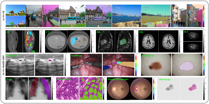
Medical image segmentation plays a crucial role in diagnosing, radiotherapy planning, treating, and further medical research huang2023stunet ; isensee2021nnu ; NEURIPS2022_ee604e1b . Accurate segmentation for medical images is a challenging task as data modalities and targets can vary significantly in clinical practice. To deal with the complex scenarios in medical image segmentation, building medical vision foundation models is essential. Foundation models are usually trained on large-scale datasets and can excel in various data modalities and targets. As such, these models can help doctors in precisely identifying and locating areas of pathology in complex clinical scenarios, enabling more accurate diagnosis and treatment. The key to successful general-purpose foundation models is broad and diverse datasets, which has been verified in natural image recognition (e.g., CLIP radford2021learning , SAM kirillov2023segment ), nature language processing (NLP, e.g., LLaMA touvron2023llama ; touvron2023llama2 ), and vision-language modeling (e.g., LLaVa liu2023visual , GPT4 openai2023gpt4 ). For instance, the Segment Anything Model (SAM) is trained on large datasets (1.1 billion masks) of which data are scraped from the Internet. However, due to the significant domain gap between natural images and medical ones, SAM cannot achieve satisfying segmentation results on multi-modal medical datasets. The huge domain gap can be attributed to the data collection methods: medical images are collected from specific protocols and scanners and are presented as different modalities (electrons, lasers, X-rays, ultrasound, nuclear physics, and magnetic resonance) due to their particular clinical purpose. As a result, the visual appearance of natural images and medical images is substantially different as reviewed in Figure 1(a).
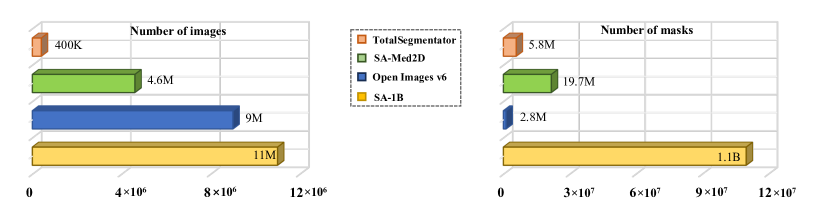
Building large-scale medical image datasets with annotated masks is the key to training a robust medical vision foundation model, which can accurately segment the target region that doctors or researchers are interested in. However, publicly available datasets for medical image segmentation are often limited in data size. Even the largest medical datasets are a fraction of the size and diversity of benchmark datasets in general computer vision kirillov2023segment ; wu2019tencent and NLP lehmann2015dbpedia ; muhleisen2012web . Figure 1(b) shows the data size gap between different research areas. More importantly, these public datasets often focus on a single environment, modality, or anatomical structure, thus with limited diversity. It is vital to build a large-scale dataset with diverse medical images, which will help to advance medical image analysis as successfully as computer vision and NLP.
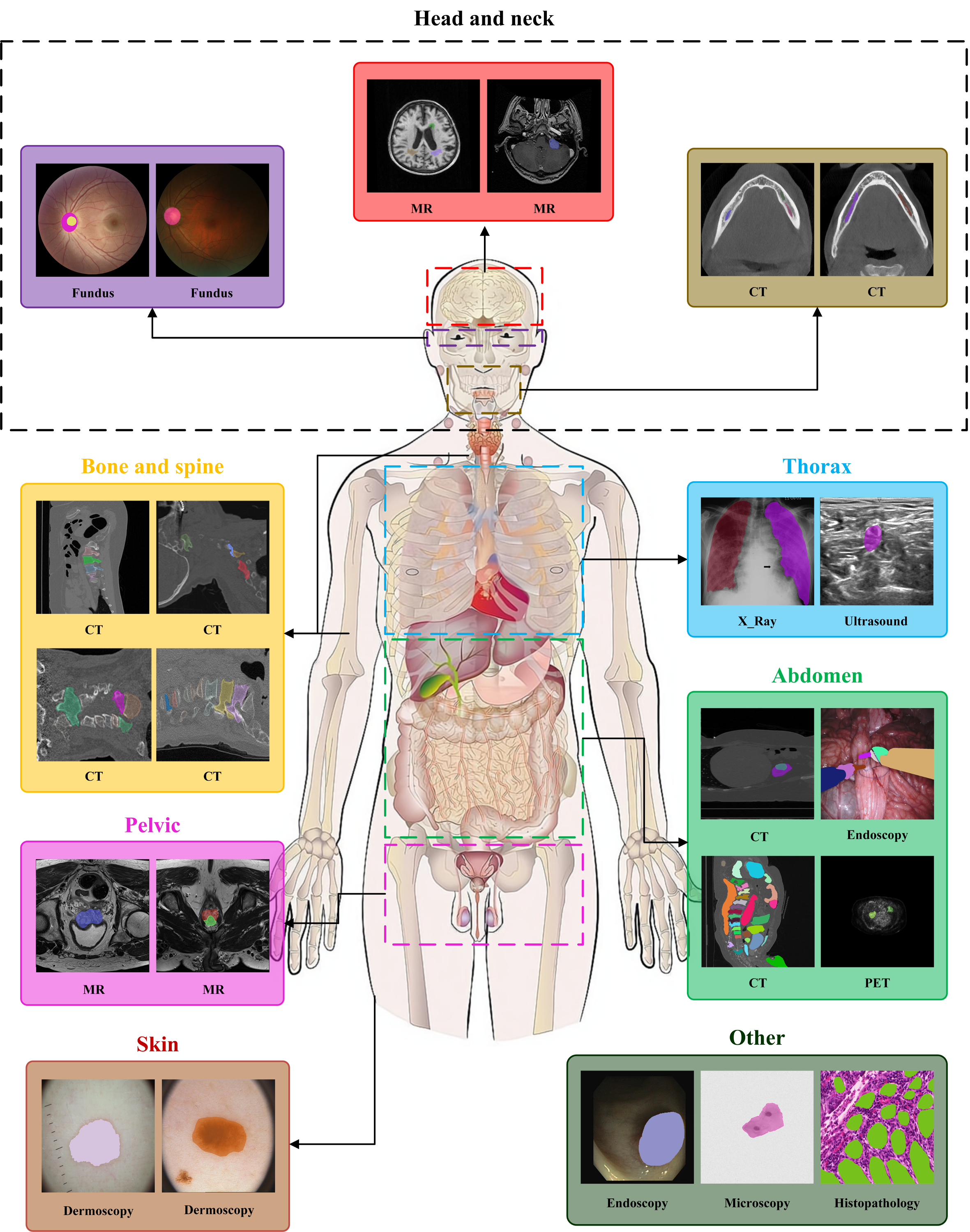
Collecting a large-scale medical image dataset from scratch and annotating them carefully by specialists is infeasible PARK202035 because accessing medical image data is not as easy as natural images and processing per-voxel annotations is expensive and time-consuming. To avoid the problems of data access and handcraft annotation, we introduce a new medical image dataset called “SA-Med2D-20M” which is built upon numerous existing datasets for the segmentation of molecules and cells to organ systems and the full body. Specifically, we take advantage of numerous public datasets on the web sources (TCIA 111https://www.cancerimagingarchive.net, OpenNeuro 222https://openneuro.org, NITRC 333https://www.nitrc.org, Grand Challenge 444https://grand-challenge.org, Synapse 555https://www.synapse.org, CodaLab 666https://codalab.lisn.upsaclay.fr, GitHub 777https://github.com, etc) to collect as many public medical segmentation datasets as possible. Thanks to these resources, SA-Med2D-20M is built to be a broad and diverse dataset, which consists of 4.6 million medical images and 19.7 million corresponding masks. To the best of our knowledge, it is the largest medical segmentation dataset by far. SA-Med2D-20M is also expected to encompass a wide range of modalities and categories. As shown in Figure 2 (a summary of statistics of SA-Med2D-20M): the dataset contains 10 modalities, 31 main organs, and 271 labeled classes. This covers almost all object types in the currently available public datasets, aiming to address the deficiency of data in medical imaging. We hope that a large-scale dataset of medical segmentation is a helpful resource for developing advanced foundation models in medical image analysis.
The paper is organized as follows: we first provide an overview of SA-Med2D-20M in Section 3, detailing what the dataset consists of and its properties from multiple perspectives. In section 4, we introduce the data collection and processing step by step. In section 5, we present one successful application by exploiting the current SA-Med2D-20M and discuss the potential values and limitations of the dataset.
2 Related Works
2.1 Large-scale Vision Models (LVMs)
LVMs seggpt ; sam ; clip ; dalle ; seem ; dosovitskiy2020image ; jia2021scaling usually have large-scale parameters which are trained on massive vision datasets. Owing to their impressive generalization abilities, they have demonstrated huge success in various tasks, such as image generation, classification, segmentation, etc. Among the LVMs, CLIP clip is renowned for bridging the vision and language domain by jointly pre-training the model on substantial image-text pairs. It can be applied to visual question answering, video understanding, and image manipulation. Similarly, DALL·E 2 dalle2 is trained on enormous data but targets generating images from text. Other notable LVMs include SegGPT seggpt , SEEM seem , and SAM sam , all trained on millions of images for segmentation tasks. Due to the generalizable knowledge learned from big data, they can also be extended for captioning, inpainting, and tracking tasks. These LVMs highlight the importance of large-scale datasets for general-purpose models, but there lack of large-scale medical image datasets for developing LVMs for medical image analysis. Without a large number of medical images for model development, these LVMs, usually trained on natural images only, cannot excel in medical tasks. This observation thus motivates us to collect the SA-Med2D-20M dataset for the medical domain.
2.2 Medical Image Segmentation
One of the most classical models for medical image segmentation is U-Net ronneberger2015u and its variants, including U-Net++ zhou2018unet++ , ResU-Net drozdzal2016importance , nnU-Net isensee2021nnu . These methods are usually task-specific, which means that they can excel on a specific modality (e.g., CT) or target class (e.g., organs), but may fail on the others. The task-specific property may be a major limitation because modalities and target classes can vary significantly in clinical medical images. As such, the latest research focuses on general-purpose segmentation models for medical images. For instance, extensive studies medlsam ; sam3d ; 3dsamAdapt ; masam ; msa have concentrated on applying SAM sam to medical image segmentation tasks. Since SAM sam is trained on natural images, it is necessary to incorporate medical knowledge into it so as to adapt it to the medical domain. To this end, some research resort to 1) fine-tuning the decoder of SAM 17 ; 20 , 2) designing adapters to the encoder of SAM 21 , or 3) manipulating the prompt of SAM 16 ; 18 . These strategies are conducted on a limited number of medical images, which may affect their effectiveness. To further advance the development of medical image segmentation, large-scale medical datasets are crucial.
2.3 Large datasets on medical imaging
Collecting medical image datasets is labor-intensive and time-consuming. Thus, the image numbers of medical datasets are usually much less than those in natural image datasets. By far, some public large-scale medical image datasets, though containing multiple modalities and diverse organs/lesions, have only thousands of cases. For example, the AbdomenCT-1K ma2021abdomenct , BraTS21 baid2021rsna , AutoPET gatidis2022whole , and TotalSegmentator wasserthal2022totalsegmentator comprise over 1,000 annotated CT, MRI, or PET images, with the largest TotalSegmentator consisting of 1204 CT images of 104 annotated organs (1228 CT cases and 117 categories in “v2” version 888https://github.com/wasserth/TotalSegmentator) for segmentation tasks. However, compared with millions of natural images used for LVM training, 1204 images can hardly support the development of advanced large-scale medical models. Our SA-Med2D-20M aims to fill this gap.
3 Overview of SA-Med2D-20M
We curated SA-Med2D-20M by collecting medical images from public segmentation datasets on the web sources such as TCIA, OpenNeuro, NITRC, Grand Challenge, Synapse, CodaLab, GitHub, etc. The SA-Med2D-20M encompasses 4.6 million 2D medical images and 19.7 million corresponding masks. In addition, it involves over 200 categories and 10 modalities, representing a comprehensive coverage of nearly the whole body and various imaging modalities. Figure 2 provides a selection of exemplary images from the dataset.
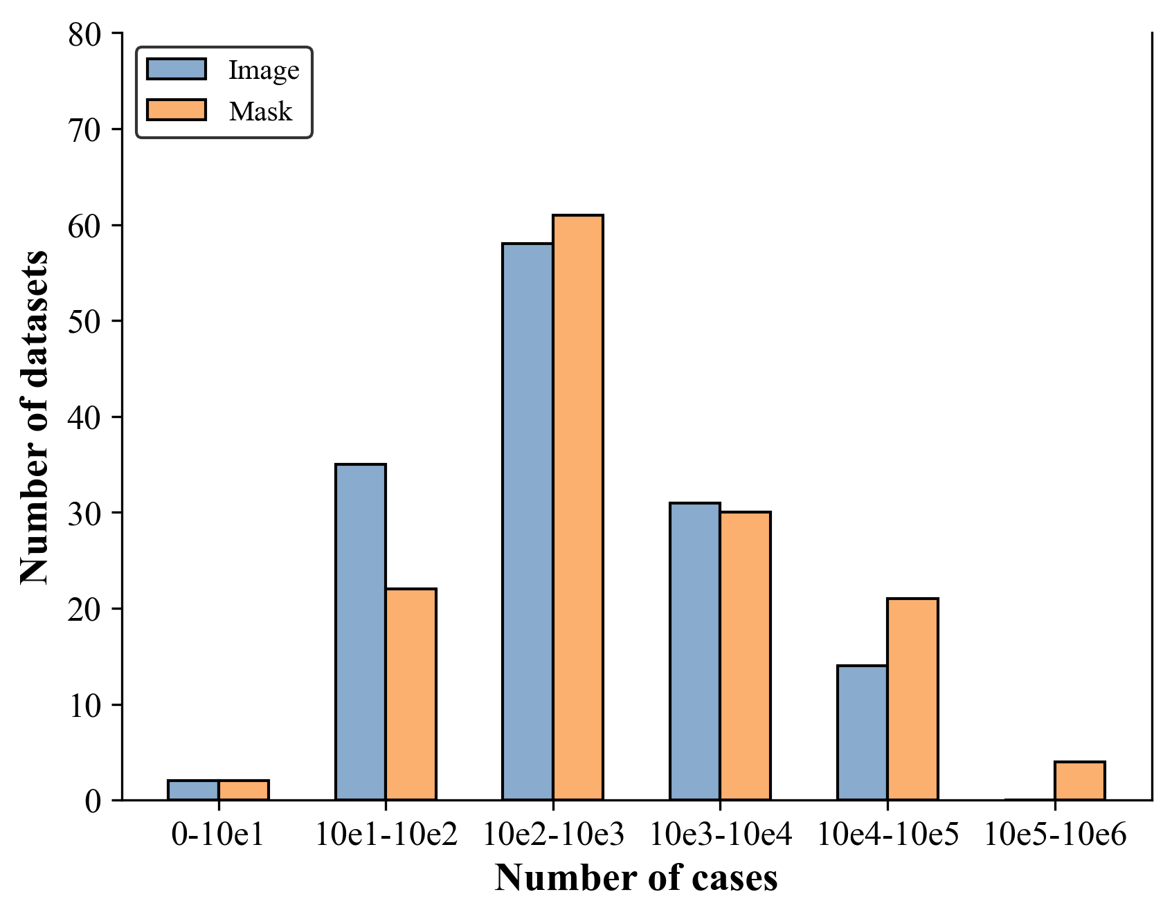
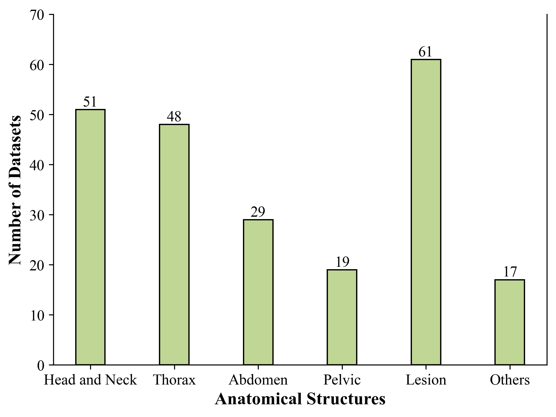
Datasets. The distribution of the datasets is depicted in Figure 3. Despite the abundance of public medical image datasets available online, a significant proportion of them encompass only a limited number of cases. As illustrated in Figure 3(a), over 120 datasets fall short of reaching 10,000 images or 100,000 masks. Furthermore, even the most extensive dataset in our collection contains fewer than 100,000 images and 1,000,000 masks, respectively. As a result, it is challenging to collect and release such a large and diverse medical image dataset. Figure 3(b) presents the case distribution across various body parts in the SA-Med2D-20M dataset. The “Lesion” datasets constitute the largest proportion among all categories, suggesting a significant interest in automating lesion or tumor segmentation in medical images which is vital in assisting medical professionals. It is noteworthy that 17 datasets categorized under “Other” are difficult to assign to specific anatomical structures due to the lack of detailed anatomical information from the original dataset.
| Modality | CT | Endoscopy | PET | Fundus | Microscopy |
| Images | 2338753 | 5838 | 11956 | 3501 | 945 |
| Masks | 12547037 | 19388 | 14129 | 7242 | 5894 |
| Modality | MR | Dermoscopy | X-ray | Ultrasound | Histopathology |
| Images | 2217633 | 7935 | 6407 | 2968 | 866 |
| Masks | 7147784 | 8000 | 9357 | 2968 | 1056 |
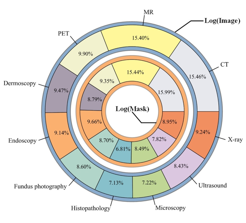
Modality. SA-Med2D-20M includes 10 modalities, as detailed in Table 1, with their distribution illustrated in Figure 4(b). CT and MR modalities are predominant in both the number of images and masks, it is mainly attributed to their widespread presence in public medical image segmentation datasets and the 3D dimension of CT and MR scans, which yields a high volume of slices when segmented across three axes. Following CT and MR, most modalities range between 1,000 to 10,000 images and 1,000 to 20,000 masks. Notably, Microscopy and Histopathology currently comprise fewer than 1,000 images each. We plan to add more data for these less-presented modalities in future updates of SA-Med2D-20M.
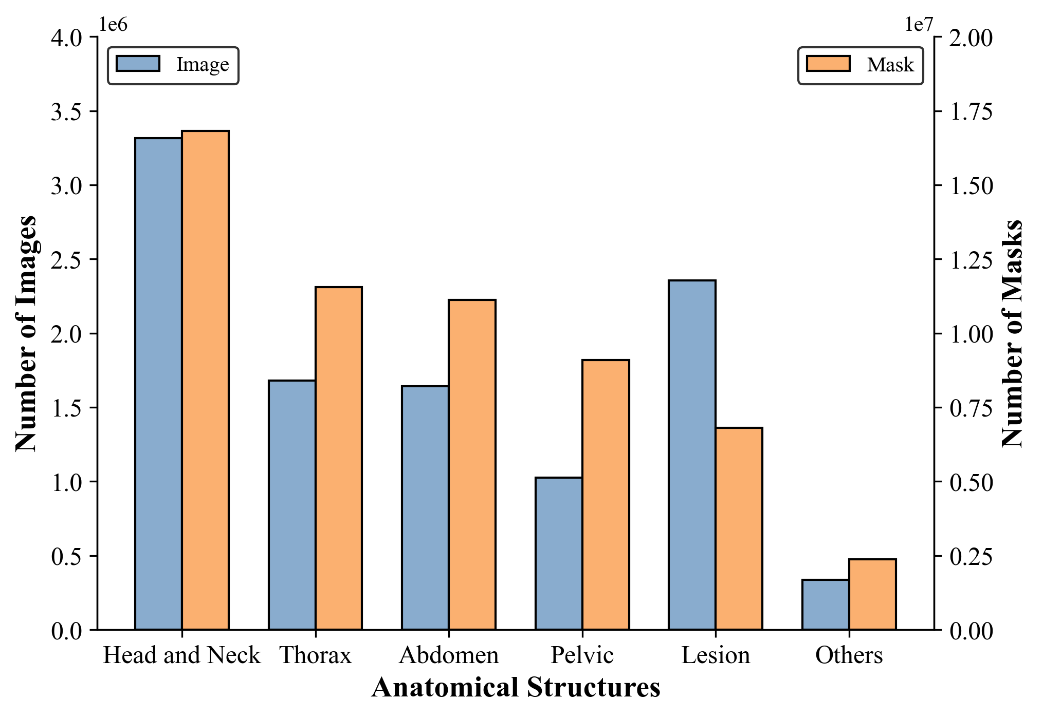
Anatomical structures. The dataset is classified into different categories based on anatomical structures and lesion presence, including Head and Neck, Thorax, Abdomen, Pelvic, Lesion, and Others, as illustrated in Figure 4(a). The approximate ratio of the number of masks to images ranges from 3 to 10. The Head and Neck category contains the largest number of both images and masks for the abundance of multi-modality brain-related data, such as from the BraTS and ISLES series. In contrast, the Others category includes the fewest images and masks. As corroborated by Figure 3(b), tumor segmentation emerges as a primary focus in medical imaging. The Others category refers to datasets not fitting into specific anatomical classes, such as those involving cell and skin segmentation.
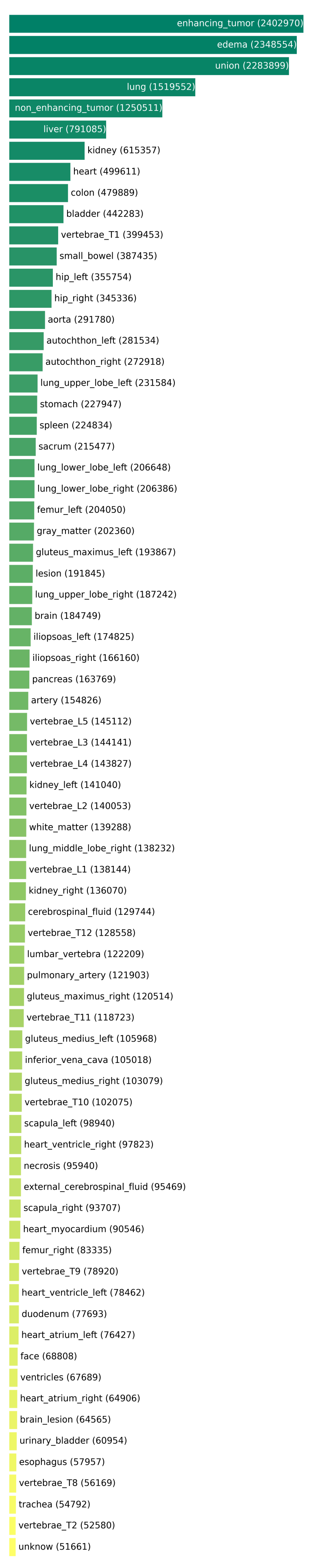
Categories. SA-Med2D-20M consists of 219 labels, with a detailed distribution illustrated in Figure 5. The categories “enhancing_tumor” and “edema” from the BraTS datasets have the highest count. The “union” category is specifically designed to address the issue of pixel overlap across multiple classes whose masks cover more than two categories. Furthermore, the label “unknown” is assigned to instances where the original dataset does not provide specific label information. In terms of label distribution, the long-tailed problem exists in SA-Med2D-20M. Specifically, 47 categories fall within the range of 100,000 to 1,000,000 masks, and 51 categories between 1,000 and 10,000. The most common range is 10,000 to 100,000, with 88 categories lying in it. Additionally, there are also 28 categories with fewer than 1,000 masks.
4 Construction of SA-Med2D-20M
In this section, we describe how to collect and process cases to construct such a large-scale and diverse dataset in detail.
4.1 Collecting data from public resources
The first stage of the construction of SA-Med2D-20M is to collect candidate medical images. Different from general images that are abundant and easy to access on the Internet, we can only find medical images on specialized portal websites, such as TCIA, OpenNeuro, Grand Challenge, Synapse, GitHub, etc. After searching all medical-related open resources and selecting medical image segmentation datasets, we list detailed dataset information in Table LABEL:tab:all_datasets. However, The raw datasets cannot be directly used for developing models because the dimension, modality, and intensity values of raw data are various. The next step is to normalize all datasets.
4.2 Image normalization
There are two aspects to be considered to normalize data. One is that the voxel or pixel values in medical images vary widely because of various modalities and collection methods. For example, the intensity range of CT is over 2,000, and MR is over 10,000. Another is how to unify the data dimension. Some modalities are 3D, like CT, MR, and PET, while some are 2D, like X-ray, Fundus, and Ultrasound. Figure 6 illustrates the whole process of normalizing different datasets. In the first step, we normalize the voxel or pixel values to the “PNG” format, which is detailed below. An original image is first normalized into with the Self-Min-Max normalization (see Equation (1)), and then taken the ceiling value after multiplying the values by 255. The process can be formulated as:
| (1) |
where and are the min value and max value in the image. In the second step, we split all 3D images along three axes and discard images with extreme aspect ratios. Specifically, slice images with the shortest edge less than half the length of the longest edge were discarded to prevent target areas from being extremely blurry. After that, all processed images are saved in “PNG” format.
4.3 Mask processing
We split multi-label masks into single foregrounds, the final representations are similar to SAM. Figure 7 shows the process flow. It can be summarized as three steps. (1) split semantic mask to binary mask. (2) separate binary foreground to multiple connected components and consolidate overlapped areas. (3) remove masks that do not meet our criteria. Then we will describe them in detail.
The first step is to split original labels into binary foregrounds. As shown in Figure 8(a), given an original mask image, we extract the foreground images according to label IDs. Then all separated foregrounds are assigned the value 1 while backgrounds are assigned 0. The second step involves two operations as two examples illustrated in Figure 8(b): one operation is the separation of foregrounds into distinct connected components, each representing a unique category such as individual cells in pathology images or discrete organs in CT scans. The other operation is to consolidate smaller segmentation targets (e.g., a tumor) contained in a larger target (e.g., a kidney containing that tumor) so that the overlap area of different classes can have only a single label (e.g., a kidney). For instance, the ((c)(d)) process in the first case shown in Figure 8(b) illustrates how to consolidate foreground elements into a larger entity when a single kidney contains tumors. In the third step as shown in Figure 8(c), masks are filtered out if the target area accounts for less than 0.153% () of the total image area – equivalent to 100 pixels in a resolution. This criterion ensures that only regions of sufficient size are considered, enhancing the precision and decreasing the noise of our analysis.
After the whole processing, each image is associated with multiple masks, and the original category information for each mask is discarded. To address this, we create a “JSON” file to record the category information for each mask systematically. Then, we establish a uniform naming convention for images and masks, separately. The name format can be formulated as:
| (2) | ||||
| (3) | ||||
| (4) | ||||
| (5) |
where refers to the specific dataset name that the case is from. is the original case name in its dataset. is mainly for 3D images: If we split a 3D case with axis x and the current slice is 100, then the term can be “”. Besides, we assign “” to all 2D cases in this field. is unique to masks and encapsulates both category information and instance id, and the detailed information is stored in “JSON” file. For instance, if the category “liver” is assigned the ID “0003” and there is only one instance of this category in the case, the can be denoted as “” and the category “liver” in “JSON” file is formulated as key-value pair with python-dict format: {“liver”: “0003”}. Finally, all validated masks are saved in the “PNG” format for consistency and ease of use.
5 Applications and Discussions
Medical Vision Foundation Models. The vision foundation model has evolved from the pre-training and fine-tuning paradigm which is a model that is “trained on broad data at scale and is adaptable to a wide range of downstream tasks” bommasani2021opportunities . On the one hand, though vision foundation models in natural imaging like SAM kirillov2023segment and CLIP radford2021learning have had impressive performance in various vision fields ji2023sam ; tang2023sam , they cannot achieve satisfying performance on medical imaging for the significant domain gap between nature images and medical ones. On the other hand, the relative scarcity of medical imaging data hinders the development of medical foundation models. Our work, however, provides the possibility to bridge the gap between natural and medical imaging by providing a large-scale medical image dataset, which can serve as a training resource for both supervised training and self-supervised learning. For example, self-supervised techniques (MoCo he2020momentum , MAE he2022masked ) are readily utilized on 4.6 million 2D medical images in our dataset, and training a medical SAM becomes feasible with 19.7 million masks.
Exploiting SA-Med2D-20M. While transfer learning techniques on pre-trained models are useful for adapting knowledge to downstream tasks with limited data, directly training models from scratch using massive task-specific datasets is more popular and effective in medical image analysis. SA-Med2D-20M is well-organized by modality, anatomical structure, and label, which allows researchers to efficiently select data pertinent to their specific applications, significantly reducing the time required for data collection. Finally, we believe it can significantly alleviate the challenge of data acquisition and potentially accelerate the process of data iteration in medical image analysis after SA-Med2D-20M has been released.
Limitations and Future work. Despite the vast number of images and masks in SA-Med2D-20M, the initial version of the dataset meets several limitations. The first limitation is mask integrity: SA-Med2D-20M may lack masks for some segmentation targets. Since we rely on the original labels for each image in the preliminary release which only provides annotated masks of interest for a specific task, the remaining “regions of non-interest” do not have masks but can be the segmentation targets in SA-Med2D-20M. In this case, we may miss the masks for these regions (i.e., regions of non-interest for such a specific task but of interest in our label system). The second one is data imbalance: SA-Med2D-20M currently faces a marked long-tailed problem due to data imbalance, especially in certain modalities and categories where public segmentation datasets are limited. The third is data format unification: our current approach is to simply convert the original data into RGB format using Self-Min-Max normalization. However, this approach cannot fully preserve the data information, and finding an effective way to normalize data formats across diverse modalities remains a significant and ongoing challenge.
In future work, we will try to pursue this work with the following aspects. For mask integrity, we intend to adopt a methodology similar to the data engine in SAM kirillov2023segment , which tags all data with pseudo labels and refines them manually. For data imbalance, we prepare to enrich SA-Med2D-20M by collecting additional data where there are current shortages in the initial version and applying the pseudo-labeling strategy, used to address mask integrity issue, on publicly available unlabeled data from open sources. Moreover, we encourage the community to contribute towards building a more comprehensive medical image segmentation dataset.
6 Acknowledgements
We thank all medical workers and dataset owners for making public datasets available to the community.
Appendix
| ID | Dataset Name | Modality | Dim | Anatomical structures | Release |
|---|---|---|---|---|---|
| 1 | ACDC 19 | MR | 3D | Thorax | ✔ |
| 2 | AMOS22 ji2022amos | CT, MR | 3D | Thorax, Abdomen, Pelvic | ✔ |
| 3 | ATM22 zhang2023multi | CT | 3D | Thorax | ✔ |
| 4 | AbdomenomenCT-1K ma2021abdomenct | CT | 3D | Abdomen | ✔ |
| 5 | ASC18 xiong2021global | MR | 3D | Thorax | ✔ |
| 6 | COSMOS22 chen2022carotid | MR | 3D | H&N | ✔ |
| 7 | BTCV landman2015miccai | CT | 3D | Thorax, Abdomen, Pelvic | ✔ |
| 8 | BraTS2013 menze2014multimodal ; info:doi/10.2196/jmir.2930 | MR | 3D | H&N, | ✔ |
| 9 | BraTS2015 menze2014multimodal ; info:doi/10.2196/jmir.2930 | MR | 3D | H&N, | ✔ |
| 10 | BraTS2018 menze2014multimodal ; bakas2017advancing ; bakas2019identifying | MR | 3D | H&N, | ✔ |
| 11 | BraTS2019 menze2014multimodal ; bakas2017advancing ; bakas2019identifying | MR | 3D | H&N, | ✔ |
| 12 | BraTS2020 menze2014multimodal ; bakas2017advancing ; bakas2019identifying | MR | 3D | H&N, | ✔ |
| 13 | BraTS2021 bakas2017advancing ; bakas2019identifying ; baid2021rsnaasnrmiccai | MR | 3D | H&N, | ✔ |
| 14 | BrainPTM2021 avital2019neural ; nelkenbaum2020automatic | MR | 3D | H&N, | ✔ |
| 15 | CAD-PE gonzález2020computer | CT | 3D | Thorax, | ✔ |
| 16 | CAUSE07 van20073d | MR | 3D | H&N | - |
| 17 | CHAOS CHAOS2021 ; CHAOSdata2019 ; kavur2019 | MR | 3D | Abdomen | ✔ |
| 18 | CMRxMotion wang2022extreme | MR | 3D | Thorax | ✔ |
| 19 | COVID-19 CT scans paiva2020helping ; jun2020covid | CT | 3D | Thorax, | ✔ |
| 20 | COVID-19-20 roth2022rapid | CT | 3D | Thorax, | ✔ |
| 21 | COVID-19-Image cohen2020covid ; cohen2020covidProspective | X-ray | 2D | Thorax, | ✔ |
| 22 | CRASS hogeweg2012clavicle | X-ray | 2D | Thorax | ✔ |
| 23 | CTPelvic1K liu2021deep | CT | 3D | Pelvic | ✔ |
| 24 | CTSpine1K deng2021ctspine1k | CT | 3D | H&N, Thorax, Abdomen | ✔ |
| 25 | CVC-ClinicDB bernal2015wm | EGD | 2D | Other, | ✔ |
| 26 | MosMedData COVID19 soham1024 | CT | 3D | Thorax, | ✘ |
| 27 | Chestimage tianchi83075 | X-ray | 2D | Thorax, | ✔ |
| 28 | Cranium hssayeni2020computed | CT | 2D | H&N | ✔ |
| 29 | CrossMoDA2021 dorent2023crossmoda | MR | 3D | H&N, | ✔ |
| 30 | CrossMoDA2022 shusharina2021cross | MR | 3D | H&N, | ✔ |
| 31 | DRISHTI-GS sivaswamy2015comprehensive ; sivaswamy2014drishti | FP | 2D | H&N | - |
| 32 | EMIDEC lalande2022deep | MR | 3D | Thorax, | ✔ |
| 33 | EndoVis15 bernal2017comparative | EGD | 2D | Other, | ✔ |
| 34 | EndoCV2020 ali2020endoscopy | EGD | 2D | Other, | - |
| 35 | FLARE21 MedIA-FLARE21 | CT | 3D | Abdomen | ✔ |
| 36 | FLARE22 FLARE22 | CT | 3D | Thorax, Abdomen | ✔ |
| 37 | FeTA2021 payette2021automatic | MR | 3D | H&N, Thorax | ✘ |
| 38 | FeTA2022 payette2021automatic | MR | 3D | H&N, Thorax | ✘ |
| 39 | Fusc2021 wang2022fuseg | DS | 2D | Other, | ✔ |
| 40 | GLaS sirinukunwattana2015stochastic ; sirinukunwattana2017gland | HP | 2D | Other, | ✘ |
| 41 | HVSMR2016 pace2015interactive | MR | 3D | Thorax | ✔ |
| 42 | Heart Seg MRI tobon2015benchmark | MR | 3D | Thorax | ✔ |
| 43 | ADAM fu2020adam | FP | 2D | H&N | ✔ |
| 44 | REFUGE2 orlando2020refuge ; li2020development | FP | 2D | H&N | - |
| 45 | PALM19 huazhu2019palm | FP | 2D | H&N | ✔ |
| 46 | GAMMA 8252743 ; orlando2020refuge ; fu2020age | FP | 2D | H&N | ✔ |
| 47 | ISLES2015- maier2017isles | MR | 3D | H&N, | ✔ |
| 48 | ISLES2016 winzeck2018isles | MR | 3D | H&N, | ✔ |
| 49 | ISLES2017 winzeck2018isles | MR | 3D | H&N, | ✔ |
| 50 | ISLES2018 cereda2016benchmarking ; hakim2021predicting | CT | 3D | H&N, | ✔ |
| 51 | ISLES2022 hernandez2022isles | MR | 3D | H&N, | ✔ |
| 52 | InSTANCE2022 li2023stateoftheart ; 9511297 | CT | 3D | H&N, | ✔ |
| 53 | KiPA22 HE2021102055 ; HE2020101722 ; SHAO2011849 ; SHAO20121001 | CT | 3D | Abdomen, | - |
| 54 | KiTS19 heller2019kits19 | CT | 3D | Abdomen, | ✔ |
| 55 | KiTS21 zhao2021coarse | CT | 3D | Abdomen, | ✔ |
| 56 | LAScarQS2022 LI2022102303 ; LI2022102360 ; li2021atrialgeneral | MR | 3D | Thorax, | - |
| 57 | LNDb pedrosa2019lndb | CT | 3D | Thorax, | ✔ |
| 58 | LUNA16 setio2017validation | CT | 3D | Thorax | ✔ |
| 59 | STACOM SLAWT karim2018algorithms | MR | 3D | Thorax | - |
| 60 | LiTS bilic2019liver | CT | 3D | Abdomen, | - |
| 61 | LMSLS CARASS201777 | MR | 3D | Other, | ✔ |
| 62 | M&Ms-2 campello2021multi | MR | 3D | Thorax | ✔ |
| 63 | MM-WHS zhuang2018multivariate ; zhuang2016multi ; luo2022mathcal | CT, MR | 3D | Thorax | ✔ |
| 64 | MRBrains13 mendrik2015mrbrains | MR | 3D | H&N | - |
| 65 | NEATBrainS15 mendrik2015mrbrains | MR | 3D | H&N | - |
| 66 | MRBrains18 mrbrains18 | MR | 3D | H&N | - |
| 67 | MSD01_BrainTumor 77antonelli2022medical | MR | 3D | H&N, | ✔ |
| 68 | MSD02_Heart 78simpson2019large | MR | 3D | Pelvic, | ✔ |
| 69 | MSD03_Liver 77antonelli2022medical | CT | 3D | Abdomen, | ✔ |
| 70 | MSD05_Prostate 77antonelli2022medical | MR | 3D | Pelvic | ✔ |
| 71 | MSD06_Lung 77antonelli2022medical | CT | 3D | Pelvic, | ✔ |
| 72 | MSD07_Pancreas 77antonelli2022medical | CT | 3D | Abdomen, | ✔ |
| 73 | MSD08_HepaticVessel 77antonelli2022medical | CT | 3D | Abdomen, | ✔ |
| 74 | MSD09_Spleen 77antonelli2022medical | CT | 3D | Abdomen | ✔ |
| 75 | MSD10_Colon 77antonelli2022medical | CT | 3D | Abdomen, | ✔ |
| 76 | MSSEG2016 85COMMOWICK2021118589 | MR | 3D | Other, | - |
| 77 | MSseg08 msseg2008 | MR | 3D | Thorax, Abdomen, | ✘ |
| 78 | MyoPS2020 87luo2022xmetric ; 87MyoPS-Net | MR | 3D | Thorax, | - |
| 79 | CT-ORG 88rister2020ct | CT | 3D | H&N, Thorax, Abdomen | ✔ |
| 80 | PICAI 89saha2021end | MR | 3D | Pelvic, | ✔ |
| 81 | PROMISE09 bharatha2001evaluation | MR | 3D | Pelvic | ✔ |
| 82 | PROMISE12 91litjens2014evaluation | MR | 3D | Pelvic | ✔ |
| 83 | Parse2022 92luo2023efficient | CT | 3D | Thorax | ✔ |
| 84 | PH2 93mendoncca2013ph | DS | 2D | Other, | - |
| 85 | Siim-acr-pneumothorax 94siim-acr-pneumothorax-segmentation | X-ray | 2D | Thorax, | ✔ |
| 86 | SAML 95liu2020saml ; 95liu2020ms | MR | 3D | Pelvic | ✔ |
| 87 | PCXA 96jaeger2013automatic ; 96candemir2013lung | X-ray | 2D | Thorax, | ✔ |
| 88 | QUBIQ2020 pal2021holistic | CT | 2D | H&N, Pelvic | ✔ |
| 89 | SLIVER07 99heimann2009comparison | CT | 3D | Abdomen | - |
| 90 | SegTHOR 100lambert2020segthor | CT | 3D | Thorax, Abdomen | - |
| 91 | StructSeg2019 structseg2019 | CT | 3D | H&N, Thorax, Abdomen, | ✔ |
| 92 | TN-SCUI2020 zhou2020thyroid | US | 2D | H&N, | ✘ |
| 93 | TotalSegmentator Wasserthal_2023 | CT | 3D | H&N, Thorax, Abdomen, Pelvic | ✔ |
| 94 | Nerve ultrasound_nerve_segmentation | US | 2D | Other | ✔ |
| 95 | VESSEL12 rudyanto2014comparing | CT | 3D | Thorax, | ✔ |
| 96 | VerSe20 sekuboyina2021verse ; loffler2020vertebral | CT | 3D | H&N, Thorax, Abdomen | ✔ |
| 97 | VerSe19 sekuboyina2021verse ; loffler2020vertebral | CT | 3D | H&N, Thorax, Abdomen | ✔ |
| 98 | WMH kuijf2019standardized | MR | 3D | H&N, | - |
| 99 | WORD luo2022word | CT | 3D | Thorax, Abdomen | ✔ |
| 100 | autoPET gatidis2022whole | CT, PET | 3D | Pelvic, | ✔ |
| 101 | braimMRI braimMRI | MR | 2D | H&N, | ✔ |
| 102 | iSeg2017 wang2019benchmark | MR | 3D | H&N | - |
| 103 | iSeg2019 sun2021multi | MR | 3D | H&N | - |
| 104 | RIM-ONE 5999143 | FP | 2D | Other, | - |
| 105 | BUSI al2019dataset | US | 2D | Thorax, | ✔ |
| 106 | KvasirCapsule-SEG jha2021nanonet | EGD | 2D | Other, | ✔ |
| 107 | JSRT Shiraishi2000Development | X-ray | 2D | Thorax, | - |
| 108 | SZ-CXR 8477564 | X-ray | 2D | Thorax | ✔ |
| 109 | EndoVis2017 allan20192017 | EGD | 2D | Other, | ✔ |
| 110 | OCCISC 7386573 ; 7005499 | MS | 2D | Other | - |
| 111 | Kvasir-SEG jha2020kvasir | EGD | 2D | Other, | ✔ |
| 112 | ISIC18 tschandl2019ham10000 ; codella2019skin | DS | 2D | Other, | ✔ |
| 113 | ISIC17 codella2018skin | DS | 2D | Other, | ✔ |
| 114 | ISIC16 gutman2016skin | DS | 2D | Other, | ✔ |
| 115 | Private01 | CT | 3D | Abdomen | ✘ |
| 116 | Private02 | CT | 3D | H&N | ✘ |
| 117 | Private03 | CT | 3D | H&N | ✘ |
| 118 | Private04 | CT | 3D | H&N | ✘ |
| 119 | Private05 | CT | 3D | H&N | ✘ |
| 120 | Private06 | CT | 3D | H&N | ✘ |
| 121 | Private07 | CT | 3D | H&N | ✘ |
| 122 | Private08 | CT | 3D | H&N | ✘ |
| 123 | Private09 | CT | 3D | Thorax | ✘ |
| 124 | Private10 | CT | 3D | Thorax | ✘ |
| 125 | Private11 | CT | 3D | H&N, Thorax, Abdomen, Pelvic | ✘ |
| 126 | Private12 | CT | 3D | Thorax | ✘ |
| 127 | Private13 | CT | 3D | Thorax | ✘ |
| 128 | Private14 | CT | 3D | Pelvic | ✘ |
| 129 | Private15 | CT | 3D | H&N, Thorax, Abdomen, Pelvic | ✘ |
| 130 | Private16 | CT | 3D | H&N | ✘ |
| 131 | Private17 | CT | 3D | H&N | ✘ |
| 132 | Private18 | CT | 3D | Thorax | ✘ |
| 133 | Private19 | CT | 3D | Abdomen | ✘ |
| 134 | Private20 | CT | 3D | Pelvic | ✘ |
| 135 | Private21 | CT | 3D | Pelvic | ✘ |
| 136 | Private22 | CT | 3D | H&N | ✘ |
| 137 | Private23 | CT | 3D | H&N | ✘ |
| 138 | Private24 | CT | 3D | H&N | ✘ |
| 139 | Private25 | CT | 3D | H&N | ✘ |
| 140 | Private26 | CT | 3D | Pelvic | ✘ |
References
- [1] Walid Al-Dhabyani, Mohammed Gomaa, Hussien Khaled, and Aly Fahmy. Dataset of breast ultrasound images. data brief 28, 104863 (2020), 2019.
- [2] Sharib Ali, Noha Ghatwary, Barbara Braden, Dominique Lamarque, Adam Bailey, Stefano Realdon, Renato Cannizzaro, Jens Rittscher, Christian Daul, and James East. Endoscopy disease detection challenge 2020. arXiv preprint arXiv:2003.03376, 2020.
- [3] Max Allan, Alex Shvets, Thomas Kurmann, Zichen Zhang, Rahul Duggal, Yun-Hsuan Su, Nicola Rieke, Iro Laina, Niveditha Kalavakonda, Sebastian Bodenstedt, et al. 2017 robotic instrument segmentation challenge. arXiv preprint arXiv:1902.06426, 2019.
- [4] Michela Antonelli, Annika Reinke, Spyridon Bakas, Keyvan Farahani, Annette Kopp-Schneider, Bennett A Landman, Geert Litjens, Bjoern Menze, Olaf Ronneberger, Ronald M Summers, et al. The medical segmentation decathlon. Nature communications, 13(1):4128, 2022.
- [5] Itzik Avital, Ilya Nelkenbaum, Galia Tsarfaty, Eli Konen, Nahum Kiryati, and Arnaldo Mayer. Neural segmentation of seeding rois (srois) for pre-surgical brain tractography. IEEE transactions on medical imaging, 39(5):1655–1667, 2019.
- [6] Ujjwal Baid, Satyam Ghodasara, Suyash Mohan, Michel Bilello, Evan Calabrese, Errol Colak, Keyvan Farahani, Jayashree Kalpathy-Cramer, and Felipe C. Kitamura. The rsna-asnr-miccai brats 2021 benchmark on brain tumor segmentation and radiogenomic classification, 2021.
- [7] Ujjwal Baid, Satyam Ghodasara, Suyash Mohan, Michel Bilello, Evan Calabrese, Errol Colak, Keyvan Farahani, Jayashree Kalpathy-Cramer, Felipe C Kitamura, Sarthak Pati, et al. The rsna-asnr-miccai brats 2021 benchmark on brain tumor segmentation and radiogenomic classification. arXiv preprint arXiv:2107.02314, 2021.
- [8] Spyridon Bakas, Hamed Akbari, Aristeidis Sotiras, Michel Bilello, Martin Rozycki, Justin S Kirby, John B Freymann, Keyvan Farahani, and Christos Davatzikos. Advancing the cancer genome atlas glioma mri collections with expert segmentation labels and radiomic features. Scientific data, 4(1):1–13, 2017.
- [9] Spyridon Bakas, Mauricio Reyes, Andras Jakab, Stefan Bauer, Markus Rempfler, Alessandro Crimi, Russell Takeshi Shinohara, and Christoph Berger. Identifying the best machine learning algorithms for brain tumor segmentation, progression assessment, and overall survival prediction in the brats challenge, 2019.
- [10] Jorge Bernal, F Javier Sánchez, Gloria Fernández-Esparrach, Debora Gil, Cristina Rodríguez, and Fernando Vilariño. Wm-dova maps for accurate polyp highlighting in colonoscopy: Validation vs. saliency maps from physicians. Computerized medical imaging and graphics, 43:99–111, 2015.
- [11] Jorge Bernal, Nima Tajkbaksh, Francisco Javier Sanchez, Bogdan J Matuszewski, Hao Chen, Lequan Yu, Quentin Angermann, Olivier Romain, Bjørn Rustad, Ilangko Balasingham, et al. Comparative validation of polyp detection methods in video colonoscopy: results from the miccai 2015 endoscopic vision challenge. IEEE transactions on medical imaging, 36(6):1231–1249, 2017.
- [12] Olivier Bernard, Alain Lalande, Clement Zotti, Frederick Cervenansky, Xin Yang, Pheng-Ann Heng, Irem Cetin, Karim Lekadir, Oscar Camara, Miguel Angel Gonzalez Ballester, et al. Deep learning techniques for automatic mri cardiac multi-structures segmentation and diagnosis: is the problem solved? IEEE transactions on medical imaging, 37(11):2514–2525, 2018.
- [13] Aditya Bharatha, Masanori Hirose, Nobuhiko Hata, Simon K Warfield, Matthieu Ferrant, Kelly H Zou, Eduardo Suarez-Santana, Juan Ruiz-Alzola, Anthony D’amico, Robert A Cormack, et al. Evaluation of three-dimensional finite element-based deformable registration of pre-and intraoperative prostate imaging. Medical physics, 28(12):2551–2560, 2001.
- [14] Patrick Bilic, Patrick Ferdinand Christ, Eugene Vorontsov, Grzegorz Chlebus, Hao Chen, Qi Dou, Chi-Wing Fu, Xiao Han, and Pheng-Ann Heng. The liver tumor segmentation benchmark (lits), 2019.
- [15] Rishi Bommasani, Drew A Hudson, Ehsan Adeli, Russ Altman, Simran Arora, Sydney von Arx, Michael S Bernstein, Jeannette Bohg, Antoine Bosselut, Emma Brunskill, et al. On the opportunities and risks of foundation models. arXiv preprint arXiv:2108.07258, 2021.
- [16] Nhat-Tan Bui, Dinh-Hieu Hoang, Minh-Triet Tran, and Ngan Le. Sam3d: Segment anything model in volumetric medical images. arXiv preprint arXiv:2309.03493, 2023.
- [17] Victor M Campello, Polyxeni Gkontra, Cristian Izquierdo, Carlos Martin-Isla, Alireza Sojoudi, Peter M Full, Klaus Maier-Hein, Yao Zhang, Zhiqiang He, Jun Ma, et al. Multi-centre, multi-vendor and multi-disease cardiac segmentation: the m&ms challenge. IEEE Transactions on Medical Imaging, 40(12):3543–3554, 2021.
- [18] Sema Candemir, Stefan Jaeger, Kannappan Palaniappan, Jonathan P Musco, Rahul K Singh, Zhiyun Xue, Alexandros Karargyris, Sameer Antani, George Thoma, and Clement J McDonald. Lung segmentation in chest radiographs using anatomical atlases with nonrigid registration. IEEE transactions on medical imaging, 33(2):577–590, 2013.
- [19] Aaron Carass, Snehashis Roy, Amod Jog, Jennifer L. Cuzzocreo, Elizabeth Magrath, Adrian Gherman, Julia Button, James Nguyen, Ferran Prados, and Carole H. Sudre. Longitudinal multiple sclerosis lesion segmentation: Resource and challenge. NeuroImage, 148:77–102, 2017.
- [20] Carlo W Cereda, Søren Christensen, Bruce CV Campbell, Nishant K Mishra, Michael Mlynash, Christopher Levi, Matus Straka, Max Wintermark, Roland Bammer, Gregory W Albers, et al. A benchmarking tool to evaluate computer tomography perfusion infarct core predictions against a dwi standard. Journal of Cerebral Blood Flow & Metabolism, 36(10):1780–1789, 2016.
- [21] Cheng Chen, Juzheng Miao, Dufan Wu, Zhiling Yan, Sekeun Kim, Jiang Hu, Aoxiao Zhong, Zhengliang Liu, Lichao Sun, Xiang Li, et al. Ma-sam: Modality-agnostic sam adaptation for 3d medical image segmentation. arXiv preprint arXiv:2309.08842, 2023.
- [22] H. Chen, X. Zhao, J. Dou, C. Du, R. Yang, H. Sun, S. Yu, H. Zhao, C. Yuan, and N. Balu. Carotid vessel wall segmentation and atherosclerosis diagnosis challenge. https://vessel-wall-segmentation-2022.grand-challenge.org/, 2022.
- [23] Noel Codella, Veronica Rotemberg, Philipp Tschandl, M Emre Celebi, Stephen Dusza, David Gutman, Brian Helba, Aadi Kalloo, Konstantinos Liopyris, Michael Marchetti, et al. Skin lesion analysis toward melanoma detection 2018: A challenge hosted by the international skin imaging collaboration (isic). arXiv preprint arXiv:1902.03368, 2019.
- [24] Noel C. F. Codella, David Gutman, M. Emre Celebi, Brian Helba, Michael A. Marchetti, Stephen W. Dusza, Aadi Kalloo, Konstantinos Liopyris, Nabin Mishra, Harald Kittler, and Allan Halpern. Skin lesion analysis toward melanoma detection: A challenge at the 2017 international symposium on biomedical imaging (isbi), hosted by the international skin imaging collaboration (isic), 2018.
- [25] Joseph Paul Cohen, Paul Morrison, and Lan Dao. Covid-19 image data collection. arXiv 2003.11597, 2020.
- [26] Joseph Paul Cohen, Paul Morrison, Lan Dao, Karsten Roth, Tim Q Duong, and Marzyeh Ghassemi. Covid-19 image data collection: Prospective predictions are the future. arXiv 2006.11988, 2020.
- [27] Olivier Commowick, Michaël Kain, Romain Casey, Roxana Ameli, Jean-Christophe Ferré, Anne Kerbrat, Thomas Tourdias, Frédéric Cervenansky, Sorina Camarasu-Pop, and Tristan Glatard. Multiple sclerosis lesions segmentation from multiple experts: The miccai 2016 challenge dataset. NeuroImage, 244:118589, 2021.
- [28] Guoyao Deng, Ke Zou, Kai Ren, Meng Wang, Xuedong Yuan, Sancong Ying, and Huazhu Fu. Sam-u: Multi-box prompts triggered uncertainty estimation for reliable sam in medical image. arXiv preprint arXiv:2307.04973, 2023.
- [29] Yang Deng, Ce Wang, Yuan Hui, Qian Li, Jun Li, Shiwei Luo, Mengke Sun, Quan Quan, Shuxin Yang, You Hao, et al. Ctspine1k: A large-scale dataset for spinal vertebrae segmentation in computed tomography. arXiv preprint arXiv:2105.14711, 2021.
- [30] Reuben Dorent, Aaron Kujawa, Marina Ivory, Spyridon Bakas, Nicola Rieke, Samuel Joutard, Ben Glocker, Jorge Cardoso, Marc Modat, Kayhan Batmanghelich, et al. Crossmoda 2021 challenge: Benchmark of cross-modality domain adaptation techniques for vestibular schwannoma and cochlea segmentation. Medical Image Analysis, 83:102628, 2023.
- [31] Alexey Dosovitskiy, Lucas Beyer, Alexander Kolesnikov, Dirk Weissenborn, Xiaohua Zhai, Thomas Unterthiner, Mostafa Dehghani, Matthias Minderer, Georg Heigold, Sylvain Gelly, et al. An image is worth 16x16 words: Transformers for image recognition at scale. arXiv preprint arXiv:2010.11929, 2020.
- [32] Michal Drozdzal, Eugene Vorontsov, Gabriel Chartrand, Samuel Kadoury, and Chris Pal. The importance of skip connections in biomedical image segmentation. In International Workshop on Deep Learning in Medical Image Analysis, International Workshop on Large-Scale Annotation of Biomedical Data and Expert Label Synthesis, pages 179–187. Springer, 2016.
- [33] H Fu, F Li, JI Orlando, H Bogunovic, X Sun, J Liao, Y Xu, S Zhang, and X Zhang. Adam: Automatic detection challenge on age-related macular degeneration. IEEE Dataport, 2020.
- [34] Huazhu Fu, Jun Cheng, Yanwu Xu, Damon Wing Kee Wong, Jiang Liu, and Xiaochun Cao. Joint optic disc and cup segmentation based on multi-label deep network and polar transformation. IEEE Transactions on Medical Imaging, 37(7):1597–1605, 2018.
- [35] Huazhu Fu, Fei Li, Xu Sun, Xingxing Cao, Jingan Liao, Jose Ignacio Orlando, Xing Tao, Yuexiang Li, Shihao Zhang, Mingkui Tan, et al. Age challenge: angle closure glaucoma evaluation in anterior segment optical coherence tomography. Medical Image Analysis, 66:101798, 2020.
- [36] F. Fumero, S. Alayon, J. L. Sanchez, J. Sigut, and M. Gonzalez-Hernandez. Rim-one: An open retinal image database for optic nerve evaluation. In 2011 24th International Symposium on Computer-Based Medical Systems (CBMS), pages 1–6, 2011.
- [37] Sergios Gatidis, Tobias Hepp, Marcel Früh, Christian La Fougère, Konstantin Nikolaou, Christina Pfannenberg, Bernhard Schölkopf, Thomas Küstner, Clemens Cyran, and Daniel Rubin. A whole-body fdg-pet/ct dataset with manually annotated tumor lesions. Scientific Data, 9(1):601, 2022.
- [38] Shizhan Gong, Yuan Zhong, Wenao Ma, Jinpeng Li, Zhao Wang, Jingyang Zhang, Pheng-Ann Heng, and Qi Dou. 3dsam-adapter: Holistic adaptation of sam from 2d to 3d for promptable medical image segmentation. arXiv preprint arXiv:2306.13465, 2023.
- [39] Germán González, Daniel Jimenez-Carretero, Sara Rodríguez-López, Carlos Cano-Espinosa, Miguel Cazorla, Tanya Agarwal, Vinit Agarwal, Nima Tajbakhsh, Michael B. Gotway, Jianming Liang, Mojtaba Masoudi, Noushin Eftekhari, Mahdi Saadatmand, Hamid-Reza Pourreza, Patricia Fraga-Rivas, Eduardo Fraile, Frank J. Rybicki, Ara Kassarjian, Raúl San José Estépar, and Maria J. Ledesma-Carbayo. Computer aided detection for pulmonary embolism challenge (cad-pe), 2020.
- [40] David Gutman, Noel C. F. Codella, Emre Celebi, Brian Helba, Michael Marchetti, Nabin Mishra, and Allan Halpern. Skin lesion analysis toward melanoma detection: A challenge at the international symposium on biomedical imaging (isbi) 2016, hosted by the international skin imaging collaboration (isic), 2016.
- [41] Arsany Hakim, Søren Christensen, Stefan Winzeck, Maarten G Lansberg, Mark W Parsons, Christian Lucas, David Robben, Roland Wiest, Mauricio Reyes, and Greg Zaharchuk. Predicting infarct core from computed tomography perfusion in acute ischemia with machine learning: Lessons from the isles challenge. Stroke, 52(7):2328–2337, 2021.
- [42] Kaiming He, Xinlei Chen, Saining Xie, Yanghao Li, Piotr Dollár, and Ross Girshick. Masked autoencoders are scalable vision learners. In Proceedings of the IEEE/CVF conference on computer vision and pattern recognition, pages 16000–16009, 2022.
- [43] Kaiming He, Haoqi Fan, Yuxin Wu, Saining Xie, and Ross Girshick. Momentum contrast for unsupervised visual representation learning. In Proceedings of the IEEE/CVF conference on computer vision and pattern recognition, pages 9729–9738, 2020.
- [44] Yuting He, Guanyu Yang, Jian Yang, Yang Chen, Youyong Kong, Jiasong Wu, Lijun Tang, Xiaomei Zhu, Jean-Louis Dillenseger, Pengfei Shao, Shaobo Zhang, Huazhong Shu, Jean-Louis Coatrieux, and Shuo Li. Dense biased networks with deep priori anatomy and hard region adaptation: Semi-supervised learning for fine renal artery segmentation. Medical Image Analysis, 63:101722, 2020.
- [45] Yuting He, Guanyu Yang, Jian Yang, Rongjun Ge, Youyong Kong, Xiaomei Zhu, Shaobo Zhang, Pengfei Shao, Huazhong Shu, Jean-Louis Dillenseger, Jean-Louis Coatrieux, and Shuo Li. Meta grayscale adaptive network for 3d integrated renal structures segmentation. Medical Image Analysis, 71:102055, 2021.
- [46] Tobias Heimann, Bram Van Ginneken, Martin A Styner, Yulia Arzhaeva, Volker Aurich, Christian Bauer, Andreas Beck, Christoph Becker, Reinhard Beichel, György Bekes, et al. Comparison and evaluation of methods for liver segmentation from ct datasets. IEEE transactions on medical imaging, 28(8):1251–1265, 2009.
- [47] Nicholas Heller, Niranjan Sathianathen, Arveen Kalapara, Edward Walczak, Keenan Moore, Heather Kaluzniak, Joel Rosenberg, Paul Blake, Zachary Rengel, Makinna Oestreich, et al. The kits19 challenge data: 300 kidney tumor cases with clinical context, ct semantic segmentations, and surgical outcomes. arXiv preprint arXiv:1904.00445, 2019.
- [48] Moritz R Hernandez Petzsche, Ezequiel de la Rosa, Uta Hanning, Roland Wiest, Waldo Valenzuela, Mauricio Reyes, Maria Meyer, Sook-Lei Liew, Florian Kofler, Ivan Ezhov, et al. Isles 2022: A multi-center magnetic resonance imaging stroke lesion segmentation dataset. Scientific data, 9(1):762, 2022.
- [49] Laurens Hogeweg, Clara I Sánchez, Pim A de Jong, Pragnya Maduskar, and Bram van Ginneken. Clavicle segmentation in chest radiographs. Medical image analysis, 16(8):1490–1502, 2012.
- [50] Murtadha Hssayeni, M Croock, A Salman, H Al-khafaji, Z Yahya, and B Ghoraani. Computed tomography images for intracranial hemorrhage detection and segmentation. Intracranial Hemorrhage Segmentation Using A Deep Convolutional Model. Data, 5(1):14, 2020.
- [51] Xinrong Hu, Xiaowei Xu, and Yiyu Shi. How to efficiently adapt large segmentation model (sam) to medical images. arXiv preprint arXiv:2306.13731, 2023.
- [52] Rui Huang, Zijie Chen, Yuanyuan Chen, Hongsheng Li, and etc. Structseg2019 grand challenge dataset. Website, 2019. Retrieved from https://structseg2019.grand-challenge.org/.
- [53] Ziyan Huang, Haoyu Wang, Zhongying Deng, Jin Ye, Yanzhou Su, Hui Sun, Junjun He, Yun Gu, Lixu Gu, Shaoting Zhang, and Yu Qiao. Stu-net: Scalable and transferable medical image segmentation models empowered by large-scale supervised pre-training, 2023.
- [54] F Huazhu, L Fei, and IO José. Palm: Pathologic myopia challenge. Comput. Vis. Med. Imaging, 2019.
- [55] Fabian Isensee, Paul F Jaeger, Simon AA Kohl, Jens Petersen, and Klaus H Maier-Hein. nnu-net: a self-configuring method for deep learning-based biomedical image segmentation. Nature methods, 18(2):203–211, 2021.
- [56] Stefan Jaeger, Alexandros Karargyris, Sema Candemir, Les Folio, Jenifer Siegelman, Fiona Callaghan, Zhiyun Xue, Kannappan Palaniappan, Rahul K Singh, Sameer Antani, et al. Automatic tuberculosis screening using chest radiographs. IEEE transactions on medical imaging, 33(2):233–245, 2013.
- [57] Debesh Jha, Pia H Smedsrud, Michael A Riegler, Pål Halvorsen, Thomas de Lange, Dag Johansen, and Håvard D Johansen. Kvasir-seg: A segmented polyp dataset. In International Conference on Multimedia Modeling, pages 451–462. Springer, 2020.
- [58] Debesh Jha, Nikhil Kumar Tomar, Sharib Ali, Michael A. Riegler, Håvard D. Johansen, Dag Johansen, Thomas de Lange, and Pål Halvorsen. Nanonet: Real-time polyp segmentation in video capsule endoscopy and colonoscopy, 2021.
- [59] Ge-Peng Ji, Deng-Ping Fan, Peng Xu, Ming-Ming Cheng, Bowen Zhou, and Luc Van Gool. Sam struggles in concealed scenes – empirical study on ”segment anything”, 2023.
- [60] Yuanfeng Ji, Haotian Bai, Chongjian GE, Jie Yang, Ye Zhu, Ruimao Zhang, Zhen Li, Lingyan Zhang, Wanling Ma, Xiang Wan, and Ping Luo. Amos: A large-scale abdominal multi-organ benchmark for versatile medical image segmentation. In S. Koyejo, S. Mohamed, A. Agarwal, D. Belgrave, K. Cho, and A. Oh, editors, Advances in Neural Information Processing Systems, volume 35, pages 36722–36732. Curran Associates, Inc., 2022.
- [61] Yuanfeng Ji, Haotian Bai, Chongjian Ge, Jie Yang, Ye Zhu, Ruimao Zhang, Zhen Li, Lingyan Zhanng, Wanling Ma, Xiang Wan, et al. Amos: A large-scale abdominal multi-organ benchmark for versatile medical image segmentation. Advances in Neural Information Processing Systems, 35:36722–36732, 2022.
- [62] Chao Jia, Yinfei Yang, Ye Xia, Yi-Ting Chen, Zarana Parekh, Hieu Pham, Quoc Le, Yun-Hsuan Sung, Zhen Li, and Tom Duerig. Scaling up visual and vision-language representation learning with noisy text supervision. In International conference on machine learning, pages 4904–4916. PMLR, 2021.
- [63] Ma Jun, Ge Cheng, Wang Yixin, An Xingle, Gao Jiantao, Yu Ziqi, Zhang Minqing, Liu Xin, Deng Xueyuan, Cao Shucheng, et al. Covid-19 ct lung and infection segmentation dataset. 2020.
- [64] Rashed Karim, Lauren-Emma Blake, Jiro Inoue, Qian Tao, Shuman Jia, R James Housden, Pranav Bhagirath, Jean-Luc Duval, Marta Varela, Jonathan M Behar, et al. Algorithms for left atrial wall segmentation and thickness–evaluation on an open-source ct and mri image database. Medical image analysis, 50:36–53, 2018.
- [65] A. Emre Kavur, N. Sinem Gezer, Mustafa Barış, Sinem Aslan, Pierre-Henri Conze, Vladimir Groza, Duc Duy Pham, Soumick Chatterjee, and Philipp Ernst. CHAOS Challenge - combined (CT-MR) healthy abdominal organ segmentation. Medical Image Analysis, 69:101950, April 2021.
- [66] A Emre Kavur, Naciye Sinem Gezer, Mustafa Barış, Yusuf Şahin, Savaş Özkan, Bora Baydar, Ulaş Yüksel, Çağlar Kılıkçıer, Şahin Olut, Gözde Bozdağı Akar, et al. Comparison of semi-automatic and deep learning-based automatic methods for liver segmentation in living liver transplant donors. Diagnostic and Interventional Radiology, 26(1):11, 2020.
- [67] Ali Emre Kavur, M. Alper Selver, Oğuz Dicle, Mustafa Barış, and N. Sinem Gezer. Chaos - combined (ct-mr) healthy abdominal organ segmentation challenge data. April 2019.
- [68] Alexander Kirillov, Eric Mintun, Nikhila Ravi, Hanzi Mao, Chloe Rolland, Laura Gustafson, Tete Xiao, Spencer Whitehead, Alexander C Berg, Wan-Yen Lo, et al. Segment anything. arXiv preprint arXiv:2304.02643, 2023.
- [69] Alexander Kirillov, Eric Mintun, Nikhila Ravi, Hanzi Mao, Chloe Rolland, Laura Gustafson, Tete Xiao, Spencer Whitehead, Alexander C Berg, Wan-Yen Lo, et al. Segment anything. arXiv preprint arXiv:2304.02643, 2023.
- [70] Michael Kistler, Serena Bonaretti, Marcel Pfahrer, Roman Niklaus, and Philippe Büchler. The virtual skeleton database: An open access repository for biomedical research and collaboration. J Med Internet Res, 15(11):e245, Nov 2013.
- [71] Hugo J Kuijf, J Matthijs Biesbroek, Jeroen De Bresser, Rutger Heinen, Simon Andermatt, Mariana Bento, Matt Berseth, Mikhail Belyaev, M Jorge Cardoso, Adria Casamitjana, et al. Standardized assessment of automatic segmentation of white matter hyperintensities and results of the wmh segmentation challenge. IEEE transactions on medical imaging, 38(11):2556–2568, 2019.
- [72] Alain Lalande, Zhihao Chen, Thibaut Pommier, Thomas Decourselle, Abdul Qayyum, Michel Salomon, Dominique Ginhac, Youssef Skandarani, Arnaud Boucher, Khawla Brahim, et al. Deep learning methods for automatic evaluation of delayed enhancement-mri. the results of the emidec challenge. Medical Image Analysis, 79:102428, 2022.
- [73] Zoé Lambert, Caroline Petitjean, Bernard Dubray, and Su Kuan. Segthor: Segmentation of thoracic organs at risk in ct images. In 2020 Tenth International Conference on Image Processing Theory, Tools and Applications (IPTA), pages 1–6. IEEE, 2020.
- [74] Bennett Landman, Zhoubing Xu, J Igelsias, Martin Styner, T Langerak, and Arno Klein. Miccai multi-atlas labeling beyond the cranial vault–workshop and challenge. In Proc. MICCAI Multi-Atlas Labeling Beyond Cranial Vault—Workshop Challenge, volume 5, page 12, 2015.
- [75] Jens Lehmann, Robert Isele, Max Jakob, Anja Jentzsch, Dimitris Kontokostas, Pablo N Mendes, Sebastian Hellmann, Mohamed Morsey, Patrick Van Kleef, Sören Auer, et al. Dbpedia–a large-scale, multilingual knowledge base extracted from wikipedia. Semantic web, 6(2):167–195, 2015.
- [76] Wenhui Lei, Xu Wei, Xiaofan Zhang, Kang Li, and Shaoting Zhang. Medlsam: Localize and segment anything model for 3d medical images. arXiv preprint arXiv:2306.14752, 2023.
- [77] Fei Li, Diping Song, Han Chen, Jian Xiong, Xingyi Li, Hua Zhong, Guangxian Tang, Sujie Fan, Dennis SC Lam, Weihua Pan, et al. Development and clinical deployment of a smartphone-based visual field deep learning system for glaucoma detection. NPJ digital medicine, 3(1):123, 2020.
- [78] Lei Li, Veronika A Zimmer, Julia A Schnabel, and Xiahai Zhuang. Atrialgeneral: domain generalization for left atrial segmentation of multi-center lge mris. In Medical Image Computing and Computer Assisted Intervention–MICCAI 2021: 24th International Conference, Strasbourg, France, September 27–October 1, 2021, Proceedings, Part VI 24, pages 557–566. Springer, 2021.
- [79] Lei Li, Veronika A. Zimmer, Julia A. Schnabel, and Xiahai Zhuang. Atrialjsqnet: A new framework for joint segmentation and quantification of left atrium and scars incorporating spatial and shape information. Medical Image Analysis, 76:102303, 2022.
- [80] Lei Li, Veronika A. Zimmer, Julia A. Schnabel, and Xiahai Zhuang. Medical image analysis on left atrial lge mri for atrial fibrillation studies: A review. Medical Image Analysis, 77:102360, 2022.
- [81] Xiangyu Li, Gongning Luo, Kuanquan Wang, Hongyu Wang, Jun Liu, Xinjie Liang, Jie Jiang, Zhenghao Song, Chunyue Zheng, Haokai Chi, Mingwang Xu, Yingte He, Xinghua Ma, Jingwen Guo, Yifan Liu, and Chuanpu Li. The state-of-the-art 3d anisotropic intracranial hemorrhage segmentation on non-contrast head ct: The instance challenge, 2023.
- [82] Xiangyu Li, Gongning Luo, Wei Wang, Kuanquan Wang, Yue Gao, and Shuo Li. Hematoma expansion context guided intracranial hemorrhage segmentation and uncertainty estimation. IEEE Journal of Biomedical and Health Informatics, 26(3):1140–1151, 2022.
- [83] Geert Litjens, Robert Toth, Wendy Van De Ven, Caroline Hoeks, Sjoerd Kerkstra, Bram Van Ginneken, Graham Vincent, Gwenael Guillard, Neil Birbeck, Jindang Zhang, et al. Evaluation of prostate segmentation algorithms for mri: the promise12 challenge. Medical image analysis, 18(2):359–373, 2014.
- [84] Haotian Liu, Chunyuan Li, Qingyang Wu, and Yong Jae Lee. Visual instruction tuning, 2023.
- [85] Pengbo Liu, Hu Han, Yuanqi Du, Heqin Zhu, Yinhao Li, Feng Gu, Honghu Xiao, Jun Li, Chunpeng Zhao, Li Xiao, et al. Deep learning to segment pelvic bones: large-scale ct datasets and baseline models. International Journal of Computer Assisted Radiology and Surgery, 16:749–756, 2021.
- [86] Quande Liu, Qi Dou, and Pheng Ann Heng. Shape-aware meta-learning for generalizing prostate mri segmentation to unseen domains. In International Conference on Medical Image Computing and Computer Assisted Intervention (MICCAI), 2020.
- [87] Quande Liu, Qi Dou, Lequan Yu, and Pheng Ann Heng. Ms-net: Multi-site network for improving prostate segmentation with heterogeneous mri data. IEEE Transactions on Medical Imaging, 2020.
- [88] Maximilian T Löffler, Anjany Sekuboyina, Alina Jacob, Anna-Lena Grau, Andreas Scharr, Malek El Husseini, Mareike Kallweit, Claus Zimmer, Thomas Baum, and Jan S Kirschke. A vertebral segmentation dataset with fracture grading. Radiology: Artificial Intelligence, 2(4):e190138, 2020.
- [89] Zhi Lu, Gustavo Carneiro, and Andrew P. Bradley. An improved joint optimization of multiple level set functions for the segmentation of overlapping cervical cells. IEEE Transactions on Image Processing, 24(4):1261–1272, 2015.
- [90] Zhi Lu, Gustavo Carneiro, Andrew P. Bradley, Daniela Ushizima, Masoud S. Nosrati, Andrea G. C. Bianchi, Claudia M. Carneiro, and Ghassan Hamarneh. Evaluation of three algorithms for the segmentation of overlapping cervical cells. IEEE Journal of Biomedical and Health Informatics, 21(2):441–450, 2017.
- [91] Gongning Luo, Kuanquan Wang, Jun Liu, Shuo Li, Xinjie Liang, Xiangyu Li, Shaowei Gan, Wei Wang, Suyu Dong, Wenyi Wang, et al. Efficient automatic segmentation for multi-level pulmonary arteries: The parse challenge. arXiv preprint arXiv:2304.03708, 2023.
- [92] Xiangde Luo, Wenjun Liao, Jianghong Xiao, Jieneng Chen, Tao Song, Xiaofan Zhang, Kang Li, Dimitris N Metaxas, Guotai Wang, and Shaoting Zhang. Word: A large scale dataset, benchmark and clinical applicable study for abdominal organ segmentation from ct image. Medical Image Analysis, 82:102642, 2022.
- [93] Xinzhe Luo and Xiahai Zhuang. -metric: An n-dimensional information-theoretic framework for groupwise registration and deep combined computing. IEEE Transactions on Pattern Analysis and Machine Intelligence, 2022.
- [94] Xinzhe Luo and Xiahai Zhuang. X-metric: An n-dimensional information-theoretic framework for groupwise registration and deep combined computing. IEEE Transactions on Pattern Analysis and Machine Intelligence, 2022.
- [95] Jun Ma and Bo Wang. Segment anything in medical images. arXiv preprint arXiv:2304.12306, 2023.
- [96] Jun Ma, Yao Zhang, Song Gu, Xingle An, Zhihe Wang, Cheng Ge, Congcong Wang, Fan Zhang, and Yu Wang. Fast and low-gpu-memory abdomen ct organ segmentation: The flare challenge. Medical Image Analysis, 82:102616, 2022.
- [97] Jun Ma, Yao Zhang, Song Gu, Cheng Ge, Shihao Ma, Adamo Young, Cheng Zhu, Kangkang Meng, Xin Yang, Ziyan Huang, Fan Zhang, Wentao Liu, and YuanKe Pan. Unleashing the strengths of unlabeled data in pan-cancer abdominal organ quantification: the flare22 challenge. arXiv preprint arXiv:2308.05862, 2023.
- [98] Jun Ma, Yao Zhang, Song Gu, Cheng Zhu, Cheng Ge, Yichi Zhang, Xingle An, Congcong Wang, Qiyuan Wang, Xin Liu, et al. Abdomenct-1k: Is abdominal organ segmentation a solved problem? IEEE Transactions on Pattern Analysis and Machine Intelligence, 44(10):6695–6714, 2021.
- [99] Oskar Maier, Bjoern H Menze, Janina Von der Gablentz, Levin Häni, Mattias P Heinrich, Matthias Liebrand, Stefan Winzeck, Abdul Basit, Paul Bentley, Liang Chen, et al. Isles 2015-a public evaluation benchmark for ischemic stroke lesion segmentation from multispectral mri. Medical image analysis, 35:250–269, 2017.
- [100] Teresa Mendonça, Pedro M Ferreira, Jorge S Marques, André RS Marcal, and Jorge Rozeira. Ph 2-a dermoscopic image database for research and benchmarking. In 2013 35th annual international conference of the IEEE engineering in medicine and biology society (EMBC), pages 5437–5440. IEEE, 2013.
- [101] Adriënne M Mendrik, Koen L Vincken, Hugo J Kuijf, Marcel Breeuwer, Willem H Bouvy, Jeroen De Bresser, Amir Alansary, Marleen De Bruijne, Aaron Carass, Ayman El-Baz, et al. Mrbrains challenge: online evaluation framework for brain image segmentation in 3t mri scans. Computational intelligence and neuroscience, 2015:1–1, 2015.
- [102] Bjoern H Menze, Andras Jakab, Stefan Bauer, Jayashree Kalpathy-Cramer, Keyvan Farahani, Justin Kirby, Yuliya Burren, Nicole Porz, Johannes Slotboom, Roland Wiest, et al. The multimodal brain tumor image segmentation benchmark (brats). IEEE transactions on medical imaging, 34(10):1993–2024, 2014.
- [103] Anna Montoya, Hasnin, kaggle446, shirzad, Will Cukierski, and yffud. Ultrasound nerve segmentation. https://kaggle.com/competitions/ultrasound-nerve-segmentation, 2016.
- [104] MRBrainS18. Mrbrains18 — grand challenge on mr brain segmentation at miccai 2018. https://mrbrains18.isi.uu.nl/, 2018.
- [105] Hannes Mühleisen and Christian Bizer. Web data commons-extracting structured data from two large web corpora. LDOW, 937:133–145, 2012.
- [106] Ilya Nelkenbaum, Galia Tsarfaty, Nahum Kiryati, Eli Konen, and Arnaldo Mayer. Automatic segmentation of white matter tracts using multiple brain mri sequences. In 2020 IEEE 17th International Symposium on Biomedical Imaging (ISBI), pages 368–371. IEEE, 2020.
- [107] OpenAI. Gpt-4 technical report, 2023.
- [108] José Ignacio Orlando, Huazhu Fu, João Barbosa Breda, Karel Van Keer, Deepti R Bathula, Andrés Diaz-Pinto, Ruogu Fang, Pheng-Ann Heng, Jeyoung Kim, JoonHo Lee, et al. Refuge challenge: A unified framework for evaluating automated methods for glaucoma assessment from fundus photographs. Medical image analysis, 59:101570, 2020.
- [109] Danielle F Pace, Adrian V Dalca, Tal Geva, Andrew J Powell, Mehdi H Moghari, and Polina Golland. Interactive whole-heart segmentation in congenital heart disease. In Medical Image Computing and Computer-Assisted Intervention–MICCAI 2015: 18th International Conference, Munich, Germany, October 5-9, 2015, Proceedings, Part III 18, pages 80–88. Springer, 2015.
- [110] O Paiva. Helping radiologists to help people in more than 100 countries! coronavirus cases. CORONACASES. ORG, Ed., ed, 2020.
- [111] Jimut Bahan Pal. Holistic network for quantifying uncertainties in medical images. In International MICCAI Brainlesion Workshop, pages 560–569. Springer, 2021.
- [112] S. Park, L.C. Chu, E.K. Fishman, A.L. Yuille, B. Vogelstein, K.W. Kinzler, K.M. Horton, R.H. Hruban, E.S. Zinreich, D. Fadaei Fouladi, S. Shayesteh, J. Graves, and S. Kawamoto. Annotated normal ct data of the abdomen for deep learning: Challenges and strategies for implementation. Diagnostic and Interventional Imaging, 101(1):35–44, 2020.
- [113] Kelly Payette, Priscille de Dumast, Hamza Kebiri, Ivan Ezhov, Johannes C Paetzold, Suprosanna Shit, Asim Iqbal, Romesa Khan, Raimund Kottke, Patrice Grehten, et al. An automatic multi-tissue human fetal brain segmentation benchmark using the fetal tissue annotation dataset. Scientific data, 8(1):167, 2021.
- [114] João Pedrosa, Guilherme Aresta, Carlos Ferreira, Márcio Rodrigues, Patrícia Leitão, André Silva Carvalho, João Rebelo, Eduardo Negrão, Isabel Ramos, António Cunha, and Aurélio Campilho. Lndb: A lung nodule database on computed tomography, 2019.
- [115] Junyi Qiu, Lei Li, Sihan Wang, Ke Zhang, Yinyin Chen, Shan Yang, and Xiahai Zhuang. Myops-net: Myocardial pathology segmentation with flexible combination of multi-sequence cmr images. Medical Image Analysis, 84:102694, 2023.
- [116] Alec Radford, Jong Wook Kim, Chris Hallacy, Aditya Ramesh, Gabriel Goh, Sandhini Agarwal, Girish Sastry, Amanda Askell, Pamela Mishkin, Jack Clark, et al. Learning transferable visual models from natural language supervision. In International conference on machine learning, pages 8748–8763. PMLR, 2021.
- [117] Alec Radford, Jong Wook Kim, Chris Hallacy, Aditya Ramesh, Gabriel Goh, Sandhini Agarwal, Girish Sastry, Amanda Askell, Pamela Mishkin, Jack Clark, Gretchen Krueger, and Ilya Sutskever. Learning transferable visual models from natural language supervision, 2021.
- [118] Aditya Ramesh, Prafulla Dhariwal, Alex Nichol, Casey Chu, and Mark Chen. Hierarchical text-conditional image generation with clip latents. arXiv preprint arXiv:2204.06125, 1(2):3, 2022.
- [119] Aditya Ramesh, Mikhail Pavlov, Gabriel Goh, Scott Gray, Chelsea Voss, Alec Radford, Mark Chen, and Ilya Sutskever. Zero-shot text-to-image generation. 2021.
- [120] Blaine Rister, Darvin Yi, Kaushik Shivakumar, Tomomi Nobashi, and Daniel L Rubin. Ct-org, a new dataset for multiple organ segmentation in computed tomography. Scientific Data, 7(1):381, 2020.
- [121] Olaf Ronneberger, Philipp Fischer, and Thomas Brox. U-net: Convolutional networks for biomedical image segmentation. In Medical Image Computing and Computer-Assisted Intervention–MICCAI 2015: 18th International Conference, Munich, Germany, October 5-9, 2015, Proceedings, Part III 18, pages 234–241. Springer, 2015.
- [122] Holger R Roth, Ziyue Xu, Carlos Tor-Díez, Ramon Sanchez Jacob, Jonathan Zember, Jose Molto, Wenqi Li, Sheng Xu, Baris Turkbey, Evrim Turkbey, et al. Rapid artificial intelligence solutions in a pandemic—the covid-19-20 lung ct lesion segmentation challenge. Medical image analysis, 82:102605, 2022.
- [123] Rina D Rudyanto, Sjoerd Kerkstra, Eva M Van Rikxoort, Catalin Fetita, Pierre-Yves Brillet, Christophe Lefevre, Wenzhe Xue, Xiangjun Zhu, Jianming Liang, Ilkay Öksüz, et al. Comparing algorithms for automated vessel segmentation in computed tomography scans of the lung: the vessel12 study. Medical image analysis, 18(7):1217–1232, 2014.
- [124] Anindo Saha, Matin Hosseinzadeh, and Henkjan Huisman. End-to-end prostate cancer detection in bpmri via 3d cnns: effects of attention mechanisms, clinical priori and decoupled false positive reduction. Medical image analysis, 73:102155, 2021.
- [125] Anjany Sekuboyina, Malek E Husseini, Amirhossein Bayat, Maximilian Löffler, Hans Liebl, Hongwei Li, Giles Tetteh, Jan Kukačka, Christian Payer, Darko Štern, et al. Verse: a vertebrae labelling and segmentation benchmark for multi-detector ct images. Medical image analysis, 73:102166, 2021.
- [126] Arnaud Arindra Adiyoso Setio, Alberto Traverso, Thomas De Bel, Moira SN Berens, Cas Van Den Bogaard, Piergiorgio Cerello, Hao Chen, Qi Dou, Maria Evelina Fantacci, Bram Geurts, et al. Validation, comparison, and combination of algorithms for automatic detection of pulmonary nodules in computed tomography images: the luna16 challenge. Medical image analysis, 42:1–13, 2017.
- [127] Pengfei Shao, Chao Qin, Changjun Yin, Xiaoxin Meng, Xiaobing Ju, Jie Li, Qiang Lv, Wei Zhang, and Zhengquan Xu. Laparoscopic partial nephrectomy with segmental renal artery clamping: Technique and clinical outcomes. European Urology, 59(5):849–855, 2011.
- [128] Pengfei Shao, Lijun Tang, Pu Li, Yi Xu, Chao Qin, Qiang Cao, Xiaobing Ju, Xiaoxin Meng, Qiang Lv, Jie Li, Wei Zhang, and Changjun Yin. Precise segmental renal artery clamping under the guidance of dual-source computed tomography angiography during laparoscopic partial nephrectomy. European Urology, 62(6):1001–1008, 2012.
- [129] Shiraishi, Junji, Katsuragawa, Shigehiko, Ikezoe, Junpei, Matsumoto, Tsuneo, Kobayashi, and Takeshi and. Development of a digital image database for chest radiographs with and without a lung nodule. American Journal of Roentgenology, 2000.
- [130] Nadya Shusharina, Thomas Bortfeld, Carlos Cardenas, Brian De, Kevin Diao, Soleil Hernandez, Yufei Liu, Sean Maroongroge, Jonas Söderberg, and Moaaz Soliman. Cross-modality brain structures image segmentation for the radiotherapy target definition and plan optimization. In Segmentation, Classification, and Registration of Multi-modality Medical Imaging Data: MICCAI 2020 Challenges, ABCs 2020, L2R 2020, TN-SCUI 2020, Held in Conjunction with MICCAI 2020, Lima, Peru, October 4–8, 2020, Proceedings 23, pages 3–15. Springer, 2021.
- [131] Amber L Simpson, Michela Antonelli, Spyridon Bakas, Michel Bilello, Keyvan Farahani, Bram Van Ginneken, Annette Kopp-Schneider, Bennett A Landman, Geert Litjens, Bjoern Menze, et al. A large annotated medical image dataset for the development and evaluation of segmentation algorithms. arXiv preprint arXiv:1902.09063, 2019.
- [132] Korsuk Sirinukunwattana, Josien PW Pluim, Hao Chen, Xiaojuan Qi, Pheng-Ann Heng, Yun Bo Guo, Li Yang Wang, Bogdan J Matuszewski, Elia Bruni, Urko Sanchez, et al. Gland segmentation in colon histology images: The glas challenge contest. Medical image analysis, 35:489–502, 2017.
- [133] Korsuk Sirinukunwattana, David RJ Snead, and Nasir M Rajpoot. A stochastic polygons model for glandular structures in colon histology images. IEEE transactions on medical imaging, 34(11):2366–2378, 2015.
- [134] Jayanthi Sivaswamy, S Krishnadas, Arunava Chakravarty, G Joshi, A Syed Tabish, et al. A comprehensive retinal image dataset for the assessment of glaucoma from the optic nerve head analysis. JSM Biomedical Imaging Data Papers, 2(1):1004, 2015.
- [135] Jayanthi Sivaswamy, SR Krishnadas, Gopal Datt Joshi, Madhulika Jain, and A Ujjwaft Syed Tabish. Drishti-gs: Retinal image dataset for optic nerve head (onh) segmentation. In 2014 IEEE 11th international symposium on biomedical imaging (ISBI), pages 53–56. IEEE, 2014.
- [136] Soham. Chest ct scans with covid-19. https://www.kaggle.com/datasets/soham1024/chest-ct-scans-with-covid19, 2020.
- [137] Sergii Stirenko, Yuriy Kochura, Oleg Alienin, Oleksandr Rokovyi, Yuri Gordienko, Peng Gang, and Wei Zeng. Chest x-ray analysis of tuberculosis by deep learning with segmentation and augmentation. In 2018 IEEE 38th International Conference on Electronics and Nanotechnology (ELNANO), pages 422–428, 2018.
- [138] Martin Styner, Simon Warfield, Wiro Niessen, Theo van Walsum, Coert Metz, Michiel Schaap, Xiang Deng, Tobias Heimann, and Bram van Ginneken. Ms lesion segmentation challenge 2008. http://www.ia.unc.edu/MSseg/index.html, 2008.
- [139] Yue Sun, Kun Gao, Zhengwang Wu, Guannan Li, Xiaopeng Zong, Zhihao Lei, Ying Wei, Jun Ma, Xiaoping Yang, Xue Feng, et al. Multi-site infant brain segmentation algorithms: the iseg-2019 challenge. IEEE Transactions on Medical Imaging, 40(5):1363–1376, 2021.
- [140] Lv Tang, Haoke Xiao, and Bo Li. Can sam segment anything? when sam meets camouflaged object detection, 2023.
- [141] Aliyun Tianchi. Chest image dataset for pneumothorax segmentation. https://tianchi.aliyun.com/dataset/83075, 2020.
- [142] Aliyun Tianchi. braimmri dataset. https://tianchi.aliyun.com/dataset/127459, 2022.
- [143] Catalina Tobon-Gomez, Arjan J Geers, Jochen Peters, Jürgen Weese, Karen Pinto, Rashed Karim, Mohammed Ammar, Abdelaziz Daoudi, Jan Margeta, Zulma Sandoval, et al. Benchmark for algorithms segmenting the left atrium from 3d ct and mri datasets. IEEE transactions on medical imaging, 34(7):1460–1473, 2015.
- [144] Hugo Touvron, Thibaut Lavril, Gautier Izacard, Xavier Martinet, Marie-Anne Lachaux, Timothée Lacroix, Baptiste Rozière, Naman Goyal, Eric Hambro, Faisal Azhar, Aurelien Rodriguez, Armand Joulin, Edouard Grave, and Guillaume Lample. Llama: Open and efficient foundation language models, 2023.
- [145] Hugo Touvron, Louis Martin, Kevin Stone, Peter Albert, Amjad Almahairi, Yasmine Babaei, Nikolay Bashlykov, Soumya Batra, Prajjwal Bhargava, Shruti Bhosale, Dan Bikel, Lukas Blecher, Cristian Canton Ferrer, Moya Chen, Guillem Cucurull, David Esiobu, Jude Fernandes, Jeremy Fu, Wenyin Fu, Brian Fuller, Cynthia Gao, Vedanuj Goswami, Naman Goyal, Anthony Hartshorn, Saghar Hosseini, Rui Hou, Hakan Inan, Marcin Kardas, Viktor Kerkez, Madian Khabsa, Isabel Kloumann, Artem Korenev, Punit Singh Koura, Marie-Anne Lachaux, Thibaut Lavril, Jenya Lee, Diana Liskovich, Yinghai Lu, Yuning Mao, Xavier Martinet, Todor Mihaylov, Pushkar Mishra, Igor Molybog, Yixin Nie, Andrew Poulton, Jeremy Reizenstein, Rashi Rungta, Kalyan Saladi, Alan Schelten, Ruan Silva, Eric Michael Smith, Ranjan Subramanian, Xiaoqing Ellen Tan, Binh Tang, Ross Taylor, Adina Williams, Jian Xiang Kuan, Puxin Xu, Zheng Yan, Iliyan Zarov, Yuchen Zhang, Angela Fan, Melanie Kambadur, Sharan Narang, Aurelien Rodriguez, Robert Stojnic, Sergey Edunov, and Thomas Scialom. Llama 2: Open foundation and fine-tuned chat models, 2023.
- [146] Philipp Tschandl, Cliff Rosendahl, and Harald Kittler. The ham10000 dataset, a large collection of multi-source dermatoscopic images of common pigmented skin lesions (2018). Preprint. Available from: https://arxiv. org/ftp/arxiv/papers/1803/1803.10417. pdf. Cited, 4, 2019.
- [147] Bram Van Ginneken, Tobias Heimann, and Martin Styner. 3d segmentation in the clinic: A grand challenge. In MICCAI workshop on 3D segmentation in the clinic: a grand challenge, volume 1, pages 7–15, 2007.
- [148] Chuanbo Wang, Amirreza Mahbod, Isabella Ellinger, Adrian Galdran, Sandeep Gopalakrishnan, Jeffrey Niezgoda, and Zeyun Yu. Fuseg: The foot ulcer segmentation challenge. arXiv preprint arXiv:2201.00414, 2022.
- [149] Li Wang, Dong Nie, Guannan Li, Élodie Puybareau, Jose Dolz, Qian Zhang, Fan Wang, Jing Xia, Zhengwang Wu, Jia-Wei Chen, et al. Benchmark on automatic six-month-old infant brain segmentation algorithms: the iseg-2017 challenge. IEEE transactions on medical imaging, 38(9):2219–2230, 2019.
- [150] Shuo Wang, Chen Qin, Chengyan Wang, Kang Wang, Haoran Wang, Chen Chen, Cheng Ouyang, Xutong Kuang, Chengliang Dai, Yuanhan Mo, et al. The extreme cardiac mri analysis challenge under respiratory motion (cmrxmotion). arXiv preprint arXiv:2210.06385, 2022.
- [151] Xinlong Wang, Xiaosong Zhang, Yue Cao, Wen Wang, Chunhua Shen, and Tiejun Huang. Seggpt: Segmenting everything in context. arXiv preprint arXiv:2304.03284, 2023.
- [152] Jakob Wasserthal, Hanns-Christian Breit, Manfred T. Meyer, Maurice Pradella, Daniel Hinck, Alexander W. Sauter, Tobias Heye, Daniel T. Boll, Joshy Cyriac, Shan Yang, Michael Bach, and Martin Segeroth. Totalsegmentator: Robust segmentation of 104 anatomic structures in ct images. Radiology: Artificial Intelligence, 5(5), September 2023.
- [153] Jakob Wasserthal, Manfred Meyer, Hanns-Christian Breit, Joshy Cyriac, Shan Yang, and Martin Segeroth. Totalsegmentator: robust segmentation of 104 anatomical structures in ct images. arXiv preprint arXiv:2208.05868, 2022.
- [154] Stefan Winzeck, Arsany Hakim, Richard McKinley, José AADSR Pinto, Victor Alves, Carlos Silva, Maxim Pisov, Egor Krivov, Mikhail Belyaev, Miguel Monteiro, et al. Isles 2016 and 2017-benchmarking ischemic stroke lesion outcome prediction based on multispectral mri. Frontiers in neurology, 9:679, 2018.
- [155] Baoyuan Wu, Weidong Chen, Yanbo Fan, Yong Zhang, Jinlong Hou, Jie Liu, and Tong Zhang. Tencent ml-images: A large-scale multi-label image database for visual representation learning. IEEE Access, 7:172683–172693, 2019.
- [156] Junde Wu, Rao Fu, Huihui Fang, Yuanpei Liu, Zhaowei Wang, Yanwu Xu, Yueming Jin, and Tal Arbel. Medical sam adapter: Adapting segment anything model for medical image segmentation. arXiv preprint arXiv:2304.12620, 2023.
- [157] Junde Wu, Rao Fu, Huihui Fang, Yuanpei Liu, Zhaowei Wang, Yanwu Xu, Yueming Jin, and Tal Arbel. Medical sam adapter: Adapting segment anything model for medical image segmentation. arXiv preprint arXiv:2304.12620, 2023.
- [158] Zhaohan Xiong, Qing Xia, Zhiqiang Hu, Ning Huang, Cheng Bian, Yefeng Zheng, Sulaiman Vesal, Nishant Ravikumar, Andreas Maier, Xin Yang, et al. A global benchmark of algorithms for segmenting the left atrium from late gadolinium-enhanced cardiac magnetic resonance imaging. Medical image analysis, 67:101832, 2021.
- [159] Anna Zawacki, Carol Wu, George Shih, Julia Elliott, Mikhail Fomitchev, Mohannad Hussain, Lakhani Paras, Phil Culliton, and Shunxing Bao. Siim-acr pneumothorax segmentation, 2019.
- [160] Kaidong Zhang and Dong Liu. Customized segment anything model for medical image segmentation. arXiv preprint arXiv:2304.13785, 2023.
- [161] Minghui Zhang, Yangqian Wu, Hanxiao Zhang, Yulei Qin, Hao Zheng, Wen Tang, Corey Arnold, Chenhao Pei, Pengxin Yu, Yang Nan, et al. Multi-site, multi-domain airway tree modeling. Medical Image Analysis, page 102957, 2023.
- [162] Zhongchen Zhao, Huai Chen, and Lisheng Wang. A coarse-to-fine framework for the 2021 kidney and kidney tumor segmentation challenge. In International Challenge on Kidney and Kidney Tumor Segmentation, pages 53–58. Springer, 2021.
- [163] J. Zhou, X. Jia, Y. Dong, D. Ni, A. Noble, R. Huang, T. Tan, X. Tao, R. Li, and M. The Van. Thyroid nodule segmentation and classification. https://tn-scui2020.grand-challenge.org/Home/, 2020.
- [164] Zongwei Zhou, Md Mahfuzur Rahman Siddiquee, Nima Tajbakhsh, and Jianming Liang. Unet++: A nested u-net architecture for medical image segmentation. In Deep Learning in Medical Image Analysis and Multimodal Learning for Clinical Decision Support: 4th International Workshop, DLMIA 2018, and 8th International Workshop, ML-CDS 2018, Held in Conjunction with MICCAI 2018, Granada, Spain, September 20, 2018, Proceedings 4, pages 3–11. Springer, 2018.
- [165] Xiahai Zhuang. Multivariate mixture model for myocardial segmentation combining multi-source images. IEEE transactions on pattern analysis and machine intelligence, 41(12):2933–2946, 2018.
- [166] Xiahai Zhuang and Juan Shen. Multi-scale patch and multi-modality atlases for whole heart segmentation of mri. Medical image analysis, 31:77–87, 2016.
- [167] Xueyan Zou, Jianwei Yang, Hao Zhang, Feng Li, Linjie Li, Jianfeng Wang, Lijuan Wang, Jianfeng Gao, and Yong Jae Lee. Segment everything everywhere all at once. 2023.