Segment Anything Model with Uncertainty Rectification for Auto-Prompting Medical Image Segmentation
Abstract
The introduction of the Segment Anything Model (SAM) has marked a significant advancement in prompt-driven image segmentation. However, SAM’s application to medical image segmentation requires manual prompting of target structures to obtain acceptable performance, which is still labor-intensive. Despite attempts of auto-prompting to turn SAM into a fully automatic manner, it still exhibits subpar performance and lacks of reliability in the field of medical imaging. In this paper, we propose UR-SAM, an uncertainty rectified SAM framework to enhance the robustness and reliability for auto-prompting medical image segmentation. Our method incorporates a prompt augmentation module to estimate the distribution of predictions and generate uncertainty maps, and an uncertainty-based rectification module to further enhance the performance of SAM. Extensive experiments on two public 3D medical datasets covering the segmentation of 35 organs demonstrate that without supplementary training or fine-tuning, our method further improves the segmentation performance with up to 10.7 % and 13.8 % in dice similarity coefficient, demonstrating efficiency and broad capabilities for medical image segmentation without manual prompting.
1 Introduction
Medical image segmentation aims to delineate the interested anatomical structures like organs and tumors from the original images by labeling each pixel into a certain class, which is one of the most representative and comprehensive research topics in both communities of computer vision and medical image analysis [21, 22]. Accurate segmentation can provide reliable volumetric and shape information of target structures, so as to assist in many further clinical applications like disease diagnosis, quantitative analysis, and surgical planning [1, 10, 17, 25]. Since manual contour delineation is labor-intensive and time-consuming and suffers from inter-observer variability, it is highly desired in clinical studies to develop automatic medical image segmentation methods. With the unprecedented developments in deep learning, deep neural networks have been widely applied and achieved great success in the field of medical image segmentation due to their outstanding performance [28, 13]. However, existing deep models are often tailored for specific modalities and targets, which limits their capacity for further generalization.
The recent introduction of the Segment Anything Model (SAM) [16] has gained massive attention as a promptable foundation model capable of generating fine-grade segmentation masks using prompts like points or bounding boxes, demonstrating impressive performance on a variety of semantic segmentation tasks [20, 36]. However, recent studies have revealed SAM’s limited performance in specific domain tasks [14], such as medical image segmentation where challenges emerge in scenarios characterized by high structural complexity and low contrast, leading to weak boundaries [39, 12, 26]. Besides, most of these applications to medical image segmentation require manual prompting of target structures to obtain acceptable performance, which is still labor-intensive. Despite attempts of auto-prompting to turn SAM into a fully automatic manner [18], it still exhibits subpar performance and lacks reliability, while guaranteeing the reliability of segmentation results is of great importance, especially for medical imaging where the variability in segmentation accuracy directly contributes to safeguarding patients’ safety during clinical procedures. One promising avenue to issue this challenge is uncertainty estimation, which serves as a valuable approach to provide the reliability of medical image segmentation since it allows us to quantify the confidence of the model’s output and identify when the model may not perform well, which has demonstrated its reliability and robustness in many medical image segmentation tasks [42, 40].
In this paper, we propose an Uncertainty Rectified Segment Anything Model (UR-SAM) by estimating the segmentation uncertainty and utilizing uncertainty for rectification to enhance the reliability and improve the accuracy of SAM for medical image segmentation. Since different prompts may yield diverse results, instead of adding perturbations to input images or model parameters, we focus on prompt augmentation to introduce perturbations and obtain a series of different segmentation outputs. Then we establish pixel-level confidence evaluation through uncertainty estimation based on these results, which can be utilized to identify areas of concern and provide additional information to the clinician along with model-generated predictions. To further utilize estimated uncertainty and improve the performance, we propose a class-specific confidence-based filtering method to select out high uncertainty regions and an uncertainty rectification module to divide regions within a certain range of image intensity into target areas.
To evaluate the effectiveness of our proposed framework, we conduct experiments based on the original SAM [16] and the medical-adapted MedSAM [23] as the foundation for our framework on two public 3D datasets including the segmentation of 22 head and neck organs and 13 abdominal organs for a comprehensive evaluation. Our experiments demonstrate significant performance improvement of SAM’s segmentation result and robustness to different prompts. The main contribution of our work can be summarized as follows:
-
•
We present UR-SAM, an Uncertainty Rectified Segment Anything Model by incorporating uncertainty estimation and rectification to enhance the robustness and reliability for auto-prompting in medical image segmentation.
-
•
Since the segmentation performance of SAM is sensitive to the input prompt [2], we propose to augment given prompts by adding perturbations with pre-defined ratios to estimate segmentation uncertainty based on the predictions of multi-prompt input. We examine the influence of different augmentation settings for prompt augmentation to empirically select out the best setting.
-
•
We establish a methodology to utilize estimated segmentation uncertainty with class-specific confidence-based filtering to select out high uncertain regions for rectification to further improve the segmentation performance.
-
•
The proposed method is comprehensively validated on two public 3D medical datasets including the segmentation of 22 head and neck organs and 13 abdominal organs with comparison to manual prompting setting simulated based on ground truth masks. It significantly improves the segmentation performance and achieves comparable results to manual prompting without the necessity for further training or tuning.
2 Related Work
Uncertainty Estimation. In deep learning, uncertainty estimation is an essential task that can lead to significant improvements in the reliability and trustworthiness of deep models, which is particularly important in medical imaging where the uncertainty can be used to identify areas of concern or to provide additional information to the clinician [7, 41]. The quantification of uncertainty involves two fundamental types: aleatoric uncertainty which represents inherent noise or variation in the data learned by the model, and epistemic uncertainty which quantifies the inherent lack of knowledge about the underlying model architecture and parameters [5]. For image segmentation, this uncertainty acknowledges that there may be uncertainty in determining whether a pixel belongs to the object or the background. The model assigns a probability value that represents the confidence or uncertainty associated with each pixel’s classification, enabling a more nuanced and informative representation of the segmentation task to capture the uncertainty inherent in the data [15]. By addressing and quantifying these uncertainties, the reliability and accuracy of deep models can be enhanced, making them more suitable for real-world applications.
Segment Anything Model for Medical Images. As the first promptable foundation model for segmentation tasks, the Segment Anything Model (SAM) [16] is trained on the large-scale SA-1B dataset with an unprecedented number of images and has shown strong zero-shot generalization for natural image segmentation. As a very important branch of image segmentation, recent studies have explored the application of SAM to medical image segmentation [39] like benchmarking SAM on different medical image segmentation tasks including pathology segmentation [4], surgical instrument segmentation [32], CT images [11, 29], MRI images [27], and several multi-modal, multi-dataset evaluations [9, 26, 2, 12]. These evaluation results on different datasets have shown that SAM has limited generalization ability when directly applied to medical image segmentation, which varies significantly across different datasets and tasks. To better adapt SAM for medical images, several studies focus on fine-tuning SAM on medical datasets [23, 35, 38, 8] or auto-prompting [18, 19, 30] to better adapt SAM to medical image segmentation. Although these approaches can improve the unsatisfactory segmentation results to some extent, the performance is still not sufficient for clinical applications where the reliability of segmentation requires further study.
3 Methods
The overall architecture of our proposed framework is shown in Fig. 1, where we aim to enhance the reliability and improve the accuracy by evaluating and incorporating uncertainty into the segmentation workflow. We first use a localization framework for the identification of the extreme points of target organs to generate bounding box prompts for the subsequent segmentation procedure. After that, the initial prompt is augmented by adding perturbations to generate a series of slightly different prompts for segmentation. Since these different prompts may cause variances in the segmentation results, we can approximate the segmentation uncertainty of the model with the predictive entropy. Finally, the estimated uncertainty is utilized for the rectification of segmentation results through confidence-based filtering to select out high uncertain regions for rectification to further improve the segmentation performance.
3.1 Landmark Localization for Auto-Prompting
Following [18], we adopt a localization framework to automatically identify the positions of target organs from the 3D volumetric medical images in a two-step approach for bounding box prompt generation. Firstly, a Relative Distance Regression module (RDR) is adopted to map input images from different individuals onto a unified anatomical coordinate system to perform a coarse localization of the point by predicting the 3D offset between the center points of the query patch and the support patch using an encoder to extract high-level features as follows:
| (1) |
where and are corresponding latent vectors of and . By incorporating the hyperbolic tangent function with the hyper-parameter , we can determine the upper and lower bound of to encompass the largest possible offset. The Mean Squared Error (MSE) loss function is employed to measure the discrepancy between the predicted offset and the ground truth. After that, to further refine the precision of coarse localization, local pixel-level features are exploited to identify the most similar feature within the vicinity of the initially localized point by extracting multi-scale feature maps from the support image and the transformed support image. During training, cross-entropy loss is adopted to align pixel features at the same positions of the input image and the augmented image. For the inference procedure, the pixel points in the query image that have the most similar features locally to the target points in the annotated template image are determined as the predicted positions. After successfully identifying the extreme points of the target, we can get the bounding box of the target as the prompt for subsequent segmentation procedure.
| Model (num/ratio) | BS | E-L | E-R | L-L | L-R | ON-L | ON-R | OC | TL-L | TL-R | P | PG-L | PG-R | IE-L | IE-R | ME-L | ME-R | J-L | J-R | SC | M-L | M-R | Avg |
|---|---|---|---|---|---|---|---|---|---|---|---|---|---|---|---|---|---|---|---|---|---|---|---|
| w/o Aug | 0.643 | 0.652 | 0.664 | 0.280 | 0.264 | 0.385 | 0.369 | 0.320 | 0.705 | 0.733 | 0.404 | 0.532 | 0.464 | 0.669 | 0.644 | 0.480 | 0.435 | 0.562 | 0.436 | 0.117 | 0.050 | 0.161 | 0.453 |
| 3 / 0.005 | 0.686 | 0.695 | 0.663 | 0.285 | 0.265 | 0.396 | 0.365 | 0.359 | 0.677 | 0.708 | 0.411 | 0.511 | 0.490 | 0.713 | 0.700 | 0.505 | 0.498 | 0.603 | 0.542 | 0.108 | 0.169 | 0.170 | 0.477 |
| 5 / 0.005 | 0.684 | 0.694 | 0.663 | 0.282 | 0.261 | 0.394 | 0.365 | 0.358 | 0.676 | 0.708 | 0.411 | 0.508 | 0.488 | 0.713 | 0.700 | 0.502 | 0.497 | 0.603 | 0.544 | 0.108 | 0.168 | 0.169 | 0.477 |
| 7 / 0.005 | 0.684 | 0.694 | 0.662 | 0.281 | 0.261 | 0.393 | 0.365 | 0.357 | 0.676 | 0.708 | 0.410 | 0.508 | 0.488 | 0.713 | 0.700 | 0.502 | 0.497 | 0.603 | 0.544 | 0.108 | 0.168 | 0.170 | 0.476 |
| 3 / 0.01 | 0.686 | 0.696 | 0.663 | 0.285 | 0.265 | 0.396 | 0.365 | 0.359 | 0.677 | 0.708 | 0.411 | 0.511 | 0.491 | 0.714 | 0.700 | 0.505 | 0.498 | 0.603 | 0.542 | 0.108 | 0.168 | 0.170 | 0.478 |
| 5 / 0.01 | 0.684 | 0.694 | 0.663 | 0.282 | 0.261 | 0.394 | 0.366 | 0.358 | 0.675 | 0.707 | 0.411 | 0.509 | 0.487 | 0.713 | 0.700 | 0.502 | 0.498 | 0.602 | 0.543 | 0.108 | 0.167 | 0.168 | 0.477 |
| 7 / 0.01 | 0.684 | 0.694 | 0.662 | 0.281 | 0.261 | 0.393 | 0.365 | 0.357 | 0.675 | 0.706 | 0.410 | 0.507 | 0.487 | 0.713 | 0.700 | 0.502 | 0.497 | 0.603 | 0.544 | 0.107 | 0.166 | 0.168 | 0.476 |
| 3 / 0.03 | 0.678 | 0.697 | 0.664 | 0.284 | 0.262 | 0.384 | 0.368 | 0.355 | 0.671 | 0.704 | 0.417 | 0.500 | 0.477 | 0.714 | 0.705 | 0.499 | 0.497 | 0.602 | 0.535 | 0.106 | 0.164 | 0.165 | 0.475 |
| 5 / 0.03 | 0.672 | 0.693 | 0.664 | 0.281 | 0.258 | 0.382 | 0.372 | 0.350 | 0.668 | 0.699 | 0.419 | 0.490 | 0.466 | 0.707 | 0.706 | 0.495 | 0.495 | 0.601 | 0.539 | 0.105 | 0.162 | 0.163 | 0.472 |
| 7 / 0.03 | 0.671 | 0.652 | 0.664 | 0.280 | 0.264 | 0.385 | 0.369 | 0.320 | 0.705 | 0.733 | 0.404 | 0.532 | 0.464 | 0.669 | 0.644 | 0.480 | 0.435 | 0.562 | 0.436 | 0.117 | 0.150 | 0.161 | 0.454 |
| 3 / 0.05 | 0.669 | 0.693 | 0.659 | 0.282 | 0.255 | 0.385 | 0.367 | 0.352 | 0.663 | 0.698 | 0.418 | 0.490 | 0.465 | 0.714 | 0.705 | 0.493 | 0.494 | 0.601 | 0.538 | 0.104 | 0.160 | 0.162 | 0.471 |
| 5 / 0.05 | 0.655 | 0.683 | 0.657 | 0.277 | 0.249 | 0.384 | 0.369 | 0.348 | 0.651 | 0.688 | 0.418 | 0.473 | 0.446 | 0.705 | 0.700 | 0.489 | 0.480 | 0.599 | 0.542 | 0.101 | 0.156 | 0.158 | 0.465 |
| 7 / 0.05 | 0.651 | 0.680 | 0.648 | 0.270 | 0.244 | 0.375 | 0.363 | 0.354 | 0.648 | 0.685 | 0.408 | 0.467 | 0.445 | 0.713 | 0.702 | 0.484 | 0.478 | 0.599 | 0.548 | 0.101 | 0.154 | 0.157 | 0.462 |
| 3 / 0.1 | 0.643 | 0.672 | 0.646 | 0.270 | 0.241 | 0.369 | 0.365 | 0.348 | 0.638 | 0.677 | 0.418 | 0.463 | 0.436 | 0.707 | 0.697 | 0.478 | 0.478 | 0.588 | 0.536 | 0.098 | 0.150 | 0.153 | 0.458 |
| 5 / 0.1 | 0.618 | 0.650 | 0.642 | 0.260 | 0.231 | 0.360 | 0.357 | 0.342 | 0.623 | 0.652 | 0.416 | 0.433 | 0.403 | 0.699 | 0.693 | 0.461 | 0.466 | 0.580 | 0.536 | 0.095 | 0.143 | 0.147 | 0.446 |
| 7 / 0.1 | 0.606 | 0.643 | 0.627 | 0.252 | 0.221 | 0.350 | 0.350 | 0.346 | 0.602 | 0.643 | 0.411 | 0.422 | 0.395 | 0.693 | 0.678 | 0.458 | 0.453 | 0.584 | 0.544 | 0.094 | 0.139 | 0.145 | 0.439 |
3.2 The SAM Architecture
The overall architecture of SAM [16] is a prompt-driven image segmentation architecture known for its impressive performance and generalization ability for image segmentation. SAM consists of three main components including an image encoder, a prompt encoder, and a mask decoder. The image encoder uses the Vision Transformer (ViT) [6] to transform original images into discrete embeddings. The prompt encoder converts diverse prompts including sparse prompts and dense prompts into compact embeddings by combining fixed positional encoding and adaptable prompt-specific embeddings. The mask decoder receives the extracted information from both the image encoder and prompt encoder and incorporates a prompt self-attention module and bidirectional cross-attention modules for prompt-to-image and image-to-prompt attention to update the image embeddings and prompt embeddings. The processed feature map is up-sampled and then passed through a multi-layer perception (MLP) to generate segmentation masks.
3.3 Prompt Augmentation for Uncertainty Estimation
To enhance the reliability and accuracy of SAM, we aim to evaluate and incorporate uncertainty into the segmentation workflow. Instead of adding perturbations to input images or model parameters, we focus on augmenting prompts to SAM for medical image segmentation, since slightly different bounding box prompts may cause variances in the segmentation results even when they refer to the same object given the same image. For model-generated bounding box prompt , we conduct a prompt augmentation procedure to add perturbations to the initial prompt by random shifting to generate augmented bounding box prompts , where is a pre-defined number for augmentation. With different prompt initializations, each bounding box prompt guides the model to generate different segmentation results . By ensembling these outputs, we can get the combined segmentation result and summarize the predictive entropy to approximate the segmentation uncertainty of the model as follows:
| (2) |
| (3) |
3.4 Uncertainty Rectification Module
When dealing with challenging scenarios like the segmentation of small organs characterized by ambiguous boundaries, SAM tends to exhibit a tendency of mis-segmenting unrelated regions primarily associated with higher uncertainty, therefore leading to poor segmentation performance. To rectify the segmentation result based on estimated uncertainty, [37] uses a straightforward approach to set a pre-defined threshold for selecting high-uncertainty areas. Then the possible false positive and false negative regions are identified based on the intersection of the high-uncertainty mask with the segmentation mask and background mask respectively. These potential false negative or false positive regions are then added or removed from the initial segmentation result to obtain the final segmentation result.
However, we argue that this simple ’correction’ may not improve the performance or even introduce additional segmentation errors, since regions with high uncertainty may not necessarily correspond to false positives or negatives, especially for complex organs with different shape and volume. In some cases, they may be ambiguous or complex regions where the target is unclear or difficult to define. To this end, we propose to rectify uncertain regions based on the assumptions that 1) pixels that have similar intensity and 2) are close to each other in the image are likely to belong to the same class. Instead of setting a fixed threshold for the selection of high uncertainty areas, we use a class-specific threshold for confidence-based filtering as follows:
| (4) |
where and represent the area of the initial segmentation mask and corresponding bounding box, respectively. Lower ratio indicates that the target occupies a smaller portion of the bounding box and there might be more uncertainty in regions surrounding the target including potential false positives or false negatives in the segmentation result. Then the high-uncertainty areas are selected.
| (5) |
By confidence-based filtering, we can divide the image into three parts, represents the certain regions of target inside high uncertainty areas, represents the certain regions of background outside the uncertainty areas, and represents the uncertainty areas with high uncertainty. For rectification of uncertain regions, we estimate the average intensity of the image within certain regions of target and background as and . If the pixel intensity of is within a certain range of image intensity included in , it is included to be part of the final segmentation result. The overall workflow is illustrated in 1.
| Dataset | StructSeg | FLARE 22 | ||
|---|---|---|---|---|
| Backbone | SAM | MedSAM | SAM | MedSAM |
| baseline | 0.394 | 0.453 | 0.527 | 0.448 |
| ensemble | 0.493 | 0.478 | 0.545 | 0.527 |
| fixed + Rec | 0.498 | 0.484 | 0.569 | 0.581 |
| class-specific + Rec | 0.501 | 0.486 | 0.572 | 0.586 |
| SAM Backbone | BS | E-L | E-R | L-L | L-R | ON-L | ON-R | OC | TL-L | TL-R | P | PG-L | PG-R | IE-L | IE-R | ME-L | ME-R | J-L | J-R | SC | M-L | M-R | Avg |
| Auto Prompting | 0.540 | 0.612 | 0.630 | 0.227 | 0.201 | 0.304 | 0.344 | 0.297 | 0.253 | 0.196 | 0.361 | 0.064 | 0.080 | 0.529 | 0.640 | 0.635 | 0.640 | 0.544 | 0.580 | 0.091 | 0.479 | 0.431 | 0.394 |
| Ensemble | 0.427 | 0.636 | 0.555 | 0.376 | 0.255 | 0.340 | 0.376 | 0.331 | 0.568 | 0.334 | 0.376 | 0.557 | 0.360 | 0.575 | 0.658 | 0.784 | 0.735 | 0.613 | 0.652 | 0.262 | 0.526 | 0.559 | 0.493 |
| Unc-FPC | 0.524 | 0.540 | 0.661 | 0.138 | 0.376 | 0.457 | 0.493 | 0.459 | 0.425 | 0.512 | 0.555 | 0.074 | 0.156 | 0.495 | 0.609 | 0.600 | 0.546 | 0.414 | 0.642 | 0.313 | 0.451 | 0.605 | 0.457 |
| Unc-FNC | 0.540 | 0.376 | 0.646 | 0.634 | 0.459 | 0.494 | 0.411 | 0.518 | 0.520 | 0.376 | 0.275 | 0.420 | 0.303 | 0.590 | 0.546 | 0.609 | 0.682 | 0.703 | 0.723 | 0.303 | 0.478 | 0.504 | 0.505 |
| Unc-FPNC | 0.491 | 0.455 | 0.558 | 0.608 | 0.325 | 0.451 | 0.376 | 0.383 | 0.572 | 0.449 | 0.598 | 0.054 | 0.044 | 0.359 | 0.559 | 0.550 | 0.528 | 0.844 | 0.660 | 0.252 | 0.494 | 0.497 | 0.460 |
| UR-SAM | 0.510 | 0.690 | 0.725 | 0.243 | 0.310 | 0.589 | 0.487 | 0.381 | 0.585 | 0.495 | 0.499 | 0.428 | 0.462 | 0.616 | 0.472 | 0.539 | 0.633 | 0.590 | 0.506 | 0.324 | 0.440 | 0.488 | 0.501 |
| Manual Prompting | 0.662 | 0.632 | 0.659 | 0.188 | 0.190 | 0.323 | 0.326 | 0.397 | 0.277 | 0.236 | 0.365 | 0.269 | 0.300 | 0.556 | 0.626 | 0.723 | 0.739 | 0.579 | 0.607 | 0.448 | 0.854 | 0.803 | 0.489 |
| MedSAM Backbone | BS | E-L | E-R | L-L | L-R | ON-L | ON-R | OC | TL-L | TL-R | P | PG-L | PG-R | IE-L | IE-R | ME-L | ME-R | J-L | J-R | SC | M-L | M-R | Avg |
| Auto Prompting | 0.643 | 0.652 | 0.664 | 0.280 | 0.264 | 0.385 | 0.369 | 0.320 | 0.705 | 0.733 | 0.404 | 0.532 | 0.464 | 0.669 | 0.644 | 0.480 | 0.435 | 0.562 | 0.436 | 0.117 | 0.050 | 0.161 | 0.453 |
| Ensemble | 0.686 | 0.696 | 0.663 | 0.285 | 0.265 | 0.396 | 0.365 | 0.359 | 0.677 | 0.708 | 0.411 | 0.511 | 0.491 | 0.714 | 0.700 | 0.505 | 0.498 | 0.603 | 0.542 | 0.108 | 0.168 | 0.170 | 0.478 |
| Unc-FPC | 0.564 | 0.503 | 0.542 | 0.154 | 0.186 | 0.631 | 0.545 | 0.464 | 0.627 | 0.542 | 0.514 | 0.663 | 0.641 | 0.592 | 0.648 | 0.499 | 0.316 | 0.393 | 0.431 | 0.234 | 0.395 | 0.280 | 0.471 |
| Unc-FNC | 0.579 | 0.594 | 0.786 | 0.390 | 0.353 | 0.563 | 0.319 | 0.581 | 0.517 | 0.578 | 0.104 | 0.708 | 0.555 | 0.659 | 0.579 | 0.477 | 0.456 | 0.610 | 0.450 | 0.254 | 0.285 | 0.259 | 0.484 |
| Unc-FPNC | 0.498 | 0.558 | 0.576 | 0.339 | 0.330 | 0.564 | 0.233 | 0.335 | 0.563 | 0.562 | 0.641 | 0.700 | 0.506 | 0.397 | 0.300 | 0.450 | 0.402 | 0.463 | 0.294 | 0.222 | 0.395 | 0.253 | 0.436 |
| UR-SAM | 0.672 | 0.773 | 0.730 | 0.272 | 0.265 | 0.552 | 0.426 | 0.376 | 0.636 | 0.628 | 0.505 | 0.494 | 0.418 | 0.573 | 0.688 | 0.443 | 0.495 | 0.526 | 0.499 | 0.174 | 0.258 | 0.287 | 0.486 |
| Manual Prompting | 0.763 | 0.620 | 0.661 | 0.247 | 0.256 | 0.417 | 0.403 | 0.416 | 0.793 | 0.831 | 0.483 | 0.605 | 0.527 | 0.704 | 0.688 | 0.531 | 0.471 | 0.572 | 0.438 | 0.576 | 0.559 | 0.616 | 0.553 |
| SAM Backbone | Liver | Kidney-R | Spleen | Pancreas | Aorta | IVC | RAG | LAG | Gall | Esophagus | Stomach | Duodenum | Kidney-L | Avg |
|---|---|---|---|---|---|---|---|---|---|---|---|---|---|---|
| Auto Prompting | 0.665 | 0.850 | 0.668 | 0.345 | 0.489 | 0.640 | 0.278 | 0.398 | 0.554 | 0.316 | 0.480 | 0.331 | 0.849 | 0.527 |
| Ensemble | 0.550 | 0.593 | 0.466 | 0.516 | 0.535 | 0.466 | 0.476 | 0.666 | 0.773 | 0.641 | 0.457 | 0.379 | 0.572 | 0.545 |
| Unc-FPC | 0.521 | 0.501 | 0.465 | 0.442 | 0.445 | 0.526 | 0.237 | 0.448 | 0.586 | 0.726 | 0.423 | 0.322 | 0.574 | 0.478 |
| Unc-FNC | 0.510 | 0.556 | 0.676 | 0.494 | 0.507 | 0.488 | 0.469 | 0.682 | 0.528 | 0.417 | 0.449 | 0.451 | 0.689 | 0.532 |
| Unc-FPNC | 0.486 | 0.466 | 0.582 | 0.332 | 0.597 | 0.284 | 0.777 | 0.630 | 0.065 | 0.589 | 0.355 | 0.404 | 0.473 | 0.457 |
| UR-SAM | 0.666 | 0.742 | 0.671 | 0.555 | 0.587 | 0.585 | 0.327 | 0.353 | 0.748 | 0.444 | 0.608 | 0.376 | 0.776 | 0.572 |
| Manual Prompting | 0.799 | 0.956 | 0.928 | 0.707 | 0.912 | 0.879 | 0.527 | 0.643 | 0.826 | 0.707 | 0.831 | 0.561 | 0.952 | 0.787 |
| MedSAM Backbone | Liver | Kidney-R | Spleen | Pancreas | Aorta | IVC | RAG | LAG | Gall | Esophagus | Stomach | Duodenum | Kidney-L | Avg |
| Auto Prompting | 0.434 | 0.546 | 0.451 | 0.299 | 0.476 | 0.499 | 0.186 | 0.425 | 0.608 | 0.608 | 0.643 | 0.126 | 0.523 | 0.448 |
| Ensemble | 0.492 | 0.494 | 0.523 | 0.332 | 0.479 | 0.724 | 0.304 | 0.731 | 0.635 | 0.533 | 0.538 | 0.581 | 0.491 | 0.527 |
| Unc-FPC | 0.504 | 0.498 | 0.548 | 0.435 | 0.495 | 0.575 | 0.164 | 0.664 | 0.441 | 0.664 | 0.500 | 0.458 | 0.576 | 0.502 |
| Unc-FNC | 0.509 | 0.618 | 0.492 | 0.558 | 0.535 | 0.628 | 0.725 | 0.692 | 0.469 | 0.742 | 0.458 | 0.462 | 0.510 | 0.569 |
| Unc-FPNC | 0.507 | 0.501 | 0.483 | 0.359 | 0.421 | 0.472 | 0.601 | 0.600 | 0.782 | 0.333 | 0.518 | 0.501 | 0.602 | 0.514 |
| UR-SAM | 0.599 | 0.653 | 0.565 | 0.444 | 0.580 | 0.599 | 0.320 | 0.752 | 0.661 | 0.680 | 0.544 | 0.513 | 0.705 | 0.586 |
| Manual Prompting | 0.685 | 0.724 | 0.782 | 0.784 | 0.714 | 0.736 | 0.466 | 0.449 | 0.651 | 0.618 | 0.638 | 0.376 | 0.693 | 0.640 |
4 Experiments
4.1 Dataset and Model
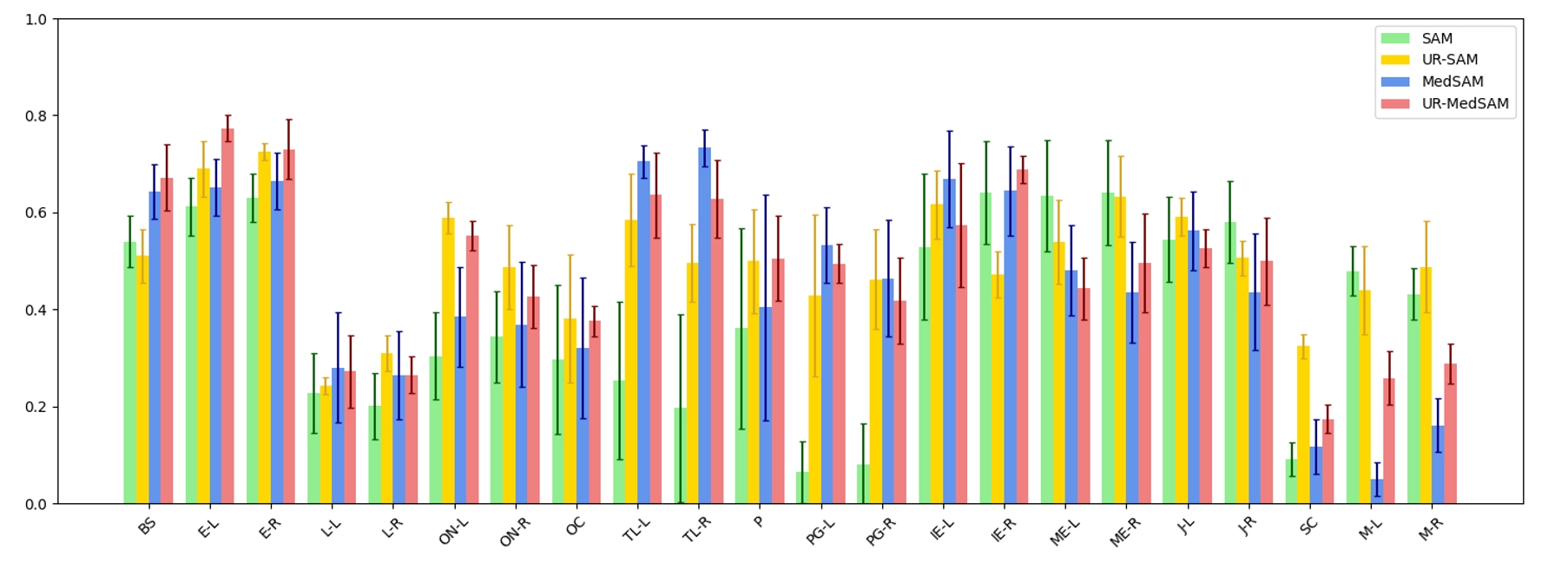
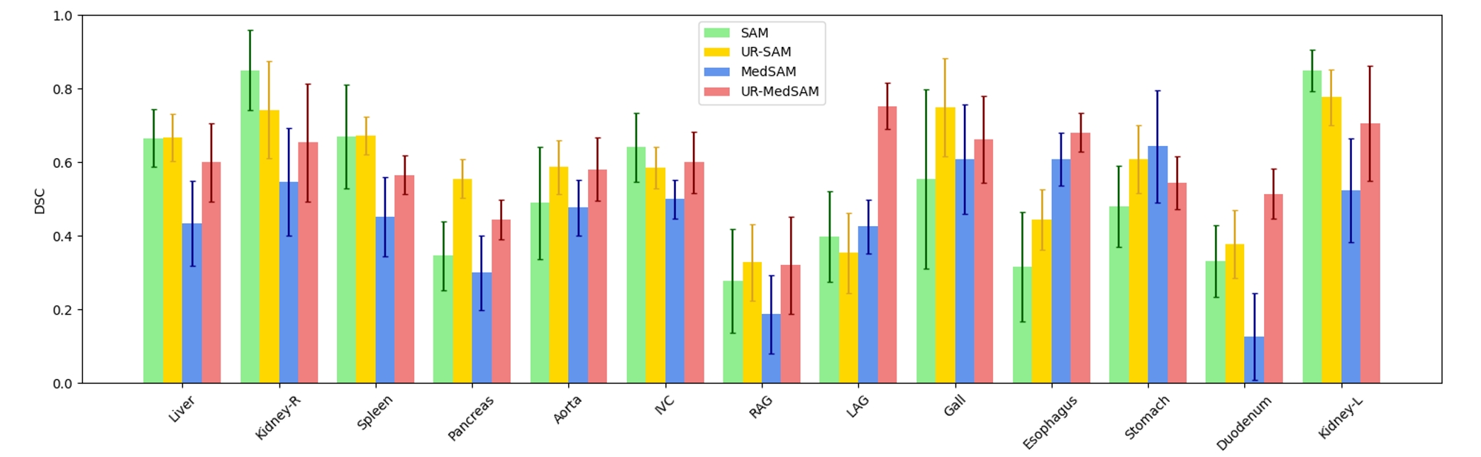
We conduct experiments on two different medical image segmentation datasets. The first dataset is Automatic Structure Segmentation for Radiotherapy Planning Challenge Task1 dataset (StructSeg), which contains 50 CT scans for the segmentation of 22 head-and-neck (HaN) organs including brain stem, left eye, right eye, left lens, right lens, left optic nerve, right optic nerve, optic chiasma, left temporal lobes, right temporal lobes, pituitary, left parotid gland, right parotid gland, left inner ear, right inner ear, left middle ear, right middle ear, left TM joint, right TM joint, spinal cord, left mandible and right mandible. The second dataset is Fast and Low-resource semi-supervised Abdominal oRgan sEgmentation Challenge labeled set (FLARE 22), which contains 50 CT scans for the segmentation of 13 abdominal organs including liver, right kidney, spleen, pancreas, aorta, inferior vena cava, right adrenal gland, left adrenal gland, gallbladder, esophagus, stomach, duodenum, left kidney. These two datasets are highly representative as they contain the vast majority of important organs in the human body, providing good coverage for organ structure segmentation tasks in common clinical scenarios. To validate the effectiveness of our proposed framework, we integrate two different segmentation models with ViT-B backbone for the experiments: the original SAM [16] and MedSAM [23], a specialized variant of SAM fine-tuned on medical image datasets.
4.2 Implementation Details
Following the settings in [18], we randomly select five scans as support images for both datasets. For each organ in these scans, we compute the maximum and minimum coordinates and take the average of the coordinates and features across the support images to generate an average representation of latent coordinates and feature extreme points for auto-prompting. Specifically, the voxel spacing of input images is re-scaled to with cropping patch sizes of pixels. We extend the 3D bounding box obtained from localization by [2, 10, 10] pixels in the z, x, and y directions, respectively to ensure that the targeted organ was completely encapsulated within the box. For data preprocessing, we perform the same procedure as described in [23], which includes adjusting the slice resolution to 3 × 1024 × 1024 and normalizing all images to the range [-500, 1000] as this range effectively encompasses most tissues. In addition to the automatic localization to generate prompts, we also conduct a series of experiments for manual prompting-based interactive segmentation, which are simulated based on the ground truth. To mimic the potential inaccuracies in manually drawn bounding boxes, we introduce a random perturbation of 0-20 pixels. In our experiments, we use the Dice Similarity Coefficient (DSC) as the evaluation metric of the segmentation task.
| (6) |
4.3 Ablation Analysis
In this section, we aim to evaluate the effectiveness of our uncertainty rectification approach. As shown in Table 1, we first conduct experiments using different augmentation numbers and perturb ratios for prompt augmentation to select the optimal parameters for uncertainty estimation. From the results, we observe that incorporating perturbations into input prompts can improve the segmentation performance to some extent and enhance the model’s overall robustness. However, the benefits of additional augmentations were not observed when the number of augmentations exceeded three, or in some cases, even led to decreased segmentation performance. It is noteworthy that high perturbation ratios may cause some parts of the target to fall outside of the bounding box, resulting in decreased segmentation performance. Therefore, the augmentation ratio should be set in a suitable range. Table 1 presents the ablation analysis of different components of our framework. It can be seen that using class-specific thresholds for rectification increases the segmentation performance in both datasets.
4.4 Comparison Experiments
We first conduct experiments on auto-prompting setting where the landmark detection model introduced in 3.1 is utilized to detect extreme points and generate bounding boxes as prompts for segmentation. We compare our rectification method with the FN/FP correction strategy in [37], where high-uncertainty areas are assumed to be potential false negative and positive regions for correction by removing potential false positive regions (Unc-FPC), adding potential false negative regions (Unc-FNC) and correcting both potential false negative and positive regions (Unc-FPNC) from the initial segmentation result. In addition to the auto-prompting, we also include manual prompting simulated based on the ground-truth masks for comparison.
The experimental results of 3D head-and-neck organ segmentation in shown in Table 3. The head-and-neck region typically comprises relatively small organs with irregular shapes. We observe that directly applying SAM fails to segment target organs like the parotid gland (PG-L/PG-R) and spinal cord (SC), with an average dice coefficient lower than 10%, while ensembled results with augmented prompts can significantly improve the performance of these targets. For these small organs, the precise location of region of interest is crucial for the accuracy of subsequent segmentation and prompt augmentation can somehow ease the negative influence of generated inaccurate prompts. For 3D abdominal organ segmentation in Table 4, most target organs can be successfully segmented when directly applying SAM, especially for organs with neat shapes and well-defined boundaries like the liver and kidneys. However, we observe that prompt augmentation cannot consistently enhance performance and may even decrease the performance for some targets, since part of the organ may extend beyond the augmented bounding box in some instances.
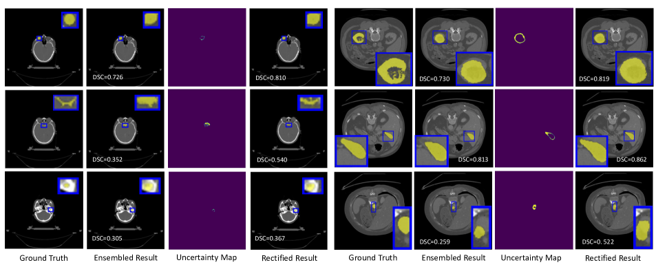
For rectification results, we observe that the performance of comparing strategies varied for different target organs due to the complex shape differences. Given that regions with high uncertainty may not necessarily correspond to false positives or negatives, especially for complex organs with differing shapes and volumes, this approach did not consistently improve performance. In comparison, our rectification strategy demonstrates more robust improvements for most segmentation targets, resulting in overall superior performance. We compare the performance of original and uncertainty rectified segmentation results in Fig 2 and 3. It can be observed that our framework further improves the segmentation performance with up to 10.7 % for head-and-neck organ segmentation and 13.8 % for abdominal organ segmentation in average dice coefficient. Fig. 4 presents the visual analysis of our proposed framework. The four columns represent the ground truth, ensembled segmentation result, high uncertainty mask, and rectified segmentation result, respectively. We can observe that rectified segmentation results have superior performance with less segmentation error compared with ensembled segmentation results, demonstrating the effectiveness of our proposed framework.
5 Discussion and Conclusion
In this work, we present UR-SAM, an uncertainty rectified SAM framework for auto-prompting medical image segmentation by leveraging prompt augmentation of generated bounding box prompts for uncertainty evaluation and utilizing estimated uncertainty for rectification of segmentation results to improve the accuracy. Furthermore, the uncertainty map can help identify potential segmentation errors and support further analysis, offering valuable guidance in areas where manual focus and refinement are required for clinicians. The framework could also be integrated into interactive mechanisms, allowing users to select and modify specific regions that require refinement. Besides, we observe that MedSAM underperforms SAM to some extent, which is in line with the observations in [18]. This indicates that despite being fine-tuned on relatively large-scale medical datasets, the model does not consistently surpass classic SAM when applied to previously unseen datasets.
While our method has demonstrated significant improvements, there are aspects where further enhancements could be made. One notable limitation is that we use a relatively simple strategy to classify pixels in regions of high uncertainty, which relied solely on the intensity of the pixel and did not consider information from neighboring pixels. Consequently, it still yields unsatisfactory results when it comes to segmenting some challenging classes. Future work will focus on incorporating contrast learning of pixels between different classes and integrating more recent medical adaptations of SAM [3, 33] for more comprehensive evaluations. Besides, it would be interesting to utilize uncertainty in the training procedure to select out more representative regions to further enhance model performance [34, 31].
References
- [1] Olivier Bernard, Alain Lalande, Clement Zotti, Frederick Cervenansky, Xin Yang, Pheng-Ann Heng, Irem Cetin, Karim Lekadir, Oscar Camara, Miguel Angel Gonzalez Ballester, et al. Deep learning techniques for automatic mri cardiac multi-structures segmentation and diagnosis: is the problem solved? IEEE transactions on medical imaging, 37(11):2514–2525, 2018.
- [2] Dongjie Cheng, Ziyuan Qin, Zekun Jiang, Shaoting Zhang, Qicheng Lao, and Kang Li. Sam on medical images: A comprehensive study on three prompt modes. arXiv preprint arXiv:2305.00035, 2023.
- [3] Junlong Cheng, Jin Ye, Zhongying Deng, Jianpin Chen, Tianbin Li, Haoyu Wang, Yanzhou Su, Ziyan Huang, Jilong Chen, Lei Jiang, et al. Sam-med2d. arXiv preprint arXiv:2308.16184, 2023.
- [4] Ruining Deng, Can Cui, Quan Liu, Tianyuan Yao, Lucas W Remedios, Shunxing Bao, Bennett A Landman, Lee E Wheless, Lori A Coburn, Keith T Wilson, et al. Segment anything model (sam) for digital pathology: Assess zero-shot segmentation on whole slide imaging. arXiv preprint arXiv:2304.04155, 2023.
- [5] Armen Der Kiureghian and Ove Ditlevsen. Aleatory or epistemic? does it matter? Structural safety, 31(2):105–112, 2009.
- [6] Alexey Dosovitskiy, Lucas Beyer, Alexander Kolesnikov, Dirk Weissenborn, Xiaohua Zhai, Thomas Unterthiner, Mostafa Dehghani, Matthias Minderer, Georg Heigold, Sylvain Gelly, et al. An image is worth 16x16 words: Transformers for image recognition at scale. In International Conference on Learning Representations, 2020.
- [7] Jakob Gawlikowski, Cedrique Rovile Njieutcheu Tassi, Mohsin Ali, Jongseok Lee, Matthias Humt, Jianxiang Feng, Anna Kruspe, Rudolph Triebel, Peter Jung, Ribana Roscher, et al. A survey of uncertainty in deep neural networks. Artificial Intelligence Review, pages 1–77, 2023.
- [8] Shizhan Gong, Yuan Zhong, Wenao Ma, Jinpeng Li, Zhao Wang, Jingyang Zhang, Pheng-Ann Heng, and Qi Dou. 3dsam-adapter: Holistic adaptation of sam from 2d to 3d for promptable medical image segmentation. arXiv preprint arXiv:2306.13465, 2023.
- [9] Sheng He, Rina Bao, Jingpeng Li, P Ellen Grant, and Yangming Ou. Accuracy of segment-anything model (sam) in medical image segmentation tasks. arXiv preprint arXiv:2304.09324, 2023.
- [10] Nicholas Heller, Fabian Isensee, Klaus H Maier-Hein, Xiaoshuai Hou, Chunmei Xie, Fengyi Li, Yang Nan, Guangrui Mu, Zhiyong Lin, Miofei Han, et al. The state of the art in kidney and kidney tumor segmentation in contrast-enhanced ct imaging: Results of the kits19 challenge. Medical image analysis, 67:101821, 2021.
- [11] Chuanfei Hu and Xinde Li. When sam meets medical images: An investigation of segment anything model (sam) on multi-phase liver tumor segmentation. arXiv preprint arXiv:2304.08506, 2023.
- [12] Yuhao Huang, Xin Yang, Lianli Liu, Hangyu Zhou, Ao Chang, Xinrui Zhou, Rusi Chen, Junxuan Yu, Jiongquan Chen, Chaoyu Chen, Haozhe Chi, Xindi Hu, Deng-Ping Fan, Fajin Dong, and Dong Ni. Segment anything model for medical images? arXiv preprint arXiv:2304.14660, 2023.
- [13] Fabian Isensee, Paul F Jaeger, Simon AA Kohl, Jens Petersen, and Klaus H Maier-Hein. nnu-net: a self-configuring method for deep learning-based biomedical image segmentation. Nature methods, 18(2):203–211, 2021.
- [14] Wei Ji, Jingjing Li, Qi Bi, Wenbo Li, and Li Cheng. Segment anything is not always perfect: An investigation of sam on different real-world applications. arXiv preprint arXiv:2304.05750, 2023.
- [15] Alex Kendall and Yarin Gal. What uncertainties do we need in bayesian deep learning for computer vision? Advances in neural information processing systems, 30, 2017.
- [16] Alexander Kirillov, Eric Mintun, Nikhila Ravi, Hanzi Mao, Chloe Rolland, Laura Gustafson, Tete Xiao, Spencer Whitehead, Alexander C Berg, Wan-Yen Lo, et al. Segment anything. arXiv preprint arXiv:2304.02643, 2023.
- [17] Alain Lalande, Zhihao Chen, Thibaut Pommier, Thomas Decourselle, Abdul Qayyum, Michel Salomon, Dominique Ginhac, Youssef Skandarani, Arnaud Boucher, Khawla Brahim, et al. Deep learning methods for automatic evaluation of delayed enhancement-mri. the results of the emidec challenge. Medical Image Analysis, 79:102428, 2022.
- [18] Wenhui Lei, Xu Wei, Xiaofan Zhang, Kang Li, and Shaoting Zhang. Medlsam: Localize and segment anything model for 3d medical images. arXiv preprint arXiv:2306.14752, 2023.
- [19] Chengyin Li, Prashant Khanduri, Yao Qiang, Rafi Ibn Sultan, Indrin Chetty, and Dongxiao Zhu. Auto-prompting sam for mobile friendly 3d medical image segmentation. arXiv preprint arXiv:2308.14936, 2023.
- [20] Feng Li, Hao Zhang, Peize Sun, Xueyan Zou, Shilong Liu, Jianwei Yang, Chunyuan Li, Lei Zhang, and Jianfeng Gao. Semantic-sam: Segment and recognize anything at any granularity. arXiv preprint arXiv:2307.04767, 2023.
- [21] Geert Litjens, Thijs Kooi, Babak Ehteshami Bejnordi, Arnaud Arindra Adiyoso Setio, Francesco Ciompi, Mohsen Ghafoorian, Jeroen Awm Van Der Laak, Bram Van Ginneken, and Clara I Sánchez. A survey on deep learning in medical image analysis. Medical image analysis, 42:60–88, 2017.
- [22] Charles J. Lynch and Conor Liston. New machine-learning technologies for computer-aided diagnosis. Nature Medicine, 24:1304–1305, 2018.
- [23] Jun Ma and Bo Wang. Segment anything in medical images. arXiv preprint arXiv:2304.12306, 2023.
- [24] Jun Ma, Yao Zhang, Song Gu, Cheng Ge, Shihao Ma, Adamo Young, Cheng Zhu, Kangkang Meng, Xin Yang, Ziyan Huang, et al. Unleashing the strengths of unlabeled data in pan-cancer abdominal organ quantification: the flare22 challenge. arXiv preprint arXiv:2308.05862, 2023.
- [25] Jun Ma, Yao Zhang, Song Gu, Cheng Zhu, Cheng Ge, Yichi Zhang, Xingle An, Congcong Wang, Qiyuan Wang, Xin Liu, Shucheng Cao, Qi Zhang, Shangqing Liu, Yunpeng Wang, Yuhui Li, Jian He, and Xiaoping Yang. Abdomenct-1k: Is abdominal organ segmentation a solved problem? IEEE Transactions on Pattern Analysis and Machine Intelligence, 44(10):6695–6714, 2022.
- [26] Maciej A Mazurowski, Haoyu Dong, Hanxue Gu, Jichen Yang, Nicholas Konz, and Yixin Zhang. Segment anything model for medical image analysis: an experimental study. Medical Image Analysis, 89:102918, 2023.
- [27] Sovesh Mohapatra, Advait Gosai, and Gottfried Schlaug. Sam vs bet: A comparative study for brain extraction and segmentation of magnetic resonance images using deep learning. arXiv preprint arXiv:2304.04738, 2023.
- [28] Olaf Ronneberger, Philipp Fischer, and Thomas Brox. U-net: Convolutional networks for biomedical image segmentation. In International Conference on Medical image computing and computer-assisted intervention, pages 234–241. Springer, 2015.
- [29] Saikat Roy, Tassilo Wald, Gregor Koehler, Maximilian R Rokuss, Nico Disch, Julius Holzschuh, David Zimmerer, and Klaus H Maier-Hein. Sam. md: Zero-shot medical image segmentation capabilities of the segment anything model. arXiv preprint arXiv:2304.05396, 2023.
- [30] Tal Shaharabany, Aviad Dahan, Raja Giryes, and Lior Wolf. Autosam: Adapting sam to medical images by overloading the prompt encoder. arXiv preprint arXiv:2306.06370, 2023.
- [31] Pin Tang, Pinli Yang, Dong Nie, Xi Wu, Jiliu Zhou, and Yan Wang. Unified medical image segmentation by learning from uncertainty in an end-to-end manner. Knowledge-Based Systems, 241:108215, 2022.
- [32] An-Chi Wang, Mobarakol Islam, Mengya Xu, Yang Zhang, and Hongliang Ren. Sam meets robotic surgery: An empirical study in robustness perspective. arXiv preprint arXiv:2304.14674, 2023.
- [33] Haoyu Wang, Sizheng Guo, Jin Ye, Zhongying Deng, Junlong Cheng, Tianbin Li, Jianpin Chen, Yanzhou Su, Ziyan Huang, Yiqing Shen, Bin Fu, et al. Sam-med3d. arXiv preprint arXiv:2310.15161, 2023.
- [34] Tao Wang, Jianglin Lu, Zhihui Lai, Jiajun Wen, and Heng Kong. Uncertainty-guided pixel contrastive learning for semi-supervised medical image segmentation. In Proceedings of the Thirty-First International Joint Conference on Artificial Intelligence, IJCAI, pages 1444–1450, 2022.
- [35] Junde Wu, Rao Fu, Huihui Fang, Yuanpei Liu, Zhaowei Wang, Yanwu Xu, Yueming Jin, and Tal Arbel. Medical sam adapter: Adapting segment anything model for medical image segmentation. arXiv preprint arXiv:2304.12620, 2023.
- [36] Jinyu Yang, Mingqi Gao, Zhe Li, Shang Gao, Fangjing Wang, and Feng Zheng. Track anything: Segment anything meets videos. arXiv preprint arXiv:2304.11968, 2023.
- [37] Xing Yao, Han Liu, Dewei Hu, Daiwei Lu, Ange Lou, Hao Li, Ruining Deng, Gabriel Arenas, Baris Oguz, Nadav Schwartz, et al. False negative/positive control for sam on noisy medical images. arXiv preprint arXiv:2308.10382, 2023.
- [38] Kaiwen Zhang and Dong Liu. Customized segment anything model for medical image segmentation. arXiv preprint arXiv:2304.13785, 2023.
- [39] Yichi Zhang and Rushi Jiao. How segment anything model (sam) boost medical image segmentation? arXiv preprint arXiv:2305.03678, 2023.
- [40] Yichi Zhang, Rushi Jiao, Qingcheng Liao, Dongyang Li, and Jicong Zhang. Uncertainty-guided mutual consistency learning for semi-supervised medical image segmentation. Artificial Intelligence in Medicine, 138:102476, 2023.
- [41] Ke Zou, Zhihao Chen, Xuedong Yuan, Xiaojing Shen, Meng Wang, and Huazhu Fu. A review of uncertainty estimation and its application in medical imaging. Meta-Radiology, 1(1):100003, 2023.
- [42] Ke Zou, Xuedong Yuan, Xiaojing Shen, Meng Wang, and Huazhu Fu. Tbrats: Trusted brain tumor segmentation. In International Conference on Medical Image Computing and Computer-Assisted Intervention, pages 503–513. Springer, 2022.