Surface Enhanced Infrared Absorption mechanism and modification of the plasmonic response
Abstract
Surface Enhanced Infrared Absorption (SEIRA) is an experimental method where trace amount of a compound can be detected with high sensibility. This high detection sensibility is the result of the interaction of the molecules with a localized plasmon, usually from a metallic nano-particle. In this study we numerically investigate by discrete dipole approximation the origin of the Fano-like response of the system, including the induced transparency when the plasmon resonance and the molecular vibrational mode coincide. The detailed analysis of the localization of the absorption show that the modification of the absorption cross-section when the molecule is present comes from a change of the plasmonic resonance, not from the direct molecular response which is negligible. This sheds a new light on the SEIRA mechanism. In particular, it demonstrates that the sensibility is associated with the influence of the molecule on the plasmon resonance rather than with the local field enhancement itself.
Keywords : SEIRA, plasmonics , LSPR , nanorods, DDA, infrared absorption, Vibrational properties
1 Introduction
Vibrational spectroscopies are powerful optical characterization techniques that offer the advantages of being label free and non-destructive. In addition to Raman spectroscopy, infrared (IR) absorption is widely used for direct analysis of molecular functional groups and in situ analysis of surface reactions [1, 2, 3, 4, 5, 6]. However, its small absorption cross-section, although much larger than that of Raman spectroscopy, limits its utility for large quantities of materials. In contrast, surface-enhanced infrared absorption (SEIRA), based on the coupling with a plasmonic system, increases the sensibility of the detection by a factor of 101 to 107, enabling optical sensing of trace amounts of molecules [2, 3, 4, 5, 7, 8, 6].
SEIRA necessitates that the plasmonic system resonates at the same frequency as that of the specific molecular vibrational mode under consideration. In practice, gold nanoparticles are frequently utilized due to their high chemical stability and their localized surface plasmon resonance (LSPR) in the IR range. Gold nanorods are particularly popular due to the good control of the synthesis or fabrication processes and the high tunability of their optical resonance, through adjusting their length and radius [9, 10, 11, 6]. Alternative nanoparticle shapes with sharper edges such as gold bowtie [8] or trimers [12] have been considered. Besides gold nanoparticles, graphene is also a substrate of choice due to its tunable IR plasmon via doping [2, 13, 14, 15].
The interpretation of SEIRA relies on the comparison of the absorbance or transmittance associated with the LSPR with or without the presence of the probed molecules. When both resonances coincide, the optical response diminishes with the presence of the molecules, resulting in a negative differential cross-section and an induced transparency phenomenon. If the resonances are slightly mismatched, a Fano-like behavior is observed [9, 10, 8, 14, 15, 16, 17, 18, 19, 20, 7, 21, 22]. The remarkable sensibility of SEIRA is linked to the fact that the magnitude of the differential cross-section is orders of magnitude larger than that of isolated molecules [2, 3, 4, 5, 7, 8]. This observation is often associated with the enhancement of the local electromagnetic field at the molecule’s position [18, 16, 14, 6]. However, this interpretation does not account for the induced transparency, or the Fano-like shape [23, 10]. The relationship of the SEIRA with the square of the local electric field which follows this first analysis has also never been clearly evidenced.
In an early investigation [5] and in a more recent review [23], it was suggested that the influence of the molecules on the polarizability of the metal nanoparticles at the LSPR is the underlying mechanism for SEIRA. This argument which was based on simulations of the optical response of ellipsoidal metallic nanoparticles coated with a dielectric layer, posits that the plasmonic particles act as an antenna, relaying the signal of the molecule, rather than just exalting the electromagnetic fields.
In this study, we present a comprehensive examination of the SEIRA mechanism through numerical simulations. The manuscript is organized as follows. Our numerical simulations show that the SEIRA effect is associated with a surprisingly high influence of the molecules on the LSPR resonance. They demonstrate that the molecule differential cross-section does not originate from changes of the absorption cross-section of the molecule. Furthermore, our findings corroborate that the scattering cross-section exhibits a similar behavior as the absorption cross-section in SEIRA, despite the fact that molecules themselves do not scatter light [10].
In Sec.II, we introduce discrete dipole approximation (DDA) [24, 25] which is used throughout this study. While DDA has proven to be effective and accurate in describing the optical properties of nanomaterials, it has not been previously applied in the context of SEIRA, to the best of our knowledge. Previous numerical investigations of SEIRA, mainly utilizing the Finite Difference Time Domain approach, have successfully replicated the Fano-like spectra but have not provided definitive conclusions regarding the origin of this phenomenon.
In Sec.III, we delve into the SEIRA response of a model molecular system, featuring a vibrational mode corresponding to the CO stretching mode, positioned within the hot spot created by two gold nanorods.
2 Methods
In DDA, the materials are modeled by a set of coupled polarizable dipoles excited by an external field, a monochromatic plane wave in our case. The formal derivation from Maxwell’s equations and boundary conditions [26] reveals that the discretization of the bulk materials and the definition of the local polarizability play a crucial role in determining the simulation accuracy. For the gold nanorod, we derived the local polarizability from lattice dispersion relation, which is a correction of Clausius-Mossotti’s formula [24], from the experimental bulk refractive index [27]. The simulations have been performed with the DDSCAT code [24]
In our simulations, we consider nano-rods with a nm diameter and a length of nm, modeled by dipoles placed on a cubic grid with a periodicity of nm, ensuring the convergence (see also Ref [28] for an investigation of the response of similar nano-object by DDA).
After the coupling with the external fields, the polarization of each dipole is computed and the local properties such as the electromagnetic near-field is determined. Far-field properties such as the absorption and scattering cross-sections are calculated by summing the contribution of each individual dipole and can be attributed to specific regions of the system.
To describe the response of the probed molecules in SEIRA, we take advantage of the versatility of the DDA by representing them as point dipoles with a polarizability associated with their vibrational modes described by a Drude-Lorentz model. The vibrational mode wavenumber is varied from cm-1 to cm-1, corresponding to the CO stretching modes in different chemical environments, to investigate the detuning between the LSPR and one of the molecular vibrational modes, with a damping parameter set at 16 cm-1. Of course, in experimental systems, the vibrational frequency is fixed by the molecular composition and cannot be tuned. In the same way, if the plasmon resonance can be tuned by a modification of the geometry of the system, it cannot be modified dynamically. Our main goal in this study is to investigate the SEIRA effect with both the metallic nanoparticle and the molecules presenting an optical resonance at the same frequency or at nearby frequencies. For practical reasons, the metallic plasmonic system will be kept fixed in the following, while the molecular mode will be tuned. Note that the optical resonance of a nanorod can be modified thanks to LASER engraving [28]. The molecules are modeled by 88 dipoles dispersed in the gap with a distance of at least nm with their closest neighbor which diminishes self-interacting phenomenon.
3 Results and Discussion
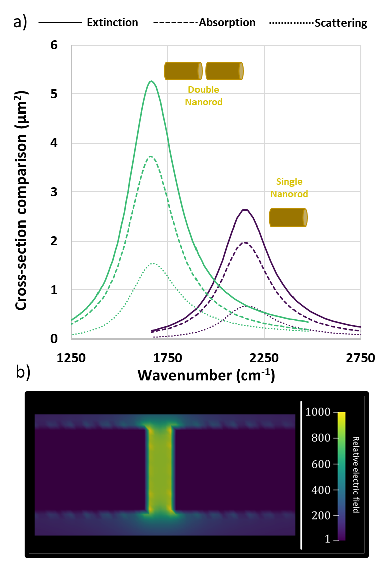
The electromagnetic response of the gold nanorods in the infrared is due to the plasmon resonance that can be described, for an infinite nanorod, by the azimuthal number m and the wavevector along the nanorod axis [28]. For finite nanorod of length , the wavector is quantified () and the m=1 mode is excited by light polarized perpendicular to the nanorod axis and the m=0 mode for parallel polarization [29]. The optical response of our gold nanorod is shown in Fig. 1a for a longitudinal excitation in the model wavenumber range of interest for this study. The resonance at cm-1 to corresponds to the plasmon with the longest wavelength that can take place in the nanorod ().
The coupling with a second nanorod modifies the optical response and creates a hot spot of intense local electromagnetic field, depending strongly on the separation distance. In Fig. 1a, a redshift to cm-1 is obtained for nanorods separated by nm, a distance small enough to get a strong hot spot but that can be described by our discretization grid. The corresponding near-field amplitude in the hot spot region at the resonance wavenumber is, in our case, enhanced by more than three orders of magnitude compared to the incident field (Fig. 1b).
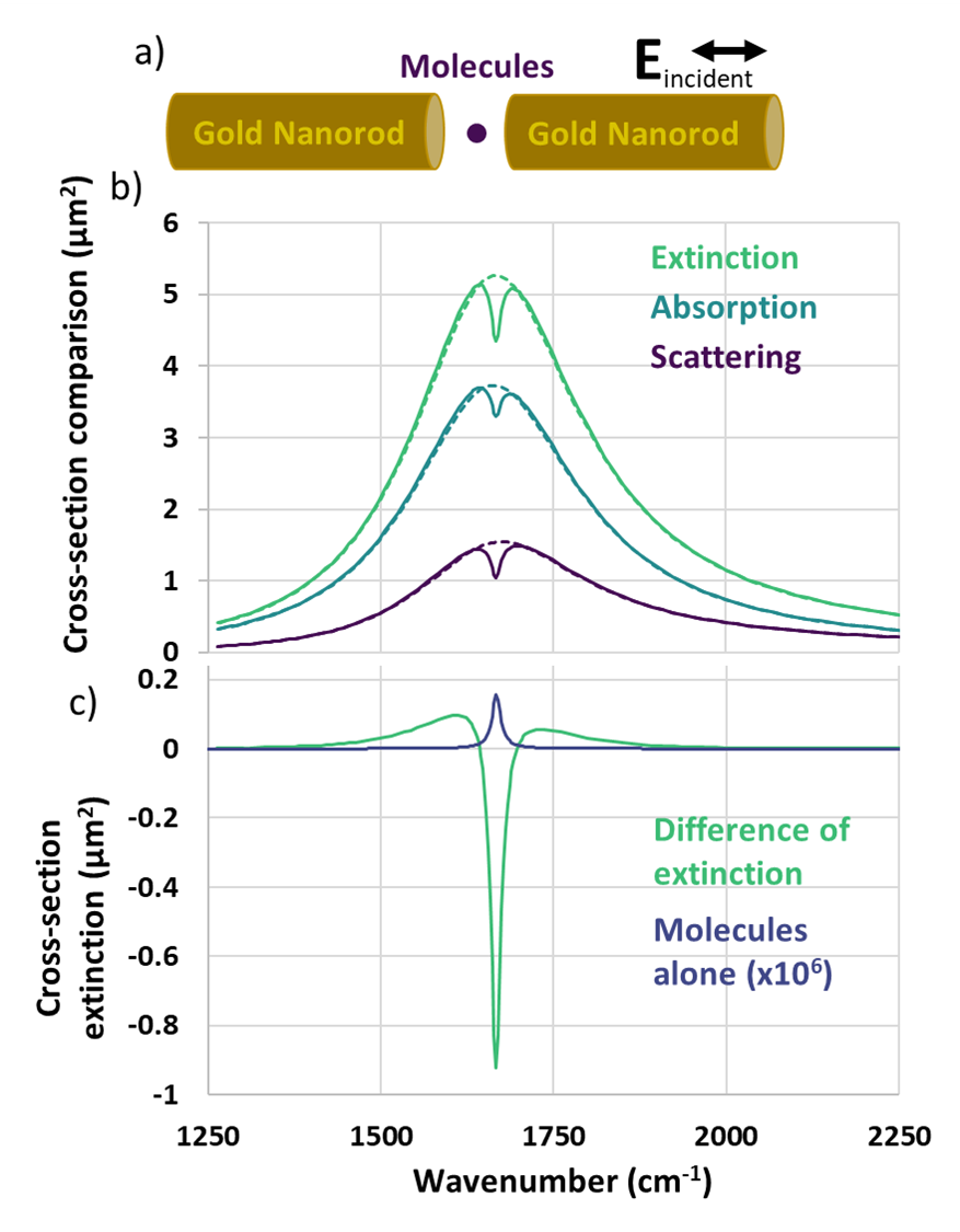
We investigate the SEIRA response of the molecule lying in the hot spot, as illustrated on Fig. 2a. The wavenumber of the vibrational mode is chosen to be cm-1 to corresponding to the maximum of the plasmon resonance of the coupled nanorods.
The absorption, scattering and extinction cross-sections are shown with and without molecules in Fig. 2b. We recover here the well-described decrease of the total cross-section in the presence of the molecule [23, 16, 30, 9, 13, 14, 15, 1, 18, 4, 19, 10, 20, 8, 7, 31, 21, 2, 22, 12, 32, 17, 11] that is observed for both absorption and scattering of light [10]. The difference of the extinction cross-sections (Fig. 2c) evidences this transparency induced by the molecules more clearly but also displays the Fano-like profile with an increase of the extinction on the two sides of the main negative peak.
It is hardly justified to speak about enhancement for SEIRA due to the very different shape of the response of the isolated molecule and of the SEIRA signal. This is even clearer for the Fano line shape when the resonance frequencies of the plasmonic system and of the molecules do not coincide (see later). We then prefer the term sensibility that we define by the difference between the extrema of the differential cross-section, divided by the maximum absorption cross-section of the isolated molecule, following Ref [3]. In the example above, the sensibility is . Experimentally, the sensibility is difficult to evaluate as the optical cross-section of the exact same plasmonic system without the molecules is not available and the number of molecules in the hot spot is not known.
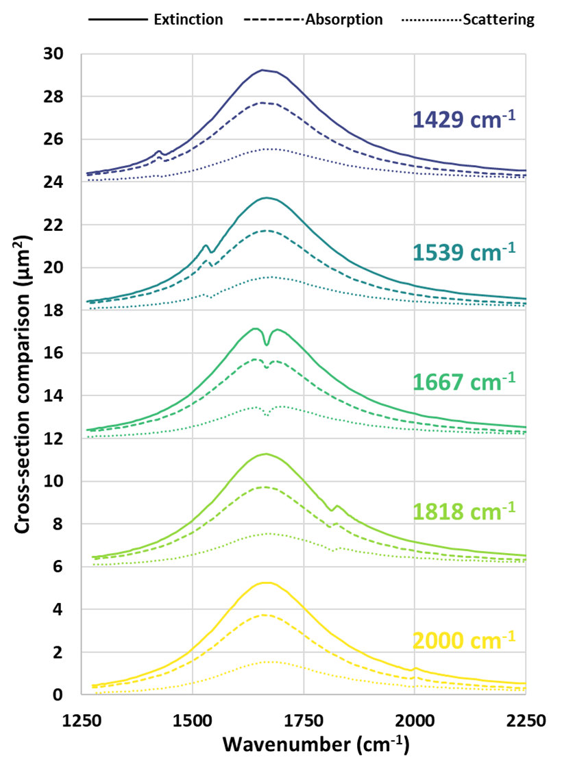
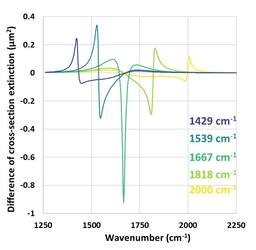
The case where both resonances coincide is very specific and does not correspond to most of the experimental results. Fig. 3 shows the extinction, absorption and scattering for five different vibrational wavenumbers below and above the plasmonic resonance. Fig. 4 presents the Fano-like difference of cross-section, with a curve shape in agreement with previous studies [9, 10, 8, 14, 15, 16, 17, 18, 19, 20, 7, 21, 22]. The overall shape of the SEIRA signal depends strongly on the difference of the resonances between the particles and the molecules [33], ranging from an induced transparency described above to, mainly, an increase of the extinction when the resonances do not coincide.
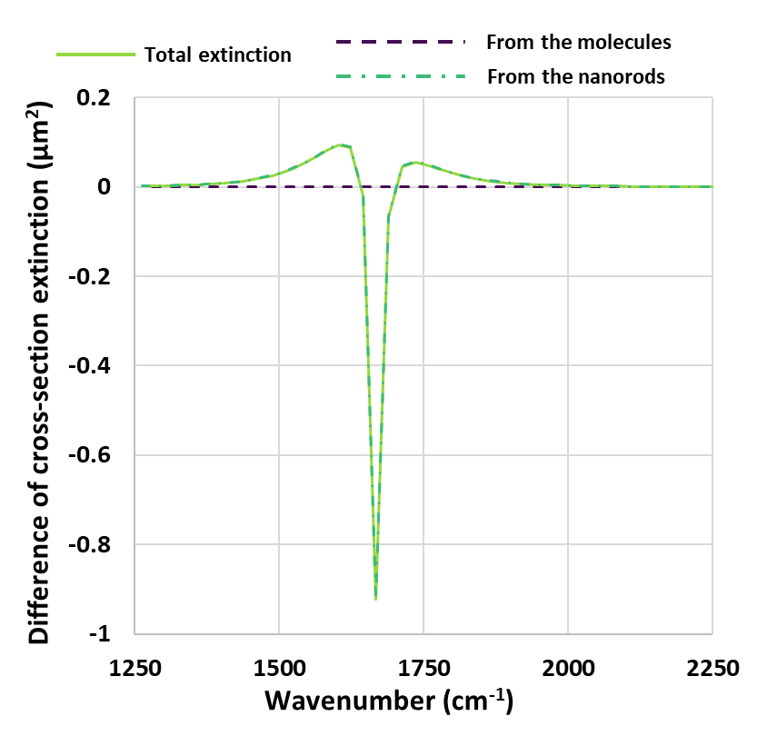
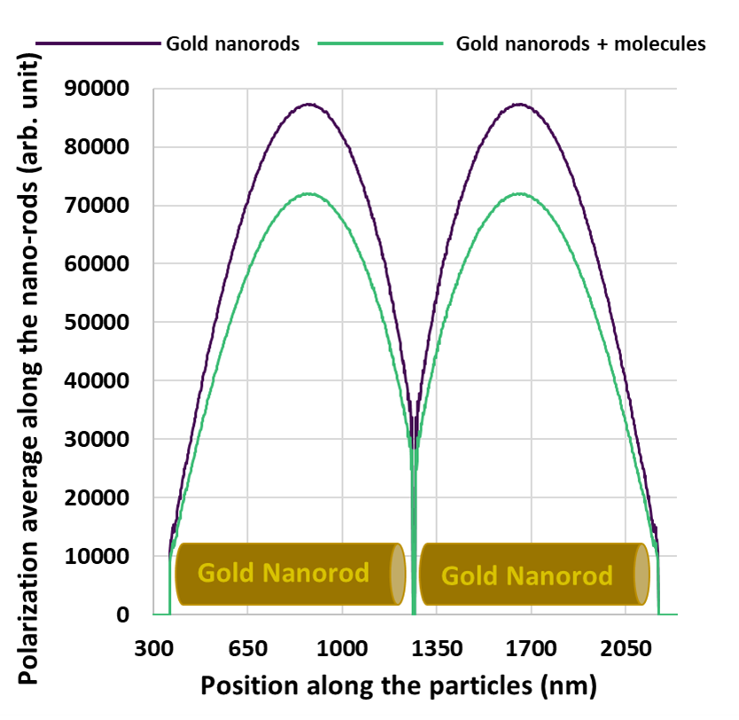
In a DDA calculation, we can discriminate the spatial origin of the optical response as it is expressed as a sum over the local dipoles [24]. It turns out that the change of extinction originates almost entirely from the nanorods, as it is illustrated for the case of the strongest SEIRA response in Fig. 5 (the same conclusion can be drawn for the other computed spectra). This conclusion is also supported by the analysis of the polarization profile of the nanorod along its axis with and without the molecules (Fig. 6). The polarization profile associated with the LSPR mode is very similar in both cases but with a global drop of the response. This behavior is similar to the one expected if a dielectric medium were added between the interacting nanorods which would make the LSPR resonance to be less pronounced, tying in with previous qualitative explanations [5, 23].
Finally, it is interesting to note that the SEIRA response is observed in both the absorption and the scattering spectra (Fig. 3 and Ref [10]). As there is no scattering from a single molecule (or from a point dipole in our model), the change in scattering comes entirely from the nanorods, going in the same direction as our previous conclusions. SEIRA is then associated with a modification of the LSPR resonance due to the presence of the molecules rather than to a direct contribution of the molecule to the spectra.
4 Conclusion
SEIRA is a very sensitive vibrational spectroscopy. Its modeling is not an easy task at it requires to describe at the same time, the plasmonic response of a metallic system and the vibrational response of a molecule. In this paper we have proposed to investigate SEIRA within a DDA approach, reproducing its main features such as the induced transparency and the Fano-like shape of the resonance for different mismatch of the resonance frequencies.
We attribute the sensibility of SEIRA signature to a modification of the plasmon excitation due to the presence of the molecules rather than to a response of the molecules interacting with the local field. This is of prime importance for the design of new SEIRA samples as the more adapted plasmonic systems will be the ones that are more sensitive to a change of environment (the molecules) rather than the ones with the highest local-field enhancement.
References
- [1] Hwang I, Kim M, Yu J, Lee J, Choi J H, Park S A, Chang W S, Lee J and Jung J Y 2021 Small Methods 5 2100277 ISSN 2366-9608
- [2] Hu H, Yang X, Guo X, Khaliji K, Biswas S R, García de Abajo F J, Low T, Sun Z and Dai Q 2019 Nature Communications 10 1131 ISSN 2041-1723 number: 1 Publisher: Nature Publishing Group URL https://www.nature.com/articles/s41467-019-09008-0
- [3] Baillieul M, Rinnert E, Lemaitre J, Michel K, Colas F, Bodiou L, Demésy G, Kakuta S, Rumyantseva A, Lerondel G, Boukerma K, Renversez G, Toury T, Charrier J and Nazabal V 2022 ACS Omega 7 47840–47850 publisher: American Chemical Society URL https://doi.org/10.1021/acsomega.2c05502
- [4] Ishikawa A, Hara S, Tanaka T, Hayashi Y and Tsuruta K 2017 Scientific Reports 7 3205 ISSN 2045-2322 number: 1 Publisher: Nature Publishing Group URL https://www.nature.com/articles/s41598-017-03545-8
- [5] Osawa M, Ataka K I, Yoshii K and Nishikawa Y 1993 Applied Spectroscopy 47 1497–1502 ISSN 0003-7028 publisher: SAGE Publications Ltd STM URL https://doi.org/10.1366/0003702934067478
- [6] Kozuch J, Ataka K and Herbele J 2023 Nat Rev Methods Primers 3 ISSN 71 URL https://www.nature.com/articles/s43586-023-00261-8
- [7] Neubrech F, Huck C, Weber K, Pucci A and Giessen H 2017 Chemical Reviews 117 5110–5145 ISSN 0009-2665 URL https://doi.org/10.1021/acs.chemrev.6b00743
- [8] Dong L, Yang X, Zhang C, Cerjan B, Zhou L, Tseng M L, Zhang Y, Alabastri A, Nordlander P and Halas N J 2017 Nano Letters 17 5768–5774 ISSN 1530-6984 URL https://doi.org/10.1021/acs.nanolett.7b02736
- [9] Giordano M C, Tzschoppe M, Barelli M, Vogt J, Huck C, Canepa F, Pucci A and Buatier de Mongeot F 2020 ACS Applied Materials & Interfaces 12 11155–11162 ISSN 1944-8244 publisher: American Chemical Society URL https://doi.org/10.1021/acsami.9b19719
- [10] Neuman T, Huck C, Vogt J, Neubrech F, Hillenbrand R, Aizpurua J and Pucci A 2015 The Journal of Physical Chemistry C 119 26652–26662 ISSN 1932-7447 URL https://doi.org/10.1021/acs.jpcc.5b08344
- [11] Abb M, Wang Y, Papasimakis N, de Groot C H and Muskens O L 2014 Nano Letters 14 346–352 ISSN 1530-6984 publisher: American Chemical Society URL https://doi.org/10.1021/nl404115g
- [12] Mackin R T, Cohn B, Engelman B, Goldner A, Rubtsov I V and Chuntonov L 2019 The Journal of Physical Chemistry C 123 24731–24739 ISSN 1932-7447 publisher: American Chemical Society URL https://doi.org/10.1021/acs.jpcc.9b07045
- [13] Farmer D B, Avouris P, Li Y, Heinz T F and Han S J 2016 ACS Photonics 3 553–557 publisher: American Chemical Society URL https://doi.org/10.1021/acsphotonics.6b00143
- [14] Li Y, Yan H, Farmer D B, Meng X, Zhu W, Osgood R M, Heinz T F and Avouris P 2014 Nano Letters 14 1573–1577 ISSN 1530-6984 publisher: American Chemical Society URL https://doi.org/10.1021/nl404824w
- [15] Yan H, Low T, Guinea F, Xia F and Avouris P 2014 Nano Letters 14 4581–4586 ISSN 1530-6984 publisher: American Chemical Society URL https://doi.org/10.1021/nl501628x
- [16] Zvagelsky R, Chubich D, Pisarenko A, Bedran Z and Zhukova E 2021 The Journal of Physical Chemistry C 125 4694–4703 ISSN 1932-7447 publisher: American Chemical Society URL https://doi.org/10.1021/acs.jpcc.0c10433
- [17] Cerjan B, Yang X, Nordlander P and Halas N J 2016 ACS Photonics 3 354–360 publisher: American Chemical Society URL https://doi.org/10.1021/acsphotonics.6b00024
- [18] Langer J, Jimenez de Aberasturi D, Aizpurua J, Alvarez-Puebla R A, Auguié B, Baumberg J J, Bazan G C, Bell S E J, Boisen A, Brolo A G, Choo J, Cialla-May D, Deckert V, Fabris L, Faulds K, García de Abajo F J, Goodacre R, Graham D, Haes A J, Haynes C L, Huck C, Itoh T, Käll M, Kneipp J, Kotov N A, Kuang H, Le Ru E C, Lee H K, Li J F, Ling X Y, Maier S A, Mayerhöfer T, Moskovits M, Murakoshi K, Nam J M, Nie S, Ozaki Y, Pastoriza-Santos I, Perez-Juste J, Popp J, Pucci A, Reich S, Ren B, Schatz G C, Shegai T, Schlücker S, Tay L L, Thomas K G, Tian Z Q, Van Duyne R P, Vo-Dinh T, Wang Y, Willets K A, Xu C, Xu H, Xu Y, Yamamoto Y S, Zhao B and Liz-Marzán L M 2020 ACS Nano 14 28–117 ISSN 1936-0851 publisher: American Chemical Society URL https://doi.org/10.1021/acsnano.9b04224
- [19] Neubrech F and Pucci A 2013 IEEE Journal of Selected Topics in Quantum Electronics 19 4600809–4600809 ISSN 1558-4542 conference Name: IEEE Journal of Selected Topics in Quantum Electronics
- [20] Neubrech F, Beck S, Glaser T, Hentschel M, Giessen H and Pucci A 2014 ACS Nano 8 6250–6258 ISSN 1936-0851 URL https://doi.org/10.1021/nn5017204
- [21] Wei J, Li Y, Chang Y, Hasan D M N, Dong B, Ma Y, Qiu C W and Lee C 2019 ACS Applied Materials & Interfaces 11 47270–47278 ISSN 1944-8244 publisher: American Chemical Society URL https://doi.org/10.1021/acsami.9b18002
- [22] Tanaka T, Yano T a and Kato R 2022 Nanophotonics 11 2541–2561 ISSN 2192-8614 publisher: De Gruyter URL https://www.degruyter.com/document/doi/10.1515/nanoph-2021-0661/html
- [23] Yang X, Sun Z, Low T, Hu H, Guo X, García de Abajo F J, Avouris P and Dai Q 2018 Advanced Materials 30 1704896 ISSN 1521-4095
- [24] Draine B T and Flatau P J 1994 J. Opt. Soc. Am. A 11 1491–1499 URL http://josaa.osa.org/abstract.cfm?URI=josaa-11-4-1491
- [25] Flatau P J and Draine B T 2012 Opt. Express 20 1247–1252 URL http://www.opticsexpress.org/abstract.cfm?URI=oe-20-2-1247
- [26] Yurkin M A and Hoekstra A G 2007 Journal of Quantitative Spectroscopy and Radiative Transfer 106 558–589 ISSN 0022-4073 URL https://www.sciencedirect.com/science/article/pii/S0022407307000556
- [27] Olmon R L, Slovick B, Johnson T W, Shelton D, Oh S H, Boreman G D and Raschke M B 2012 Physical Review B 86 235147 URL https://link.aps.org/doi/10.1103/PhysRevB.86.235147
- [28] Pelaez-Fernandez M, Majerus B, Dufour R, Funes-Hernando D, Duvail J L, Henrard L and Arenal R 2023 Microscopy and Microanalysis 29 378–379 ISSN 1431-9276 URL https://doi.org/10.1093/micmic/ozad067.178
- [29] Gómez-Medina R, Yamamoto N, Nakano M and Abajo F J G d 2008 New Journal of Physics 10 105009 ISSN 1367-2630 URL https://dx.doi.org/10.1088/1367-2630/10/10/105009
- [30] Durmaz H, Li Y and Cetin A E 2018 Sensors and Actuators B: Chemical 275 174–179 ISSN 0925-4005 URL https://www.sciencedirect.com/science/article/pii/S0925400518314564
- [31] Wang T, Dong Z, Koay E H H and Yang J K W 2019 ACS Photonics 6 1272–1278 publisher: American Chemical Society URL https://doi.org/10.1021/acsphotonics.9b00229
- [32] Barbillon G 2022 International Journal of Molecular Sciences 23 10592 ISSN 1422-0067 number: 18 Publisher: Multidisciplinary Digital Publishing Institute URL https://www.mdpi.com/1422-0067/23/18/10592
- [33] Tu X, Mario L Y and Mei T 2010 Optics Express 18 18820–18831 ISSN 1094-4087 URL https://opg.optica.org/oe/abstract.cfm?uri=oe-18-18-18820