Enhancing Epileptic Seizure Detection
with EEG Feature Embeddings
Abstract
Epilepsy is one of the most prevalent brain disorders that disrupts the lives of millions worldwide. For patients with drug-resistant seizures, there exist implantable devices capable of monitoring neural activity, promptly triggering neurostimulation to regulate seizures, or alerting patients of potential episodes. Next-generation seizure detection systems heavily rely on high-accuracy machine learning-based classifiers to detect the seizure onset. Here, we propose to enhance the seizure detection performance by learning informative embeddings of the EEG signal. We empirically demonstrate, for the first time, that converting raw EEG signals to appropriate embeddings can significantly boost the performance of seizure detection algorithms. Importantly, we show that embedding features, which converts the raw EEG into an alternative representation, is beneficial for various machine learning models such as Logistic Regression, Multi-Layer Perceptron, Support Vector Machines, and Gradient Boosted Trees. The experiments were conducted on the CHB-MIT scalp EEG dataset. With the proposed EEG feature embeddings, we achieve significant improvements in sensitivity, specificity, and AUC score across multiple models. By employing this approach alongside an SVM classifier, we were able to attain state-of-the-art classification performance with a sensitivity of 100% and specificity of 99%, setting a new benchmark in the field.
Index Terms:
Seizure detection, Feature embeddings, EEG classification, Machine learningI Introduction
A large population globally is affected by epilepsy, a neurological disorder that is recognized by the repetitive occurrence of seizures. The frequency and intensity of seizures have a profound impact on an individual’s quality of life, affecting their ability to work, participate in social activities, and overall well-being. Early detection and management of seizures are crucial to minimize their devastating effect on patients. Seizures are represented by episodes of high-amplitude low-frequency, or low-amplitude high-frequency neural activity in the brain [1, 2, 3]. Current seizure detection systems operate by constantly acquiring neural data from either scalp (i.e., EEG) or intracranial electrodes (i.e., iEEG), followed by signal processing to identify the onset of seizures. This technology offers substantial benefits for individuals with epilepsy and enables them to seek assistance promptly, mitigate the risk of injury, or receive a closed-loop therapy (e.g., brain stimulation).
Today, machine learning (ML) is being used to develop predictive models for disease diagnosis and treatment in various neurological and psychiatric disorders [4, 5, 6, 7, 8, 9]. The majority of the current approaches use handcrafted EEG/iEEG biomarkers (e.g., band power or time-domain features) directly as input to the ML models [6, 10, 11, 12]. However, the learning capabilities of such methods tend to be limited due to spectral bias that arises during the training process, as reported in [13]. Recently, [14] introduced a modern solution to this problem by encoding the input space of ML models with an embedding module. The embedding module could effectively resolve these optimization complexities and enhance the learning power of Multi-Layer Perceptrons (MLPs) as a universal approximator [15, 16]. Inspired by this line of work, [17] applied a similar technique to general tabular datasets by mapping numerical features into feature embeddings to enhance the learning capabilities of Deep Neural Networks (DNNs).
Building upon these influential works in computer science [17, 14], we aimed to study the effectiveness of embedding approach within the realm of seizure detection for epilepsy. Based on our findings, we empirically demonstrate that EEG feature embeddings are highly beneficial for identifying seizure events. We tested this technique on commonly used classifiers for seizure detection such as Logistic Regression (LR), Multi-Layer Perceptron (MLP), Support Vector Machine (SVM), and Light Gradient Boosted Machine (LGBM). We observed notable improvements in sensitivity, specificity, and AUC score on all the aforementioned models, using the scalp EEG dataset of the Children’s Hospital Boston. Remarkably, the utilization of feature embeddings alongside an SVM classifier yielded an unprecedented performance, achieving an average sensitivity of and specificity of on 24 patients. Our findings have important implications for the development of future seizure warning systems, as well as for other studies involving neural signal classification and closed-loop stimulation.
The remainder of this paper is organized as follows. Section II presents the proposed feature embedding framework. Model evaluations and results are discussed in Section III, and Section IV concludes the paper.
II Methods
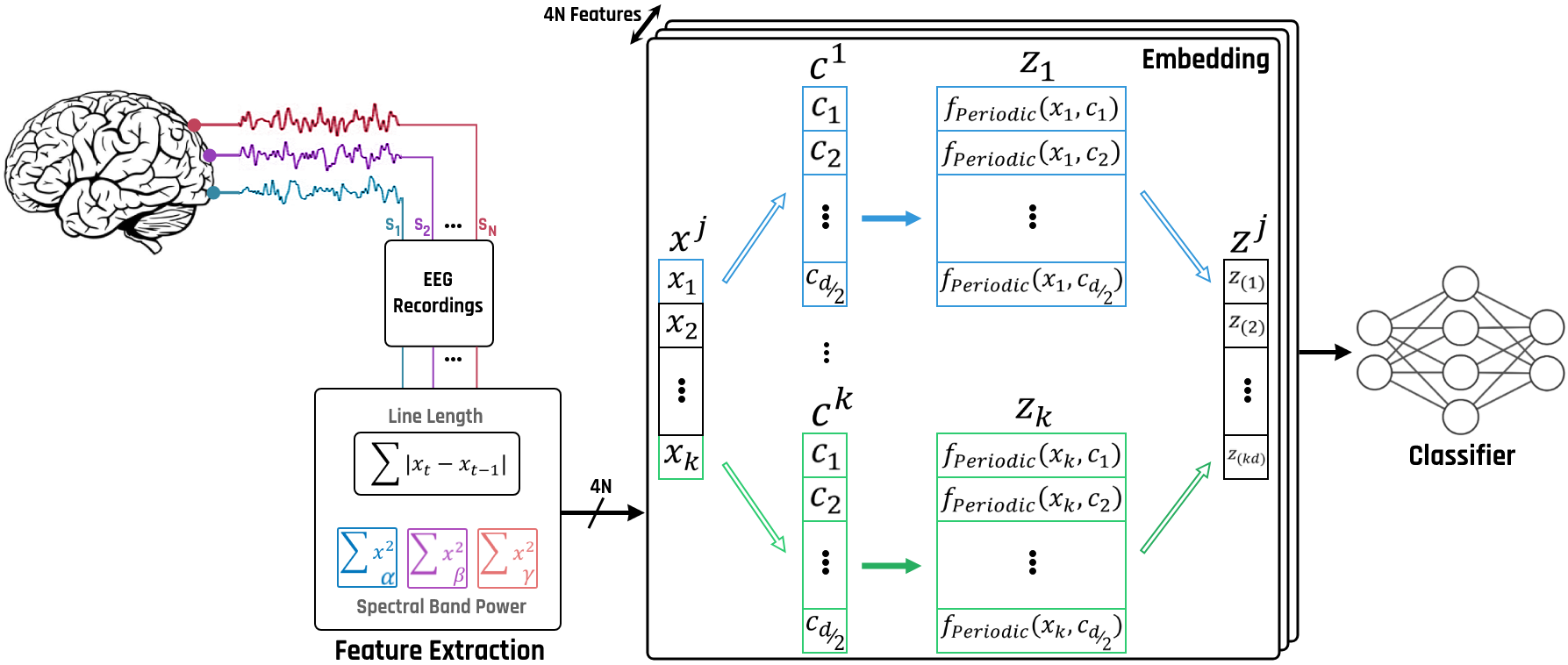
In this Section, we present the overall framework for learning EEG feature embeddings, which can be applied to MLP and other classifiers. We extract multi-band spectral powers and line-length [12] from 1-second EEG epochs to construct a feature vector. Rather than using the raw features as input to the ML models, we encode the scalar values of EEG biomarkers and convert each numerical feature to an EEG embedding [17]. We later show that the embedding step can lead to superior performance in seizure detection.
We denote the dataset as where represents the EEG features extracted from the -th epoch (as discussed in detail in Section III-B) and denotes the binary annotation of seizure events (seizure or non-seizure). Thus, would represent the -th feature of the -th epoch. For simplicity, we omit the index in the following. We then formalize the notion of EEG feature embeddings as:
where is the embedding vector, refers to the embedding function, and is the dimension of the embedding space for the -th feature. Equation (1) highlights the fact that the calculation of embeddings for each feature is performed independently. In this study, different embedding functions () share the same dimension in the embedding space (). However, the parameters of are not shared across different EEG biomarkers.
After calculating for each , the EEG feature embeddings are flattened and concatenated into a -D vector which is passed to a classification model. More specifically, we define the EEG seizure classifier as:
where is the total number of features extracted from each EEG epoch, and with being calculated from Equation (1).
As discussed in [17], there are multiple variations of to calculate the embedding of the input vector. Here, we employ the Periodic Activation Functions which are shown to be optimal [17]. Thus, we define as follows:
where is an even number and is defined as:
The parameters can be either constant or variables that are trained during the model training process. In this study, we assume they are constants initialized from a normal distribution . Figure 1 illustrates the pipeline of the proposed method. The forward pass is initiated by the calculation of EEG feature embeddings. These embeddings are then concatenated and fed to a machine learning classifier (e.g., LR or MLP) for predicting seizure onsets.
III Results
In this Section, we present the experimental details of the proposed EEG feature embedding method followed by a thorough evaluation of the seizure detection performance.
| Classifier | Conventional Approach | with Embedding Module | ||||
|---|---|---|---|---|---|---|
| Sensitivity | Specificity | AUC | Sensitivity | Specificity | AUC | |
| LR | ||||||
| MLP | ||||||
| SVM | ||||||
| KNN | ||||||
| GNB | ||||||
| BNB | ||||||
| LGBM | ||||||
III-A Dataset
We validated our approach on the widely-used CHB-MIT [18] EEG dataset that includes 24 epileptic patients with 165 annotated seizures. We use 1-second epochs for extracting EEG biomarkers. Each epoch is annotated as either “seizure” (i.e., ) or “non-seizure” (i.e., ) by expert neurologists. We exclude those epochs that include both seizure and non-seizure periods. This step is crucial as it ensures that the resulting windows contain either a clear label of 1 indicating the presence of a seizure event, or a label of 0 in the absence of a seizure. Overall, we used seizure epochs and non-seizure epochs for training, and seizure epochs and non-seizure epochs for testing our models.
III-B Feature Extraction
We compute four EEG biomarkers from each channel, which are later converted to feature embeddings. Specifically, the feature extraction unit calculates the line-length (i.e., the sum of absolute differences between consecutive samples), as well as the ( Hz), ( Hz) and ( Hz) band powers [19, 20]. Multiple studies have demonstrated the effectiveness of these features in successfully discriminating between seizure and non-seizure periods [6, 11, 20, 12].
III-C EEG Feature Embedding
As discussed in the Methods Section, we opted to use the Periodic Activation Function as the embedding function. We set the parameter (dimension of the embedding) to , while is sampled from a normal distribution in all of our experiments. Moreover, we use the QuantileTransformer (from scikit-learn) to process the EEG biomarkers [17], with the number of quantiles set to . This preprocessing step along with the Periodic module improve the seizure detection performance. The hyperparameters used in the experiments were optimized for a particular patient. The same hyperparameters were then applied to the remaining patients to demonstrate the generalizability of our proposed approach. It should be noted that the performance can be enhanced further by optimizing the hyperparameters for each subject.
| JSSC’15 [6] | ISSCC’20 [21] | JSSC’22 [11] | IEEE Access’20 [22] | TNSRE’22 [23] | This work | |||
| Dataset (# of Patients) | CHB-MIT (14) | CHB-MIT (23) | CHB-MIT (24) | CHB-MIT (24) | CHB-MIT (24) | CHB-MIT (24) | ||
| Classifier | D2A-LSVM |
|
LR + SGD | CNN + SVM | Bi-GRU | Embed. + SVM | ||
| Sensitivity (%) | 95.7 | 97.8 | 97.5 | 92.7 | 95.49 | 100 | ||
| Specificity (%) | 98.0 | 99.7 | 98.2 | 90.8 | 98.49 | 99.0 |
III-D ML Models
The experiments were conducted on six diverse ML models including Logistic Regression (LR), Multi-Layer Perceptron (MLP), Support Vector Machine (SVM), Gaussian Naive Bayes (GNB), Bernoulli Naive Bayes (BNB), K-Nearest Neighbors (KNN), and Light Gradient Boosting Machine (LGBM), which is an efficient implementation of the gradient boosting tree ensembles. The MLP model has two hidden layers of size and , while the SVM employs a polynomial kernel with a degree of . The LGBM was trained with estimators and a maximum depth of . The remaining model parameters were set to their default values provided in scikit-learn [24] and LightGBM library.
III-E Seizure Detection Results
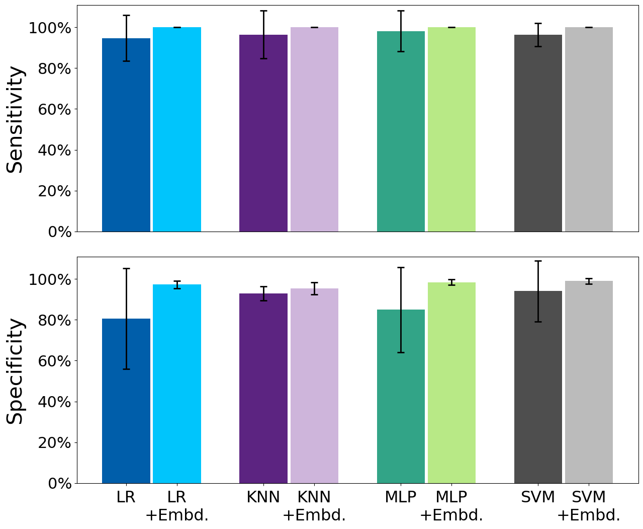
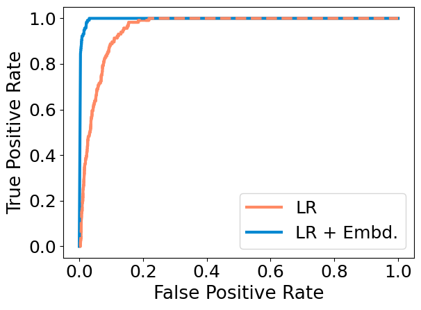
Figure 2 shows the performance of the widely used ML models for epileptic seizure detection, with and without feature embeddings. The average sensitivity and specificity of the LR are improved by and , respectively, using the proposed EEG feature embeddings. This approach is not limited to simple linear models and can be employed with more advanced algorithms such as MLP, non-linear SVM, and KNN. As demonstrated in Fig. 2, we achieve an average improvement of in sensitivity and in specificity, using an MLP classifier. This figure also demonstrates the improvements achieved on SVM and KNN. Furthermore, the numerical improvements in sensitivity, specificity, and AUC score for the models discussed in Section III-D can be seen in Table I. Here, the bold numbers represent the enhanced performance achieved by the proposed feature embedding technique compared to the conventional models. We achieve an average improvement of , , and in the AUC score for the LR, MLP, and SVM, respectively. While gradient boosted trees achieve the highest performance prior to embedding, SVM and MLP with embedding outperform the LightGBM. Furthermore, we achieve improvements of for SVM, for MLP, for LR, for KNN, for GNB, for BNB, and for LGBM in terms of epoch-based sensitivity. Since the majority of prior work reported event-based sensitivity, we use this measure throughout the paper. In addition, Fig. 3 shows a comparison of the ROC curves of the LR on Patient #5, with and without embedding. Our analysis shows that the AUC score of Logistic Regression for this specific patient increases from 0.954 to 0.997 when used in combination with the embedding module.
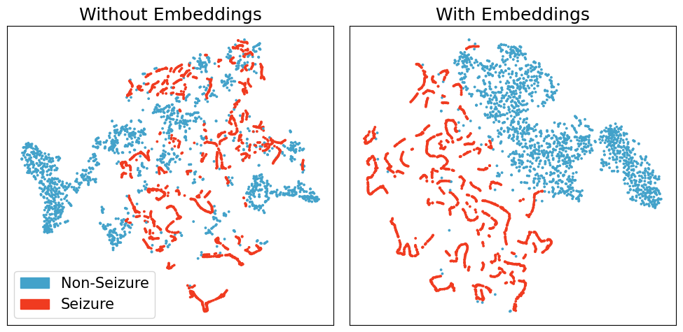
Figure 5 provides a visual representation of EEG epochs for patient #21 of the CHB-MIT dataset. Here, the t-SNE is employed to project the high-dimensional feature space onto two dimensions [25]. Seizure epochs are shown by red dots, while blue dots represent the non-seizure epochs. It is evident that the inclusion of the proposed embedding module significantly improves the separation between seizure and non-seizure states. The improved separability enhances the performance of ML models, thus explaining the higher performance achieved by the integration of the embedding module.
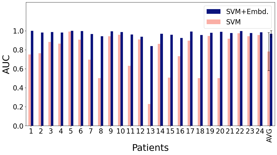
Finally, we demonstrate the effectiveness of our proposed method by comparing the performance achieved by an SVM model integrated with the embedding module, against the state-of-the-art seizure detectors. As depicted in Table II, our model achieves a superior performance, exhibiting a remarkable sensitivity of 100% by accurately detecting all seizure events and a specificity of 99%. Figure 4 further illustrates the model’s AUC score on each patient of the CHB-MIT dataset, unequivocally highlighting the substantial performance enhancement yielded by the embedding module.
Given the universality of the proposed method (i.e., its effectiveness regardless of the ML model) and its generalizability across subjects, we plan to consider this approach for future hardware implementation. In our design, feature embeddings only require 720 parameters for a patient with 18 EEG channels (4 features per channel, embedding dimension of 20), which is significantly less compared to models such as CNN [26]. By sharing the embedding computational module across features (e.g., utilizing a TDM approach as in [20]), training the model to utilize only the top-performing features, and reducing preprocessing steps such as QuantileTransformer, we will improve the hardware efficiency of our approach. Nevertheless, this work serves as a compelling proof-of-concept for the potential impact of embedding techniques in enhancing the accuracy of neural signal classification.
IV CONCLUSIONS
In this work, we present, for the first time, the concept of learning EEG feature embeddings to enhance the performance in the epileptic seizure detection task. By evaluating the proposed model on the CHB-MIT dataset of 24 patients, we show that EEG embeddings substantially enhance the performance of seizure detection systems, using a variety of machine learning models. Our results demonstrate the efficacy of this method and emphasize its potential in assisting patients with epilepsy and other neurological disorders.
References
- [1] K. Schindler, H. Leung, C. E. Elger, and K. Lehnertz, “Assessing seizure dynamics by analysing the correlation structure of multichannel intracranial eeg,” Brain, vol. 130, no. 1, pp. 65–77, 2007.
- [2] M. Shoaran, C. Pollo, K. Schindler, and A. Schmid, “A fully integrated ic with 0.85-w/channel consumption for epileptic ieeg detection,” IEEE Transactions on Circuits and Systems II: Express Briefs, vol. 62, no. 2, pp. 114–118, 2015.
- [3] M. Shoaran, M. H. Kamal, C. Pollo, P. Vandergheynst, and A. Schmid, “Compact low-power cortical recording architecture for compressive multichannel data acquisition,” IEEE transactions on biomedical circuits and systems, vol. 8, no. 6, pp. 857–870, 2014.
- [4] A. H. Shoeb and J. V. Guttag, “Application of machine learning to epileptic seizure detection,” in Proceedings of the 27th international conference on machine learning (ICML-10), 2010, pp. 975–982.
- [5] L. Yao, P. Brown, and M. Shoaran, “Improved detection of parkinsonian resting tremor with feature engineering and kalman filtering,” Clinical Neurophysiology, vol. 131, no. 1, pp. 274–284, 2020.
- [6] M. A. Bin Altaf, C. Zhang, and J. Yoo, “A 16-channel patient-specific seizure onset and termination detection soc with impedance-adaptive transcranial electrical stimulator,” IEEE Journal of Solid-State Circuits, vol. 50, no. 11, pp. 2728–2740, 2015.
- [7] L. Yao, J. L. Baker, N. D. Schiff, K. P. Purpura, and M. Shoaran, “Predicting task performance from biomarkers of mental fatigue in global brain activity,” Journal of neural engineering, vol. 18, no. 3, p. 036001, 2021.
- [8] L. Yao, P. Brown, and M. Shoaran, “Resting tremor detection in parkinson’s disease with machine learning and kalman filtering,” in 2018 IEEE Biomedical Circuits and Systems Conference (BioCAS). IEEE, 2018, pp. 1–4.
- [9] B. Zhu, G. Coppola, and M. Shoaran, “Migraine classification using somatosensory evoked potentials,” Cephalalgia, vol. 39, no. 9, pp. 1143–1155, 2019.
- [10] G. O’Leary, D. M. Groppe, T. A. Valiante, N. Verma, and R. Genov, “Nurip: Neural interface processor for brain-state classification and programmable-waveform neurostimulation,” IEEE Journal of Solid-State Circuits, vol. 53, no. 11, pp. 3150–3162, 2018.
- [11] A. Chua, M. I. Jordan, and R. Muller, “Soul: An energy-efficient unsupervised online learning seizure detection classifier,” IEEE Journal of Solid-State Circuits, vol. 57, no. 8, pp. 2532–2544, 2022.
- [12] B. Zhu, M. Farivar, and M. Shoaran, “Resot: Resource-efficient oblique trees for neural signal classification,” IEEE Transactions on Biomedical Circuits and Systems, vol. 14, no. 4, pp. 692–704, 2020.
- [13] N. Rahaman, A. Baratin, D. Arpit, F. Draxler, M. Lin, F. Hamprecht, Y. Bengio, and A. Courville, “On the spectral bias of neural networks,” in International Conference on Machine Learning. PMLR, 2019, pp. 5301–5310.
- [14] M. Tancik, P. Srinivasan, B. Mildenhall, S. Fridovich-Keil, N. Raghavan, U. Singhal, R. Ramamoorthi, J. Barron, and R. Ng, “Fourier features let networks learn high frequency functions in low dimensional domains,” Advances in Neural Information Processing Systems, vol. 33, pp. 7537–7547, 2020.
- [15] K. Hornik, “Approximation capabilities of multilayer feedforward networks,” Neural networks, vol. 4, no. 2, pp. 251–257, 1991.
- [16] G. Cybenko, “Approximation by superpositions of a sigmoidal function,” Mathematics of control, signals and systems, vol. 2, no. 4, pp. 303–314, 1989.
- [17] Y. Gorishniy, I. Rubachev, and A. Babenko, “On embeddings for numerical features in tabular deep learning,” Advances in Neural Information Processing Systems, vol. 35, pp. 24 991–25 004, 2022.
- [18] A. L. Goldberger, L. A. Amaral, L. Glass, J. M. Hausdorff, P. C. Ivanov, R. G. Mark, J. E. Mietus, G. B. Moody, C.-K. Peng, and H. E. Stanley, “Physiobank, physiotoolkit, and physionet: components of a new research resource for complex physiologic signals,” circulation, vol. 101, no. 23, pp. e215–e220, 2000.
- [19] M. Shoaran, B. A. Haghi, M. Taghavi, M. Farivar, and A. Emami-Neyestanak, “Energy-efficient classification for resource-constrained biomedical applications,” IEEE Journal on Emerging and Selected Topics in Circuits and Systems, vol. 8, no. 4, pp. 693–707, 2018.
- [20] U. Shin, C. Ding, B. Zhu, Y. Vyza, A. Trouillet, E. C. M. Revol, S. P. Lacour, and M. Shoaran, “Neuraltree: A 256-channel 0.227-j/class versatile neural activity classification and closed-loop neuromodulation soc,” IEEE Journal of Solid-State Circuits, vol. 57, no. 11, pp. 3243–3257, 2022.
- [21] Y. Wang, Q. Sun, H. Luo, X. Chen, X. Wang, and H. Zhang, “A closed-loop neuromodulation chipset with 2-level classification achieving 1.5vpp cm interference tolerance, 35db stimulation artifact rejection in 0.5ms and 97.8 sensitivity seizure detection,” in 2020 IEEE International Solid- State Circuits Conference - (ISSCC), 2020, pp. 406–408.
- [22] S. M. Usman, S. Khalid, and M. H. Aslam, “Epileptic seizures prediction using deep learning techniques,” Ieee Access, vol. 8, pp. 39 998–40 007, 2020.
- [23] Y. Zhang, S. Yao, R. Yang, X. Liu, W. Qiu, L. Han, W. Zhou, and W. Shang, “Epileptic seizure detection based on bidirectional gated recurrent unit network,” IEEE Transactions on Neural Systems and Rehabilitation Engineering, vol. 30, pp. 135–145, 2022.
- [24] F. Pedregosa, G. Varoquaux, A. Gramfort, V. Michel, B. Thirion, O. Grisel, M. Blondel, P. Prettenhofer, R. Weiss, V. Dubourg et al., “Scikit-learn: Machine learning in python,” the Journal of machine Learning research, vol. 12, pp. 2825–2830, 2011.
- [25] L. Van der Maaten and G. Hinton, “Visualizing data using t-sne.” Journal of machine learning research, vol. 9, no. 11, 2008.
- [26] M. S. Hossain, S. U. Amin, M. Alsulaiman, and G. Muhammad, “Applying deep learning for epilepsy seizure detection and brain mapping visualization,” ACM Transactions on Multimedia Computing, Communications, and Applications (TOMM), vol. 15, no. 1s, pp. 1–17, 2019.