Picosecond pump pulses probe the relevance of hot electrons for the laser-induced phase transition in FeRh
Abstract
Recent ultrafast photoemission experiments showed signatures of an ultrafast modification of the electronic band structure in FeRh indicative of a ferromagnetic (FM) state that is initiated by a non-equilibrium occupation of the electronic states upon femtosecond laser excitation. We use ultrafast x-ray diffraction to examine the impact of hot electrons on the antiferromagnetic (AFM) to FM phase transition. By increasing the pump-pulse duration up to , we eliminate hot electrons and see that the nucleation of FM domains still proceeds at the intrinsic timescale of , which starts when the deposited energy surpasses the threshold energy. For long pulses, the phase transition proceeds considerably faster than predicted by a convolution of the dynamics observed for ultrafast excitation with the long pump pulse duration. We predict that quite generally, slow photoexcitation can result in a fast response, if the non-linear threshold behavior of a first-order phase transition is involved.
First-order phase transitions are characterized by an abrupt change of structural, electronic or/and magnetic properties and a co-existence of multiple phases when the deposited energy in thermal equilibrium[1, 2, 3, 4] or on ultrafast timescales[5, 6, 7, 8, 9, 10, 11, 12] exceeds a threshold. The abruptly emerging phase is a consequence of a fine interplay of spin, charge and lattice degrees of freedom[6, 13]. Since the optical excitation often affects only the electrons directly, they heat up far beyond the transition temperature, rendering the driving mechanism of the laser-induced phase transition a formidable question.
Since the discovery of the first-order magneto-structural antiferromagnetic-to-ferromagnetic (AFM-FM) phase transition of FeRh at different mechanisms such as expansion-induced sign change of the exchange constant[14], excitation of spin waves[15] and dominant FM exchange of the Fe moments mediated by an induced Rh moment[16, 8, 17, 18, 13, 19] were proposed.
On ultrafast timescales, direct signatures of the ferromagnetic phase transition of FeRh are the rise of the increased FM lattice constant[10, 20, 11, 21] and the emergence of a net magnetization[8, 22, 9, 23, 24]. While the rise of the magnetization is most significant after the coalescence of the nucleated FM domains[22, 23, 24], probing the structural order parameter via ultrafast x-ray diffraction (UXRD) directly yields insights into their nucleation and growth[10, 20, 11, 21], where the intrinsic nucleation time[11] of applies for a wide range of fluences, external magnetic fields and even for FeRh nanostructures[21].
The local configuration of magnetic moments has been described as a competition between bilinear and higher-order four spin exchange terms in atomistic spin dynamics [18], a combination of Heisenberg exchange of Fe and a Stoner model for Rh [8] and a modification of the Rh-Fe hybridization[17, 13, 19]. Recently, time-resolved photoelectron spectroscopy identified a sub-picosecond formation of an electronic ferromagnetic state[19], related to a photo-induced change of the band structure: Charge transfer from Rh to Fe and an intersite spin transfer between Fe sites was found to induce a Rh moment during the relaxation of optically excited non-equilibrium electrons. However, it remains unclear if the non-equilibrium character of the photoexcited electrons is required for the laser-induced phase transition.
Here, we investigate the role of hot electrons concomitant with ultrashort laser pulse excitation for this prototypical phase transition. By ultrafast x-ray diffraction (UXRD), we directly measure how the kinetics of domain nucleation and the threshold of the AFM-FM phase transition depend on the duration of optical pump-pulses. For -long pulses, the pronounced electron-phonon non-equilibrium present upon femtosecond laser excitation is effectively suppressed. Thus, we gradually deposit the energy in all subsystems and recover the energy threshold for the laser-induced phase transition known from equilibrium. The transient FM volume fraction extracted from the laser-induced strain response is found to exclusively depend on the total deposited energy. It reaches the same final value at the intrinsic nucleation timescale irrespective of the pump-pulse duration. As a consequence of the non-linear threshold behavior, the rise of the FM phase is delayed but faster than the convolution of the pump pulse duration with the signal upon femtosecond pulse excitation. In total, a laser-induced hot Fermi-distribution is not necessary to drive the phase transition on the intrinsic nucleation timescale.
Figure 1(a) sketches the epitaxial thick FeRh(001) film grown by magnetron sputtering from an equiatomic FeRh target[25] on an MgO(001) substrate. We used synchrotron radiation from the KMC-3 XPP endstation at BESSY II[26] to determine the film thickness via x-ray reflectivity (XRR) and to characterize the first order AFM-FM phase transition via the concomitant change of the mean out-of-plane lattice constant (symbols in Fig. 1(b)). The hysteresis for this locally probed lattice constant is narrower than the global temperature-dependent magnetization (solid line) determined by Vibrating sample magnetometry (VSM) using a QuantumDesign VersaLab magnetometer. The magnetization data indicates the presence of a residual FM phase of around originating from interface effects[27, 28, 29] consistent with the reduced out-of-plane expansion of compared to observed in thicker films[11, 21].
Figure 1(c) illustrates the influence of increasing the pump-pulse duration on the optically induced electron-phonon non-equilibrium. We model the transient mean electron and phonon temperature of the FeRh film as function of the pump-pulse duration in the framework of a diffusive two-temperature model[30]. We use the modular Python library udkm1Dsim [31] and literature values for the Sommerfeld constant[32] , the phononic heat capacity[33] and the electron-phonon coupling constant [34] . The thermophysical properties of the MgO substrate were used as reported previously[11]. The excitation by a pump pulse increases the electron temperature that stays significantly higher than the slowly rising phonon temperature within the first , with the maximum . Increasing the pump pulse duration to drastically reduces the maximum electron temperature, as a considerable amount of energy already dissipated to the phonons during the optical deposition. For a pump pulse of duration , the electron temperature barely exceeds the phonon temperature. A strong electron-phonon non-equilibrium with a substantial amount of hot electrons is absent.
In the following, we experimentally apply such pump-pulses to investigate the kinetics of the laser-induced phase transition in FeRh by UXRD. The thin film is excited by p-polarized pump pulses with a central wavelength of that are incident under with respect to the sample normal. We probe the transient out-of-plane strain response of the FeRh layer via symmetric scans [35] around the FeRh(002) Bragg peak at our table-top laser-driven plasma x-ray source [36] providing hard x-ray pulses with a photon energy of approx. . The Bragg peak position along the reciprocal space coordinate encodes the mean out-of-plane lattice constant of the FeRh films via . The lattice strain is the relative change of the lattice constant with respect to its value before excitation. We independently determined the pump-probe overlap and calibrated the duration of the pump-pulse by the strictly linear laser-induced response of a metal-insulator superlattice serving as reference sample[36].
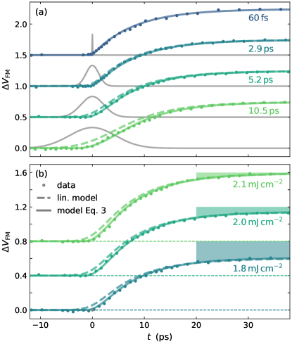
Figure 2(a) displays the laser-induced strain response of the thin FeRh film for a fluence of and different pump pulse durations ranging from to . The excitation fluence exceeds the previously identified critical threshold[11] of for this sample at room temperature and drives the magnetostructural phase transition that is associated with an out-of-plane expansion of FeRh. In total, the strain response is the superposition of an expansion due to the phase transition, a quasi-static expansion due to heating and a propagating strain pulse reflected at the surface and the FeRh-MgO interface, where it is partially transmitted into the substrate. In case of the excitation by a pulse, this results in a a decaying oscillation [30] with a period of given by the layer thickness and the sound velocity [37] (see Figure 2(a)). Increasing the pump-pulse duration successively suppresses this oscillation of the mean out-of-plane strain until pump-pulses only drive a slow expansion. The strain at is identical for all pump-pulse durations. This indicates that all strain contributions including the expansion associated with the AFM-FM phase transition exclusively depend on the deposited energy and not on the details of the optical excitation.
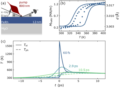
Fig. 2(b) compares the transient strain for pump pulse durations of (symbols) and (grey solid curves) for various super- and sub-threshold fluences. At first sight, the long pump pulses seem just to smear out the oscillations resulting from coherent longitudinal acoustic phonons. We obtain better insight by disentangling the linear acoustic response of the sample from the expansion driven by the phase transition, which depends non-linearly on the excitation fluence. The sub-threshold fluence of does not induce the AFM-FM phase transition[11]. This simplifies the strain response to a superposition of a quasi-static expansion and propagating strain pulses, which both depend linearly on the deposited pulse energy. Therefore, the acoustic strain contribution upon a super-threshold excitation is given by this sub-threshold strain response (grey solid line) scaled by the ratio of the fluences and convoluted with the respective pump-pulse duration.
The dotted line in Fig. 2(b) depicts the acoustic strain contribution for given by with . The deviation from the strain response to the super-threshold excitation is essentially due to the additional expansion induced by the phase transition, which directly provides insights into the rise of the FM volume fraction . To determine its absolute value, we consider the residual FM phase present before the laser excitation due to interface effects and the latent heat of the first-order phase transition. The energy required for transforming FeRh into the FM phase reduces the local temperature[38] by , which reduces the quasi-static expansion of FeRh by with the expansion coefficient in the AFM phase. This relates the deviation in the transient strain to the laser-induced FM volume fraction via:
| (1) |
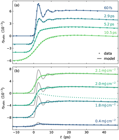
Figure 3 displays the laser-induced volume fraction derived from the measured transient strain via Eq. \tagform@1. The variation of the excitation fluence in Fig. 3(b) exemplifies the non-linear response at the phase transition: A tiny increase of the fluence from to changes from to a complete phase transition, which corresponds to . The data for pulse excitation in Fig. 3(a) is very well reproduced by the nucleation of domains on a timescale by [30]:
| (2) |
with the Heaviside function and the final FM volume fraction increase that depends on the fluence with if and if as reported previously[11].
Equation \tagform@2 yields excellent agreement with for in Fig. 3(a). As a first attempt, the dashed lines for the longer pump-pulses in Fig. 3 represent from Eq. \tagform@2 convoluted with a Gaussian representing the pump-pulse in the experiment. However, the deviation of the measured transient FM volume fraction from this simple estimation becomes larger with increasing pump-pulse duration (see Fig. 3(a)). Interestingly, the data rise faster than the convolution although the rise starts later, which must be a consequence of the non-linearity associated with the threshold for the phase transition.
As an improved model of the rising for long pump-pulses, we explicitly consider the successively deposited energy that leads to the unlocking of the AFM-FM phase transition in an increasing volume fraction of the film during the pump-pulse. This explicit treatment of the threshold character of first-order phase transitions extends Eq. \tagform@2 in the case of picosecond pump-pulses to:
| (3) |
Here, the FM volume fraction is unlocked at delay by the increase of the deposited energy and rises on the nucleation timescale. This transiently unlocked phase transition adds to the already present FM phase driven at delays . The Heaviside function ensures a start of the phase transition at . To model the transient FM volume fraction we assume a linear increase of from to between the fluences and , which corresponds to a full phase transition as characterized previously[11].
Under this assumption, Eq. \tagform@3 yields excellent agreement (see solid lines) with the experimentally determined transients in Fig. 3(a and b) for various pump-pulse durations and super-threshold fluences. Our model including the critical threshold of the first-order phase transition reproduces both the delayed start of the domain nucleation relative to the beginning of the pump pulse for longer pump-pulses and the earlier start of the domain nucleation with increasing fluence, which highlights the central role of the threshold for the laser-induced phase transition.
In summary, we studied the laser-induced magnetostructural AFM-FM phase transition in FeRh driven by picosecond pump-pulses via UXRD. The extracted transient FM volume fraction highlights the crucial role of the threshold for the first-order phase transition in FeRh. The insensitivity of the final FM volume fraction on varying the pump-pulse duration from 60 fs up to reveals that the threshold is exclusively determined by the amount of deposited energy and that the laser-induced AFM-FM phase transition does not need to proceed though the generation of hot non-equilibrium electrons. With increasing pump-pulse duration we observe an increasing deviation of the FM volume fraction from a linear response model. We successfully model the data using the intrinsic nucleation of FM domains on an timescale, by simply considering the slow deposition of energy by picosecond pump-pulses, which successively overcomes the critical threshold that makes the phase transition dynamics nonlinear.
We acknowledge the DFG for financial support via Project-No. 328545488 – TRR 227, project A10 and the BMBF for funding via 05K22IP1. Access to the CEITEC Nano Research Infrastructure was supported by the Ministry of Education, Youth and Sports (MEYS) of the Czech Republic under the project CzechNanoLab (LM2023051).
References
- Qazilbash et al. [2007] M. M. Qazilbash, M. Brehm, B.-G. Chae, P.-C. Ho, G. O. Andreev, B.-J. Kim, S. J. Yun, A. Balatsky, M. Maple, F. Keilmann, et al., “Mott transition in vo2 revealed by infrared spectroscopy and nano-imaging,” Science 318, 1750–1753 (2007).
- Roy et al. [2004] S. Roy, G. Perkins, M. Chattopadhyay, A. Nigam, K. Sokhey, P. Chaddah, A. Caplin, and L. Cohen, “First order magnetic transition in doped cefe 2 alloys: Phase coexistence and metastability,” Physical review letters 92, 147203 (2004).
- Uhlíř, Arregi, and Fullerton [2016] V. Uhlíř, J. A. Arregi, and E. E. Fullerton, “Colossal magnetic phase transition asymmetry in mesoscale ferh stripes,” Nature communications 7, 13113 (2016).
- Baldasseroni et al. [2012] C. Baldasseroni, C. Bordel, A. Gray, A. Kaiser, F. Kronast, J. Herrero-Albillos, C. Schneider, C. Fadley, and F. Hellman, “Temperature-driven nucleation of ferromagnetic domains in ferh thin films,” Applied Physics Letters 100, 262401 (2012).
- Randi et al. [2016] F. Randi, I. Vergara, F. Novelli, M. Esposito, M. Dell’Angela, V. Brabers, P. Metcalf, R. Kukreja, H. A. Dürr, D. Fausti, et al., “Phase separation in the nonequilibrium verwey transition in magnetite,” Physical Review B 93, 054305 (2016).
- De Jong et al. [2013] S. De Jong, R. Kukreja, C. Trabant, N. Pontius, C. Chang, T. Kachel, M. Beye, F. Sorgenfrei, C. Back, B. Bräuer, et al., “Speed limit of the insulator–metal transition in magnetite,” Nature materials 12, 882–886 (2013).
- Wegkamp et al. [2014] D. Wegkamp, M. Herzog, L. Xian, M. Gatti, P. Cudazzo, C. L. McGahan, R. E. Marvel, R. F. Haglund Jr, A. Rubio, M. Wolf, et al., “Instantaneous band gap collapse in photoexcited monoclinic vo 2 due to photocarrier doping,” Physical review letters 113, 216401 (2014).
- Ju et al. [2004] G. Ju, J. Hohlfeld, B. Bergman, R. J. van de Veerdonk, O. N. Mryasov, J.-Y. Kim, X. Wu, D. Weller, and B. Koopmans, “Ultrafast generation of ferromagnetic order via a laser-induced phase transformation in ferh thin films,” Physical review letters 93, 197403 (2004).
- Radu et al. [2010] I. Radu, C. Stamm, N. Pontius, T. Kachel, P. Ramm, J.-U. Thiele, H. Dürr, and C. Back, “Laser-induced generation and quenching of magnetization on ferh studied with time-resolved x-ray magnetic circular dichroism,” Physical Review B 81, 104415 (2010).
- Mariager et al. [2012] S. O. Mariager, F. Pressacco, G. Ingold, A. Caviezel, E. Möhr-Vorobeva, P. Beaud, S. Johnson, C. Milne, E. Mancini, S. Moyerman, et al., “Structural and magnetic dynamics of a laser induced phase transition in ferh,” Physical Review Letters 108, 087201 (2012).
- Mattern et al. [2023a] M. Mattern, J. Jarecki, J. A. Arregi, V. Uhlíř, M. Rössle, and M. Bargheer, “Disentangling nucleation and domain growth during a laser-induced phase transition,” (2023a), 10.48550/arXiv.2305.02094.
- Mattern et al. [2023b] M. Mattern, J.-E. Pudell, K. Dumesnil, A. von Reppert, and M. Bargheer, “Towards shaping picosecond strain pulses via magnetostrictive transducers,” Photoacoustics 30, 100463 (2023b).
- Polesya et al. [2016] S. Polesya, S. Mankovsky, D. Ködderitzsch, J. Minár, and H. Ebert, “Finite-temperature magnetism of ferh compounds,” Physical Review B 93, 024423 (2016).
- Kittel [1960] C. Kittel, “Model of exchange-inversion magnetization,” Physical Review 120, 335 (1960).
- Gu and Antropov [2005] R. Gu and V. Antropov, “Dominance of the spin-wave contribution to the magnetic phase transition in ferh,” Physical Review B 72, 012403 (2005).
- Gruner, Hoffmann, and Entel [2003] M. Gruner, E. Hoffmann, and P. Entel, “Instability of the rhodium magnetic moment as the origin of the metamagnetic phase transition in - ferh,” Physical Review B 67, 064415 (2003).
- Sandratskii and Mavropoulos [2011] L. M. Sandratskii and P. Mavropoulos, “Magnetic excitations and femtomagnetism of ferh: A first-principles study,” Physical Review B 83, 174408 (2011).
- Barker and Chantrell [2015] J. Barker and R. W. Chantrell, “Higher-order exchange interactions leading to metamagnetism in ferh,” Physical Review B 92, 094402 (2015).
- Pressacco et al. [2021] F. Pressacco, D. Sangalli, V. Uhlíř, D. Kutnyakhov, J. A. Arregi, S. Y. Agustsson, G. Brenner, H. Redlin, M. Heber, D. Vasilyev, et al., “Subpicosecond metamagnetic phase transition in ferh driven by non-equilibrium electron dynamics,” Nature Communications 12, 5088 (2021).
- Quirin et al. [2012] F. Quirin, M. Vattilana, U. Shymanovich, A.-E. El-Kamhawy, A. Tarasevitch, J. Hohlfeld, D. von der Linde, and K. Sokolowski-Tinten, “Structural dynamics in ferh during a laser-induced metamagnetic phase transition,” Physical Review B 85, 020103 (2012).
- Mattern et al. [2023c] M. Mattern, J.-E. Pudell, J. A. Arregi, J. Zlámal, R. Kalousek, V. Uhlíř, M. Rössle, and M. Bargheer, “Accelerating the laser-induced phase transition in nanostructured ferh via plasmonic absorption,” (2023c), 10.48550/arXiv.2309.12683.
- Bergman et al. [2006] B. Bergman, G. Ju, J. Hohlfeld, R. J. van de Veerdonk, J.-Y. Kim, X. Wu, D. Weller, and B. Koopmans, “Identifying growth mechanisms for laser-induced magnetization in ferh,” Physical Review B 73, 060407 (2006).
- Li et al. [2022] G. Li, R. Medapalli, J. Mentink, R. Mikhaylovskiy, T. Blank, S. Patel, A. Zvezdin, T. Rasing, E. Fullerton, and A. Kimel, “Ultrafast kinetics of the antiferromagnetic-ferromagnetic phase transition in ferh,” Nature Communications 13, 2998 (2022).
- Dolgikh et al. [2022] I. Dolgikh, T. Blank, G. Li, K. Prabhakara, S. Patel, A. Buzdakov, R. Medapalli, E. Fullerton, O. Koplak, J. Mentink, et al., “Ultrafast emergence of ferromagnetism in antiferromagnetic ferh in high magnetic fields,” (2022), 10.48550/arXiv.2202.03931.
- Arregi, Caha, and Uhlíř [2020] J. A. Arregi, O. Caha, and V. Uhlíř, “Evolution of strain across the magnetostructural phase transition in epitaxial ferh films on different substrates,” Physical Review B 101, 174413 (2020).
- Rössle et al. [2021] M. Rössle, W. Leitenberger, M. Reinhardt, A. Koç, J. Pudell, C. Kwamen, and M. Bargheer, “The time-resolved hard x-ray diffraction endstation kmc-3 xpp at bessy ii,” Journal of Synchrotron Radiation 28, 948–960 (2021).
- Pressacco et al. [2016] F. Pressacco, V. Uhlř, M. Gatti, A. Bendounan, E. E. Fullerton, and F. Sirotti, “Stable room-temperature ferromagnetic phase at the ferh (100) surface,” Scientific reports 6, 22383 (2016).
- Fan et al. [2010] R. Fan, C. J. Kinane, T. Charlton, R. Dorner, M. Ali, M. De Vries, R. M. Brydson, C. H. Marrows, B. J. Hickey, D. A. Arena, et al., “Ferromagnetism at the interfaces of antiferromagnetic ferh epilayers,” Physical Review B 82, 184418 (2010).
- Chen et al. [2017] X. Chen, J. Feng, Z. Wang, J. Zhang, X. Zhong, C. Song, L. Jin, B. Zhang, F. Li, M. Jiang, et al., “Tunneling anisotropic magnetoresistance driven by magnetic phase transition,” Nature Communications 8, 449 (2017).
- Mattern et al. [2023d] M. Mattern, A. von Reppert, S. P. Zeuschner, M. Herzog, J.-E. Pudell, and M. Bargheer, “Concepts and use cases for picosecond ultrasonics with x-rays,” Photoacoustics , 100503 (2023d).
- Schick [2021] D. Schick, “udkm1dsim–a python toolbox for simulating 1d ultrafast dynamics in condensed matter,” Computer Physics Communications 266, 108031 (2021).
- Tu et al. [1969] P. Tu, A. Heeger, J. Kouvel, and J. Comly, “Mechanism for the first-order magnetic transition in the ferh system,” Journal of Applied Physics 40, 1368–1369 (1969).
- Richardson, Melville, and Ricodeau [1973] M. Richardson, D. Melville, and J. Ricodeau, “Specific heat measurements on an fe rh alloy,” Physics Letters A 46, 153–154 (1973).
- Günther et al. [2014] S. Günther, C. Spezzani, R. Ciprian, C. Grazioli, B. Ressel, M. Coreno, L. Poletto, P. Miotti, M. Sacchi, G. Panaccione, et al., “Testing spin-flip scattering as a possible mechanism of ultrafast demagnetization in ordered magnetic alloys,” Physical Review B 90, 180407 (2014).
- Schick et al. [2013] D. Schick, R. Shayduk, A. Bojahr, M. Herzog, C. v. Korff Schmising, P. Gaal, and M. Bargheer, “Ultrafast reciprocal-space mapping with a convergent beam,” Journal of Applied Crystallography 46, 1372–1377 (2013).
- Schick et al. [2012] D. Schick, A. Bojahr, M. Herzog, C. v. K. Schmising, R. Shayduk, W. Leitenberger, P. Gaal, and M. Bargheer, “Normalization schemes for ultrafast x-ray diffraction using a table-top laser-driven plasma source,” Review of Scientific Instruments 83, 025104 (2012).
- Palmer, Dentschuk, and Melville [1975] S. Palmer, P. Dentschuk, and D. Melville, “Elastic properties of an iron-rhodium alloy,” physica status solidi (a) 32, 503–508 (1975).
- Ahn et al. [2022] Y. Ahn, M. J. Cherukara, Z. Cai, M. Bartlein, T. Zhou, A. DiChiara, D. A. Walko, M. Holt, E. E. Fullerton, P. G. Evans, et al., “X-ray nanodiffraction imaging reveals distinct nanoscopic dynamics of an ultrafast phase transition,” Proceedings of the National Academy of Sciences 119, e2118597119 (2022).