Now at: ]European Synchrotron Radiation Facility, 71 Avenue des Martyrs, Grenoble 38000, France
Nanoscale transient polarization gratings
Abstract
We present the generation of transient polarization gratings at the nanoscale, achieved using a tailored accelerator configuration of the FERMI free electron laser. We demonstrate the capabilities of such a transient polarization grating by comparing its induced dynamics with the ones triggered by a more conventional intensity grating on a thin film ferrimagnetic alloy. While the signal of the intensity grating is dominated by the thermoelastic response of the system, such a contribution is suppressed in the case of the polarization grating. This exposes helicity-dependent magnetization dynamics that have so-far remained hidden under the large thermally driven response. We anticipate nanoscale transient polarization gratings to become useful for the study of any physical, chemical and biological systems possessing chiral symmetry.
Manipulating light at the nanoscale is a major challenge for modern science, with the potential to unveil fundamental aspects of light-matter interactions and to enable advances in key technologies such as light harvesting, imaging, biosensing or catalysis. In the visible range, nanoscale control of properties of the radiation such as intensity and phase is often achieved via artificial structures with dimensions comparable to or shorter than the wavelength, such as metasurfaces, photonic crystals and plasmonic nanostructures [1, 2, 3]. Instead, nanoscale control of light polarization remains particularly challenging. Spatially variable inhomogeneous vector beams can for instance be generated by using specially designed metasurfaces [4], but a straightforward approach at short wavelengths is yet to be found.
The use of coherent extreme ultraviolet (EUV) and X-ray radiation in a transient grating (TG) scheme provides an alternative way to control light fields at the nanoscale. In fact, the brightness of EUV free electron laser (FEL) pulses allows to efficiently generate sinusoidal patterns of light intensity, without requiring any physical modification of the sample. With spatial periodicity in the \qtyrange[range-units = single]10100nm range [5, 6], such an EUV TG excitation is then capable of driving ultrafast nanoscale dynamics in different kinds of materials in a controlled way. It has recently proven to be an effective tool for investigating the thermal and mechanical properties of matter in a length-scale range previously inaccessible [7, 8]. Additionally, the access to core resonances provides unprecedented insights in the electronic and magnetic dynamics at such a lengthscale [9, 10].
In this Letter, we show how the EUV TG approach can be used to generate ultrafast modulations of light polarization in the tens of nm scale. This unique capability can be broadly applied to study helicity-dependent dynamics at the nanoscale and with ultrafast time resolution. We show the potential of the polarization transient grating by investigating the ultrafast demagnetization of a thin film ferrimagnetic alloy, where we are able to trigger non-thermal magnetization dynamics that are otherwise hidden by the thermoelastic response.
In a TG experiment, two pulses of the same wavelength and intensity , overlapped in time and space and at a given crossing angle at the sample, generate a transient interference pattern. If the two excitation beams are parallelly-polarized (PP), as in Figure 1(a), the intensity at the sample is fully modulated and given by , where is the grating wavevector. The spatial periodicity is given by , while the polarization remains uniform. Instead, when the two beams are orthogonally-polarized (OP), the polarization is modulated with the same periodicity , ranging from circular left to circular right, while the intensity remains uniform [11, 12, 13, 14], as depicted in Figure 1(b). Therefore, analogously to EUV intensity TGs, EUV polarization TGs enable the generation of ultrafast modulations of light polarization with values of comparable to those of .
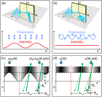
Both intensity and polarization gratings can be revealed by the diffraction of an EUV probe beam. The diffraction intensity as a function of the time delay () with respect to the EUV TG excitation encodes information on the sample dynamics. In the case of an EUV intensity TG, after the electronic grating generated by photo-absorption transfers its energy to the lattice via electron-phonon coupling, leading to a temperature grating [7]. This induces a time- and space-dependent density modulation via thermal expansion and, in magnetic samples, also a magnetization modulation [9]. These two thermal effects contribute to an effective periodic variation of the refractive index between the unexcited areas, which retain equilibrium density and magnetization , and the expanded and demagnetized photo-excited stripes. This periodic variation leads to the diffraction of the EUV probe beam, provided that at the wavelength of the probe . Additionally, an intensity grating is associated to a surface displacement, (see 1(c)), resulting from thermal expansion.111The figure neglects the ultrafast, sub-ps, electronic contribution to since the whole discussion concentrates on the \unitps dynamics.. In forward diffraction, this latter contribution is typically negligible with respect to the signal arising from , which can be enhanced via propagation through the bulk of the sample [7, 8]. In backward diffraction, on the contrary, the contribution of surface displacement typically dominates and vanishes only in the absence of a temperature grating. This is indeed the case of the polarization grating, where the uniform excitation intensity, and thus uniform temperature, prevents both surface displacement and bulk density modulations. Instead, a pure polarization grating can still modulate (see 1(d)), provided that the sample shows a non-thermal chiral response. Such a response can be caused, for instance, by the inverse Faraday effect (IFE) or the magnetization induced by light absorption (MILA) mechanism in magnetic samples [16]. Thus, in backward diffraction and in a magnetic sample, a polarization grating suppresses the contribution and reveals the remaining helicity-dependent magnetic contribution to . More generally, pure nanoscale polarization gratings can be used to isolate the chiral contributions to .
We demonstrate this experimentally in a thin film CoGd alloy, where Ksenzov et al. [9] have previously observed a strong TG signal in forward diffraction when probing resonantly at the Co M-edge. For larger than 100s of \unitfs, the diffraction intensity is mainly determined by , i.e. [9]. That magnetic response has a clear non-sinusoidal time-dependence and should be observed also in backward diffraction. However, the contribution is expected to be dominant, according to previous results [17] and considering the typical thermal expansion coefficients of metallic films. The exact temporal evolution of the surface-induced signal is generally unknown a priori. Nevertheless, in these \unitnm-thick layered samples on a bulk substrate, it typically consists of a combination of surface acoustic waves (SAW), Lamb modes and leaky waves that are all associated to sinusoidal waveforms in time. Thus, the sinusoidal thermoelastic and the non-sinusoidal magnetic dynamics can be easily discerned on the \unitps timescale. When exciting with OP beams, the suppression of the oscillatory signal and the isolation of the non-sinusoidal signal from indicate the generation of the nanoscale polarization grating.
Practically however, realizing EUV polarization gratings is not a trivial endeavor. Polarization control after the EUV source has been demonstrated using phase retardation upon reflection off metallic mirrors [18]. Such a setup would allow for independent OP beams, but it sacrifices the overall efficiency and complicates greatly the experimental scheme. On the other hand, polarization control of the EUV source [19, 20, 21, 22, 23, 24, 25, 26, 27] does not allow the simultaneous generation of multiple EUV pulses with different polarization and direction. Instead, in this Letter we present a special configuration of the FERMI FEL (Trieste, Italy), which permits to simultaneously obtain a pair of EUV pulses with independent linear polarization emitted along slightly different trajectories. Moreover, it allows to switch from PP to OP beams without changing any other experimental parameter. Further details on the accelerator setup are provided in the Appendix. The FEL is tuned at an operating wavelength of \qty20.8, to match the Co M-edge resonance. The two FEL beams are sent each on one of the two pump branch lines of the focusing system of the TIMER beamline, which is used to cross them at the sample with an angle , resulting in . The TIMER setup is also used to generate a variably-delayed probe beam [28] with wavelength and linear polarization parallel to the y-axis as defined in Fig. 1.
The sample is a \qty15 thick film of \ceCo_0.78Gd_0.22 alloy with perpendicular magnetic anisotropy, deposited within a metallic stack (\qty1.5 \ceTa/\qty1.5 \cePt/ \qty15 \ceCo_0.78Gd_0.22/ \qty3 \cePt) on a lead-germanate glass substrate, analogous to the one used in reference [9] (see Ref. [[SeeSupplementalMaterialat][fordetailsonthesamplepreparationandcharacterization, andadiscussiononthefittingprocedureandthermoelasticpropertiesofthesample, whichincludesRefs.[30, 31]]Suppl] and Ref. [30, 31] therein). The film is magnetized to saturation with the magnetic field normal to the film surface. The excitation fluence at the sample is about \qty5\milliJ\per\squared, the penetration depth at \qty20.8nm in the magnetic layer is calculated to be \qty16nm and we estimate a local sample heating due to FEL excitation of about \qty400. Each acquisition is integrated over 1500 shots per delay point with a FEL repetition rate of \qty50Hz and a pulse duration of . We measured the total diffracted signal without polarization analysis.
Figure 2(a) depicts the backward-diffracted intensity TG signal (PP pump beams) as blue circles, clearly showing the sinusoidal oscillatory waveform typical of the acoustic response. Fitting with a sum of co-sinusoidal terms results in two dominating phonon modes. Their frequencies ( and ) are compatible with the expected frequencies of the SAW and longitudinal acoustic (LA) phonon, respectively. A more detailed discussion of the thermoelastic properties of the sample and the fitting procedure is given in the Supplemental Material [29].
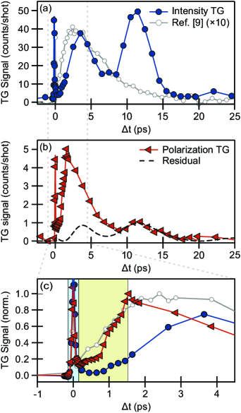
We compare this signal with the one obtained with OP pump beams, plotted as red triangles in Figure 2(b). The two dynamics strongly differ up to \qty10, since the latter signal exhibits a clear non-sinusoidal behavior with a \qty2 rise and a sub-\qty10 decay. Qualitatively, these dynamics strongly resemble the magnetic TG signal observed in Ref. [9]. This is evidenced by the comparison with the gray dotted trace in panel (a) and (c). Following this decay, the signal exhibits oscillatory dynamics, suggesting the presence of a residual density modulation as discussed below.
The early-time dynamics, shown in Fig. 2(c), confirm the different functional dependence of the signals, normalized to their maximum. As discussed above, the disappearance of the sinusoidal signal from the surface displacement , and the persistence of a signal associated with a refractive index variation of magnetic nature , is consistent with the working hypothesis of the generation of a nanoscale transient polarization grating.
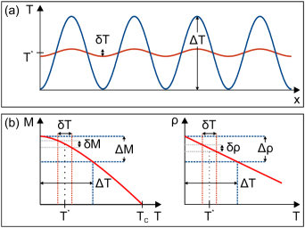
The dashed line in panel (b) is the fit to the backward-diffracted signal from the intensity grating scaled by (See Supplemental Material [29]). This residual intensity contrast can have a two-fold origin: i) a non-perfect orthogonal polarization of the two FEL pulses (See Appendix) and ii) a remaining temperature grating due to dichroic absorption , i.e. the dichroic contribution to the imaginary part of the EUV refractive index: . As an order of magnitude estimate of the second mechanism, we write the amplitude of the fully modulated thermal grating resulting from intensity TG excitation as , where and are the FEL spot size and the average absorption lengths, respectively. In the case of the polarization grating, the residual temperature grating has an amplitude around the average temperature . Here account for the different absorption lengths for left and right circular polarization. The two temperature modulations are schematically depicted in Figure 3(a). Since a) the spot size and overlap conditions are the same for intensity and polarization gratings, b) the amplitude of the grating is linearly proportional to the temperature modulation and c) the TG signal is proportional to , the ratio between the acoustic (sinusoidal) signal in intensity and polarization TGs can be written as . This is on the same order of magnitude as the residual thermoelastic contribution extracted from the data analysis [29].
In order to further elaborate on the nature of the helicity-dependent excitation mechanism, we first recall the interpretation of the nanoscale magnetic TG data from Ref. [9]. On an initial, ultrafast time scale, the intensity TG excitation modulates the electronic temperature and thereby also the spin temperature, leading to ultrafast demagnetization of the photoexcited stripes. This results in a modulation of the magnetic circular dichroism and consequent observation of the diffracted signal. The signal grows on a timescale in agreement with the literature on ultrafast demagnetization of \ceCo [32, 33] before it decays exponentially within \qty10. Due to the dependence on , the decay was attributed to thermal diffusion washing away the magnetic contrast.
Similar temporal regimes are distinguished in the case of the polarization TG and are evidenced respectively by the different shaded areas in Fig. 2(c). The longer time scale is associated, also in this case, to the decay of the magnetic contrast once the magnetic grating is established.
More intriguing are the initial and intermediate regimes in Fig. 2(c), where the chiral response and subsequent growth of the magnetic contrast occur. Several concurrent processes can lead to an helicity-dependent change of magnetization within this initial timescale. Any process inducing a temperature contrast after a sub-ps thermalization, such as dichroic absorption, can be ruled out. Indeed, as sketched in Fig. 3(b), not only leads to a demagnetization but also to a change in density , which results in the excitation of the SAW. For small temperature variations, both and can be approximated to be linear with temperature. Hence, one could expect to observe a time-dependent signal resembling the intensity TG response, scaled to the much smaller induced .
However, we do not observe such behavior. Therefore, the mechanism behind the observed ps-dynamics in the polarization TG must be related to the magnetization dynamics, but not to the electronic and lattice temperature. This could be the case, for example, of MILA, a purely optical effect that is due to electron-photon scattering, and which induces, via spin-orbit coupling, transitions between electronic states of opposite spins [34, 35]. Alternatively, recent works attributed to the IFE helicity-dependent magnetization dynamics across the Fe M-edge even for photon energies where [36]. Moreover, in the case of the polarization TG, we can expect a cooperation between the thermally driven changes in the magnetization induced by the uniform heating and the magnetic torque induced by the circularly polarized periodic pattern [37]. Although a complete understanding of the underlying excitation mechanism goes beyond the scope of the current demonstrative experiment, these results already reveal how fluence, wavelength or pulse-length dependent studies comparing intensity and polarization TGs on the same sample and in the same experimental conditions could provide relevant insights on the intricate physics of light-induced nanoscale magnetic dynamics.
In conclusion, we applied a tailored FEL setting and the unique TG instrument available at FERMI to demonstrate the generation of an ultrafast polarization grating with a \qty43.6 period, by selectively switching off the thermoelastic response of a magnetic system. We showed how this method is ideal to investigate ultrafast light-induced magnetization changes at the nanoscale, where the comparison of intensity and polarization TG allows to differentiate between absorption- and helicity-dependent processes. Indeed, the use of EUV pulses does not only allow to access the nanoscale but also provides the unique playground to tune the relative weight of absorption, dispersion and circular dichroism around resonances. Our approach is not limited to magnetic systems but can be extended to all systems that have chiral response, both in soft and hard condensed matter.
Examples include experiments targeting element-specific dichroic absorption in chiral molecules [38, 39] and molecular crystals [40]. In other examples, the polarization grating can be used to access processes that would be otherwise hidden, as it is the case of electron drift mobility or spin Coulomb drag in semiconductor quantum wells [41, 14] or exciton spin relaxation in quantum dots [42]. Finally, we anticipate that polarization TG experiments could become a unique tool to investigate chiral opto-magnetic properties and the valley degree of freedom of emerging topological systems such as Weyl semimetals [43], metal dichalchogenides [44] or twisted bilayer graphene [45].
Acknowledgements.
K.Y. and C. v. K. S. acknowledge financial support by the Deutsche Forschungsgemeinschaft (DFG, German Research Foundation) – Project-ID 328545488 – TRR 227, project A02. E. P. acknowledges funding from the European Union’s Horizon 2020 research and innovation programme under the Marie Skłodowska-Curie grant agreement No 860553. B.W. and S.B. acknowledge support from the European Research Council, Starting Grant 715452 “MAGNETIC-SPEED-LIMIT”.Appendix A Accelerator setup
The FERMI free electron laser was tuned in an ad-hoc tailored setup aiming at delivering orthogonal polarized pulses displaced in the horizontal plane. Figure 4(a) schematically depicts the machine geometry. The undulator chain of FERMI FEL 1 [46] was divided in two sections of 3 undulators each that were set to a different, orthogonal, linear polarization [47]. Additionally, the trajectory of the electron bunch is tilted to separate the pulses horizontally [48]. Both pulses were emitted off-axis with respect to the undulator line with the idea of partially compensating the reduction of FEL gain and balancing the relative pulse energy of the two beams.
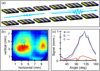
Figure 4(b) shows a CCD image of the two spots acquired \qty70m after the end of the undulator chain, with the linear vertical (LV) polarized beam on the left and the linear horizontal (LH) one on the right. The degree of polarization of the two pulses has been measured at the beamline using a polarimeter consisting of a mirror operating at the Brewster angle and a photodiode, both rotating azimuthally around the beam axis. The geometry was such that the maximum reflectivity for LH polarization corresponds to an angle of and for LV to . It is important to remark that these measurements have been performed posterior to the experiment, albeit with a similar machine configuration. The results are shown in Figure 4(c). The violet curve (a) has been measured with the polarimeter placed along the LV trajectory and both undulator sections tuned, i.e. both LH and LV beams were emitted by the FEL. The light blue curve (b) was measured with the polarimeter in the same position but only the first section of the undulators tuned, i.e. only the LV beam was emitted. Finally, to quantify the background LV light propagating collinear to the LH beam the polarimeter was centered on the second beam but the LH emission was kept detuned. All traces have been normalized to the same factor (), corresponding to the counts at the maximum of trace (a). The left spot in fig. 2(b) showed an almost pure vertical polarization, with the minimum of trace (b) being centered exactly on and the one of trace (a) slighly shifted towards lower angles. The difference of intensity between the two traces is at most \qty6. The LV contamination along the LH trajectory instead was found to be \qty15.
References
- Rotenberg and Kuipers [2014] N. Rotenberg and L. Kuipers, Nature Photonics 8, 919 (2014).
- Manjavacas et al. [2019] A. Manjavacas, L. Zundel, and S. Sanders, ACS Nano 13, 10682 (2019).
- Burresi et al. [2009] M. Burresi, R. J. P. Engelen, A. Opheij, D. van Oosten, D. Mori, T. Baba, and L. Kuipers, Physical Review Letters 102, 033902 (2009).
- Zhao et al. [2018] R. Zhao, L. Huang, C. Tang, J. Li, X. Li, Y. Wang, and T. Zentgraf, Advanced Optical Materials 6, 1800490 (2018).
- Bencivenga et al. [2015] F. Bencivenga, R. Cucini, F. Capotondi, A. Battistoni, R. Mincigrucci, E. Giangrisostomi, A. Gessini, M. Manfredda, I. P. Nikolov, E. Pedersoli, E. Principi, C. Svetina, P. Parisse, F. Casolari, M. B. Danailov, M. Kiskinova, and C. Masciovecchio, Nature 520, 205 (2015).
- Bencivenga et al. [2019] F. Bencivenga, R. Mincigrucci, F. Capotondi, L. Foglia, D. Naumenko, A. A. Maznev, E. Pedersoli, A. Simoncig, F. Caporaletti, V. Chiloyan, R. Cucini, F. Dallari, R. A. Duncan, T. D. Frazer, G. Gaio, A. Gessini, L. Giannessi, S. Huberman, H. Kapteyn, J. Knobloch, G. Kurdi, N. Mahne, M. Manfredda, A. Martinelli, M. Murnane, E. Principi, L. Raimondi, S. Spampinati, C. Spezzani, M. Trovò, M. Zangrando, G. Chen, G. Monaco, K. A. Nelson, and C. Masciovecchio, Science Advances 5, 10.1126/sciadv.aaw5805 (2019).
- Foglia et al. [2023] L. Foglia, R. Mincigrucci, A. Maznev, G. Baldi, F. Capotondi, F. Caporaletti, R. Comin, D. De Angelis, R. Duncan, D. Fainozzi, G. Kurdi, J. Li, A. Martinelli, C. Masciovecchio, G. Monaco, A. Milloch, K. Nelson, C. Occhialini, M. Pancaldi, E. Pedersoli, J. Pelli-Cresi, A. Simoncig, F. Travasso, B. Wehinger, M. Zanatta, and F. Bencivenga, Photoacoustics 29, 100453 (2023).
- Bencivenga et al. [2023] F. Bencivenga, F. Capotondi, L. Foglia, R. Mincigrucci, and C. Masciovecchio, Advances in Physics: X 8, 10.1080/23746149.2023.2220363 (2023).
- Ksenzov et al. [2021] D. Ksenzov, A. A. Maznev, V. Unikandanunni, F. Bencivenga, F. Capotondi, A. Caretta, L. Foglia, M. Malvestuto, C. Masciovecchio, R. Mincigrucci, K. A. Nelson, M. Pancaldi, E. Pedersoli, L. Randolph, H. Rahmann, S. Urazhdin, S. Bonetti, and C. Gutt, Nano Letters 21, 2905 (2021).
- Yao et al. [2022] K. Yao, F. Steinbach, M. Borchert, D. Schick, D. Engel, F. Bencivenga, R. Mincigrucci, L. Foglia, E. Pedersoli, D. De Angelis, M. Pancaldi, B. Wehinger, F. Capotondi, C. Masciovecchio, S. Eisebitt, and C. von Korff Schmising, Nano Letters 22, 4452 (2022).
- Eichler et al. [1986] H. J. Eichler, P. Günter, and D. W. Pohl, Laser-Induced Dynamic Gratings, Springer Series in Optical Sciences, Vol. 50 (Springer Berlin Heidelberg, Berlin, Heidelberg, 1986).
- Terazima [1995] M. Terazima, The Journal of Physical Chemistry 99, 1834 (1995).
- Yang et al. [2012] L. Yang, J. D. Koralek, J. Orenstein, D. R. Tibbetts, J. L. Reno, and M. P. Lilly, Physical Review Letters 109, 246603 (2012).
- Weber et al. [2005] C. P. Weber, N. Gedik, J. E. Moore, J. Orenstein, J. Stephens, and D. D. Awschalom, Nature 437, 1330 (2005).
- Note [1] The figure neglects the ultrafast, sub-ps, electronic contribution to since the whole discussion concentrates on the \unitps dynamics.
- Scheid et al. [2022] P. Scheid, Q. Remy, S. Lebègue, G. Malinowski, and S. Mangin, Journal of Magnetism and Magnetic Materials 560, 169596 (2022).
- Maznev et al. [2021] A. A. Maznev, R. Mincigrucci, F. Bencivenga, V. Unikandanunni, F. Capotondi, G. Chen, Z. Ding, R. A. Duncan, L. Foglia, M. G. Izzo, C. Masciovecchio, A. Martinelli, G. Monaco, E. Pedersoli, S. Bonetti, and K. A. Nelson, Applied Physics Letters 119, 044102 (2021).
- von Korff Schmising et al. [2017] C. von Korff Schmising, D. Weder, T. Noll, B. Pfau, M. Hennecke, C. Strüber, I. Radu, M. Schneider, S. Staeck, C. M. Günther, J. Lüning, A. el dine Merhe, J. Buck, G. Hartmann, J. Viefhaus, R. Treusch, and S. Eisebitt, Review of Scientific Instruments 88, 10.1063/1.4983056 (2017).
- Allaria et al. [2014] E. Allaria, B. Diviacco, C. Callegari, P. Finetti, B. Mahieu, J. Viefhaus, M. Zangrando, G. De Ninno, G. Lambert, E. Ferrari, J. Buck, M. Ilchen, B. Vodungbo, N. Mahne, C. Svetina, C. Spezzani, S. Di Mitri, G. Penco, M. Trovó, W. M. Fawley, P. R. Ribič, D. Gauthier, C. Grazioli, M. Coreno, B. Ressel, A. Kivimäki, T. Mazza, L. Glaser, F. Scholz, J. Seltmann, P. Gessler, J. Grünert, A. De Fanis, M. Meyer, A. Knie, S. P. Moeller, L. Raimondi, F. Capotondi, E. Pedersoli, O. Plekan, M. B. Danailov, A. Demidovich, I. Nikolov, A. Abrami, J. Gautier, J. Lüning, P. Zeitoun, and L. Giannessi, Phys. Rev. X 4, 41040 (2014).
- Roussel et al. [2017] E. Roussel, E. Allaria, C. Callegari, M. Coreno, R. Cucini, S. D. Mitri, B. Diviacco, E. Ferrari, P. Finetti, D. Gauthier, G. Penco, L. Raimondi, C. Svetina, M. Zangrando, A. Beckmann, L. Glaser, G. Hartmann, F. Scholz, J. Seltmann, I. Shevchuk, J. Viefhaus, and L. Giannessi, Photonics 4, 10.3390/photonics4020029 (2017).
- Lutman et al. [2016] A. A. Lutman, J. P. MacArthur, M. Ilchen, A. O. Lindahl, J. Buck, R. N. Coffee, G. L. Dakovski, L. Dammann, Y. Ding, H. A. Dürr, L. Glaser, J. Grünert, G. Hartmann, N. Hartmann, D. Higley, K. Hirsch, Y. I. Levashov, A. Marinelli, T. Maxwell, A. Mitra, S. Moeller, T. Osipov, F. Peters, M. Planas, I. Shevchuk, W. F. Schlotter, F. Scholz, J. Seltmann, J. Viefhaus, P. Walter, Z. R. Wolf, Z. Huang, and H.-D. Nuhn, Nature Photonics 10, 468 (2016).
- Deng et al. [2014] H. Deng, T. Zhang, L. Feng, C. Feng, B. Liu, X. Wang, T. Lan, G. Wang, W. Zhang, X. Liu, J. Chen, M. Zhang, G. Lin, M. Zhang, D. Wang, and Z. Zhao, Phys. Rev. ST Accel. Beams 17, 20704 (2014).
- Schmidt and Calvi [2018] T. Schmidt and M. Calvi, Synchrotron Radiation News 31, 35 (2018).
- Schneidmiller and Yurkov [2018] E. Schneidmiller and M. V. Yurkov, in Proc. of International Free Electron Laser Conference (FEL’17), Santa Fe, NM, USA, August 20-25, 2017, International Free Electron Laser Conference No. 38 (JACoW, Geneva, Switzerland, 2018) pp. 106–108.
- Li et al. [2017] P. Li, T. Wei, Y. Li, and J. Pflueger, Nuclear Instruments and Methods in Physics Research Section A: Accelerators, Spectrometers, Detectors and Associated Equipment 870, 103 (2017).
- Yakopov et al. [2022] M. Yakopov, M. Calvi, S. Casalbuoni, U. Englisch, S. Karabekyan, X. Liang, and T. Schmidt, Journal of Physics: Conference Series 2380, 12019 (2022).
- Perosa et al. [2023] G. Perosa, J. Wätzel, D. Garzella, E. Allaria, M. Bonanomi, M. B. Danailov, A. Brynes, C. Callegari, G. De Ninno, A. Demidovich, M. Di Fraia, S. Di Mitri, L. Giannessi, M. Manfredda, L. Novinec, N. Pal, G. Penco, O. Plekan, K. C. Prince, A. Simoncig, S. Spampinati, C. Spezzani, M. Zangrando, J. Berakdar, R. Feifel, R. J. Squibb, R. Coffee, E. Hemsing, E. Roussel, G. Sansone, B. W. J. McNeil, and P. R. Ribič, Phys. Rev. Lett. 131, 45001 (2023).
- Mincigrucci et al. [2018] R. Mincigrucci, L. Foglia, D. Naumenko, E. Pedersoli, A. Simoncig, R. Cucini, A. Gessini, M. Kiskinova, G. Kurdi, N. Mahne, M. Manfredda, I. P. Nikolov, E. Principi, L. Raimondi, M. Zangrando, C. Masciovecchio, F. Capotondi, and F. Bencivenga, Nuclear Instruments and Methods in Physics Research Section A: Accelerators, Spectrometers, Detectors and Associated Equipment 907, 132 (2018).
- [29] [URL_will_be_inserted_by_publisher].
- Ceballos et al. [2021] A. Ceballos, A. Pattabi, A. El-Ghazaly, S. Ruta, C. P. Simon, R. F. L. Evans, T. Ostler, R. W. Chantrell, E. Kennedy, M. Scott, J. Bokor, and F. Hellman, Physical Review B 103, 024438 (2021).
- Shirakawa et al. [1985] K. Shirakawa, K. Fukamichi, K. Aoki, T. Masumoto, and T. Kaneko, Journal of Physics F: Metal Physics 15, 961 (1985).
- Bergeard et al. [2014] N. Bergeard, V. López-Flores, V. Halté, M. Hehn, C. Stamm, N. Pontius, E. Beaurepaire, and C. Boeglin, Nature Communications 5, 3466 (2014).
- López-Flores et al. [2013] V. López-Flores, N. Bergeard, V. Halté, C. Stamm, N. Pontius, M. Hehn, E. Otero, E. Beaurepaire, and C. Boeglin, Physical Review B 87, 214412 (2013).
- Scheid et al. [2019] P. Scheid, G. Malinowski, S. Mangin, and S. Lebègue, Physical Review B 100, 214402 (2019).
- Scheid et al. [2021] P. Scheid, S. Sharma, G. Malinowski, S. Mangin, and S. Lebègue, Nano Letters 21, 1943 (2021).
- Hennecke et al. [2023] M. Hennecke, C. v. K. Schmising, K. Yao, E. Jal, B. Vodungbo, V. Chardonnet, K. Légaré, F. Capotondi, D. Naumenko, E. Pedersoli, I. Lopez-Quintas, I. P. Nikolov, L. Raimondi, G. De Ninno, L. Salemi, S. Ruta, R. Chantrell, T. Ostler, B. Pfau, D. Engel, P. M. Oppeneer, S. Eisebitt, and I. Radu, arXiv:2303.08564 (2023).
- Stanciu et al. [2007] C. D. Stanciu, F. Hansteen, A. V. Kimel, A. Kirilyuk, A. Tsukamoto, A. Itoh, and T. Rasing, Physical Review Letters 99, 047601 (2007).
- Mincigrucci et al. [2023] R. Mincigrucci, J. R. Rouxel, B. Rossi, E. Principi, C. Bottari, S. Catalini, J. S. Pelli-Cresi, D. Fainozzi, L. Foglia, A. Simoncig, A. Matruglio, G. Kurdi, F. Capotondi, E. Pedersoli, A. Perucchi, F. Piccirilli, A. Gessini, M. Giarola, G. Mariotto, M. Oppermann, S. Mukamel, F. Bencivenga, M. Chergui, and C. Masciovecchio, Nature Communications 14, 386 (2023).
- Zhang et al. [2017] Y. Zhang, J. R. Rouxel, J. Autschbach, N. Govind, and S. Mukamel, Chemical Science 8, 5969 (2017).
- Peacock and Stewart [2001] R. D. Peacock and B. Stewart, The Journal of Physical Chemistry B 105, 351 (2001).
- Cameron et al. [1996] A. R. Cameron, P. Riblet, and A. Miller, Physical Review Letters 76, 4793 (1996).
- Scholes et al. [2006] G. D. Scholes, J. Kim, and C. Y. Wong, Physical Review B 73, 195325 (2006).
- Yan and Felser [2017] B. Yan and C. Felser, Annual Review of Condensed Matter Physics 8, 337 (2017).
- Schaibley et al. [2016] J. R. Schaibley, H. Yu, G. Clark, P. Rivera, J. S. Ross, K. L. Seyler, W. Yao, and X. Xu, Nature Reviews Materials 1, 16055 (2016).
- Liu and Dai [2020] J. Liu and X. Dai, npj Computational Materials 6, 57 (2020).
- Allaria et al. [2012] E. Allaria, R. Appio, L. Badano, W. A. Barletta, S. Bassanese, S. G. Biedron, A. Borga, E. Busetto, D. Castronovo, P. Cinquegrana, S. Cleva, D. Cocco, M. Cornacchia, P. Craievich, I. Cudin, G. D’Auria, M. Dal Forno, M. B. Danailov, R. De Monte, G. De Ninno, P. Delgiusto, A. Demidovich, S. Di Mitri, B. Diviacco, A. Fabris, R. Fabris, W. Fawley, M. Ferianis, E. Ferrari, S. Ferry, L. Froehlich, P. Furlan, G. Gaio, F. Gelmetti, L. Giannessi, M. Giannini, R. Gobessi, R. Ivanov, E. Karantzoulis, M. Lonza, A. Lutman, B. Mahieu, M. Milloch, S. V. Milton, M. Musardo, I. Nikolov, S. Noe, F. Parmigiani, G. Penco, M. Petronio, L. Pivetta, M. Predonzani, F. Rossi, L. Rumiz, A. Salom, C. Scafuri, C. Serpico, P. Sigalotti, S. Spampinati, C. Spezzani, M. Svandrlik, C. Svetina, S. Tazzari, M. Trovo, R. Umer, A. Vascotto, M. Veronese, R. Visintini, M. Zaccaria, D. Zangrando, and M. Zangrando, Nature Photonics 6, 699 (2012).
- Ferrari et al. [2019] E. Ferrari, E. Roussel, J. Buck, C. Callegari, R. Cucini, G. De Ninno, B. Diviacco, D. Gauthier, L. Giannessi, L. Glaser, G. Hartmann, G. Penco, F. Scholz, J. Seltmann, I. Shevchuk, J. Viefhaus, M. Zangrando, and E. M. Allaria, Phys. Rev. Accel. Beams 22, 80701 (2019).
- MacArthur et al. [2018] J. P. MacArthur, A. A. Lutman, J. Krzywinski, and Z. Huang, Phys. Rev. X 8, 41036 (2018).