[orcid=0000-0003-4769-4313]
[1]
[1]Corresponding author
1]organization=The University of British Columbia, addressline=2329 West Mall, city=Vancouver, postcode=V6T 1Z4, state=British Columbia, country=Canada 2]organization=The University of California San Diego, addressline=9500 Gilman Dr, city=La Jolla, postcode=92093, state=California, country=USA 3]organization=Intuitive Surgical, addressline=1020 Kifer Rd, city=Sunnyvale, postcode=94086, state=California, country=United States
Tracking and Mapping in Medical Computer Vision: A Review
Abstract
As computer vision algorithms are becoming more capable, their applications in clinical systems will become more pervasive. These applications include diagnostics such as colonoscopy and bronchoscopy, guiding biopsies and minimally invasive interventions and surgery, automating instrument motion and providing image guidance using pre-operative scans. Many of these applications depend on the specific visual nature of medical scenes, and require designing and applying algorithms to perform in this environment.
In this review, we provide an update to the field of camera-based tracking and scene mapping in surgery and diagnostics in medical computer vision. We begin with describing our review process, which results in a final list of 515 papers that we cover. We then give a high-level summary of the state of the art and provide relevant background for those who need tracking and mapping for their clinical applications. We then review datasets provided in the field and the clinical needs therein. Then, we delve in depth into the algorithmic side, and summarize recent developments, which should be especially useful for algorithm designers and to those looking to understand the capability of off-the-shelf methods. We focus on algorithms for deformable environments while also reviewing the essential building blocks in rigid tracking and mapping since there is a large amount of crossover in methods. Finally, we discuss the current state of the tracking and mapping methods along with needs for future algorithms, needs for quantification, and the viability of clinical applications in the field. We conclude that new methods need to be designed or combined to support clinical applications in deformable environments, and more focus needs to be put into collecting datasets for training and evaluation.
keywords:
Nonrigid tracking \sepReconstruction\sepMapping \sepTissue tracking \sepSLAM1 Introduction
To begin, we will define camera-based tracking and mapping in medical computer vision (MCV). By tracking, we mean the problem of taking the environment using camera data, and understanding the motion and position of objects in it. This motion includes be that of the camera, instruments, or tissue in the environment. By mapping we denote methods that take in data and create an persistent underlying representation that can be used for other applications. This underlying representation essentially provides memory state to tracking and mapping methods. In mosaicking for example, the map is an image, or for Simultaneous Localization and Mapping (SLAM) it is often a point cloud. Tracking and mapping of course often go hand in hand. By tracking and mapping, we mean methods which both create a map of the and perform tracking on the same scene. Finally, we note that we focus on methods which use cameras such as endoscopes, bronchoscopes, etc., but we omit methods such as microscopy.
To motivate the importance of these methods in medical applications, we will provide some brief background. For medical intervention involving cameras, be it controlling a robot, scanning autonomously, or using scans to guide the surgeon, it is extremely important to know where tissue is, and how it is moving. Likewise for diagnostics: in colonoscopy and bronchoscopy it is important to localize (find position) the camera in order to enable accurate surveys of the tissue and guide biopsies. This is important in easing the process for clinicians, in addition to improving outcomes for patients (e.g., for colonoscopy: earlier detection of cancers, and for minimally invasive surgery (MIS): better margins in tumor resection). The technical challenges to enable this require addressing the difficulty encountered in medical environments, some of which include low texture, specularity, deformation, blood, artifacts and smoke. With successful application of robust and efficient tracking and mapping algorithms, we could help improve patient outcomes, ease clinical tasks, and reduce cost of care.
Research in medical computer vision is fast-moving given the concepts (and progress) it shares with multiple intersecting fields. Some of these fields include: human tracking, SLAM for robotics and self driving cars, mosaicking, and panorama creation. That said, the medical field has specific needs that others may not have, such as requirements for precise tracking of specific points, need for models that deal well with deformation, and means to generate useful results with small amounts of training data. Since medical data often has a distinct appearance it is important to address this with a specific framing of losses and models suited to the medical environment (the outside world does not deform, or bleed, and looks… different). Thus, in this review we limit the search in this to applications in the medical field that use cameras for measurement. With the specifics of MCV now noted, it is still important to consider relevant works in the non-medical computer vision field, especially since this is where many of the new developments and algorithms come from (after adaptation to deal with the specifics of medical data). Works outside of our search will be mentioned if they are relevant, or are technical concepts built on in medical applications.
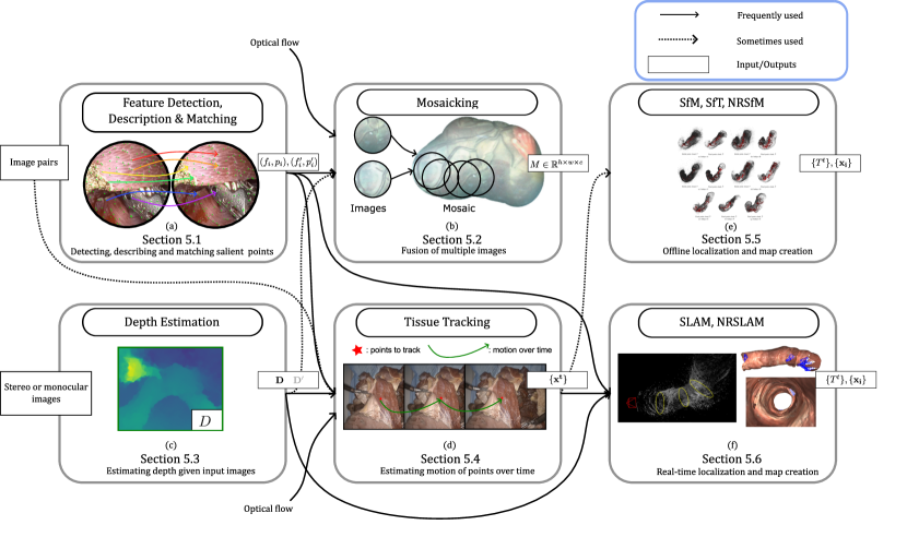
Our review will begin with a detailed explanation of the review process (Section 2) where we explain our literature search process (Section 2.1) followed by detailing prior relevant reviews and what makes ours necessary in Section 2.2. Then, we will summarize a broad list of medical specialties and the relevant algorithms that are useful to them in Section 3. This should give algorithm designers, researchers, and clinicians a high-level overview of the clinical applications along with some example algorithmic needs. Following that, we will explain the datasets relevant to MCV in Section 4, which are of great importance for both training and evaluating algorithms. In Section 5 we will delve deep into the algorithms and cover relevant works to help the reader understand the benefits, approaches, and designs decisions for the applications that were mentioned in Section 3. The flowchart in Fig. 8 provides a good high level overview of the relevant methods. Finally, in Section 6, we will provide a discussion on the features and drawbacks of algorithms, along with future needs and discussion points as we draw connections between the different algorithms. We will follow this discussion up with some ideas, questions, and needs that still need to be addressed in tracking and mapping in MCV. Finally, we will conclude and summarize the state of the field.
2 Review Process
2.1 Literature Search
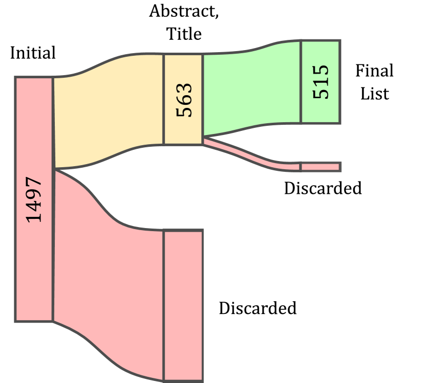
We review all papers which perform any sort of camera-based mapping or tracking in medical computer vision (MCV). These can include mosaicking (Section 5.2), depth estimation (Section 5.3), tissue tracking (Section 5.4), structure from motion (SfM) (Section 5.5.1), shape from template (SfT) (Section 5.5.3), simultaneous localization and mapping (SLAM) (Section 5.6), and nonrigid variants (which are in explained in their respective sections). Refer to the referenced sections for more details on each method. We survey any of these methods that use a clinical camera (eg. endo/colono/bronchoscope/etc). With these specifics, we perform a SCOPUS search to get a preliminary initial paper list. Our search term reflects our criterion: (( TITLE-ABS-KEY ( ( mosaicing, OR mosaicking, OR "simultaneous localization and mapping" OR slam, OR (surface* w/6 reconstruction) OR "structure from motion" OR sfm OR (stereo w/6 reconstruction) OR (tissue w/6 track*) OR ( deform AND tracking OR mapping ) OR ( deformable AND tracking OR mapping ) OR ( deformation AND tracking OR mapping ) OR ( deforming AND tracking OR mapping ) ) AND ( endoscop* OR bronchoscop* OR colonoscop* OR "surgical" OR surgery OR (capsule w/6 robot*) OR (capsule w/6 camera) ) ) )) AND ( LIMIT-TO ( SUBJAREA,"COMP" ) OR LIMIT-TO ( SUBJAREA,"ENGI" ) ) This term is a combination of tracking and mapping terms (reconstruction, mosaicking, SLAM) paired with (AND) surgery-related terms such as endoscopy, capsule cameras, etc. On July 15th, 2023 this search returned 1497 results, and after culling irrelevant results based off title and abstract we were left with 563 papers. Culling irrelevant papers was performed by removing items which included:
-
•
Surgeon performance evaluation works
-
•
Registration of multimodal images as the paper’s primary topic. eg. MR to CT. Image guidance with multimodal imagery which uses camera data is still included.
-
•
Endoscope or camera system designs (structured light, Lidar, etc.)
-
•
Non-medical applications (sewer/pipe defect mapping, metal analysis, human hand pose)
-
•
Video retrieval
-
•
Segmentation methods
-
•
OCT and pCLE
-
•
Needle steering and guidance
-
•
Simulation platforms
-
•
Surgical interventions (eg. clinical grafting methods for eye surgery)
After this, we filtered out the papers that could not be decided on based solely on the abstract. This was performed via reading the paper itself, which reduced the list to a final count of 516 papers. After this, we separated the papers into groupings by application and algorithms, which helped to define the structure of this review. Additional frequently encountered citations were added, along with recent papers that cite prior review papers. See Fig. 2 for a figure summarizing this process, and Fig. 1 for a histogram plotting the number of included publications over time.
2.2 Prior Reviews
To justify the necessity of this review and assert proper coverage in our list of included papers, we also performed a comprehensive search through all reviews from the last decade in the field. By noting prior reviews, we help to motivate the need for a recent review in medical camera tracking and mapping.
In 2013, Maier-Hein et al. (2013) provide an in depth review of optical techniques for surface reconstruction covering: stereo, structured light, SfM, SLAM, Time-of-Flight, models, toolkits, and intra-operative registration. Much has happened since then with the adoption of machine learning. More recently, providing more detail on devices, Fu et al. (2021b) reviews devices in optical and flourescence imaging, along with providing a brief coverage of surgical tool tracking and SLAM methods.
Focusing on image stitching, surface reconstruction and view enhancement, Bergen and Wittenberg (2016) provide a review that covers technology readiness and provide a useful classification of different methods and their clinical feasibility.
At a similar time,Lin et al. (2016) review the complementary problem of deformation recovery and surface reconstruction. They concluded that deformation recovery and localization remain an open challenge.
In surgical data management and processing, Münzer et al. (2018) focus on content-based processing (specularity removal, compression, retrieval) methods for endoscopic images. Later, Maier-Hein et al. (2022) review the field of surgical data science, detailing infrastructure, data annotation, and analytics.
In augmented reality (AR), Bernhardt et al. (2017) provide an in depth review of the uses of augmented reality in laparoscopic surgery, which serves to motivate many image guidance applications. Qian et al. (2020) provide a review of AR for robotic assisted surgery, summarizing methods and AR content used for each application (eg. heart model, kidney, pre-op imaging). Malhotra et al. (2023) further review AR for surgical navigation but do not delve into models or deformation. More broadly, Chadebecq et al. (2023) provide a review of artificial intelligence and automation in surgery, with a summary including robotic control, and other applications.
With clinical focus, Schneider et al. (2021) perform a systematic review on image guided liver surgery, focusing on interventions. They provide motivation for improving image guidance (and thus tracking algorithms). Acidi et al. (2023) survey clinical applications of AR in liver surgery, concluding that the application is limited due to insufficient precision, but stating that it is likely to become more effective with increased usage. These are of specific relevance to algorithmic applications in image guidance.
In summary, these reviews either cover specific subfields or do not have more recent technical details on deformation models, neural networks, etc. used for tracking and mapping. In contrast to the the mentioned reviews, we will be more algorithmically focused without constraining our discussions to devices or sensors such as Time-of-Flight. Thus, our review fills the position as: a guide for recent algorithmic advances through the entire tracking and mapping process, a coverage of quantification and data, and finally a thorough discussion of needs for this field in the future.
3 Medical Specialties and Relevant Applications
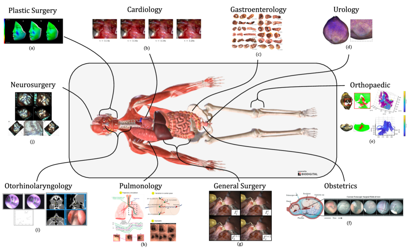
In this section we will briefly cover different medical specialties that have clinical applications requiring tracking or mapping. Alongside this, we summarize the algorithms that are relevant to said specialty. This should serve as a quick reference of sample works for clinicians and those implementing algorithms. To those researching algorithms and MCV, this should serve as a overview of how broadly applicable some of the methods can be. We separate the sections by clinical application: cardiology, orthopaedic, obstetrics, otorhinolaryngology (ear, nose, throat (ENT)), plastic surgery, pulmonology, gastroenterology, neurosurgery, urology, and general surgery. For locating which body regions are relevant to each specialty, along with what the data looks like, the diagram in Fig. 3 should be of use. For every mentioned application, see the flowchart in Fig. 8 for a description of the algorithm and its dependencies. For a quick summary of specialties, Table 1 provides an overview. We note that opthamology and dermatology also could have relevant applications, but we do not provide sections as there limited works with algorithmic focus.
3.1 Orthopaedics
In orthopaedics, guiding navigation via aligning models to the camera feed requires localizing the arthroscope’s position relative to the body. This helps achieve the clinical objectives of better registration for implants and orthrothpaedic surgery for reconstructing bone (Marmol et al., 2017, 2019; Zhang et al., 2022), or automating interventions such as milling of bone. The algorithms useful for this field are rigid mapping (SLAM, SfM), and of course all dependent algorithms (see Fig. 8).
3.2 Obstetrics
In obstetrics, twin-to-twin transfusion syndrome is treated via anastamosing placental vessels between twins. Visualizing the surface of the placenta is difficult due to a small field of view, thus algorithms look to extend the field of view via mosaicking (Li et al., 2021; Bano et al., 2019, 2020b). Additionally, De Smet et al. (2019) show that pelvic repair could benefit from stereo reconstruction via enabling better visualization than a 2D screen. Thus the relevant algorithms are mosaicking and stereo reconstruction.
3.3 Otorhinolaryngology (ENT)
In otorhinolaryngology, enabling tracheal robot steering using cameras on the tip of a robotic device could help ease deployment and avoid damage to critical structures (Girerd et al., 2020). Additionally, maps of the nasal passage can help in sinus surgery by registering pre-operative data to aid in avoiding critical structures (Liu et al., 2020a). In these environments, SLAM, SfM, feature description, and depth estimation are of particular importance.
3.4 Plastic Surgery
Plastic surgery includes, but is not limited to reconstructive operations on the face. Predicting facial outcome for planning in maxillofacial surgery requires surface reconstruction (Buchart et al., 2009). Deformation modelling is also important to help create accurate craniofacial models for surgery (Suputra et al., 2020). Stereo reconstruction can also be used for efficiently evaluating grafting outcomes after surgery (Baserga et al., 2020). Thus, both stereo reconstruction and nonrigid reconstruction are both relevant to plastic surgery.
3.5 Neurosurgery
In neurosurgery, brain shift between the time when the MRI scan is acquired and surgery affects the usability of the MRI-determined landmarks. Deformable tracking is important here since the brain can undergo complex nonrigid deformation (Hartkens et al., 2003). Therefore, methods that can visually track the surface of the brain could prove useful for deforming the preoperative scan (De Momi et al., 2016). This is especially true if they can be performed using camera video without markers (Jiang et al., 2016). Recently, in neurosurgery, convolutional neural networks (CNNs) have been used to quantify vascular structures and track regions (Martin et al., 2023).
3.6 Gastroenterology
In gastroenterology, medical computer vision is useful for extending field of view with the purpose of ensuring coverage in colonoscopy screening. This enables better detection of polyps or cancer by helping all regions be seen and surveyed (Ma et al., 2019; Zhang et al., 2021a; Turan et al., 2017). It is similarly helpful for stomach reconstruction reconstruction where it can again help for detecting detect ulcers or cancer. To enable the reconstruction of a 3D surface such as the colon, successful localization of the camera is key (Widya et al., 2021). Thus, methods that are important in this field are SfM, NRSfM, SLAM, NR SLAM, and mosaicking. These environments are nonrigid, so the accuracy of rigid methods when they are applied depends on the rigidity of capture and length of video.
3.7 Cardiology
In cardiology, being able to compensate for motion during heart surgery is a promising application of medical computer vision. This is called motion compensation, where the goal is to give the surgeon the impression that the heart is stationary, by moving the camera observing the heart in a synchronized manner with the heart motion, and moving the robotic instruments relative to the heart’s surface. This requires accurately measuring the motion of the heart surface, which has been addressed algorithmically (Richa et al., 2011; Schoob et al., 2017). Tissue tracking, stereo reconstruction and deformable SLAM are the particular methods that are useful for this.
3.8 Pulmonology
In pulmonology, the primary image modality using medical computer vision is bronchoscopy. In bronchoscopy, a camera is inserted into the lungs. Thus depth estimation and mapping are important for visually guiding the scope to a nodule biopsy (Visentini-Scarzanella et al., 2017; Wang et al., 2020a) rather than using fluoroscopy (live x-ray) (or CT) which have ionizing radiation. Thus, SLAM, and deformable SLAM methods are of specific importance, as we would like to recover the pose of the bronchoscope to then be able to correctly localize the biopsy site.
3.9 Urology
Bladder cancer screening can require surveying the entire bladder to ensure all lesions can be found. Therefore, creating panoramas could help aid in diagnostics (Soper et al., 2012). Designing algorithms to aid navigation can also make procedures easier by providing a map when inspecting the kidneys or ureters. Kidney stone removal is an application of flexible ureteroscopy where it can be hard to orient the instrument. SLAM methods have been introduced here as potential solutions (Fu et al., 2021a; Oliva Maza et al., 2023). Thus mosaicking and deformable SLAM are of relevance in urology.
3.10 General Surgery
In minimally invasive surgery, tracking and mapping would help improve surgical perception, thus providing better image guidance. This could help to: improve margins in surgery by deforming pre-operative scans to track the movement of tissue, enable autonomous scanning and suturing, and ease proctoring Maier-Hein et al. (2014); Chadebecq et al. (2023). In this field, the main algorithmic applications are mosaicking, NR SLAM, and Nonrigid SfM, with NR SLAM being the one suited to use during surgery, since it is real-time.
| Location | Use | Algorithms |
| Ortho. | Auto., Guid. | SfM, SLAM (Marmol et al., 2017; Ma et al., 2020; Zhang et al., 2022) |
| Obste. | TTTS., Pelv. surg. | DM, Mos. (De Smet et al., 2019; Bano et al., 2020b) |
| ENT | Guid., Auto., | SfM, SLAM (Girerd et al., 2020; Liu et al., 2020a) |
| Plastics. | Recon. | DM, NRSfM (Suputra et al., 2020; Baserga et al., 2020) |
| Neuro. | Img. Guid. | NRSLAM, tis. track. (Jiang et al., 2016; Martin et al., 2023) |
| Gastro. | Recon., Diag. | Mos., (NR)SfM, (NR)SLAM (Ma et al., 2019; Widya et al., 2021) |
| Cardio. | Mo. Comp., Meas. | DM, tis. track., NRSLAM (Richa et al., 2011; Schoob et al., 2017) |
| Pulmo. | Diag., Biopsy | NRSLAM (Visentini-Scarzanella et al., 2017; Wang et al., 2020a) |
| Uro. | Cancer, Uretoscopy | Mos., NRSfM, NRSLAM (Soper et al., 2012; Oliva Maza et al., 2023) |
| Gen. surg. | Auto., Guid., Meas., Recon. | DM, tis. track., NRSLAM (Maier-Hein et al., 2014; Chadebecq et al., 2023) |
4 Datasets
4.1 Introduction
In this section, we will detail datasets that have been released and are available for quantifiying tracking and mapping methods in MCV. As a sample, some of these datasets include labelled data for evaluating: image stitching, stereo, reconstruction, or tracking. Datasets which are for segmentation or classification are excluded. Datasets which have been used for tracking but do not have labels will be mentioned in brief.
We will begin in Section 4.2 with a summary of datasets that do not have any ground truth which are primarily useful for training unsupervised methods. In Section 4.3, we delve into datasets with algorithmically generated ground truth. The algorithmically generated datasets are in their own section because they depend on reconstruction accuracy of stereo algorithms or SfM, and are limited to be at best as good as the classical reconstruction methods. We will then follow with summarizing simulated ground truth datasets, generated via rendered 3D models, in Section 4.4, and physical phantoms, e.g., silicone tissue models, in Section 4.5. We finally close with ground truth that uses real tissue in Section 4.6. We separate these into these classes as different data types can be vulnerable to different biases. For example, simulation or phantom data might not carry over to real data. Alongside the sections, we have a table of datasets to reference in Table 2, and a figure showing their availability over time in Fig. 4. This table should provide a means to get a high level summary of different algorithmic approaches and dataset generation. For our discussion on the datasets, please go to Section 6.1 near the end of this review.
| Dataset | R/S/P | Location | Rigid | Truth | Use |
| Stoyanov et al. (2010); Pratt et al. (2010) | P | Heart | Nonrigid | Stereo (CT) | Recon. |
| Maier-Hein et al. (2015) | R | Abd. | Nonrigid | Annotated | Stereo Recon. |
| Ye et al. (2016) | R | Abd. | Nonrigid | Annotated (BB) | Tracking, Retargeting |
| Visentini-Scarzanella et al. (2017) | S | Lung | Rigid | Sim/Phantom CT | DM, Transf., Navigation |
| Penza et al. (2018a) | P | Abd. | Rigid | Laser Scan | Stereo Recon. |
| Rau et al. (2019) | S | Colon | Rigid | Simulation | Stereo Recon. |
| Li et al. (2020) | R | Abd. | Nonrigid | Annotated | Tracking |
| Fulton et al. (2020) | P | Colon | Nonrigid | Phantom, Pose | Localization, Navigation |
| Bano et al. (2020a) | R | Fet. | Nonrigid | Labels | Mosaicing |
| Zhang et al. (2021a) | S | Colon | Nonrigid | Simulation | Deformable 3D Recon. |
| Recasens et al. (2021) | R | Abd. | Nonrigid | LibELAS | Training/Tracking |
| Ozyoruk et al. (2021) | R | GI | Rigid | Scanner | SLAM, DM |
| Ozyoruk et al. (2021) | S | GI | Rigid | Rendering | SLAM, DM |
| Xi et al. (2021) | R | Abd. | Rigid | Neural* | Monocular Recon. |
| Zhang et al. (2021b) | S | Colon | Rigid | Sim | SLAM, DM |
| Allan et al. (2021) | R | Abd. | Rigid | Structured Light | Stereo Recon. |
| Edwards et al. (2022) | R | Abd. | Rigid | CT | Stereo Recon. |
| Guy et al. (2022) | S | Abd. | Ridid | Simulation | Stitching |
| Rau et al. (2022) | S | Colon | Rigid | Simulation | Depth and SLAM |
| Azagra et al. (2022) | S | Colon | Nonrigid | Simulation | SLAM |
| Azagra et al. (2022) | R | Colon | Nonrigid | Colmap | SLAM |
| Bobrow et al. (2022) | P | Colon | Rigid | Phantom | Recon., Localization |
| Cartucho et al. (2024) | R | Abd. | Nonrigid | Annotated | Tracking |
| Hayoz et al. (2023) | R | Abd. | Nonrigid | Kinematics | Rel. pose est. |
| Lin et al. (2023) | R | Abd. | Nonrigid | Vis Markers | Tracking, Recon. |
| Schmidt et al. (2023b) | R | Abd. | Nonrigid | IR Markers | Tracking, Recon. |
4.2 Unlabelled Datasets
As described in our review process (Section 2), we focus on literature related to tracking and mapping. We exclude unlabelled datasets that are useful in other domains, or are designed for tasks such as segmentation, since they are seldom used in tracking work. The Hamlyn Centre datasets include many unlabelled sequences from procedures using both monocular and stereo cameras (Mountney et al., 2010), in addition to some stereo sequences with a deforming heart (Stoyanov et al., 2005). Additionally, they provide some datasets designed for qualitatively evaluating tissue tracking in varied environments and with different artifacts such as smoke, blood, and lens smudge (Giannarou et al., 2013). They also provide a dataset of unlabelled stereo image pairs for the purpose of evaluating unsupervised methods using photometric reconstruction error (Ye et al., 2017). Photometric reconstruction evaluates how accurately a depth estimation works for reproducing an image, using photometric error, which compares colors at image pixels, but does not provide actual measurements of reconstruction accuracy. The Hamlyn datasets that provide labels will be referenced in later sections.
4.3 Algorithmic ground truth
By algorithmic ground truth, we mean data that is generated via a reconstruction algorithm, and can act as a pseudo ground truth. Reconstruction algorithms include stereo algorithms, SLAM, or SfM. By using reconstruction algorithms to generate ground truth data, we have to assume that they are accurate. This limits the performance evaluation of new algorithms. For example, a classical SfM method will only obtain sparse points in a rigid manner and does not deal with lighting effects such as specularities, and thus cannot be used to evaluate a new method that addresses these issues.
Many works have used stereo depth networks to evaluate accuracy, with EndoDepthAndMotion (Recasens et al., 2021) being one. They release a dataset with ground truth generated by LibELAS (Geiger et al., 2011) in abdominal sequences. This dataset is intended for training depth models and evaluating tracking methods. Xi et al. (2021) generate pseudo-ground truth using autoencoders. They design a network for monocular depth learning along with a method for point cloud completion. They evaluate their algorithm on the EndoAbS (Penza et al., 2018a) dataset, and then release the point clouds created with their network.
4.4 Simulated data
MCV scenes can be generated by rendering from simulation. Recent methods have been improving the photorealism of these simulations, bringing simulation closer to the real environment. Some of these methods use CT scans and phantoms, but they are still grouped into being simulated if they use rendered data for ground truth. In Visentini-Scarzanella et al. (2017), the authors generated 32 video sequences with ground truth depth and renders in a simulated bronchoscopy. These sequences are generated using a rigid realistic lung phantom with rendering performed using a model from paired CT scans. The rendering contains frames that act as depth ground truth. To align the physical phantom with the simulation model, they use SLAM and follow it with Iterative Closest Point (ICP), a point cloud alignment method. The dataset is designed for transfer learning in depth networks for modelling to-and-from rendered to real, and for depth estimation and mapping in bronchoscopy. Rau et al. (2019) also release rendered ground truth depth frames in monocular colonoscopy that are generated via simulation based on CT scans.
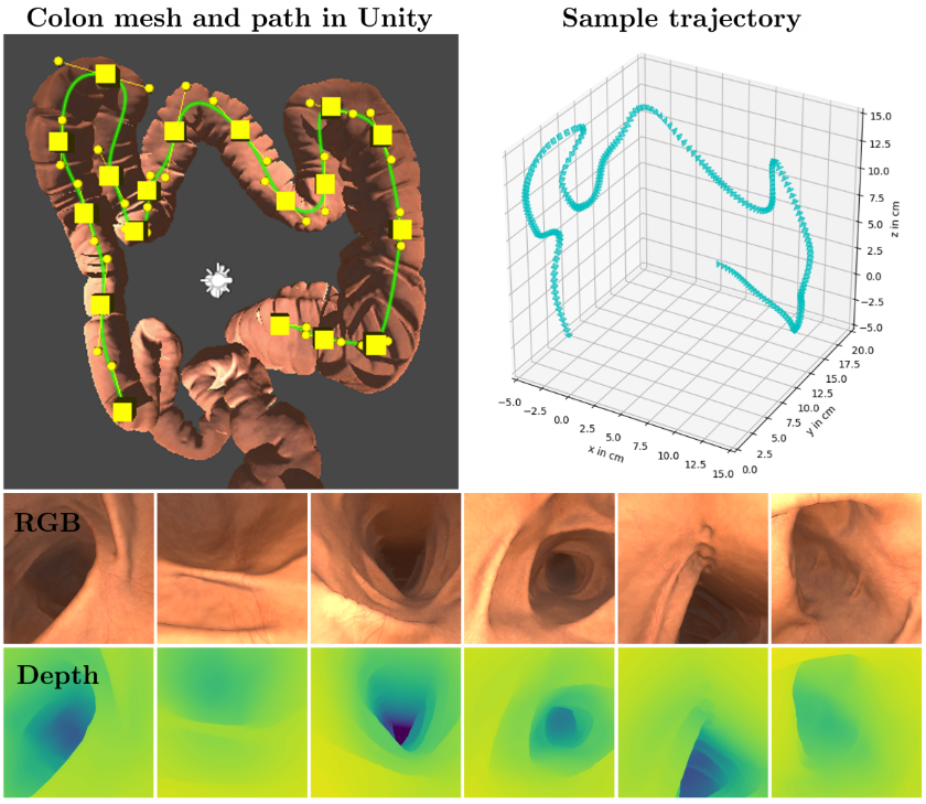
Since it is very difficult to obtain ground truth in colonoscopy because it is nonrigid, Zhang et al. (2021a) opt to use simulated colonoscopies. In order to construct realistic models, they texture four different CT scans by applying colors and lighting parameters to a mesh. To simulate nonrigid motion, they deform their simulated tubular colon model about the centerline. They release depth maps and monocular frames from their dataset for evaluation of reconstruction algorithms. They also release a similar dataset with fifteen colon models, but with a rigid model instead (Zhang et al., 2021b). Instead of using a monocular camera, this dataset provides stereo pairs, and also includes ground truth camera poses.
Moving on to systems for simulation in minimally invasive surgery (MIS), VisionBlender (Cartucho et al., 2021) propose and publish code for creating simulated endoscopic data along with a utility for creating depth maps, optical flow, poses, and normals. Later on, in a similar vein of simulation, but for image stitching instead of depth estimation and flow, Guy et al. (2022) generate a dataset for evaluating image stitching (merging images taken at the same time in multi-camera setups). They look to address difficulties that occur in stitching such as the duplication of or disappearing of objects in the surgical field. Their simulation framework can generate tools and organs with varying camera models, and is shown in Fig. 5. In C3VD (Colonoscopy 3D Video Dataset), Bobrow et al. (2022) release many video sequences with video from 3D printed phantoms alongside sequences from the rendered simulated models. In SimCol, Rau et al. (2022) release another colonoscopy dataset but with the additions of monocular pose and depth images. This can help for evaluating SLAM and depth mapping frameworks, although this dataset does not include deformation. Alongside this submission, they propose a novel pose estimation network. Reconstructions using their framework are demonstrated in Fig. 6 They provide depth, pose, flow, and the 3D models as a part of their dataset.
In the EndoMapper dataset (Azagra et al., 2022), release data from both real and simulated scenarios. The real dataset comprises videos and camera calibrations without ground truth labels. For the real dataset, they provide some algorithmically generated ground truth via 3D reconstructions generated with COLMAP (Schönberger et al., 2016a; Schonberger and Frahm, 2016)–a publicly available library for generating point clouds using SfM. This data is released for a couple partial colon chunks since SfM can fail in colonoscopy on larger environments. For the simulated section of their dataset release, they artificially deform their model to better represent motions of a real colon. In the simulated dataset, they release depth, video frames and the camera trajectory (pose over time). The dataset is available with a release request for nonprofit institutions.
4.5 Phantoms
These datasets are designed to quantify performance using phantoms, which are physically printed or sculpted models of organs or different environments. One of the Hamlyn datasets (Stoyanov et al., 2010; Pratt et al., 2010) includes a beating heart phantom. Using CT, 3D ground truth is registered to the stereo camera. This dataset can be used for evaluating stereo algorithms, and tracking performance. In EndoAbS (Penza et al., 2018a) release a dataset for evaluating stereo reconstruction which comprises 120 stereo pairs with camera calibration. Their ground truth is generated on abdominal organ phantoms using a laser scanner. They collect stereo frames over multiple different distances, lighting, and smoke conditions. Fulton et al. (2020) release a dataset with a deformable phantom colon The ground truth they provide is camera pose generated via a magnetic tracker. They collect sequences with multiple different levels of deformation. They additionally survey the performance of different visual odometry (VO) systems in correctly estimating pose using their dataset. Edwards et al. (2022) introduce a methodology for evaluating stereo algorithms via paired CT scans. Their dataset comprises 16 stereo image pairs of varying organ phantoms along with CT-generated 3D ground truth.
4.6 Real tissue
In summarizing real tissue datasets, we include works that focus on surgical tissue and organs, both in vivo and ex vivo. Beginning with tissue tracking and deformable mapping datasets, Maier-Hein et al. (2015) introduce crowd-sourcing to labelling endoscopic data (users use software to select points), and release a methodology for generating validation sets. Alongside the methodology, they release a set of one hundred annotated stereo pairs. Ye et al. (2016) also release a dataset for evaluating tracking in endoscopy with data generated via having users label bounding boxes of tracked regions throughout a video clip. SuPer (Li et al., 2020) and SurgT (Cartucho et al., 2024) perform a similar labelling procedure for tissue in stereo endoscopy. Later, Semantic SuPer instead uses green pins to mark the tissue surface (Lin et al., 2023) rather than requiring user labelling. With Surgical Tattoos in Infrared (STIR), Schmidt et al. (2023b) introduce a dataset for evaluating tissue tracking and SLAM methods. It comprises labeled points in infrared and stereo video clips, with the benefit being that it neither requires software labelling, nor visible markers that can affect algorithm evaluation.
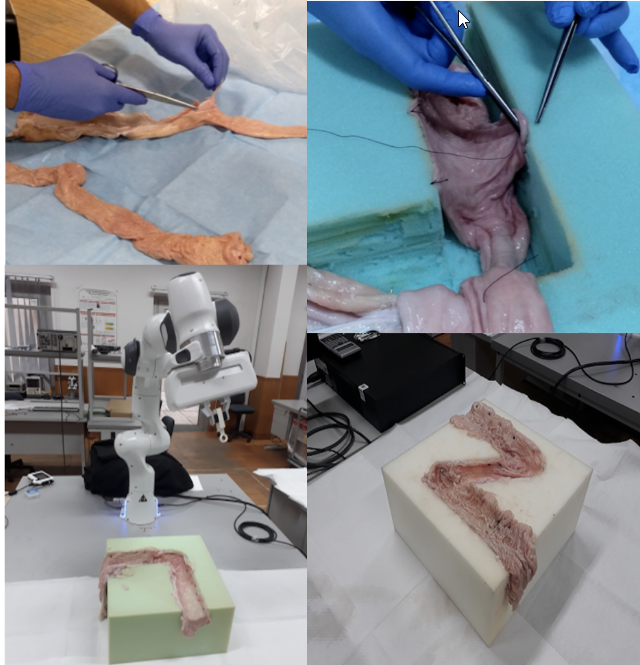
For pose estimation and depth mapping in the gastrointestinal tract, EndoSLAM (Ozyoruk et al., 2021) provides a rigid dataset with video from many different capsule cameras and endoscopes. The ground truth is obtained as point clouds generated from a 3D scanner that are aligned to the camera frame with ICP. Ex vivo sequences are acquired by attaching tissue to a foam scaffold. Fig. 7 demonstrates their collection methodology. They provide a synthetic dataset as well.
Addressing accurate depth generation in MIS, SCARED (Allan et al., 2021) provides a dataset of depth maps calculated using structured light alongside stereo endoscopic videos. Focusing on pose, Hartwig et al. (2022) release the MITI dataset which includes stereo video and camera pose from a surgical intervention. The pose is calculated using an IMU (inertial measurement unit) and infrared (IR) markers. StereoMIS (Hayoz et al., 2023) also address the problem of quantifying pose estimation, focusing specifically on relative pose between images. Their dataset releases relative pose calculated using kinematics alongside stereo videos from porcine models. In a different vein, Bano et al. (2020a) provide segmentation of hundreds of frames of vessels in fetoscopic procedures for mosaicking.
As seen, the datasets using real tissue vary in the truths they provide (pose, depth, motion), and the authors use many different methods for collecting this ground truth.
5 Algorithms
In this section we will begin with the important technical building blocks in tracking and mapping, and then move into more complex methods that manage deformation. First, feature detection, description, and matching in MCV will be reviewed in Section 5.1. Then, mosaicking, in which features are used to fuse images into panoramas is covered in Section 5.2 In Section 5.3 we cover depth mapping which calculates the 3D position of 2D image pixels. Then, we will summarize surgical tissue tracking which looks to track points in the surgical scene in Section 5.4 (for using a map as well, see SLAM). After which, in Section 5.5.1 we will explain rigid and nonrigid (NR) Structure from Motion (SfM), and Shape-from-* methods that estimate shape using a model or a set of points. Finally, in Section 5.6 we will cover rigid and nonrigid (NR) Simultaneous Localization and Mapping (SLAM) which aims to create a real-time map from a video of the surgical environment. In the SLAM section, we will also include related methods that address the mapping problem without a localization focus. See Fig. 8 for an illustration of how all these methods depend on one another.
5.1 Feature Description and Detection
The purpose of image features is to provide a numerical means to create correspondences between images. Therefore, having well-defined image features has been a well established goal for enabling methods in tracking deformation. Image features assign numerical vectors to positions, and can be either sparse or dense. By comparing these vectors, features can be matched to create data correspondences. The feature error comparison vectors are used to create data association terms, which are terms in the cost function for optimization models such as SLAM or relative pose estimation. In this section we will summarize sparse features (Section 5.1.1)) and feature matching (Section 5.1.2) followed by dense features (Section 5.1.3)) used in MCV. Sparse features are often used for image alignment/mapping, or other problems that require computational efficiency. The follow-up task of feature matching is often performed only for sparse features, since for the dense ones we are able to perform a pixelwise search over the entire image. Dense features are calculated over a whole image grid, and provide higher resolution at the cost of efficiency.
5.1.1 Sparse Features
Sparse features are generated by two main components: detection and description. Detection is the process of finding locations for each keypoint in an image . Description assigns each keypoint a d-dimensional numerical vector (could also be binary). SIFT (Lowe, 1999), SURF (Bay et al., 2008), and ORB (Rublee et al., 2011) are examples of classical descriptors. Classical in this sense means they are hand engineered and use intensity histograms, decision trees/etc., to create the numerical descriptor values.
Classical descriptors are still frequently used in SLAM works (Lamarca et al., 2021; Song et al., 2018). Early descriptors for surgical environments used feature histograms and decision trees along with LK (Lucas-Kanade) optical flow (Mountney and Yang, 2008). Giannarou et al. (2009) propose an affine-invariant detector that detects points over scales, assigning ellipses to them to better deal with varying angle and scale. Classical descriptors still remain in use, with many applications such as registering pre-operative brain images (Jiang et al., 2015). On usage in the brain, Jiang et al. (2016) use segmented Frangi features (Frangi et al., 1998) – which detect tube-like structures – for non-rigid registration of brains using vessel/sulci surface features. Classical features have also been evaluated in arthroscopy, which deals with a fairly rigid environment (Marmol et al., 2017).
Moving onto neural applications, there are learned sparse features that are applicable to surgery. For example, ReTRo (Schmidt and Salcudean, 2021) proposes a lightweight real-time descriptor, trained using camera-pose self-supervision (Wang et al., 2020b) in surgical environments. The authors use classical motivations to train a neural network that samples and rotates like ORB. Although this does not include tissue deformation in training pairs, it contains the same point from different views. To work in deformable spaces, although not trained on surgical data, Potje et al. propose training deformable features using data augmentation with a thin plate spline. More specifically to surgery, Barbed et al. (2023) present a SuperPoint style descriptor and detector using COLMAP reconstruction for training. Rather than depending on homographies (a re-projection that treats an image as planar), they propose tracking adaptation, which trains on the re-projections of the 3D points. This should help the descriptors perform in surgical environments. With the same goal of improving performance in surgical environments, Karaoglu et al. (2023) note that the surgical environments differ from real-world images which are often oriented vertically, and they propose RIDE which builds rotation equivariance into the network design.
5.1.2 Feature Matching
After obtaining a sparse set of features with their positions in an image, we need to match them to corresponding features and positions in the other image : , . This is often done via performing a dot product between features, , to obtain a similarity score. Modern methods can better match features using more than just their descriptor values. By using descriptors along with the relative motion estimated motion, for example, they can design efficient and accurate feature matching schemes. GMSMatch (Grid-Based Motion Statistics (Bian et al., 2020)) aids matching using heuristics based on the motion of surrounding matches. Recent neural network based matchers such as SuperGlue (Sarlin et al., 2020) and LightGlue (Lindenberger et al., 2023) learn matching based on a graph-neural network of points. The principle of these modern methods is to take in a point and, rather than brute-force match, use the motion of the surrounding points, their features, or both. For example, if a match is in a different motion direction than all of its surrounding matches then it can be discarded. GMSMatch uses a heuristic for this, while SuperGlue uses a learned Graph Neural Network trained using homographies and large-scale outdoor depth reconstruction scenes. These methods could be trained for surgical environments as well given ground truth, or robust reconstructions such as SfM.
For match filtering specifically in surgical environments, Chu et al. (2020) use A-SIFT descriptors for laparoscopy and gastroscopy. They assume that features move smoothly and slowly in these environments, and perform match filtering via expectation maximization (EM).
Again using EM, Zhang et al. (2023) refine matches under the assumption that the environment can be represented with Dual-Quaternion Blending (DQB). This does not allow for discontinuities or transformations that do not fit the smooth DQB deformation field. We note that detected points need not necessarily be discarded entirely, because they can still provide useful information, e.g., texture.
Since feature matching is very sensitive to position, in Schmidt et al. (2022a), the authors chose to keep all keypoint matches, but train to refine (instead of discard) the detections to best improve downstream photometric reconstruction using Graph Neural Networks (GNNs).
5.1.3 Research in Dense Image Descriptors
Research in dense image descriptors is less common, likely due to computational costs. Indirectly, some models could be said to create dense descriptors (eg. stereo or optical flow networks mentioned later), but these models directly use the features as part of the model, so it comes down to a question of semantics. Models that are designed primarily as a means for feature description, e.g., Liu et al. (2020b), train a CNN-based descriptor model in a novel way. They use SfM to generate ground truth for their dense descriptor. To find matches, the detected point searches for matches over the entire image. This takes ms to match a set of descriptors on a image. Since this is a convolutional search method, we can expect the costs to scale by the amount of additional keypoints and the increase in image size. For a full-resolution image (), we can expect it to take anywhere from ms if the CNN is the bottleneck (16x the data) to ms if the bottleneck is in the matching step (16x the matches and 16x the data).
5.2 Mosaicking
5.2.1 Introduction
Mosaicking is the process of creating a compound image using a collection of images over time. This is performed by first matching and aligning similar features in the images by warping the images, followed by color-correcting via blending (Burt and Adelson, 1983). Having a mosaicked image, , can help in interventions or diagnostics where the camera only provides a small field of view. Mosaicking is different from stitching, which looks to fuse images taken at the same time using multiple different cameras. Mosaicking can be done in a 2D manner, or in 3D on a surface such as a sphere or mesh. Although they still use mosaicking, we omit works in pCLE and microscopy (as per our literature search methodology), to maintain our focus on work that uses video images for tracking and mapping. For more information, Bano and Stoyanov (2024) provide a very recent summary chapter on mosaicking.
5.2.2 Mosaicking in MCV
In early works on retinal and catadioptric endometrial videos, Seshamani et al. (2006) propose using mosaicking to create a broader field of view. They do this by aligning images photometrically with an affine transformation for each image, and provide an algorithm that can run at native camera frame rate (30fps). Mosaicking using images from fibroscopes is challenging because of the many artifacts and specularities. To address this, Atasoy et al. (2008) propose a method that uses SIFT features to match between images. They additionally solve for a global alignment where the relative transformation is calculated by optimizing over all frames. This better allows consensus and reduces the drift that can be caused when just aligning on a frame-to-frame basis (since errors can compound). This is similar in principle to bundle adjustment that happens in SLAM (Section 5.6). They evaluate their method on ex vivo kidney tissue. In order to account for image differences, they merge images with multi-band blending Burt and Adelson (1983) which partitions the images to remove low frequency variations while preserving high frequency details. A similar method is proposed and evaluated on in vivo experiments with applications for bladder mosaicking in urology (Miranda-Luna et al., 2008). For endoscopy, Bergen et al. (2009) generate a mosaic using a Kanade-Lucas Tomasi tracker (KLT) with RANSAC (Random Sample Consensus) for outlier removal. A homography transformation is estimated between frames, and specularity removal is performed via masking. In cytoscopy, mosaicking using dense cross-correlation is also used for aligning images, and the results are evaluated clinically in research by Hernández-Mier et al. (2010). Here, the tissue surface can be ill-featured, and this can make robust matching difficult. Because of poor features, it can be helpful to perform mosaicking in the fluorescent imaging spectrum, where features of interest such as tumors are better illustrated. By mosaicking on fluorescent images, diagnostics can be improved by providing a wider field of view (Behrens et al., 2011). Even though most mosaicking work has been performed in 2D, images can be projected and blended on 3D surfaces as well, for example 3D spherical models have been created to provide 360° views of the bladder (Soper et al., 2012). None of these methods account for re-aligning features when a camera loops back to the same location, and Weibel et al. (2012) solve this by providing a way to close and align loops (via detecting when features are seen again). For more details on stitching and mosaicking, Bergen and Wittenberg (2016) provide an in-depth review of works before 2016.
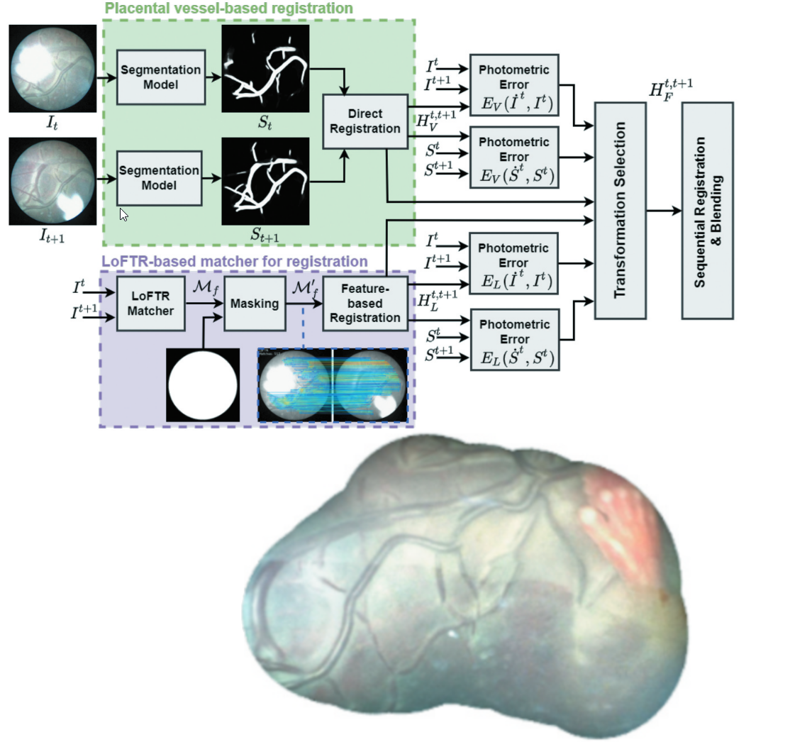
More recently, methods have begun utilizing machine learning. Bano et al. (2019) use a CNN-based homography estimation network that takes in image pairs and estimates a homographic transformation. They adapt it to fetoscopy via adding data augmentation and outlier rejection for artifacts such as specularity. Recently, they found that a combination of deep learning for homography along with matching vessel segmentation maps creates a hybrid method that outperforms either on their own (Bano et al., 2023) (see Fig. 9 for their mosaicking architecture).
The concepts of loop closure and pose graphs (connected set of camera locations with measurements or co-observance of features acting as connections) from SLAM are also used in fetoscopic mosaicking. This allows for better global alignment and bundle adjustment (Li et al., 2021). By combining a neural method along with the idea of a pose graph in endoscopy, Li et al. (2023) mosaick using both neural optical flow and SIFT keypoint matches by optimizing the image transformations in an underlying pose graph.
5.3 Stereo and Monocular Depth Estimation
5.3.1 Introduction
In order to track surfaces in 3D, we must have a notion of their depth. This requires a depth estimation network which estimates the disparity value of each pixel in the image, denoted as a disparity map, . The disparity is the relative difference in distance from a point to the camera center between each image in the stereo pair. This disparity of a point can then be used along with the camera matrix to calculate the 3D position of that point. Many depth estimation methods exist that have been applied to endoscopy (GA-Net (Zhang et al., 2019), LibELAS (Geiger et al., 2011), etc.), that are relevant to both the surgical and non-surgical applications. Additionally, we note that although the methods we review in this section calculate depth densely, some SLAM or SfM methods instead efficiently back-project the points from feature matching to estimate their 3D position sparsely, although this does not provide the smoothness regularization that dense methods do.
5.3.2 Stereo Depth Mapping
Stereo disparity estimation algorithms work as follows. For each pixel in one image of the stereo pair (), we search along the epipolar (usually horizontal) line to find the most similar pixel in the other image, (). A patch based similarity metric is often used. This can be built into a neural network, or performed classically using an optimization framework. Neural networks are often trained using an image reconstruction loss, which measures how well the network warps the left image into the right image using image-level photometric errors such as L1 distance and structural similarity index measure (SSIM). This is an indirect, or unsupervised, approach since when training on datasets in MCV we seldom have ground truth depth. Since it is indirect method, visual effects in MCV such as specularity will cause artifacts in the algorithmic reconstruction. Simulated ground truth, or ground truth generated using scans is also feasible for training models without requiring photometric supervision.
Motion compensation and stabilization are often goals in endoscopy work. Stoyanov et al. (2004) propose a depth estimation method that could be used to estimate motion by calculating depth over time. Depth is solved for by using multi-resolution Normalized Cross Correlation (NCC) between rectified images, and BFGS (Broyden-Fletcher-Goldfarb-Shanno) as the iterative optimization algorithm. Later, Lo et al. (2008) propose a hybrid that combines stereo depth and Shape from Shading (SfS). SfS uses lighting cues to estimate the normal of a point in space. For example, a viewing ray that intersects with the tissue surface normal to the light source will appear brighter. The authors use a Markov Random Field (MRF) to fuse these methods, where the SfS measurement and the stereo measurements influence the true depth in a Bayesian form. Nearby points on the depth map grid are also connected in this model to provide a smoothness constraint.
With a focus on increased efficiency, sparse means for depth prediction have also been proposed. Stoyanov et al. (2010) use a sparse set of feature matches is used to propagate measurements around said matches. The method can take any set of feature descriptors and matches () as input, and the method propagates depth around each match according to color and distance difference. The very popular LibELAS (Efficient Large-scale Stereo (Geiger et al., 2011)) work uses a similar idea with more details on refinement in neighborhoods around each sparse match. They propose a model for the probability distribution of a depth point given (conditioned on) sparse matches of point features (support points), and image features. With this Bayesian model, they can propose a procedure for estimating depth. The model takes in matches which use Sobel feature as their descriptors. These act as support points. To densify the matches onto an image grid, they refine the estimated points in regions surrounding the support points by fitting them to the maximum probability in their model, which combines the distance from support points with a regularizing distance to keep pixel estimates close to the neighboring support points. LibELAS (Geiger et al., 2011) is often used for pseudo ground truth in surgical tracking and mapping (Recasens et al., 2021; Gomez Rodriguez et al., 2022; Gómez-Rodríguez et al., 2021).
Some classical computer vision methods have been adjusted for surgical video. Chang et al. (2013) use ZNCC (Zero-mean Normalized Cross Correlation) to help accommodate for brightness differences when comparing patches along an edge. They evaluate their method using 3D data from CT scans.
With machine learning beginning to make an impact, Luo et al. (2019) train an encoder/decoder model for each image in a stereo pair, fusing the results from each view afterwards. They train using proxy labels from classical stereo algorithms along with an image reconstruction loss for enforcing left-right consistency. By warping the left image according to the left disparity map, it should look visually similar to the right image. They measure performance using CT ground truth on heart phantoms from the Hamlyn dataset (Stoyanov et al., 2010; Pratt et al., 2010) and compare performance against pseudo-ground truth from classical algorithms. As mentioned in Section 4, a drawback of using the pseudo-ground truth means it is not possible to see if the method outperforms classical methods. More recently, StaSiSNet (Bardozzo et al., 2022) use a Siamese network for real-time depth estimation. On another note, since accurate calibration is essential for quality depth estimation, Luo et al. (2022) propose a machine learning method that can deal with imperfect rectification due to an incorrect stereo camera model. They first use a network to perform vertical correction estimated to better align the image pair (so the epipolar lines match). They follow with a disparity estimation CNN which uses a Generative Adversarial Network (GAN) to differentiate between warped stereo frames from left to right (and right to left) and the true frame on the right (left). They add a mask based on the residual between the reconstructed image and the true image to reduce the influence from outlier points such as specularities. Even more specific to surgical tissues, and, specifically, their contiguity, Zhao et al. (2022) estimate depth by incorporating a constraint that takes into account the surface smoothness in camera space (3D) instead of just using image-space based photometric loss. Like many other methods, they run a specularity removal step. For quantifying their method, they use EndoDepthAndMotion (Recasens et al., 2021) for ground truth, which in turn uses LibELAS. Coming back to earlier work which used hybrids of methods, Cao et al. (2022) combine SfS with a classical stereo algorithm for stereo MIS, again demonstrating the benefits of joint methods. Pushing classical methods further forward, Song et al. (2023) propose a classical method using conditional random fields and a coarse-to fine methodology, similar to LibELAS but with faster performance: taking 72ms for sized images (LibELAS 291ms Geiger et al. (2011), PSMNet 566ms Chang and Chen (2018)).
Bringing in more modern machine learning, contrastive learning also improves endoscopic stereo when used in combination with photometric loss, outperforming other self-supervised models (Tukra and Giannarou, 2022). Finally, Wei et al. (2023) use a pre-trained HSM-Net and then fine tune it on the SERV-CT dataset. Their goal is to perform localization and 3D reconstruction of dense scenes.
In brief, many different methods exist for stereo depth, but most of the baseline networks come from computer vision field. The gap that MCV algorithms fill compared to broader CV is primarily how to train and design losses for the visual appearance in MCV scenes along with designing models to incorporate priors from this environment.
5.3.3 Monocular Depth Mapping
Monocular depth estimation uses images from one camera alongside visual cues to estimate depth. Since there is no known relative distance (e.g., a camera baseline), estimating scale is not done unless there is a reference. Monocular depth estimation is necessary in bronchoscopy, for example, where the cameras are often monocular due to size constraints. Addressing monocular reconstruction, Visentini-Scarzanella et al. (2017) train a CNN to estimate relative depth (up to scale) by using ground truth renderings. In essence, the network learns the visual cues from lighting to estimate depth, similar to SfS. Of course, areas with no texture or lighting will have to be inpainted or estimated by infilling with whatever those regions looked like in training. To train in a realistic environment, Liu et al. (2018) train a monocular depth estimation network using SfM point clouds for ground truth. This is a sparse, albeit accurate, form of supervision that works in rigid environments such as sinus surgery. They later extend their loss formulation and demonstrate generalization to other environments (Liu et al., 2020a). In MCV, there is often additional information given the camera and lighting that are present. Batlle et al. (2022) propose a monocular photometric reconstruction method, which uses known positions of the camera and light as a means to shape under a Lambertian assumption. The Lambertian lighting model models a a surface as perfectly matte (its appearance is not view-dependent, unlike a mirror, for example). Due to this assumption, pixels that do not follow it have to be masked or adjusted, so they opt to remove specularities with in-painting. They use an iterative optimization method to solve for depth. Although their method is offline, it opens the door to model-based methods for MCV.
5.4 Tissue Tracking
Tissue tracking entails methods that estimate motion of tissue surfaces or organs in MCV. These are useful for any applications that require tracking of specific points (or regions). These applications includes autonomous scanning, image guidance, automation, and measurement of marked points. Tissue tracking often uses optical flow (dense) or temporal feature matching (sparse). This can be paired with feature management in order to maintain features over time and to find features after they disappear. We will begin with a summary of classical methods that are still used in endoscopy to this day. Summarizing a classical computer vision based tracking method, Lucas and Kanade (1981) introduce a tracker that (for each tracked patch) uses image similarity to find the best aligned patch and optimize its position until convergence using image intensity metrics (eg. L1, SSD). Tomasi and Kanade (1991) extend this with salient detections, creating the frequently used the Kanade–Lucas–Tomasi (KLT) tracker.
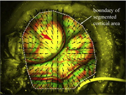
Now, moving into surgical algorithms for tissue tracking, Richa et al. (2008) perform tracking for motion compensation on beating heart surgery. They use an underlying thin plate spline (TPS) model to fit motion. In other work that does not require an underlying model, Yip et al. (2012) maintain features over time using a STAR detector (Agrawal et al., 2008) and BRIEF (Binary Robust Independent Elementary Features (Hutchison et al., 2010)) descriptor. To perform their tracking in 3D, they match stereo pairs to triangulate points. Using their method, they also propose a region tracking framework that allows tracking of user-selected regions. Regions are then tracked with a rigid transformation according to the motion of feature points lying within them. This is limited in cases with deformation, specularity, or occlusion. For tracking with novel features designed for surgery, Giannarou et al. (2013) track detected elliptical regions in real-time with an extended Kalman filter (EKF) to improve noise tolerance. We note that tissue tracking methods are also useful for image guidance in other environments, such as in brain surgery for registration of MR images under brain shift. Ji et al. (2014) track a cortical surface using LK optical flow and use stereo reconstruction to estimate 3D positions (Fig. 10).
In less featured regions, sparse correspondences enable alignment between salient features while denser optical flow can prove useful in less textured regions. Du et al. (2015) combine the benefits of sparse correspondences with LK optical flow. For estimating displacement they represent the scene using a triangular mesh. They choose to use Sum of Conditional Variance (SCV) instead of SSD as their similarity metric for optical flow. This enables better performance under non-linear variations in the images. Schoob et al. (2017) use a similar tracking algorithm, but with application in laser ablation for microsurgery. Using the classic KLT, Penza et al. (2018b) match regions with additional specularity filtering for tracking Safety Areas (structures to avoid damaging) in surgery. Their method importantly estimates when tracking fails. When tracking fails, they find SURF (Bay et al., 2006) matches in the image and compare to those in lost region to re-localize. Failure estimation of tracked regions is performed via a hand-engineered probability dependent on features in the area, percentage of features lost, validity of the transformation, and standard deviation of optical flow distribution.
In a different approach that uses neither LK optical flow nor sparse features, Collins et al. (2016) estimate flow by solving for the deformation that a model’s texture map has to undergo to match the current image via rendering the model. This requires a pre-acquisition of a model with texture, and is close in principle to Shape-from-Template (SfT, Section 5.5.3).
For applications which only require tracking a few points, tracking-by-detection can prove useful. In tracking-by-detection, a location is set as the center of initial patch to track, and then this patch region is detected in following frames to perform tracking. Ye et al. (2016) perform tracking-by-detection using a descriptor like the Haar descriptor (Viola and Jones, 2001), searching in local windows around the patch for candidate matches. They train in an unsupervised manner by sampling patches near each tracked patch as positive, and those far away as negatives.
With the advent of neural networks, CNN-based optical flow methods have begun to be used in MCV. Ihler et al. (2020) train a CNN using FlowNetL. They fine tune their network in an unsupervised manner using synthetic image warps and a zero-flow regularization (the same image tested against itself should result in zero movement). Since FlowNet is relatively efficient, their fine tuning enables a fast convolutional tissue tracking model. Other fast methods include Schmidt et al. (2022a), where neural networks are used in a sparse manner. A tracking algorithm is proposed that works by conditioning motion on a graph neural network of salient sparse correspondences. The authors later extend their work with a recurrence model Schmidt et al. (2022b), and then to 3D Schmidt et al. (2023a) Returning to tracking-by-detection, but in a neural manner, Kam et al. (2023) present a neural network for detecting six points around a vaginal cuff for cuff closure using autonomous suturing. Finally, Liu et al. (2023) use an MRF to mask surgical instruments, and they then perform tissue tracking with an underlying piecewise affine deformation model (triangular mesh) representing motions.
5.5 Structure-from-Motion (SfM), Nonrigid Structure-from-Motion (NRSfM), Shape-from-Template (SfT)
In this section, we will cover three sets of methods for offline reconstruction. These are Structure from Motion, Nonrigid Structure from Motion, and Shape from Template. Structure from motion (SfM) estimates a map in a rigid scene given a set of measurements (images in our case). Nonrigid structure from motion (NRSfM) does the same, except with an underlying map that can be non-rigid. Shape from Template (SfT) uses a learned template to estimate the template’s position given observations. These all differ from depth estimation since they look to create and maintain a usable map over time. For a detailed and wide survey in computer vision, see (Tretschk et al., 2022) for a review on dense monocular non-rigid 3D reconstruction. Here we will focus on the specific applications in MCV.
5.5.1 Structure from Motion (SfM)
SfM aims to reconstruct a rigid environment, often a set of points in 3D space, and estimate camera poses, , given a set of images, . This performed offline with the images collected beforehand. The 3D point set is called the map (a map could also be a mesh, or other representation, but for MCV we primarily see 3D points). Refer to Section 5.6 (SLAM) for the real-time counterpart which, for efficiency, differs in optimization and mapping methods. SfM is designed for rigid environments, and often entails optimizing a map and a set of poses in tandem until convergence. SfM can be used for dataset generation, but also for creating maps that surgeons can use for decision-making and planning. Most methods in SfM for MCV use the same base algorithm but adjust algorithms and terms to suit the medical environment, such as by filtering outliers or specularity.
Beginning with an early example, Hu et al. (2007) propose to use SfM for creating a larger field-of-view for surgeons. To make these methods more robust by accounting for specularity and other artifacts, Hu et al. (2012) extend their work by adding outlier removal and bundle adjustment (alignment of the 3D point positions and camera pose to reduce re-projective error of measured points in the image over times indexed by and map points indexed by ).
| (1) |
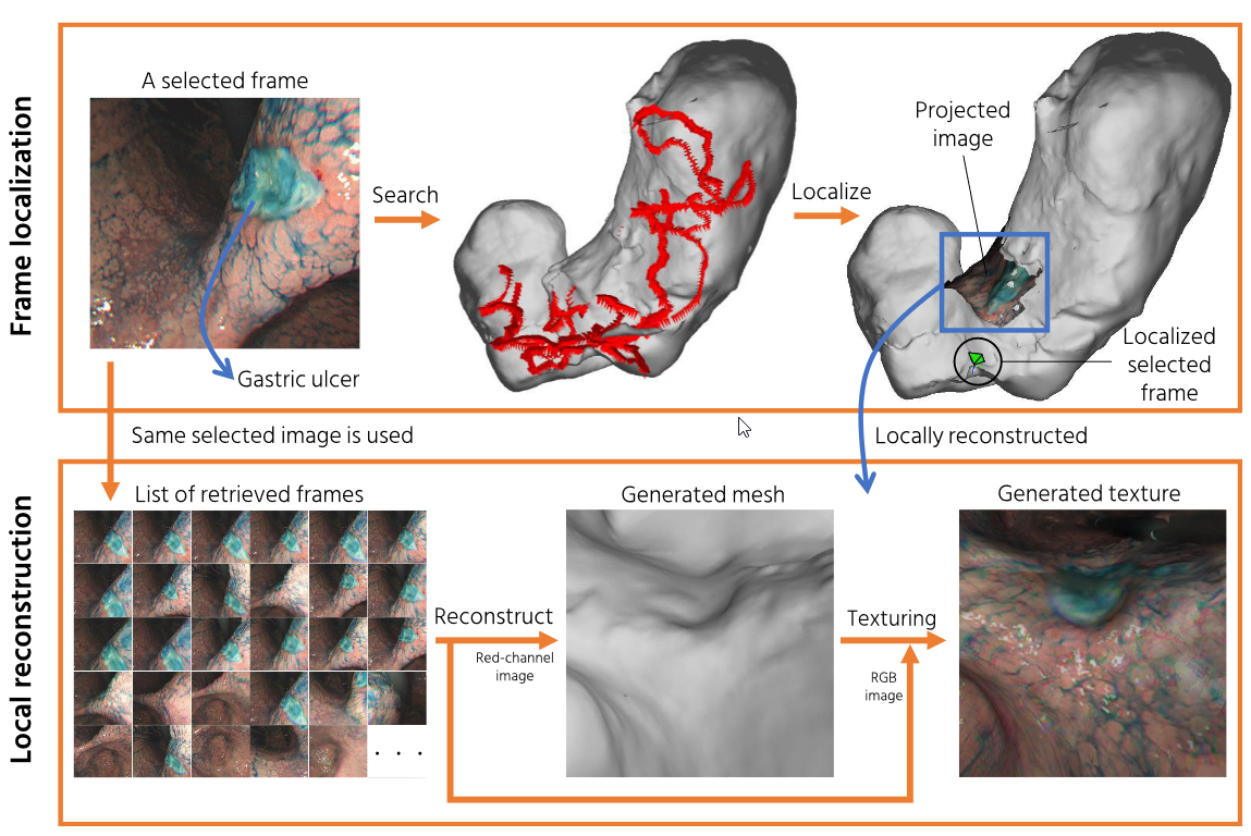
Since endoscopic environments can often be ill featured in addition to having artifacts, Widya et al. (2019) increase visible features via dying tissue with indigo carmine (IC) dye, and then demonstrating the comparative performance increase by using dye for helping SfM reconstruction. They remove outliers from the point cloud map (generated with SfM) using local plane fitting, and then create a mesh of the SfM point cloud. With this mesh, they can perform another outlier removal step for points that do not align well with the mesh. Both these steps account for the physical surface consistency priors we often have in MCV. To localize where the camera is in a current map, they use NetVLAD (Arandjelović et al., 2016) which is a CNN-based model that provides a distance metrics between image pairs. Then, given similar pairs, they can reconstruct higher detail images of these regions. This process is shown in Fig. 11. This approach is computationally intensive (runs offline) and can not be used for interactive clinical applications. Interestingly, the authors take the concept of IC-dye improving texture, and carry it on to design a model to perform virtual generation of textures using a CycleGAN. This allows them to generate IC-images artificially from non-IC images, and they demonstrate how their GAN-based method outperforms the non-augmented images for reconstruction applications (Widya et al., 2020).
5.5.2 Nonrigid Structure from Motion
Nonrigid Structure from Motion (NRSfM) is Structure from Motion without the assumption that the world has a rigid state. This means there are many more parameters to be solved for in optimization, adding to both the computational expense and modelling difficulty. To make it so not every point in the map is a degree of freedom, these methods make assumptions about tissue motion. Since we do not have an underlying model to fit to, by assuming priors on the types of motion that can happen, we provide a way to regularize. Two ways in which this is performed are via low rank shape models (LRSM) (Torresani et al., 2008), or isometric priors. Isometric priors depend on assuming that locally connected (nearby) points are isometric (distance-preserving), and enforce this constraint by between point neighbors during optimization. Example: A sheet of paper is isometric, while an exercise band is not. Low rank shape models assume shape can be represented as a linear combination of multiple basis shapes. A shape at time , can be represented as a mean shape plus basis shapes . At each point in time, the shape is represented as a combination of these shapes with a set of weights, :
| (2) |
Using LRSM, Hu et al. (2009) reconstruct a beating heart model. To improve results, they take advantage of the periodic motion of the heart, and use the same times in the heart cycle as additional samples to reduce dimensionality (ie. 10ms into a heartbeat should look the same every time). Although we do not have the same periodic motion in colonoscopy, we do have priors on the colon being a tubular structure. To utilize this, Sengupta and Bartoli (2021) add an underlying model to NRSfM and demonstrate improved performance on simulated tubular structures. They begin with calculating 3D point locations by performing NRSfM using an isometric prior. After calculating 3D point locations, they fit a tubular model to these locations. They model the tubular structure with harmonic splines. Optimization considers the tradeoff between being close to the 3D locations and smoothness regularization of the model. This is actually an example of a mixture of NRSfM with Shape-from-Template (SfT) which will be detailed in more detail in the following section. In a similar vein (mixing NRSfM and SfT), Golyanik et al. (2020) learn a dynamic shape prior using NRSfM. They collect this prior over a fixed representative set of frames, collecting a set of shape states. That is, they have different instances of what the shape can look like. Then for performing tracking of their dynamic shape prior, they match images to the nearest pre-calculated state.
The choice of prior that NRSfM methods rely on is particularly important in MCV since the priors dictate the transformations that the map can undergo, and the motion that can be accurately represented. The following section will detail further information on priors that come in the form of templates rather than the regularization that is used in NRSfM via low rank or isometry constraints.
5.5.3 Shape-from-Template (SfT)
In Shape-from-Template (SfT), we first calculate a template of the scene (or design a predetermined canonical one, e.g., a sheet or tube), and then in the following frames we align the template to match the current frame. Malti et al. (2012) construct a template using rigid SfM and then combine Deformable Shape from motion (DSfM) with Shape from Shading (estimating shape using a lighting model). Their template is initialized using video from a rigid scene. Then, they calculate the albedo of this template by using a Lambertian model of a BRDF (bidirectional reflectance distribution function, which models how a surface emits incoming light). To initialize a coarse reconstruction of their shape at a certain time they match sparse points between the template and the current image. They perform the coarse matching step using SIFT correspondences. Then, with the calculated albedo, they can then refine the coarsely aligned shape by matching lighting effects given their lighting model and using SfS.
Malti and Bartoli (2014) extend this work and use a more realistic lighting model than a Lambertian one. They select a Cook-Torrance model with a Beckmann distribution rather than Lambertian shading for the SfS refinement step and show that this performs better than modelling using Lambertian or Oren-Nayar distributions.
Cheema et al. (2019) use SfS as well, but with an additional incorporation of a pre-operative CT model of the liver as a prior. In the colon, Zhang et al. (2021a) also use a known 3D template generated using a CT scan. They use SGM (semi-global matching) for disparity estimation from stereo camera data. They generate a large video dataset from a colonoscopy simulator for evaluation. They choose SIFT for feature description, and represent deformation using an embedded deformation (ED) model (Sumner et al., 2007). In embedded deformation, the motion of any point, , is a function of neighboring control-point nodes and their positions . Each node essentially controls a rotation and translation . A weight function is used to increase influence by nodes that are closer, with weights, , that are normalized to sum to 1. As can be seen from the equation, this model supports a smooth deformation:
| (3) |
As neural networks have become popular, they have also taken a hold of SfT research. The general idea of using neural networks in SfT is that a template can also be represented by a set of neural network weights or latent codes. Golyanik et al. (2018) train a CNN on images using varying known ground truth deformations. Given an input image, their CNN estimates sets of 3D points to represent a triangular mesh grid. This can be seen as a form of template-based reconstruction, where the learning step learns the template, and an inference model evaluates template position given an image. For fitting novel views, they simply input a new image. The difficulty is that this model requires ground truth training data along with a full training step to solve for the parameters in the template-fitting network.
A step in this direction that no longer requires ground truth artificial data and uses optical flow as training signals is proposed by Sidhu et al. (2020). The authors propose Neural NRSfM by learning a latent space function that adds CNN-estimated offsets to a mean shape using an autodecoder, similarly to an LRSM. They additionally enforce a high-frequency regularization on the Fourier transform of the latent codes over time which – in addition to regularizing – allows for latent measurement of periodic signals. This helps recognize motions such as a heartbeat. The drawbacks for possible clinical application are the sensitivity to optical flow outliers, the need to initialize a rigid mean shape, and the model training taking multiple hours.
5.6 Simultaneous Localization and Mapping (SLAM)
In visual Simultaneous Localization and Mapping (SLAM), the goal is to create a map of the environment, often a set of points, ), and at the same time localize the sensor position within said environment (represented as poses in time, ). In this section, we will review methods that do so given video data. We note that for different environments, the means of mapping can vary. In implementation, SLAM is often implemented with multi-threaded methods that both optimize a map over a large set of keyframes, along with a current localization thread that estimates the position of the camera relative to the most current map state. By having separate threads this enables real-time operation since the slower (bundle adjustment and re-localization) optimizations will not affect near-term pose estimation. A map can be represented by anything from a point cloud with features to a triangular mesh. The optimization is often done as a nonlinear least squares – NLLS, where Levenberg-Marquardt is frequently used as the optimization method – problem, whereby a set of error terms are minimized over what is called a pose graph. The pose graph acts as overarching graph that connects nodes (poses) with data association terms (losses) (e.g., co-visible camera views are connected with feature matches). The components that change between methods in MCV are primarily the underlying map representation, the error terms used, and the means for re-localization, which finds out where in the environment the camera or the features are, once they have been lost. We will investigate rigid SLAM in Section 5.6.1 and nonrigid SLAM in Section 5.6.2. In the rigid SLAM section we will additionally mention some of the works that address a subset of the SLAM problem, such as relative pose estimation. In the nonrigid SLAM section we also include the problem of nonrigid mapping (SLAM without the camera localization) since these works are closely related. We will provide an overview with a focus on the evolution of algorithms over time along with the particular reasons for different proposed solutions.
5.6.1 Rigid SLAM
Rigid SLAM has been applied in endoscopy for decades, with applications in fields such as CT-guided sinus surgery (Burschka et al., 2004). In the rigid SLAM problem, we are looking to estimate a set of camera poses along with a map of the environment. Thankfully, if we would like to track individual points in the environment, we can calculate their motions easily as the entire motion is explained by the rigid 6DoF transformation the camera undergoes. A sparse map (set of points in space) can provide us with localization, rigid 6DoF motion estimation, and sparse measurements. Sometimes we would also like a dense map (rather than sparse); the primary reasons for this are for enabling visualisation or dense surface reconstruction for applications such as scanning. Here we will summarize the methods in Rigid SLAM for MCV, followed by some sections on map densification, localization, and dealing with texture. Table 3 provides an overarching summary of rigid SLAM methods.
| Authors | LC | Dens. | Base | Uses NN |
| Burschka et al. (2004) | Y | N | N/A | N |
| Mountney et al. (2006) | N | N | EKF-SLAM | N |
| Grasa et al. (2014) | N | N | EKF-SLAM | N |
| Mahmoud et al. (2017) | Y | Y | ORB-SLAM (dens. off keyframes) | N |
| Chen et al. (2018) | Y | Y | ORB-SLAM | N |
| Mahmoud et al. (2019) | Y | Y | ORB-SLAM | N |
| Ma et al. (2019) | N | Y | DSO | For dens. and pose-init (Wang et al., 2019) |
| Vasconcelos et al. (2019) | Y | N | N/A | N |
| Zhou and Jagadeesan (2020) | N | Y | N/A | N |
| Wang et al. (2020a) | N | N | ORB-SLAM | N |
| Jia et al. (2021) | Y | N | N/A | N |
| Liu et al. (2022) | Y | Y | N/A | For depth and im. features |
| Huo et al. (2023) | Y | Y | ORB-SLAM | Khamis et al. (2018) |
ORB-SLAM (Mur-Artal et al., 2015) is the most commonly used approach and will be seen throughout these MCV works, with some modifications for each environment. ORB-SLAM is appealing as it uses sparse ORB features to maintain real-time performance. It provides relocalization using the same features (using a bag-of-words method called DBoW2) and refines pose with bundle adjustment in both a local and global manner. Since many video frames look similar, these methods use keyframes (individual frames with separation in time and poses) so bundle adjustment and loop closure (recognizing when we have looped back to the same location, thus reducing drift) steps only have to look at a smaller set of frames. For estimating camera pose in the real-time thread, it can be efficiently performed using the prior pose along with a constant velocity estimation. Points are detected and matched to the map, and the current pose is refined to best match them.
Mountney et al. (2006) initially approach SLAM for estimating laparascope motion using patch based Shi-Tomasi features (Shi and Tomasi, 1994) and a Kalman filter (called EKF-SLAM, which is another SLAM methodology that uses Kalman filtering). Grasa et al. (2014) extend EKF-SLAM in a monocular environment, using randomized list relocalization (RLR) to estimate pose after tracking loss. For training the RLR, features of warped patches are used to train the classifier at initialization, and patches are also sampled online during operation. The RLR patches are classified by performing binary comparison tests, and creating the class given the binary result (similar to the ORB descriptor). They use a feature matching method that searches for feature matches between images using image correlation, emphasizing that the environment can be textureless (enabled by using image correlation search).
The difficulty with using EKF-SLAM is that it can be limited in cases with large numbers of landmarks due to requiring a full map update. Thus, recent methods often use ORB-SLAM to track over longer periods.
Densifying a map: ORB-SLAM only provides a sparse map, which limits applications to surface reconstruction. To address this, Mahmoud et al. (2017) extend ORB-SLAM to create a denser map by using an epipolar NCC search along with Lucas-Kanade optical flow. Extending this further, they (Mahmoud et al., 2019) add in a separate thread to densify over whole sets of keyframes, validating against CT scan ground truth.
This can provide a happy medium between sparse tracking and dense reconstruction.
In arthroscopic navigation, ArthroSLAM (Marmol et al., 2018) uses an external camera in addition to an arthroscopic camera and odometry to localize the arthroscope in space. This method still results in a sparse reconstruction, so in DenseArthroSLAM (Marmol et al., 2019) they extend ArthroSLAM to densely optimize a multi-view stereo method that estimates normals and points (Schönberger et al. (2016b)). They can then create a mesh from this oriented point cloud using a screened Poisson reconstruction (Kazhdan and Hoppe, 2013). In endoscopy, another reconstruction method (Chen et al., 2018) uses ORB-SLAM with a Poisson surface reconstruction to create a dense surface. The mesh surface can then be used for annotation and measurement (Fig. 12).
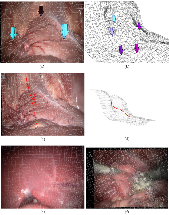
Using a different SLAM method called direct sparse odometry (DSO Engel et al. (2018)), which directly uses depth rather than image features, Ma et al. (2019) aim to densify maps in colonoscopy coverage estimation. They extend DSO by adding in a depth estimation RNN (recurrent neural network) which is trained on SfM in the colon. They only can only reconstruct partial regions of the colon likely due to limitations in long term mapping using rigid models, but they do provide dense reconstructions for some regions. In order to obtain a denser surface representation, Huo et al. (2023) utilize ORB-SLAM in combination with a StereoNet (Khamis et al., 2018) method to infill depth in a dense manner.
Localization: Here, we cover methods which look to improve localization through improved pose estimation, bundle adjustment, or outlier rejection. Some are not necessarily SLAM, but are designed to be usable as a component in SLAM models. In laparoscopy, we know the camera has specific motion constraints as it needs to pass through a fixed trocar hole. Vasconcelos et al. (2019) integrate these known constraints to constrain possible poses, creating RCM-SLAM.
To estimate pose or depth between frames as a part of a network, Ozyoruk et al. (2021) introduce a large dataset along with a pose and depth learning neural network, Endo-SFMLearner, which learns relative pose and monocular depth using unsupervised losses. Specifically, they use an affine-adjusted photometric loss along with geometric losses for training. Fig. 13 shows the dataset they use for evaluation.
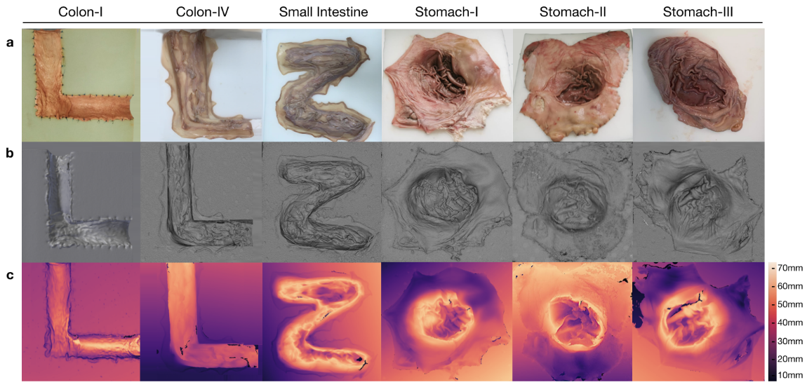
Poor texture: The following articles deal with poorly textured regions, for reconstruction and also localization. To address the low texture present in endoscopic environments, Zhou and Jagadeesan (2020) propose a rigid SLAM methodology with modifications for low texture. They use ZNCC for stereo estimation, and ORB for feature matching. They discard points using a RANSAC outlier rejection method and use an ICP loss for matching frames. A Truncated Signed Distance Field (TSDF) is used for visualization of the point cloud as a watertight surface. Again, addressing poor matches/texture, Wang et al. (2020a) extend ORB-SLAM by proposing a specific feature matching criteria for new frames based on bronchoscopic priors (e.g., by limiting inliers to a smaller filtering window), and evaluate their method on ex vivo bronchoscopies.
Jia et al. (2021), address stereo endoscopy by matching stereo ORB features using an epipolar search in combination with GMSMatch (Bian et al., 2020). They assume a rigid environment, and thus can use keyframes to re-localize, ORB features to match points, and ICP to find pose for new frames. Their method performs bundle adjustment in a background thread to refine the poses and 3D map point locations. Specifically, their method operates on a masked (separated from the background) kidney surface under the assumption it is rigid enough to track.
SAGESLAM (Liu et al., 2022) designs depth, feature, and descriptor networks to improve monocular SLAM methods in weakly textured regions. They propose feature and depth estimation models that can then be integrated into SLAM. They estimate a depth map as a combination of depth bases parameters (similar to how LRSMs reduce parameters). These parameters can be optimized in the SLAM process. They train their model using a differentiable Levenberg Marquardt relative-pose estimation method with ground truth generated by rigid SfM. They use a bundle adjustment network (BA-Net Tang and Tan (2019)) and extremely-dense point correspondences for a feature metric and sparse keypoint based loss. For evaluation, they evaluate relative camera pose estimation performance.
Hayoz et al. (2023) learn to estimate relative camera pose using back-projected stereo and optical flow using the recent Recurrent All-Pairs Field Transform (RAFT Teed and Deng (2020). Their model addresses poor texture by learning a confidence map to discard pixels that are not useful for pose estimation. Their optimization uses a Deep Declarative Network (DDN Gould et al. (2021)) which enables easier embedding of optimization problems into neural networks (with the goal here being pose optimization)). Their network architecture is shown in Fig 14.
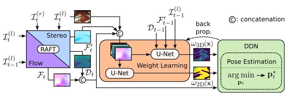
5.6.2 Non-Rigid SLAM
Now, we will cover non-rigid SLAM, where, similar to NRSfM, the difficulties lie in how to constrain the problem to represent motion, but not noise. Differences between camera motion and object scaling cannot be resolved in a totally unconstrained environment. Thus, these approaches have to regularize. As an example, camera pose and tissue movement cannot be decoupled without a fixed reference, so one way methods can deal with this is to model the camera as accounting for the rigid movement in the scene. Alternatively, measurements of camera pose can be made using other means like robot kinematics. Another difficulty in NRSLAM is that the environment can move in multiple different ways, so there needs to be a model of the underlying motion. This can be done using a mesh, FEM, etc., or an implicit model that regularizes map points (e.g., point sets should be As-Rigid-As-Possible, similar to regularization presented in Section 5.5.2). This makes non-rigid SLAM an algorithm design problem that depends on the needs that are the focus. Finally, the problem of localization and bundle adjustment are much more difficult in NRSLAM as points in the images are not fixed, and the environment can move even when it is out of view. We will summarize models that make contributions to NRSLAM, grouping by methods and dependence on one another. We begin with motion fields, then address mesh models, sparse methods, and finally tracking and mapping without localization. Specific drawbacks of each method are noted in brief as well. See Table 4 for an overarching summary of all the methods introduced that work for deformable environments as you read through this section.
| Authors | TT | Map | LC | Dis. | Uses NN | Base | Repr. |
| NRSLAM | |||||||
| Mountney and Yang (2010) | Per. | Y | N | N | N | EKFSlam | Pts. |
| Song et al. (2018) | Y | Y (R.) | Y (R.) | N (ED) | N | ORB-SLAM | Pts. |
| Schule et al. (2022) | Y | Y | Y | N | N | ORB-SLAM | Pts.+FEM |
| Lamarca et al. (2021) | Y | Y | N | N | N | N/A | Pts.+Tri. Mesh |
| Gómez-Rodríguez et al. (2021) | Y | Y | Y | N | N | N/A | Pts.+Tri. Mesh |
| Lamarca et al. (2022) | kpts. | Y | N | Y | N | N/A | Pts. |
| Gomez Rodriguez et al. (2022) | kpts. | Y | N | Y | N | N/A | Pts. |
| Zhou and Jayender (2021) | Y | Y | N | N | N | N/A | Pts. + EMDQ |
| Surgical Perception (No localization) | |||||||
| Schoob et al. (2017) | Y | Y | N | N | N | LK | Tri. Mesh |
| Li et al. (2020) | Y | Y | N | N | N | Gao and Tedrake (2019) | Pts./Surfels |
| Lu et al. (2021) | Y | Y | N | N | Y | Gao and Tedrake (2019) | Pts./Surfels |
| Lin et al. (2023) | Y | Y | N | N | Y | Gao and Tedrake (2019) | Pts./Surfels |
| Tissue Tracking | |||||||
| Yip et al. (2012) | Y | N | N | N | N | N/A | Pts. |
| Ye et al. (2016) | Y | N | N | Y | N | N/A | Pts. |
| Liu et al. (2023) | Y | N | N | N | N | MRF | Mesh |
| Schmidt et al. (2023a) | Y | N | N | Y | Y | Schmidt et al. (2022a) | Pts. |
A preliminary solution to NRSLAM in MCV (Mountney and Yang, 2010) uses a periodic motion model to account for deformation with a learned tracker and EKF SLAM. This prescribes specific motion to the environment, and requires exact periodicity, so later methods work to better accommodate changes and track all deformable tissue. Far later on, Schule et al. (2022) introduce a model-based method integrated with ORB-SLAM. They constrain map points by projecting them onto a FEM (finite element method) mesh. They create a map in 3D under the assumption of fully known forces and physical models. This 3D map can then be passed into the SLAM algorithm. The limitation of this approach is that forces exerted on tissue and finite-element models are often unknown in surgery. In order to escape these limitations, there needs to be a more flexible underlying representation.
Motion Fields: Instead of ascribing a physical model, we can instead represent the motion as a function of underlying control points with regularization (Embedded Deformation (ED Sumner et al. (2007)), or Expectation Maximization Dual Quaternion (EMDQ Zhou and Jayender (2022)) are some examples). MIS-SLAM (Song et al., 2018) performs rigid ORB-SLAM along with a separate deformable tracking thread. The deformable tracking uses embedded deformation Sumner et al. (2007) as the underlying motion model. These methods can deform a separate model of back-projected stereo points, but are unable to track or re-localize under large deformation since ORB-SLAM map points are not warped, only the live model is. Thus they are of limited use in applications that involve loop closure and longer term tracking since the underlying map does not deform and would not be able to be localized under visual changes.
EMDQ-SLAM (Zhou and Jayender, 2021) uses SURF features with an underlying motion field represented with dual quaternions. A TSDF is used for surface visualization, where surface color is calculated by blending multiple images. No re-localization or bundle adjustment is provided, and tracking is only frame-to-frame. They evaluate their method qualitatively.
Mesh models: We will now cover models which use a surface mesh as the underlying map representation. Such models create a dense surface but can have difficulties in modelling discontinuous motion.
DefSLAM (Lamarca et al., 2021) use a triangular mesh template for surface representation. To fit this template they minimize the 2D image re-projection error over detected keypoints. Bending and stretching energy is used on said points as a regularization.
They perform data association using ORB matches within a local search region. For near-term warping estimation, they use SfT. They re-estimate the template in a slower mapping thread as needed. A NRSfM optimization is performed on co-visible frames to calculate template updates. This method uses a mesh representation which can limit the possible deformations, and only uses ORB features for data association. Additionally, it does not provide loop closure. For evaluation they use stereo methods to evaluate the performance. These do not provide direct information on tracking accuracy in deformable environments.
SD-DefSLAM (Gómez-Rodríguez et al., 2021) adds LK optical flow to DefSLAM. They add a ORB bag-of-words model (BoW) for enabling relocalization under mild deformation conditions. To estimate initial pose they use Perspective-n-Point. Limitations again lie in the use of a mesh for the map and quantification using RMSE on stereo maps. Additionally, LK optical flow can fail in cases with large displacement.
Sparse Models: Recently, to escape the limitations of discontinuity, methods have been proposed that track sparse map points only.
Direct and sparse deformable tracking (Lamarca et al., 2022) proposes a mostly-monocular (requires stereo for initialization) method to track surfel points independently in a deformable manner in space. It detects surfels with Shi-Tomasi features (Shi and Tomasi, 1994) and tracks their re-projections in 3D space. To deal with ambiguity, they regularize surfels to lie near an equilibrium position. Covariance parameters can then be tuned to adjust regularization. The benefit of sparse tracking is that it enables processing scenes with discontinuity. Some limitations are that there are no constraints in between surfels, the requirement of stereo for initialization, and the cost of tracking sized patches. The regularizers used can cause issues in representing motion (according to Gomez Rodriguez et al. (2022)).
Gomez Rodriguez et al. (2022) present a method to track a sparse set of detected points in a monocular camera. For data association, they use LK optical flow and photometric error on patches. For regularization, they use a deformation graph connecting nearby points with a radial basis weight (see embedded deformation Sumner et al. (2007)). Additional temporal regularization limits the size of motion over time. This allows for discontinuities which is particularly demonstrated in a video from the Hamlyn dataset where a liver lobe moves separately from the background. For some drawbacks, they again quantify using LibELAS (Geiger et al., 2011) as stereo ground truth.
The method does not localize new map points, nor does it recover points, making it difficult to apply for long term tracking.
Tracking and Mapping: Here we will summarize works that perform tracking and mapping, but either assume a static camera, or do not estimate camera position. The work SuPer (Li et al., 2020) addresses surgical perception, which entails tracking instruments and tissue along with creating a map. They track tissue with a surfel ED model (Gao and Tedrake, 2019) using point-to-plane error and SURF features for data association. Painted markers are applied to the surgical instruments to aid in instrument detection and masking. SuPerDeep (Lu et al., 2021) extends the flexibility of SuPer by using CNNs to detect instrument keypoints and calculate disparity maps. The primary limitation of this work is computational cost and efficiency from having to run two neural networks along with a deformation optimization. Semantic SuPer (Lin et al., 2023) approaches the same problem, but they remove the keypoint loss. They add in constraints for semantic segmentation regions to match, in addition to considering image features by using differentiable rendering in their optimization. With a semantic separation of the embedded deformation warp field, this work could be extended to support discontinuity with some simple changes.
6 Discussion
We will begin with a summary discussion of each section (Section 6.2), followed by some key points we believe are important to consider in Section 6.3. We will then pose a couple future directions and open questions in Section 6.4, and we will finally wrap it up with a conclusion in Section 6.5.
6.1 Datasets
In summary, datasets can appear in all manners from entirely unlabelled to meticulously hand-labeled or modelled, all of which are useful for different applications and stages of an algorithm’s evaluation. Dataset generation in MCV is difficult because of the needed proximity to the real clinical environment and the difficulty of adding measurement equipment to medical procedures. Thus, methods sometimes generate ground truth via algorithms. This is in turn dependent on, and limited by, the clinical accuracy of the ground truth generation algorithm, which must then also be validated. This is likewise the case for synthetic data, except instead of the quality of algorithmic reconstruction, we need to account for the generalization capability of models that are trained on simulated data. We also do not know how well phantom models will generalize to real data in MCV. The benefits of these approaches are that if we can use a smaller amount of real labelled data for final testing, we can still use large amounts of synthetic or algorithmically trained ground truth for validation or training.
Thus, having a real dataset is important. Even with this, there are still difficulties. Many real datasets are rigid as seen in Table 2. Even if non-rigid, they can still be different from what we would find clinically. Just as we cannot know how well synthetic data can be used, the same argument stands in a weaker manner for porcine tissue instead of human tissue. A similar argument goes for visible markers. In short the question comes down to, how can we reduce both the bias and difficulty that occurs in generating ground truth. Some methods use non-medical datasets to evaluate their algorithms Du et al. (2019). Concepts such as crowd sourcing, or means to ease the labelling process with markers or IR tattoos are relevant here. To conclude, more work needs to be done with relevant clinical experiments to know what data we have and need.
Additionally, the data is biased towards general surgery and colonoscopy, with fewer available datasets in mosaicking and fetoscopy. No relevant datasets are found in neurosurgery, orthopedics, and plastic surgery. Thus in order to better continue enabling methods to be applied in this field, we need to move to release and collect data for these specialties.
As is often the case, we emphasize the importance of having both better data and more data in this field. Methods often have to resort to quantifying on small datasets that do not exactly address the problem (e.g., photometric reconstruction or depth for quantifying deformable tracking), or data that is outside their domain. Of course, hand-in-hand with this is the problem of quantifying methods that can train in an unsupervised manner and then perform on in-domain data. Synthetic data is helpful, but it does not remove the necessity of having labelled data for evaluating performance clinically. In the field of synthetic data, designing better methods of generating synthetic data is also a useful goal since SfM for example can only reconstruct partial regions (chunks) of the colon in methods thus far, so the capability of ground truth is limited by the size of reconstruction and the quality of the classical-methods for generating ground truth.
6.2 Discussions of Algorithms
In this summary discussion, we will discuss the algorithms covered in this review, starting with that of sparse features, dense features, and matching in Section 6.2.1. After which, we will cover mosaicking (Section 6.2.2) and depth estimation (Section 6.2.3). Followed by this, we will cover the problem of tissue tracking in Section 6.2.4. Then we will cover offline reconstruction (SfM, Section 6.2.5), and Simultaneous Localization and Mapping (Section 6.2.6).
6.2.1 Feature Detection and Description
Although many still use classical descriptors in the surgical environment, the newer methods which incorporate machine learning improve performance using medical data and are trained in an unsupervised manner, or via algorithmic reconstructions. A question here is if there are better ways to reconstruct training data for this environment. Since the algorithmic reconstructions depend on classical algorithms, we could be limited in the capability of the descriptors we are learning. Training on deformable models or neural reconstructions generated offline could be a promising avenue here.
Even with good descriptors, matching can still be difficult, and better constraints could be generated instead of using classical methods such as GMS. One way to improve this would be to design a means to train a GNN like SuperGlue on surgical scenes. Alternatively we can avoid discarding matches and losing information, in the manner described in Schmidt et al. (2022a). This still leaves us possibly limited by feature detection. With good matching, there is no way to ensure that the matches are pixel accurate (e.g., the same detection may be a couple pixels off due to lighting conditions). Thus a follow up question to be answered is how to deal with slight inconsistencies. At this point, these small errors are likely not realizable as there are more assumptions to be addressed in downstream processes (e.g., spline/mesh modelling) that can add to more error. Once these improve, an investigation into detection quality is likely to further increase performance. We note that in the case of rigid methods, this issue becomes less relevant under the assumption that detection noise is normal, as the least-squares fitting should average this error out. Dense description Liu et al. (2020b) provides a possible solution, as the matches can be searched over all possible pixel (or subpixel) locations. How to fuse the benefits of sparse matching and dense descriptors efficiently forms another research direction.
6.2.2 Mosaicking
Methods from mosaicking share many difficulties also encountered in tracking and mapping. These include means for matching and dealing with artifacts, blending images, and optimizing underlying maps (or pose graphs) for better global state estimation. In mosaicking in the medical environment, many of the articles reviewed aim to deal with poor texture, artifacts, or specularity. To address these, combinations of dense methods with sparse keypoint based methods look promising, allowing for hybrid benefits from both. Some questions that remain are how to better merge images that have different light distances or angles, along with how best to learn from machine learning methods for better matching of points (e.g., SuperGlue (Sarlin et al., 2020)). Additionally, mosaicking methods do not train custom descriptors, which could be due to the need for more data. (Section 4 has a recent dataset for the closely-related problem of stitching). Note that as far as tracking or 3D mapping are concerned, mosaicking does not aim to estimate the underlying 3D state or motion as the primary goal.
6.2.3 Depth estimation
Since depth estimation is very important for enabling accurate reconstruction, evaluating how these methods perform is critical. Datasets such as SCARED (Allan et al., 2021) provide means to better quantify depth methods, although classical algorithms such as LibELAS (Geiger et al., 2011) are still frequently used as ground truth. This is often due to other environments or domains not having the same ground truth data available, so evaluating on a different dataset is required, e.g., different cameras, or surgical field. Thus, we believe it is important to generate more data in this space, or if using generated data, to provide a strong proof of artificial data generation being able to generalize, which would still require some in-domain data.
Most of the methodological improvements to depth estimation in MCV are through in-painting, removing artifacts, or noting lighting priors. Physical priors such as smoothness have a similar impact. These ways (masking, lighting adjustment, …) to deal with specifics of MCV will be seen throughout this discussion.
6.2.4 Tissue Tracking
For tissue tracking, as in mosaicking, some methods choose to use hybrids of both dense optical flow and sparse feature matching. For the same reason as provided in mosaicking, this enables matches in salient locations (sparse) along with performance in ill-textured regions (dense optical flow). When using frame-to-frame motion estimates, some works propose means for filtering and managing features using classical methods. An avenue for improvement would be to bring this feature management into a neural paradigm, but this could be a difficult problem to formulate using a differentiable cost function. This is because for discrete operations such as deciding whether or not to keep a keypoint, it can be difficult to generate good proxy gradients. The straight-through estimator (Bengio et al., 2013) is one such heuristic that deals with this by passing the gradient directly through non-linear operations such as binarization.
Again in tissue tracking, we see that ways to deal with small amounts of data available for training is important. For example, Ihler et al. (2020) use a pre-trained non-MCV model followed by fine-tuning on medical images to improve their results. In many classical methods of online training, using nearby patches as positive correspondences and far ones as negatives, provides another reasonable physical prior for designing loss functions. Adaptation of such an approach to machine learning holds promise. More physical priors could also prove useful, for example if we could enforce stability or diffeomorphism in the loss function or model this could help further performance. This is especially important when we have small quantities of training data.
Performance is very important in tissue tracking, as downstream applications will have to use additional methods and computation, and large CNNs can be prohibitive in cost. Methods that are informed by sparse feature-based matching show promise here. Seeing machine learning models influence this field, and slowly accommodate classical ideas, is an exciting development.
Broader literature computer vision trackers that can manage occlusion (Doersch et al., 2023b; Harley et al., 2022; Neoral et al., 2023; Rajič et al., 2023; Wang et al., 2023) provide additional avenues for MCV. Both real-world hand-labelled (Doersch et al., 2023a) and synthetic (Zheng et al., 2023; Butler et al., 2012) datasets are of note for quantifying future tracking algorithm design in other domains.
6.2.5 Structure from Motion, Nonrigid Structure from Motion, and Shape from Template
In addition to being used for reconstruction of anatomy and surgical planning, Structure from Motion (SfM) has also been used to create ground truth datasets (Liu et al., 2018). The primary contributions in MCV for SfM entail compensating for lighting or better integrating models, priors and applications (Widya et al., 2019). For rigid SfM, methods that perform regularization or outlier filtering are often used to manage artifacts. Neural networks are used for estimating image similarity in re-localization, but do not yet extend into replacing ways to represent a map.
In Nonrigid SfM, simplifying assumptions are frequently used to ease the problem. These include only modelling periodic motion or isometry, or using linear bases for deformation. An avenue for research here is the development of methods that no longer require these assumptions, but would still benefit from parameter reduction without constraining the modelling capability.
These limiting assumptions also occur in SfT. For example, to initialize a template model, many methods need a rigid sequence for initialization. Antother possible limitation is that lighting models are simplified to approximate the template using few parameters (e.g., Lambertian or Cook-Torrance). Neural networks can also be used for template reconstruction without the same hand-engineered limitations, but still have to manage other implicit regularization provided by loss, gradient descent, and weight decay. These neural SfT models still depend on calculating an underlying representation thus far, and require this representation to be predefined or obtained as a mean shape by fitting.
As mentioned in Section 6.2.1, the use of offline methods for the sake of training online methods is an interesting research avenue that could likely be extended to NRSfM methods (rather than just SfM) if reconstruction performance issues are solved. In conclusion, NRSfM methods bring us closer to deformable reconstruction, but they can still be limited by the underlying representation.
6.2.6 SLAM and Nonrigid SLAM
For rigid SLAM, most works focus on means to address outliers and poor texture via removing the outliers or adjusting their models to use multiple correspondence methods (hybrids of both sparse and dense, etc.) that can measure motion in non-featured regions. Some also add a goal of densifying sparse maps to better enable clinical application. They do this via providing a parallel step that does not modify the sparse map or slow down the SLAM process.
Like seen in the other sections, the priors we have in this environment can have large effects, and integrating knowledge such as camera motion constraints can better improve performance of localization (Vasconcelos et al., 2019; Batlle et al., 2023). Neural networks have begun to make an impact, with RNNs improving depth estimation by using the temporal state of the 2D disparity images. Taking temporal visual state of the 3D map points into account using neural networks has yet to be approached. Matching in SLAM has primarily been classical, but there are methods we saw in Section 5.4 that could better refine matches.
In terms of evaluation for Nonrigid SLAM, there is still work to be done. Methods often quantify non-rigid methods using stereo data or photometric reconstruction, when the ideal goal is to quantify deformation and map accuracy.
For future work, being able to optimize non-physical parameters (e.g., a depth basis as in Liu et al. (2022)) is of high interest. For example, performing the same optimization for a map represented by a neural network can lead to great efficiency since it inherits the benefits of neural networks, with the efficiency of Levenberg-Marquardt optimization (or other NLLS methods). Since we do not have accurate models for underlying deformation thus far, we seldom see re-localization/loop closure done in non-rigid SLAM, but an underlying neural representation with few control parameters is one possible way to address that problem. The question comes down to how we define these priors while maintaining efficiency.
6.3 Key Takeways
Here we will discuss some of the important takeaways such as the importance of modelling lighting (Section 6.3.1), and having models that can represent the scene faithfully (Section 6.3.2). Finally, we will cover some additional future direction in Section 6.4 with reference to some ideas from modern computer vision, and some questions to be answered in the field.
6.3.1 Lighting
Here we briefly summarize some notes on the appearance of MCV environments. Lighting models can be improved, and work has begun to do so in MCV: for example we can use GGX in BRDFs as a distribution instead of Beckmann Malti and Bartoli (2014), or even design other learned versions Batlle et al. (2023). Combining a loss with a learned lighting model is really important for being able to better train models. We often have to mask specularities or artifacts, but then after the fact we use SfS to continue to make use of these useful signals. Methods could benefit if these masked specularities and artifacts could be integrated directly into the loss rather than having to combine different algorithms. Most methods rely on hand-tuned mixtures of image-intensity based losses for training–combined L1 and SSIM for example (see Jonschkowski et al. (2020) for recommendations), or classical algorithms that estimate using similar losses. Discovering novel ways to take surface light transport into account could be useful not only in disparity estimation and deformable tracking, but also for mosaicking and learning blending methodologies.
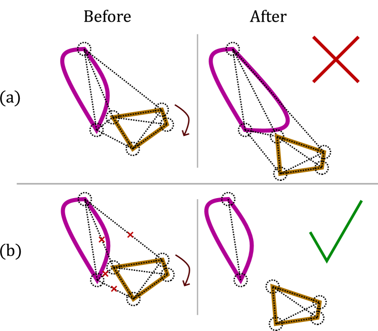
6.3.2 Underlying models
If we would like to map tissue, defining underlying models (in both NRSfM and NRSLAM) for how we map this tissue is extremely important. There are gaps in how these models are represented in that classical methods cannot faithfully represent tissue deformation (see Fig. 15), and modern machine learning is either not yet real-time capable, or does not enable tasks such as re-localization or bundle adjustment. Designing ways to represent tissue or organs in an environment as a persistent map that is also capable of adapting and changing is a very difficult problem. In the near term, even using better data association terms (e.g., frame-to-frame tissue tracking) in lieu of classical descriptors such as SIFT or ORB is a closer albeit still useful task.
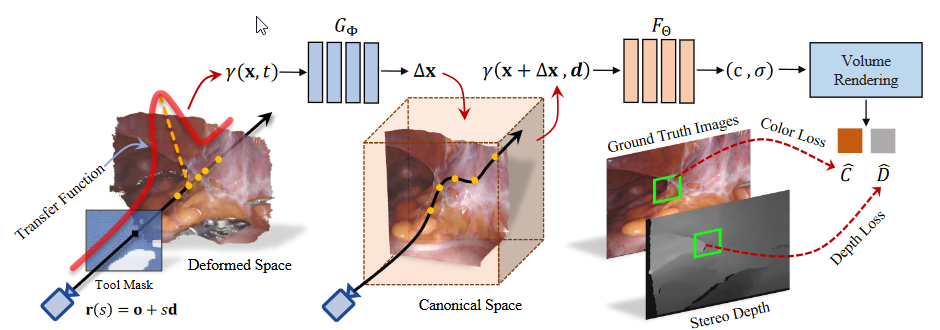
6.4 Future
Neural rendering: Neural networks for rendering such as neural radiance fields (Mildenhall et al., 2022) parameterize 3D space as a volume by using an implicit neural representation. These can be trained in a per-scene manner using multiple views for reconstruction of deformable scenes, with recent applications in endoscopy Wang et al. (2022). Although these are limited currently by having to train on the same scene, the next reasonable question if we generalize them to be conditional implicit functions (such as for flow in Schmidt et al. (2022a)). In addition to being limited by requiring training on the same dataset, they can also be slow for inference (each pixel requires multiple evaluations of a multi-layer perceptron (MLP) over samples on rays in 3D space).
More recently, methods have: approached the problem with signed distance fields (SDFs) for better surface representation Zha et al. (2023), accounted for lighting decay models in endoscopy Batlle et al. (2023), and optimized the training process (by conditioning on projected features from 2D planes) to perform in minutes Yang et al. (2023). That said, inference can still be slow. Methods such as Neural Graphics Primitives (NGP Müller et al. (2022)) or Gaussian splatting for faster rendering (Kerbl et al., 2023) or Neural Radial Basis Functions (NeuRBF Chen et al. (2023)) provide promising directions. If these are to work, we need to begin quantifying them on real tissue deformation data. Thus far, these methods use image reconstruction or stereo depth loss (on SCARED (Allan et al., 2021)) to qualify their loss, and require pre-defined instrument masks. Due to this, there is no way to determine yet if they perform well on tracking problems, so we emphasize that for any clinical application, it is important to have these measurements.
Detection: Feature-metric refinement, which has been used for pose in SfM Sarlin et al. (2023), is an appetizing alternative to having detections be repeatable, since it offers the best of both worlds - they detect in a sparse manner, but are not limited to matching sparse points. Alternative means to use detections is to not treat matching as such a one-to-one problem. With neural networks, we can learn matching as a graph function of two point sets If an object midpoint is only detected in one image, but the ends are detected in another, we should be able to softly match and use the end points to determine the midpoint in the other image (see Fig. 17).
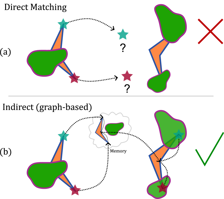
Questions:
How do we localize in changing environments? If an environment is changing, is it still helpful to try to localize against a map that is out of date? For example if tissue has been cut, we should ignore the map at all points near that region. Thus, we need to design new ways to adaptively manipulate maps.
How do we robustly deal with drift and feature change over time? For features that are initially defined to be a point, how do we track them if the feature changes – e.g., blood splatter or translucent shifting of mesentery layers. Some of these are ill-defined, but the question comes to when do we update a feature’s state and acknowledge a change in visible appearance. Of course, this depends on the application, for example on a mesentery layer, a feature could be anywhere in the translucent layer and is not easy to semantically define.
How (or should) we quantify what is happening out of frame? For maintaining a map of the environment, assuming motion happens out of frame can be useful. That said, we cannot ever truly estimate the state of tissue. The question comes with an idea of how do we deform the map in order to best improve performance and correctly update the state when tissue comes back into view. For maintaining performance in deformable bundle adjustment, loop closure, and drift-correction, maintaining a map is important, so the question comes to how do we keep these without making overly coarse assumptions.
SfM is to SLAM as NERF is to …? The works of iMAP (Sucar et al., 2021) and NICE-SLAM (Zhu et al., 2022) bring live NERF-optimization into a SLAM field. The question is how we can do this with an underlying deformable motion model as a primary goal. This again moves back to the question of how best to define models.
6.5 Conclusion
Computer vision and machine learning is taking a larger hold in Tracking and Mapping in Medical Computer Vision, but there are still many difficulties we must account for. We conclude with a couple of main points: datasets for evaluation and training are only increasing in importance in this field; in order to support deformable tracking, novel models still need to be designed; and many of the challenges in each of the subtopics we cover have shared difficulties (lighting, poor texture, dynamic scene) that can share inspiration and crossover.
References
- Acidi et al. (2023) Acidi, B., Ghallab, M., Cotin, S., Vibert, E., Golse, N., 2023. Augmented reality in liver surgery. Journal of Visceral Surgery 160, 118–126. doi:10.1016/j.jviscsurg.2023.01.008.
- Agrawal et al. (2008) Agrawal, M., Konolige, K., Blas, M.R., 2008. CenSurE: Center Surround Extremas for Realtime Feature Detection and Matching, in: Forsyth, D., Torr, P., Zisserman, A. (Eds.), Computer Vision – ECCV 2008, Springer, Berlin, Heidelberg. pp. 102–115. doi:10.1007/978-3-540-88693-8_8.
- Allan et al. (2021) Allan, M., Mcleod, J., Wang, C., Rosenthal, J.C., Hu, Z., Gard, N., Eisert, P., Fu, K.X., Zeffiro, T., Xia, W., Zhu, Z., Luo, H., Jia, F., Zhang, X., Li, X., Sharan, L., Kurmann, T., Schmid, S., Sznitman, R., Psychogyios, D., Azizian, M., Stoyanov, D., Maier-Hein, L., Speidel, S., 2021. Stereo Correspondence and Reconstruction of Endoscopic Data Challenge. arXiv:2101.01133 [cs] arXiv:2101.01133.
- Arandjelović et al. (2016) Arandjelović, R., Gronat, P., Torii, A., Pajdla, T., Sivic, J., 2016. NetVLAD: CNN architecture for weakly supervised place recognition, in: arXiv:1511.07247 [Cs]. arXiv:1511.07247.
- Atasoy et al. (2008) Atasoy, S., Noonan, D., Benhimane, S., Navab, N., Yang, G.Z., 2008. A Global Approach for Automatic Fibroscopic Video Mosaicing in Minimally Invasive Diagnosis. volume 5241 LNCS. doi:10.1007/978-3-540-85988-8_101.
- Azagra et al. (2022) Azagra, P., Sostres, C., Ferrandez, Á., Riazuelo, L., Tomasini, C., Barbed, O.L., Morlana, J., Recasens, D., Batlle, V.M., Gómez-Rodríguez, J.J., Elvira, R., López, J., Oriol, C., Civera, J., Tardós, J.D., Murillo, A.C., Lanas, A., Montiel, J.M.M., 2022. EndoMapper dataset of complete calibrated endoscopy procedures. arXiv:2204.14240.
- Bano and Stoyanov (2024) Bano, S., Stoyanov, D., 2024. Chapter 15 - Image mosaicking, in: Frangi, A.F., Prince, J.L., Sonka, M. (Eds.), Medical Image Analysis. Academic Press. The MICCAI Society Book Series, pp. 387--411. doi:10.1016/B978-0-12-813657-7.00030-3.
- Bano et al. (2023) Bano, S., Vasconcelos, F., David, A., Deprest, J., Stoyanov, D., 2023. Placental vessel-guided hybrid framework for fetoscopic mosaicking. Computer Methods in Biomechanics and Biomedical Engineering: Imaging and Visualization 11, 1166--1171. doi:10.1080/21681163.2022.2154278.
- Bano et al. (2020a) Bano, S., Vasconcelos, F., Shepherd, L.M., Poorten, E.V., Vercauteren, T., Ourselin, S., David, A.L., Deprest, J., Stoyanov, D., 2020a. Deep Placental Vessel Segmentation for Fetoscopic Mosaicking, volume 12263, pp. 763--773. doi:10.1007/978-3-030-59716-0_73, arXiv:2007.04349.
- Bano et al. (2019) Bano, S., Vasconcelos, F., Tella Amo, M., Dwyer, G., Gruijthuijsen, C., Deprest, J., Ourselin, S., Vander Poorten, E., Vercauteren, T., Stoyanov, D., 2019. Deep Sequential Mosaicking of Fetoscopic Videos. volume 11764 LNCS. doi:10.1007/978-3-030-32239-7_35.
- Bano et al. (2020b) Bano, S., Vasconcelos, F., Tella-Amo, M., Dwyer, G., Gruijthuijsen, C., Vander Poorten, E., Vercauteren, T., Ourselin, S., Deprest, J., Stoyanov, D., 2020b. Deep learning-based fetoscopic mosaicking for field-of-view expansion. International Journal of Computer Assisted Radiology and Surgery 15, 1807--1816. doi:10.1007/s11548-020-02242-8.
- Barbed et al. (2023) Barbed, O.L., Montiel, J.M.M., Fua, P., Murillo, A.C., 2023. Tracking Adaptation to Improve SuperPoint for 3D Reconstruction in Endoscopy, in: Greenspan, H., Madabhushi, A., Mousavi, P., Salcudean, S., Duncan, J., Syeda-Mahmood, T., Taylor, R. (Eds.), Medical Image Computing and Computer Assisted Intervention – MICCAI 2023, Springer Nature Switzerland, Cham. pp. 583--593. doi:10.1007/978-3-031-43907-0_56.
- Bardozzo et al. (2022) Bardozzo, F., Collins, T., Forgione, A., Hostettler, A., Tagliaferri, R., 2022. StaSiS-Net: A stacked and siamese disparity estimation network for depth reconstruction in modern 3D laparoscopy. Medical Image Analysis 77. doi:10.1016/j.media.2022.102380.
- Baserga et al. (2020) Baserga, C., Cappella, A., Gibelli, D., Sacco, R., Dolci, C., Cullati, F., Giannì, A., Sforza, C., 2020. Efficacy of autologous fat grafting in restoring facial symmetry in linear morphea-associated lesions. Symmetry 12, 1--13. doi:10.3390/sym12122098.
- Batlle et al. (2022) Batlle, V., Montiel, J., Tardos, J., 2022. Photometric single-view dense 3D reconstruction in endoscopy, in: IEEE International Conference on Intelligent Robots and Systems, pp. 4904--4910. doi:10.1109/IROS47612.2022.9981742.
- Batlle et al. (2023) Batlle, V.M., Montiel, J.M.M., Fua, P., Tardós, J.D., 2023. LightNeuS: Neural Surface Reconstruction in Endoscopy Using Illumination Decline, in: Greenspan, H., Madabhushi, A., Mousavi, P., Salcudean, S., Duncan, J., Syeda-Mahmood, T., Taylor, R. (Eds.), Medical Image Computing and Computer Assisted Intervention – MICCAI 2023, Springer Nature Switzerland, Cham. pp. 502--512. doi:10.1007/978-3-031-43999-5_48.
- Bay et al. (2008) Bay, H., Ess, A., Tuytelaars, T., Van Gool, L., 2008. Speeded-Up Robust Features (SURF). Computer Vision and Image Understanding 110, 346--359. doi:10.1016/j.cviu.2007.09.014.
- Bay et al. (2006) Bay, H., Tuytelaars, T., Van Gool, L., 2006. SURF: Speeded Up Robust Features, in: Leonardis, A., Bischof, H., Pinz, A. (Eds.), Computer Vision – ECCV 2006. Springer Berlin Heidelberg, Berlin, Heidelberg. volume 3951, pp. 404--417. doi:10.1007/11744023_32.
- Behrens et al. (2011) Behrens, A., Bommes, M., Stehle, T., Gross, S., Leonhardt, S., Aach, T., 2011. Real-time image composition of bladder mosaics in fluorescence endoscopy. Computer Science - Research and Development 26, 51--64. doi:10.1007/s00450-010-0135-z.
- Bengio et al. (2013) Bengio, Y., Léonard, N., Courville, A., 2013. Estimating or Propagating Gradients Through Stochastic Neurons for Conditional Computation. arXiv:1308.3432.
- Bergen et al. (2009) Bergen, T., Ruthotto, S., Münzenmayer, C., Rupp, S., Paulus, D., Winter, C., 2009. Feature-based real-time endoscopic mosaicking, in: ISPA 2009 - Proceedings of the 6th International Symposium on Image and Signal Processing and Analysis, pp. 695--700. doi:10.1109/ispa.2009.5297633.
- Bergen and Wittenberg (2016) Bergen, T., Wittenberg, T., 2016. Stitching and surface reconstruction from endoscopic image sequences: A review of applications and methods. IEEE Journal of Biomedical and Health Informatics 20, 304--321. doi:10.1109/JBHI.2014.2384134.
- Bernhardt et al. (2017) Bernhardt, S., Nicolau, S.A., Soler, L., Doignon, C., 2017. The status of augmented reality in laparoscopic surgery as of 2016. Medical Image Analysis 37, 66--90. doi:10.1016/j.media.2017.01.007.
- Bian et al. (2020) Bian, J.W., Lin, W.Y., Liu, Y., Zhang, L., Yeung, S.K., Cheng, M.M., Reid, I., 2020. GMS: Grid-Based Motion Statistics for Fast, Ultra-robust Feature Correspondence. International Journal of Computer Vision 128, 1580--1593.
- Bobrow et al. (2022) Bobrow, T.L., Golhar, M., Vijayan, R., Akshintala, V.S., Garcia, J.R., Durr, N.J., 2022. Colonoscopy 3D Video Dataset with Paired Depth from 2D-3D Registration. doi:10.48550/arXiv.2206.08903, arXiv:2206.08903.
- Borrego-Carazo et al. (2023) Borrego-Carazo, J., Sanchez, C., Castells-Rufas, D., Carrabina, J., Gil, D., 2023. BronchoPose: An analysis of data and model configuration for vision-based bronchoscopy pose estimation. Computer Methods and Programs in Biomedicine 228, 107241. doi:10.1016/j.cmpb.2022.107241.
- Buchart et al. (2009) Buchart, C., Vicente, G., Amundarain, A., Borro, D., 2009. Hybrid visualization for maxillofacial surgery planning and simulation, in: Proceedings of the International Conference on Information Visualisation, pp. 266--273. doi:10.1109/IV.2009.98.
- Burschka et al. (2005) Burschka, D., Li, M., Ishii, M., Taylor, R.H., Hager, G.D., 2005. Scale-invariant registration of monocular endoscopic images to CT-scans for sinus surgery. Medical Image Analysis 9, 413--426. doi:10.1016/j.media.2005.05.005.
- Burschka et al. (2004) Burschka, D., Li, M., Taylor, R., Hager, G., 2004. Scale-invariant registration of monocular stereo images to 3D surface models. 2004 IEEE/RSJ International Conference on Intelligent Robots and Systems (IROS) 3, 2581--2586.
- Burt and Adelson (1983) Burt, P.J., Adelson, E.H., 1983. A multiresolution spline with application to image mosaics. ACM Transactions on Graphics (TOG) 2, 217--236.
- Butler et al. (2012) Butler, D.J., Wulff, J., Stanley, G.B., Black, M.J., 2012. A Naturalistic Open Source Movie for Optical Flow Evaluation, in: Hutchison, D., Kanade, T., Kittler, J., Kleinberg, J.M., Mattern, F., Mitchell, J.C., Naor, M., Nierstrasz, O., Pandu Rangan, C., Steffen, B., Sudan, M., Terzopoulos, D., Tygar, D., Vardi, M.Y., Weikum, G., Fitzgibbon, A., Lazebnik, S., Perona, P., Sato, Y., Schmid, C. (Eds.), Computer Vision – ECCV 2012. Springer Berlin Heidelberg, Berlin, Heidelberg. volume 7577, pp. 611--625. doi:10.1007/978-3-642-33783-3_44.
- Cao et al. (2022) Cao, Z., Wang, Y., Zheng, W., Yin, L., Tang, Y., Miao, W., Liu, S., Yang, B., 2022. The algorithm of stereo vision and shape from shading based on endoscope imaging. Biomedical Signal Processing and Control 76. doi:10.1016/j.bspc.2022.103658.
- Cartucho et al. (2021) Cartucho, J., Tukra, S., Li, Y., S. Elson, D., Giannarou, S., 2021. VisionBlender: A tool to efficiently generate computer vision datasets for robotic surgery. Computer Methods in Biomechanics and Biomedical Engineering: Imaging & Visualization 9, 331--338. doi:10.1080/21681163.2020.1835546.
- Cartucho et al. (2024) Cartucho, J., Weld, A., Tukra, S., Xu, H., Matsuzaki, H., Ishikawa, T., Kwon, M., Jang, Y.E., Kim, K.J., Lee, G., Bai, B., Kahrs, L.A., Boecking, L., Allmendinger, S., Müller, L., Zhang, Y., Jin, Y., Bano, S., Vasconcelos, F., Reiter, W., Hajek, J., Silva, B., Lima, E., Vilaça, J.L., Queirós, S., Giannarou, S., 2024. SurgT challenge: Benchmark of soft-tissue trackers for robotic surgery. Medical Image Analysis 91, 102985. doi:10.1016/j.media.2023.102985.
- Chadebecq et al. (2023) Chadebecq, F., Lovat, L.B., Stoyanov, D., 2023. Artificial intelligence and automation in endoscopy and surgery. Nat Rev Gastroenterol Hepatol 20, 171--182. doi:10.1038/s41575-022-00701-y.
- Chang and Chen (2018) Chang, J.R., Chen, Y.S., 2018. Pyramid Stereo Matching Network, in: 2018 IEEE/CVF Conference on Computer Vision and Pattern Recognition, IEEE, Salt Lake City, UT. pp. 5410--5418. doi:10.1109/CVPR.2018.00567.
- Chang et al. (2013) Chang, P.L., Stoyanov, D., Davison, A., Edwards, P., 2013. Real-time dense stereo reconstruction using convex optimisation with a cost-volume for image-guided robotic surgery. Lecture Notes in Computer Science (including subseries Lecture Notes in Artificial Intelligence and Lecture Notes in Bioinformatics) 8149 LNCS, 42--49. doi:10.1007/978-3-642-40811-3_6.
- Cheema et al. (2019) Cheema, M., Nazir, A., Sheng, B., Li, P., Qin, J., Kim, J., Feng, D., 2019. Image-aligned dynamic liver reconstruction using intra-operative field of views for minimal invasive surgery. IEEE Transactions on Biomedical Engineering 66, 2163--2173. doi:10.1109/TBME.2018.2884319.
- Chen et al. (2018) Chen, L., Tang, W., John, N., Wan, T., Zhang, J., 2018. SLAM-based dense surface reconstruction in monocular Minimally Invasive Surgery and its application to Augmented Reality. Computer Methods and Programs in Biomedicine 158, 135--146. doi:10.1016/j.cmpb.2018.02.006.
- Chen et al. (2023) Chen, Z., Li, Z., Song, L., Chen, L., Yu, J., Yuan, J., Xu, Y., 2023. NeuRBF: A Neural Fields Representation with Adaptive Radial Basis Functions, in: Proceedings of the IEEE/CVF International Conference on Computer Vision, pp. 4182--4194.
- Chu et al. (2020) Chu, Y., Li, H., Li, X., Ding, Y., Yang, X., Ai, D., Chen, X., Wang, Y., Yang, J., 2020. Endoscopic image feature matching via motion consensus and global bilateral regression. Computer Methods and Programs in Biomedicine 190. doi:10.1016/j.cmpb.2020.105370.
- Collins et al. (2016) Collins, T., Bartoli, A., Bourdel, N., Canis, M., 2016. Robust,Real-Time,Dense and Deformable 3D Organ Tracking in Laparoscopic Videos. volume 9900 LNCS. doi:10.1007/978-3-319-46720-7_47.
- De Momi et al. (2016) De Momi, E., Ferrigno, G., Bosoni, G., Bassanini, P., Blasi, P., Casaceli, G., Fuschillo, D., Castana, L., Cossu, M., Lo Russo, G., Cardinale, F., 2016. A method for the assessment of time-varying brain shift during navigated epilepsy surgery. International Journal of Computer Assisted Radiology and Surgery 11, 473--481. doi:10.1007/s11548-015-1259-1.
- De Smet et al. (2019) De Smet, J., Poorten, E., Poliakov, V., Niu, K., Chesterman, F., Fornier, J., Ahmad, M., Ourak, M., Voros, V., Deprest, J., 2019. Evaluating the potential benefit of autostereoscopy in laparoscopic sacrocolpopexy through VR simulation, in: 2019 19th International Conference on Advanced Robotics, ICAR 2019, pp. 566--571. doi:10.1109/ICAR46387.2019.8981553.
- Doersch et al. (2023a) Doersch, C., Gupta, A., Markeeva, L., Recasens, A., Smaira, L., Aytar, Y., Carreira, J., Zisserman, A., Yang, Y., 2023a. TAP-Vid: A Benchmark for Tracking Any Point in a Video. arXiv:2211.03726.
- Doersch et al. (2023b) Doersch, C., Yang, Y., Vecerik, M., Gokay, D., Gupta, A., Aytar, Y., Carreira, J., Zisserman, A., 2023b. TAPIR: Tracking Any Point with per-frame Initialization and temporal Refinement. arXiv:2306.08637.
- Du et al. (2019) Du, X., Allan, M., Bodenstedt, S., Maier-Hein, L., Speidel, S., Dore, A., Stoyanov, D., 2019. Patch-based adaptive weighting with segmentation and scale (PAWSS) for visual tracking in surgical video. Medical Image Analysis 57, 120--135. doi:10.1016/j.media.2019.07.002.
- Du et al. (2015) Du, X., Clancy, N., Arya, S., Hanna, G., Kelly, J., Elson, D., Stoyanov, D., 2015. Robust surface tracking combining features, intensity and illumination compensation. International Journal of Computer Assisted Radiology and Surgery 10, 1915--1926. doi:10.1007/s11548-015-1243-9.
- Edwards et al. (2022) Edwards, P., Psychogyios, D., Speidel, S., Maier-Hein, L., Stoyanov, D., 2022. SERV-CT: A disparity dataset from cone-beam CT for validation of endoscopic 3D reconstruction. Medical Image Analysis 76. doi:10.1016/j.media.2021.102302.
- Engel et al. (2018) Engel, J., Koltun, V., Cremers, D., 2018. Direct Sparse Odometry. IEEE Transactions on Pattern Analysis and Machine Intelligence 40, 611--625. doi:10.1109/TPAMI.2017.2658577.
- Frangi et al. (1998) Frangi, A.F., Niessen, W.J., Vincken, K.L., Viergever, M.A., 1998. Multiscale vessel enhancement filtering, in: Wells, W.M., Colchester, A., Delp, S. (Eds.), Medical Image Computing and Computer-Assisted Intervention — MICCAI’98, Springer, Berlin, Heidelberg. pp. 130--137. doi:10.1007/BFb0056195.
- Fu et al. (2021a) Fu, Z., Jin, Z., Zhang, C., Dai, Y., Gao, X., Wang, Z., Li, L., Ding, G., Hu, H., Wang, P., Ye, X., 2021a. Visual-electromagnetic system: A novel fusion-based monocular localization, reconstruction, and measurement for flexible ureteroscopy. International Journal of Medical Robotics and Computer Assisted Surgery 17. doi:10.1002/rcs.2274.
- Fu et al. (2021b) Fu, Z., Jin, Z., Zhang, C., He, Z., Zha, Z., Hu, C., Gan, T., Yan, Q., Wang, P., Ye, X., 2021b. The Future of Endoscopic Navigation: A Review of Advanced Endoscopic Vision Technology. IEEE Access .
- Fulton et al. (2020) Fulton, M., Micah Prendergast, J., Ditommaso, E., Rentschler, M., 2020. Comparing visual odometry systems in actively deforming simulated colon environments, in: IEEE International Conference on Intelligent Robots and Systems, pp. 4988--4995. doi:10.1109/IROS45743.2020.9341159.
- Gao and Tedrake (2019) Gao, W., Tedrake, R., 2019. SurfelWarp: Efficient Non-Volumetric Single View Dynamic Reconstruction. doi:10.48550/arXiv.1904.13073, arXiv:1904.13073.
- Geiger et al. (2011) Geiger, A., Roser, M., Urtasun, R., 2011. Efficient Large-Scale Stereo Matching, in: Kimmel, R., Klette, R., Sugimoto, A. (Eds.), Computer Vision – ACCV 2010. Springer Berlin Heidelberg, Berlin, Heidelberg. volume 6492, pp. 25--38.
- Giannarou et al. (2009) Giannarou, S., Visentini-Scarzanella, M., Yang, G.Z., 2009. Affine-invariant anisotropic detector for soft tissue tracking in minimally invasive surgery, in: Proceedings - 2009 IEEE International Symposium on Biomedical Imaging: From Nano to Macro, ISBI 2009, pp. 1059--1062. doi:10.1109/ISBI.2009.5193238.
- Giannarou et al. (2013) Giannarou, S., Visentini-Scarzanella, M., Yang, G.Z., 2013. Probabilistic tracking of affine-invariant anisotropic regions. IEEE Transactions on Pattern Analysis and Machine Intelligence 35, 130--143. doi:10.1109/TPAMI.2012.81.
- Girerd et al. (2020) Girerd, C., Kudryavtsev, A., Rougeot, P., Renaud, P., Rabenorosoa, K., Tamadazte, B., 2020. Automatic Tip-Steering of Concentric Tube Robots in the Trachea Based on Visual SLAM. IEEE Transactions on Medical Robotics and Bionics 2, 582--585. doi:10.1109/TMRB.2020.3034720.
- Golyanik et al. (2020) Golyanik, V., Jonas, A., Stricker, D., Theobalt, C., 2020. Intrinsic Dynamic Shape Prior for Fast, Sequential and Dense Non-Rigid Structure from Motion with Detection of Temporally-Disjoint Rigidity. arXiv:1909.02468 [cs] arXiv:1909.02468.
- Golyanik et al. (2018) Golyanik, V., Shimada, S., Varanasi, K., Stricker, D., 2018. HDM-Net: Monocular Non-Rigid 3D Reconstruction with Learned Deformation Model. EuroVR doi:10.1007/978-3-030-01790-3_4, arXiv:1803.10193.
- Gomez Rodriguez et al. (2022) Gomez Rodriguez, J., Montiel, J., Tardos, J., 2022. Tracking monocular camera pose and deformation for SLAM inside the human body, in: IEEE International Conference on Intelligent Robots and Systems, pp. 5278--5285. doi:10.1109/IROS47612.2022.9981203.
- Gómez-Rodríguez et al. (2021) Gómez-Rodríguez, J.J., Lamarca, J., Morlana, J., Tardós, J.D., Montiel, J.M.M., 2021. SD-DefSLAM: Semi-Direct Monocular SLAM for Deformable and Intracorporeal Scenes, in: 2021 IEEE International Conference on Robotics and Automation (ICRA), pp. 5170--5177.
- Gould et al. (2021) Gould, S., Hartley, R., Campbell, D., 2021. Deep Declarative Networks: A New Hope. IEEE Trans. Pattern Anal. Mach. Intell. , 1--1arXiv:1909.04866.
- Grasa et al. (2014) Grasa, O., Bernal, E., Casado, S., Gil, I., Montiel, J., 2014. Visual slam for handheld monocular endoscope. IEEE Transactions on Medical Imaging 33, 135--146. doi:10.1109/TMI.2013.2282997.
- Guy et al. (2022) Guy, S., Haberbusch, J.L., Promayon, E., Mancini, S., Voros, S., 2022. Qualitative Comparison of Image Stitching Algorithms for Multi-Camera Systems in Laparoscopy. Journal of Imaging 8, 52. doi:10.3390/jimaging8030052.
- Harley et al. (2022) Harley, A.W., Fang, Z., Fragkiadaki, K., 2022. Particle Video Revisited: Tracking Through Occlusions Using Point Trajectories. arXiv:2204.04153.
- Hartkens et al. (2003) Hartkens, T., Hill, D.L.G., Castellano-Smith, A.D., Hawkes, D.J., Maurer, C.R., Martin, A.J., Hall, W.A., Liu, H., Truwit, C.L., 2003. Measurement and analysis of brain deformation during neurosurgery. IEEE Trans Med Imaging 22, 82--92. doi:10.1109/TMI.2002.806596.
- Hartwig et al. (2022) Hartwig, R., Ostler, D., Rosenthal, J.C., Feußner, H., Wilhelm, D., Wollherr, D., 2022. MITI: SLAM Benchmark for Laparoscopic Surgery. arXiv:2202.11496.
- Hayoz et al. (2023) Hayoz, M., Hahne, C., Gallardo, M., Candinas, D., Kurmann, T., Allan, M., Sznitman, R., 2023. Learning how to robustly estimate camera pose in endoscopic videos. International Journal of Computer Assisted Radiology and Surgery doi:10.1007/s11548-023-02919-w.
- Hernández-Mier et al. (2010) Hernández-Mier, Y., Blondel, W., Daul, C., Wolf, D., Guillemin, F., 2010. Fast construction of panoramic images for cystoscopic exploration. Computerized Medical Imaging and Graphics 34, 579--592. doi:10.1016/j.compmedimag.2010.02.002.
- Hu et al. (2007) Hu, M., Penney, G., Edwards, P., Figl, M., Hawkes, D., 2007. 3D reconstruction of internal organ surfaces for minimal invasive surgery 4791 LNCS, 77. doi:10.1007/978-3-540-75757-3_9.
- Hu et al. (2012) Hu, M., Penney, G., Figl, M., Edwards, P., Bello, F., Casula, R., Rueckert, D., Hawkes, D., 2012. Reconstruction of a 3D surface from video that is robust to missing data and outliers: Application to minimally invasive surgery using stereo and mono endoscopes. Medical Image Analysis 16, 597--611. doi:10.1016/j.media.2010.11.002.
- Hu et al. (2009) Hu, M., Penney, G., Rueckert, D., Edwards, P., Bello, F., Casula, R., Figl, M., Hawkes, D., 2009. Non-Rigid Reconstruction of the Beating Heart Surface for Minimally Invasive Cardiac Surgery. volume 5761 LNCS. doi:10.1007/978-3-642-04268-3_5.
- Huo et al. (2023) Huo, J., Zhou, C., Yuan, B., Yang, Q., Wang, L., 2023. Real-Time Dense Reconstruction with Binocular Endoscopy Based on StereoNet and ORB-SLAM. Sensors 23. doi:10.3390/s23042074.
- Hutchison et al. (2010) Hutchison, D., Kanade, T., Kittler, J., Kleinberg, J.M., Mattern, F., Mitchell, J.C., Naor, M., Nierstrasz, O., Pandu Rangan, C., Steffen, B., Sudan, M., Terzopoulos, D., Tygar, D., Vardi, M.Y., Weikum, G., Calonder, M., Lepetit, V., Strecha, C., Fua, P., 2010. BRIEF: Binary Robust Independent Elementary Features, in: Daniilidis, K., Maragos, P., Paragios, N. (Eds.), Computer Vision – ECCV 2010. Springer Berlin Heidelberg, Berlin, Heidelberg. volume 6314, pp. 778--792. doi:10.1007/978-3-642-15561-1_56.
- Ihler et al. (2020) Ihler, S., Kuhnke, F., Laves, M.H., Ortmaier, T., 2020. Self-supervised domain adaptation for patient-specific, real-time tissue tracking. Lecture Notes in Computer Science (including subseries Lecture Notes in Artificial Intelligence and Lecture Notes in Bioinformatics) 12263 LNCS, 54--64.
- Ji et al. (2014) Ji, S., Fan, X., Roberts, D., Hartov, A., Paulsen, K., 2014. Cortical surface shift estimation using stereovision and optical flow motion tracking via projection image registration. Medical Image Analysis 18, 1169--1183. doi:10.1016/j.media.2014.07.001.
- Jia et al. (2021) Jia, T., Taylor, Z., Chen, X., 2021. Long term and robust 6DoF motion tracking for highly dynamic stereo endoscopy videos. Computerized Medical Imaging and Graphics 94. doi:10.1016/j.compmedimag.2021.101995.
- Jiang et al. (2016) Jiang, J., Nakajima, Y., Sohma, Y., Saito, T., Kin, T., Oyama, H., Saito, N., 2016. Marker-less tracking of brain surface deformations by non-rigid registration integrating surface and vessel/sulci features. International Journal of Computer Assisted Radiology and Surgery 11, 1687--1701. doi:10.1007/s11548-016-1358-7.
- Jiang et al. (2015) Jiang, L., Zhang, S., Yang, J., Zhuang, X., Zhang, L., Gu, L., 2015. A robust automated markerless registration framework for neurosurgery navigation. International Journal of Medical Robotics and Computer Assisted Surgery 11, 436--447. doi:10.1002/rcs.1626.
- Jonschkowski et al. (2020) Jonschkowski, R., Stone, A., Barron, J.T., Gordon, A., Konolige, K., Angelova, A., 2020. What Matters in Unsupervised Optical Flow, in: Vedaldi, A., Bischof, H., Brox, T., Frahm, J.M. (Eds.), Computer Vision – ECCV 2020, Springer International Publishing, Cham. pp. 557--572.
- Kam et al. (2023) Kam, M., Wei, S., Opfermann, J., Saeidi, H., Hsieh, M., Wang, K., Kang, J., Krieger, A., 2023. Autonomous System for Vaginal Cuff Closure via Model-Based Planning and Markerless Tracking Techniques. IEEE Robotics and Automation Letters 8, 3915--3922. doi:10.1109/LRA.2023.3273416.
- Karaoglu et al. (2023) Karaoglu, M.A., Markova, V., Navab, N., Busam, B., Ladikos, A., 2023. RIDE: Self-Supervised Learning of Rotation-Equivariant Keypoint Detection and Invariant Description for Endoscopy. arXiv:2309.09563.
- Kazhdan and Hoppe (2013) Kazhdan, M., Hoppe, H., 2013. Screened poisson surface reconstruction. ACM Trans. Graph. 32, 29:1--29:13. doi:10.1145/2487228.2487237.
- Kerbl et al. (2023) Kerbl, B., Kopanas, G., Leimkuehler, T., Drettakis, G., 2023. 3D gaussian splatting for real-time radiance field rendering. ACM Trans. Graph. 42. doi:10.1145/3592433.
- Khamis et al. (2018) Khamis, S., Fanello, S., Rhemann, C., Kowdle, A., Valentin, J., Izadi, S., 2018. StereoNet: Guided Hierarchical Refinement for Real-Time Edge-Aware Depth Prediction, in: Proceedings of the European Conference on Computer Vision (ECCV), pp. 573--590.
- Lamarca et al. (2022) Lamarca, J., Gómez Rodríguez, J.J., Tardós, J.D., Montiel, J., 2022. Direct and Sparse Deformable Tracking. IEEE Robotics and Automation Letters 7, 11450--11457. doi:10.1109/LRA.2022.3201253.
- Lamarca et al. (2021) Lamarca, J., Parashar, S., Bartoli, A., Montiel, J., 2021. DefSLAM: Tracking and mapping of deforming scenes from monocular sequences. IEEE Transactions on Robotics 37, 291--303. doi:10.1109/TRO.2020.3020739.
- Li et al. (2021) Li, L., Bano, S., Deprest, J., David, A., Stoyanov, D., Vasconcelos, F., 2021. Globally optimal fetoscopic mosaicking based on pose graph optimisation with affine constraints. IEEE Robotics and Automation Letters 6, 7831--7838. doi:10.1109/LRA.2021.3100938.
- Li et al. (2023) Li, L., Mazomenos, E., Chandler, J., Obstein, K., Valdastri, P., Stoyanov, D., Vasconcelos, F., 2023. Robust endoscopic image mosaicking via fusion of multimodal estimation. Medical Image Analysis 84. doi:10.1016/j.media.2022.102709.
- Li et al. (2020) Li, Y., Richter, F., Lu, J., Funk, E., Orosco, R., Zhu, J., Yip, M., 2020. Super: A surgical perception framework for endoscopic tissue manipulation with surgical robotics. IEEE Robotics and Automation Letters 5, 2294--2301. doi:10.1109/LRA.2020.2970659.
- Lin et al. (2016) Lin, B., Sun, Y., Qian, X., Goldgof, D., Gitlin, R., You, Y., 2016. Video-based 3D reconstruction, laparoscope localization and deformation recovery for abdominal minimally invasive surgery: A survey. The International Journal of Medical Robotics and Computer Assisted Surgery 12, 158--178. doi:10.1002/rcs.1661.
- Lin et al. (2023) Lin, S., Miao, A.J., Lu, J., Yu, S., Chiu, Z.Y., Richter, F., Yip, M.C., 2023. Semantic-SuPer: A Semantic-aware Surgical Perception Framework for Endoscopic Tissue Identification, Reconstruction, and Tracking, in: 2023 IEEE International Conference on Robotics and Automation (ICRA), pp. 4739--4746. doi:10.1109/ICRA48891.2023.10160746.
- Lindenberger et al. (2023) Lindenberger, P., Sarlin, P.E., Pollefeys, M., 2023. LightGlue: Local Feature Matching at Light Speed. doi:10.48550/arXiv.2306.13643, arXiv:2306.13643.
- Liu et al. (2022) Liu, X., Li, Z., Ishii, M., Hager, G., Taylor, R., Unberath, M., 2022. SAGE: SLAM with Appearance and Geometry Prior for Endoscopy, in: Proceedings - IEEE International Conference on Robotics and Automation, pp. 5587--5593. doi:10.1109/ICRA46639.2022.9812257.
- Liu et al. (2020a) Liu, X., Sinha, A., Ishii, M., Hager, G., Reiter, A., Taylor, R., Unberath, M., 2020a. Dense Depth Estimation in Monocular Endoscopy with Self-Supervised Learning Methods. IEEE Transactions on Medical Imaging 39, 1438--1447. doi:10.1109/TMI.2019.2950936.
- Liu et al. (2018) Liu, X., Sinha, A., Unberath, M., Ishii, M., Hager, G., Taylor, R., Reiter, A., 2018. Self-supervised learning for dense depth estimation in monocular endoscopy. Lecture Notes in Computer Science (including subseries Lecture Notes in Artificial Intelligence and Lecture Notes in Bioinformatics) 11041 LNCS, 128--138. doi:10.1007/978-3-030-01201-4_15.
- Liu et al. (2020b) Liu, X., Zheng, Y., Killeen, B., Ishii, M., Hager, G., Taylor, R., Unberath, M., 2020b. Extremely dense point correspondences using a learned feature descriptor, in: Proceedings of the IEEE Computer Society Conference on Computer Vision and Pattern Recognition, pp. 4846--4855. doi:10.1109/CVPR42600.2020.00490.
- Liu et al. (2023) Liu, Z., Gao, W., Zhu, J., Yu, Z., Fu, Y., 2023. Surface deformation tracking in monocular laparoscopic video. Medical Image Analysis 86. doi:10.1016/j.media.2023.102775.
- Lo et al. (2008) Lo, B., Scarzanella, M., Stoyanov, D., Yang, G.Z., 2008. Belief propagation for depth cue fusion in minimally invasive surgery, p. 112. doi:10.1007/978-3-540-85990-1_13.
- Lowe (1999) Lowe, D., 1999. Object recognition from local scale-invariant features, in: Proceedings of the Seventh IEEE International Conference on Computer Vision, pp. 1150--1157 vol.2. doi:10.1109/ICCV.1999.790410.
- Lu et al. (2021) Lu, J., Jayakumari, A., Richter, F., Li, Y., Yip, M., 2021. SuPer Deep: A Surgical Perception Framework for Robotic Tissue Manipulation using Deep Learning for Feature Extraction, in: Proceedings - IEEE International Conference on Robotics and Automation, pp. 4783--4789. doi:10.1109/ICRA48506.2021.9561249.
- Lucas and Kanade (1981) Lucas, B.D., Kanade, T., 1981. An iterative image registration technique with an application to stereo vision, in: Proceedings of the 7th International Joint Conference on Artificial Intelligence - Volume 2, Morgan Kaufmann Publishers Inc., San Francisco, CA, USA. pp. 674--679.
- Luo et al. (2019) Luo, H., Hu, Q., Jia, F., 2019. Details preserved unsupervised depth estimation by fusing traditional stereo knowledge from laparoscopic images. Healthc Technol Lett 6, 154--158. doi:10.1049/htl.2019.0063.
- Luo et al. (2022) Luo, H., Wang, C., Duan, X., Liu, H., Wang, P., Hu, Q., Jia, F., 2022. Unsupervised learning of depth estimation from imperfect rectified stereo laparoscopic images. Computers in Biology and Medicine 140. doi:10.1016/j.compbiomed.2021.105109.
- Ma et al. (2020) Ma, C., Cui, X., Chen, F., Ma, L., Xin, S., Liao, H., 2020. Knee arthroscopic navigation using virtual-vision rendering and self-positioning technology. International Journal of Computer Assisted Radiology and Surgery 15, 467--477. doi:10.1007/s11548-019-02099-6.
- Ma et al. (2019) Ma, R., Wang, R., Pizer, S., Rosenman, J., McGill, S., Frahm, J.M., 2019. Real-Time 3D Reconstruction of Colonoscopic Surfaces for Determining Missing Regions 11768 LNCS, 582. doi:10.1007/978-3-030-32254-0_64.
- Ma et al. (2021) Ma, R., Wang, R., Zhang, Y., Pizer, S., McGill, S., Rosenman, J., Frahm, J.M., 2021. RNNSLAM: Reconstructing the 3D colon to visualize missing regions during a colonoscopy. Medical Image Analysis 72. doi:10.1016/j.media.2021.102100.
- Mahmoud et al. (2017) Mahmoud, N., Cirauqui, I., Hostettler, A., Doignon, C., Soler, L., Marescaux, J., Montiel, J., 2017. ORBSLAM-based endoscope tracking and 3d reconstruction 10170 LNCS, 83. doi:10.1007/978-3-319-54057-3_7.
- Mahmoud et al. (2019) Mahmoud, N., Collins, T., Hostettler, A., Soler, L., Doignon, C., Montiel, J., 2019. Live tracking and dense reconstruction for handheld monocular endoscopy. IEEE Transactions on Medical Imaging 38, 79--89. doi:10.1109/TMI.2018.2856109.
- Maier-Hein et al. (2022) Maier-Hein, L., Eisenmann, M., Sarikaya, D., März, K., Collins, T., Malpani, A., Fallert, J., Feussner, H., Giannarou, S., Mascagni, P., Nakawala, H., Park, A., Pugh, C., Stoyanov, D., Vedula, S.S., Cleary, K., Fichtinger, G., Forestier, G., Gibaud, B., Grantcharov, T., Hashizume, M., Heckmann-Nötzel, D., Kenngott, H.G., Kikinis, R., Mündermann, L., Navab, N., Onogur, S., Roß, T., Sznitman, R., Taylor, R.H., Tizabi, M.D., Wagner, M., Hager, G.D., Neumuth, T., Padoy, N., Collins, J., Gockel, I., Goedeke, J., Hashimoto, D.A., Joyeux, L., Lam, K., Leff, D.R., Madani, A., Marcus, H.J., Meireles, O., Seitel, A., Teber, D., Ückert, F., Müller-Stich, B.P., Jannin, P., Speidel, S., 2022. Surgical data science – from concepts toward clinical translation. Medical Image Analysis 76, 102306. doi:10.1016/j.media.2021.102306.
- Maier-Hein et al. (2014) Maier-Hein, L., Groch, A., Bartoli, A., Bodenstedt, S., Boissonnat, G., Chang, P.L., Clancy, N., Elson, D., Haase, S., Heim, E., Hornegger, J., Jannin, P., Kenngott, H., Kilgus, T., Muller-Stich, B., Oladokun, D., Rohl, S., Dos Santos, T., Schlemmer, H.P., Seitel, A., Speidel, S., Wagner, M., Stoyanov, D., 2014. Comparative validation of single-shot optical techniques for laparoscopic 3-d surface reconstruction. IEEE Transactions on Medical Imaging 33, 1913--1930. doi:10.1109/TMI.2014.2325607.
- Maier-Hein et al. (2015) Maier-Hein, L., Kondermann, D., Roß, T., Mersmann, S., Heim, E., Bodenstedt, S., Kenngott, H., Sanchez, A., Wagner, M., Preukschas, A., Wekerle, A.L., Helfert, S., März, K., Mehrabi, A., Speidel, S., Stock, C., 2015. Crowdtruth validation: A new paradigm for validating algorithms that rely on image correspondences. International Journal of Computer Assisted Radiology and Surgery 10, 1201--1212. doi:10.1007/s11548-015-1168-3.
- Maier-Hein et al. (2013) Maier-Hein, L., Mountney, P., Bartoli, A., Elhawary, H., Elson, D., Groch, A., Kolb, A., Rodrigues, M., Sorger, J., Speidel, S., Stoyanov, D., 2013. Optical techniques for 3D surface reconstruction in computer-assisted laparoscopic surgery. Medical Image Analysis 17, 974--996. doi:10.1016/j.media.2013.04.003.
- Malhotra et al. (2023) Malhotra, S., Halabi, O., Dakua, S.P., Padhan, J., Paul, S., Palliyali, W., 2023. Augmented Reality in Surgical Navigation: A Review of Evaluation and Validation Metrics. Applied Sciences 13, 1629. doi:10.3390/app13031629.
- Malti and Bartoli (2014) Malti, A., Bartoli, A., 2014. Combining conformal deformation and cook-torrance shading for 3-D reconstruction in laparoscopy. IEEE Transactions on Biomedical Engineering 61, 1684--1692. doi:10.1109/TBME.2014.2300237.
- Malti et al. (2012) Malti, A., Bartoli, A., Collins, T., 2012. Template-Based Conformal Shape-from-Motion-and-Shading for Laparoscopy. volume 7330 LNCS. doi:10.1007/978-3-642-30618-1_1.
- Marmol et al. (2019) Marmol, A., Banach, A., Peynot, T., 2019. Dense-ArthroSLAM: Dense Intra-Articular 3-D Reconstruction With Robust Localization Prior for Arthroscopy. IEEE Robotics and Automation Letters 4, 918--925. doi:10.1109/LRA.2019.2892199.
- Marmol et al. (2018) Marmol, A., Corke, P., Peynot, T., 2018. ArthroSLAM: Multi-sensor robust visual localization for minimally invasive orthopedic surgery, pp. 3882--3889. doi:10.1109/IROS.2018.8593501.
- Marmol et al. (2017) Marmol, A., Peynot, T., Eriksson, A., Jaiprakash, A., Roberts, J., Crawford, R., 2017. Evaluation of Keypoint Detectors and Descriptors in Arthroscopic Images for Feature-Based Matching Applications. IEEE Robotics and Automation Letters 2, 2135--2142. doi:10.1109/LRA.2017.2714150.
- Martin et al. (2023) Martin, T., El Hage, G., Shedid, D., Bojanowski, M., 2023. Using artificial intelligence to quantify dynamic retraction of brain tissue and the manipulation of instruments in neurosurgery. International Journal of Computer Assisted Radiology and Surgery doi:10.1007/s11548-022-02824-8.
- Mildenhall et al. (2022) Mildenhall, B., Srinivasan, P.P., Tancik, M., Barron, J.T., Ramamoorthi, R., Ng, R., 2022. NeRF: Representing scenes as neural radiance fields for view synthesis. Commun. ACM 65, 99--106. doi:10.1145/3503250.
- Miranda-Luna et al. (2008) Miranda-Luna, R., Daul, C., Blondel, W., Hernandez-Mier, Y., Wolf, D., Guillemin, F., 2008. Mosaicing of bladder endoscopic image sequences: Distortion calibration and registration algorithm. IEEE Transactions on Biomedical Engineering 55, 541--553. doi:10.1109/TBME.2007.903520.
- Mountney et al. (2006) Mountney, P., Stoyanov, D., Davison, A., Yang, G.Z., 2006. Simultaneous Stereoscope Localization and Soft-Tissue Mapping for Minimal Invasive Surgery. volume 4190 LNCS - I. doi:10.1007/11866565_43.
- Mountney et al. (2010) Mountney, P., Stoyanov, D., Yang, G.Z., 2010. Three-dimensional tissue deformation recovery and tracking. IEEE Signal Processing Magazine 27, 14--24. doi:10.1109/MSP.2010.936728.
- Mountney and Yang (2008) Mountney, P., Yang, G.Z., 2008. Soft Tissue Tracking for Minimally Invasive Surgery: Learning Local Deformation Online. volume 5242 LNCS. doi:10.1007/978-3-540-85990-1_44.
- Mountney and Yang (2010) Mountney, P., Yang, G.Z., 2010. Motion compensated SLAM for image guided surgery, p. 504. doi:10.1007/978-3-642-15745-5_61.
- Müller et al. (2022) Müller, T., Evans, A., Schied, C., Keller, A., 2022. Instant neural graphics primitives with a multiresolution hash encoding. ACM Trans. Graph. 41, 1--15. doi:10.1145/3528223.3530127.
- Münzer et al. (2018) Münzer, B., Schoeffmann, K., Böszörmenyi, L., 2018. Content-based processing and analysis of endoscopic images and videos: A survey. Multimed Tools Appl 77, 1323--1362. doi:10.1007/s11042-016-4219-z.
- Mur-Artal et al. (2015) Mur-Artal, R., Montiel, J.M.M., Tardos, J.D., 2015. ORB-SLAM: A Versatile and Accurate Monocular SLAM System. IEEE Transactions on Robotics 31, 1147--1163. doi:10.1109/TRO.2015.2463671.
- Neoral et al. (2023) Neoral, M., Šerých, J., Matas, J., 2023. MFT: Long-Term Tracking of Every Pixel. arXiv:2305.12998.
- Oliva Maza et al. (2023) Oliva Maza, L., Steidle, F., Klodmann, J., Strobl, K., Triebel, R., 2023. An ORB-SLAM3-based Approach for Surgical Navigation in Ureteroscopy. Computer Methods in Biomechanics and Biomedical Engineering: Imaging and Visualization 11, 1005--1011. doi:10.1080/21681163.2022.2156392.
- Ozyoruk et al. (2021) Ozyoruk, K., Gokceler, G., Bobrow, T., Coskun, G., Incetan, K., Almalioglu, Y., Mahmood, F., Curto, E., Perdigoto, L., Oliveira, M., Sahin, H., Araujo, H., Alexandrino, H., Durr, N., Gilbert, H., Turan, M., 2021. EndoSLAM dataset and an unsupervised monocular visual odometry and depth estimation approach for endoscopic videos. Medical Image Analysis 71. doi:10.1016/j.media.2021.102058.
- Penza et al. (2018a) Penza, V., Ciullo, A., Moccia, S., Mattos, L., De Momi, E., 2018a. EndoAbS dataset: Endoscopic abdominal stereo image dataset for benchmarking 3D stereo reconstruction algorithms. International Journal of Medical Robotics and Computer Assisted Surgery 14. doi:10.1002/rcs.1926.
- Penza et al. (2018b) Penza, V., Du, X., Stoyanov, D., Forgione, A., Mattos, L., De Momi, E., 2018b. Long Term Safety Area Tracking (LT-SAT) with online failure detection and recovery for robotic minimally invasive surgery. Medical Image Analysis 45, 13--23. doi:10.1016/j.media.2017.12.010.
- (137) Potje, G., Cadar, F., Araujo, A., Martins, R., Nascimento, E.R., . Enhancing Deformable Local Features by Jointly Learning To Detect and Describe Keypoints .
- Pratt et al. (2010) Pratt, P., Stoyanov, D., Visentini-Scarzanella, M., Yang, G.Z., 2010. Dynamic Guidance for Robotic Surgery Using Image-Constrained Biomechanical Models, in: Jiang, T., Navab, N., Pluim, J.P.W., Viergever, M.A. (Eds.), Medical Image Computing and Computer-Assisted Intervention – MICCAI 2010, Springer, Berlin, Heidelberg. pp. 77--85. doi:10.1007/978-3-642-15705-9_10.
- Qian et al. (2020) Qian, L., Wu, J.Y., DiMaio, S.P., Navab, N., Kazanzides, P., 2020. A Review of Augmented Reality in Robotic-Assisted Surgery. IEEE Transactions on Medical Robotics and Bionics 2, 1--16. doi:10.1109/TMRB.2019.2957061.
- Rajič et al. (2023) Rajič, F., Ke, L., Tai, Y.W., Tang, C.K., Danelljan, M., Yu, F., 2023. Segment Anything Meets Point Tracking. arXiv:2307.01197.
- Rau et al. (2022) Rau, A., Bhattarai, B., Agapito, L., Stoyanov, D., 2022. Bimodal Camera Pose Prediction for Endoscopy. arXiv:2204.04968.
- Rau et al. (2019) Rau, A., Edwards, P.J.E., Ahmad, O.F., Riordan, P., Janatka, M., Lovat, L.B., Stoyanov, D., 2019. Implicit domain adaptation with conditional generative adversarial networks for depth prediction in endoscopy. Int J CARS 14, 1167--1176. doi:10.1007/s11548-019-01962-w.
- Recasens et al. (2021) Recasens, D., Lamarca, J., Fácil, J.M., Montiel, J.M.M., Civera, J., 2021. Endo-Depth-and-Motion: Reconstruction and Tracking in Endoscopic Videos Using Depth Networks and Photometric Constraints. IEEE Robotics and Automation Letters 6, 7225--7232. doi:10.1109/LRA.2021.3095528.
- Richa et al. (2011) Richa, R., Bó, A., Poignet, P., 2011. Towards robust 3D visual tracking for motion compensation in beating heart surgery. Medical Image Analysis 15, 302--315. doi:10.1016/j.media.2010.12.002.
- Richa et al. (2008) Richa, R., Poignet, P., Liu, C., 2008. Efficient 3D Tracking for Motion Compensation in Beating Heart Surgery. volume 5242 LNCS. doi:10.1007/978-3-540-85990-1_82.
- Rublee et al. (2011) Rublee, E., Rabaud, V., Konolige, K., Bradski, G., 2011. ORB: An efficient alternative to SIFT or SURF, in: 2011 International Conference on Computer Vision, pp. 2564--2571. doi:10.1109/ICCV.2011.6126544.
- Sarlin et al. (2020) Sarlin, P.E., DeTone, D., Malisiewicz, T., Rabinovich, A., 2020. SuperGlue: Learning Feature Matching With Graph Neural Networks, in: CVPR. arXiv:1911.11763.
- Sarlin et al. (2023) Sarlin, P.E., Lindenberger, P., Larsson, V., Pollefeys, M., 2023. Pixel-Perfect Structure-From-Motion With Featuremetric Refinement. IEEE Transactions on Pattern Analysis and Machine Intelligence , 1--12doi:10.1109/TPAMI.2023.3237269.
- Schmidt et al. (2022a) Schmidt, A., Mohareri, O., Dimaio, S., Salcudean, S., 2022a. Fast Graph Refinement and Implicit Neural Representation for Tissue Tracking, in: Proceedings - IEEE International Conference on Robotics and Automation, pp. 1281--1288. doi:10.1109/ICRA46639.2022.9811742.
- Schmidt et al. (2022b) Schmidt, A., Mohareri, O., DiMaio, S., Salcudean, S.E., 2022b. Recurrent Implicit Neural Graph for Deformable Tracking in Endoscopic Videos, in: Wang, L., Dou, Q., Fletcher, P.T., Speidel, S., Li, S. (Eds.), Medical Image Computing and Computer Assisted Intervention – MICCAI 2022, Springer Nature Switzerland, Cham. pp. 478--488. doi:10.1007/978-3-031-16440-8_46.
- Schmidt et al. (2023a) Schmidt, A., Mohareri, O., DiMaio, S., Salcudean, S.E., 2023a. SENDD: Sparse Efficient Neural Depth and Deformation for Tissue Tracking. doi:10.48550/arXiv.2305.06477, arXiv:2305.06477.
- Schmidt et al. (2023b) Schmidt, A., Mohareri, O., DiMaio, S., Salcudean, S.E., 2023b. STIR: Surgical Tattoos in Infrared. doi:10.48550/arXiv.2309.16782, arXiv:2309.16782.
- Schmidt and Salcudean (2021) Schmidt, A., Salcudean, S., 2021. Real-Time Rotated Convolutional Descriptor for Surgical Environments. volume 12904 LNCS. doi:10.1007/978-3-030-87202-1_27.
- Schneider et al. (2021) Schneider, C., Allam, M., Stoyanov, D., Hawkes, D., Gurusamy, K., Davidson, B., 2021. Performance of image guided navigation in laparoscopic liver surgery – A systematic review. Surgical Oncology 38, 101637. doi:10.1016/j.suronc.2021.101637.
- Schonberger and Frahm (2016) Schonberger, J.L., Frahm, J.M., 2016. Structure-From-Motion Revisited, in: Proceedings of the IEEE Conference on Computer Vision and Pattern Recognition, pp. 4104--4113.
- Schönberger et al. (2016a) Schönberger, J.L., Zheng, E., Frahm, J.M., Pollefeys, M., 2016a. Pixelwise View Selection for Unstructured Multi-View Stereo, in: Leibe, B., Matas, J., Sebe, N., Welling, M. (Eds.), Computer Vision – ECCV 2016, Springer International Publishing, Cham. pp. 501--518. doi:10.1007/978-3-319-46487-9_31.
- Schönberger et al. (2016b) Schönberger, J.L., Zheng, E., Frahm, J.M., Pollefeys, M., 2016b. Pixelwise View Selection for Unstructured Multi-View Stereo, in: Leibe, B., Matas, J., Sebe, N., Welling, M. (Eds.), Computer Vision – ECCV 2016. Springer International Publishing, Cham. volume 9907, pp. 501--518. doi:10.1007/978-3-319-46487-9_31.
- Schoob et al. (2017) Schoob, A., Kundrat, D., Kahrs, L., Ortmaier, T., 2017. Stereo vision-based tracking of soft tissue motion with application to online ablation control in laser microsurgery. Medical Image Analysis 40, 80--95. doi:10.1016/j.media.2017.06.004.
- Schule et al. (2022) Schule, J., Haag, J., Somers, P., Veil, C., Tarin, C., Sawodny, O., 2022. A Model-based Simultaneous Localization and Mapping Approach for Deformable Bodies, in: IEEE/ASME International Conference on Advanced Intelligent Mechatronics, AIM, pp. 607--612.
- Sengupta and Bartoli (2021) Sengupta, A., Bartoli, A., 2021. Colonoscopic 3D reconstruction by tubular non-rigid structure-from-motion. International Journal of Computer Assisted Radiology and Surgery 16, 1237--1241. doi:10.1007/s11548-021-02409-x.
- Seshamani et al. (2006) Seshamani, S., Lau, W., Hager, G., 2006. Real-Time Endoscopic Mosaicking. volume 4190 LNCS - I. doi:10.1007/11866565_44.
- Shi and Tomasi (1994) Shi, J., Tomasi, 1994. Good features to track, in: 1994 Proceedings of IEEE Conference on Computer Vision and Pattern Recognition, pp. 593--600. doi:10.1109/CVPR.1994.323794.
- Sidhu et al. (2020) Sidhu, V., Tretschk, E., Golyanik, V., Agudo, A., Theobalt, C., 2020. Neural Dense Non-Rigid Structure from Motion with Latent Space Constraints, in: Vedaldi, A., Bischof, H., Brox, T., Frahm, J.M. (Eds.), Computer Vision – ECCV 2020. Springer International Publishing, Cham. volume 12361, pp. 204--222. doi:10.1007/978-3-030-58517-4_13.
- Song et al. (2018) Song, J., Wang, J., Zhao, L., Huang, S., Dissanayake, G., 2018. MIS-SLAM: Real-Time Large-Scale Dense Deformable SLAM System in Minimal Invasive Surgery Based on Heterogeneous Computing. IEEE Robotics and Automation Letters 3, 4068--4075. doi:10.1109/LRA.2018.2856519.
- Song et al. (2023) Song, J., Zhu, Q., Lin, J., Ghaffari, M., 2023. BDIS: Bayesian Dense Inverse Searching Method for Real-Time Stereo Surgical Image Matching. IEEE Transactions on Robotics 39, 1388--1406.
- Soper et al. (2012) Soper, T., Porter, M., Seibel, E., 2012. Surface mosaics of the bladder reconstructed from endoscopic video for automated surveillance. IEEE Transactions on Biomedical Engineering 59, 1670--1680. doi:10.1109/TBME.2012.2191783.
- Stoyanov et al. (2004) Stoyanov, D., Darzi, A., Yang, G., 2004. Dense 3D depth recovery for soft tissue deformation during robotically assisted laparoscopic surgery, in: Lecture Notes in Computer Science, pp. 41--48. doi:10.1007/978-3-540-30136-3_6.
- Stoyanov et al. (2005) Stoyanov, D., Mylonas, G.P., Deligianni, F., Darzi, A., Yang, G.Z., 2005. Soft-Tissue Motion Tracking and Structure Estimation for Robotic Assisted MIS Procedures, Springer Berlin Heidelberg, Berlin, Heidelberg, pp. 139--146. doi:10.1007/11566489_18.
- Stoyanov et al. (2010) Stoyanov, D., Scarzanella, M., Pratt, P., Yang, G.Z., 2010. Real-Time Stereo Reconstruction in Robotically Assisted Minimally Invasive Surgery. volume 6361 LNCS. doi:10.1007/978-3-642-15705-9_34.
- Sucar et al. (2021) Sucar, E., Liu, S., Ortiz, J., Davison, A.J., 2021. iMAP: Implicit Mapping and Positioning in Real-Time. arXiv:2103.12352 [cs] arXiv:2103.12352.
- Sumner et al. (2007) Sumner, R.W., Schmid, J., Pauly, M., 2007. Embedded deformation for shape manipulation, in: ACM SIGGRAPH 2007 Papers, Association for Computing Machinery, New York, NY, USA. pp. 80--es. doi:10.1145/1275808.1276478.
- Suputra et al. (2020) Suputra, P., Sensusiati, A., Yuniarno, E., Purnomo, M., Purnama, I., 2020. 3D laplacian surface deformation for template fitting on craniofacial reconstruction, in: ACM International Conference Proceeding Series, pp. 27--32. doi:10.1145/3411174.3411175.
- Tang and Tan (2019) Tang, C., Tan, P., 2019. BA-Net: Dense Bundle Adjustment Network. doi:10.48550/arXiv.1806.04807, arXiv:1806.04807.
- Teed and Deng (2020) Teed, Z., Deng, J., 2020. Raft: Recurrent all-pairs field transforms for optical flow, in: European Conference on Computer Vision. Springer, pp. 402--419.
- Tomasi and Kanade (1991) Tomasi, C., Kanade, T., 1991. Detection and tracking of point. Int J Comput Vis 9, 3.
- Torresani et al. (2008) Torresani, L., Hertzmann, A., Bregler, C., 2008. Nonrigid Structure-from-Motion: Estimating Shape and Motion with Hierarchical Priors. IEEE Transactions on Pattern Analysis and Machine Intelligence 30, 878--892. doi:10.1109/TPAMI.2007.70752.
- Tretschk et al. (2022) Tretschk, E., Kairanda, N., R, M.B., Dabral, R., Kortylewski, A., Egger, B., Habermann, M., Fua, P., Theobalt, C., Golyanik, V., 2022. State of the Art in Dense Monocular Non-Rigid 3D Reconstruction. arXiv:2210.15664.
- Tukra and Giannarou (2022) Tukra, S., Giannarou, S., 2022. Stereo Depth Estimation via Self-supervised Contrastive Representation Learning. volume 13437 LNCS. doi:10.1007/978-3-031-16449-1_58.
- Turan et al. (2017) Turan, M., Almalioglu, Y., Araujo, H., Konukoglu, E., Sitti, M., 2017. A non-rigid map fusion-based direct SLAM method for endoscopic capsule robots. International Journal of Intelligent Robotics and Applications 1, 399--409. doi:10.1007/s41315-017-0036-4.
- Vasconcelos et al. (2019) Vasconcelos, F., Mazomenos, E., Kelly, J., Stoyanov, D., 2019. RCM-SLAM: Visual localisation and mapping under remote centre of motion constraints, in: Proceedings - IEEE International Conference on Robotics and Automation, pp. 9278--9284. doi:10.1109/ICRA.2019.8793931.
- Viola and Jones (2001) Viola, P., Jones, M., 2001. Rapid object detection using a boosted cascade of simple features, in: Proceedings of the 2001 IEEE Computer Society Conference on Computer Vision and Pattern Recognition. CVPR 2001, pp. I--I. doi:10.1109/CVPR.2001.990517.
- Visentini-Scarzanella et al. (2017) Visentini-Scarzanella, M., Sugiura, T., Kaneko, T., Koto, S., 2017. Deep monocular 3D reconstruction for assisted navigation in bronchoscopy. International Journal of Computer Assisted Radiology and Surgery 12, 1089--1099. doi:10.1007/s11548-017-1609-2.
- Wang et al. (2020a) Wang, C., Oda, M., Hayashi, Y., Villard, B., Kitasaka, T., Takabatake, H., Mori, M., Honma, H., Natori, H., Mori, K., 2020a. A visual SLAM-based bronchoscope tracking scheme for bronchoscopic navigation. International Journal of Computer Assisted Radiology and Surgery 15, 1619--1630. doi:10.1007/s11548-020-02241-9.
- Wang et al. (2023) Wang, Q., Chang, Y.Y., Cai, R., Li, Z., Hariharan, B., Holynski, A., Snavely, N., 2023. Tracking Everything Everywhere All at Once. arXiv:2306.05422.
- Wang et al. (2020b) Wang, Q., Zhou, X., Hariharan, B., Snavely, N., 2020b. Learning Feature Descriptors Using Camera Pose Supervision, in: Computer Vision – ECCV 2020. arXiv:2004.13324.
- Wang et al. (2019) Wang, R., Pizer, S.M., Frahm, J.M., 2019. Recurrent Neural Network for (Un-)Supervised Learning of Monocular Video Visual Odometry and Depth, in: Proceedings of the IEEE/CVF Conference on Computer Vision and Pattern Recognition.
- Wang et al. (2022) Wang, Y., Long, Y., Fan, S.H., Dou, Q., 2022. Neural Rendering for Stereo 3D Reconstruction of Deformable Tissues in Robotic Surgery, in: Wang, L., Dou, Q., Fletcher, P.T., Speidel, S., Li, S. (Eds.), Medical Image Computing and Computer Assisted Intervention – MICCAI 2022, Springer Nature Switzerland, Cham. pp. 431--441.
- Wei et al. (2023) Wei, R., Li, B., Mo, H., Lu, B., Long, Y., Yang, B., Dou, Q., Liu, Y., Sun, D., 2023. Stereo Dense Scene Reconstruction and Accurate Localization for Learning-Based Navigation of Laparoscope in Minimally Invasive Surgery. IEEE Transactions on Biomedical Engineering 70, 488--500. doi:10.1109/TBME.2022.3195027.
- Weibel et al. (2012) Weibel, T., Daul, C., Wolf, D., Rösch, R., Guillemin, F., 2012. Graph based construction of textured large field of view mosaics for bladder cancer diagnosis. Pattern Recognition 45, 4138--4150. doi:10.1016/j.patcog.2012.05.023.
- Widya et al. (2019) Widya, A., Monno, Y., Okutomi, M., Suzuki, S., Gotoda, T., Miki, K., 2019. Whole Stomach 3D reconstruction and frame localization from monocular endoscope video. IEEE Journal of Translational Engineering in Health and Medicine 7. doi:10.1109/JTEHM.2019.2946802.
- Widya et al. (2020) Widya, A., Monno, Y., Okutomi, M., Suzuki, S., Gotoda, T., Miki, K., 2020. Stomach 3D Reconstruction Based on Virtual Chromoendoscopic Image Generation, in: Proceedings of the Annual International Conference of the IEEE Engineering in Medicine and Biology Society, EMBS, pp. 1848--1852. doi:10.1109/EMBC44109.2020.9176016.
- Widya et al. (2021) Widya, A., Monno, Y., Okutomi, M., Suzuki, S., Gotoda, T., Miki, K., 2021. Stomach 3D Reconstruction Using Virtual Chromoendoscopic Images. IEEE Journal of Translational Engineering in Health and Medicine 9. doi:10.1109/JTEHM.2021.3062226.
- Xi et al. (2021) Xi, L., Zhao, Y., Chen, L., Gao, Q., Tang, W., Wan, T., Xue, T., 2021. Recovering dense 3D point clouds from single endoscopic image. Computer Methods and Programs in Biomedicine 205. doi:10.1016/j.cmpb.2021.106077.
- Yang et al. (2023) Yang, C., Wang, K., Wang, Y., Yang, X., Shen, W., 2023. Neural LerPlane Representations for Fast 4D Reconstruction of Deformable Tissues, in: Greenspan, H., Madabhushi, A., Mousavi, P., Salcudean, S., Duncan, J., Syeda-Mahmood, T., Taylor, R. (Eds.), Medical Image Computing and Computer Assisted Intervention – MICCAI 2023, Springer Nature Switzerland, Cham. pp. 46--56. doi:10.1007/978-3-031-43996-4_5.
- Ye et al. (2016) Ye, M., Giannarou, S., Meining, A., Yang, G.Z., 2016. Online tracking and retargeting with applications to optical biopsy in gastrointestinal endoscopic examinations. Medical Image Analysis 30, 144--157. doi:10.1016/j.media.2015.10.003.
- Ye et al. (2017) Ye, M., Johns, E., Handa, A., Zhang, L., Pratt, P., Yang, G.Z., 2017. Self-Supervised Siamese Learning on Stereo Image Pairs for Depth Estimation in Robotic Surgery. arXiv:1705.08260 [cs] arXiv:1705.08260.
- Yip et al. (2012) Yip, M., Lowe, D., Salcudean, S., Rohling, R., Nguan, C., 2012. Tissue tracking and registration for image-guided surgery. IEEE Transactions on Medical Imaging 31, 2169--2182. doi:10.1109/TMI.2012.2212718.
- Zha et al. (2023) Zha, R., Cheng, X., Li, H., Harandi, M., Ge, Z., 2023. EndoSurf: Neural Surface Reconstruction of Deformable Tissues with Stereo Endoscope Videos, in: Greenspan, H., Madabhushi, A., Mousavi, P., Salcudean, S., Duncan, J., Syeda-Mahmood, T., Taylor, R. (Eds.), Medical Image Computing and Computer Assisted Intervention – MICCAI 2023, Springer Nature Switzerland, Cham. pp. 13--23. doi:10.1007/978-3-031-43996-4_2.
- Zhang et al. (2019) Zhang, F., Prisacariu, V., Yang, R., Torr, P.H., 2019. GA-Net: Guided Aggregation Net for End-To-End Stereo Matching, in: 2019 IEEE/CVF Conference on Computer Vision and Pattern Recognition (CVPR), IEEE, Long Beach, CA, USA. pp. 185--194. doi:10.1109/CVPR.2019.00027.
- Zhang et al. (2023) Zhang, G., Feng, G., He, F., Jiang, Z., 2023. Robust Feature Matching for VSLAM in Non-Rigid Scenes, in: Proceedings of SPIE - The International Society for Optical Engineering. doi:10.1117/12.2668343.
- Zhang et al. (2021a) Zhang, S., Zhao, L., Huang, S., Ma, R., Hu, B., Hao, Q., 2021a. 3D Reconstruction of Deformable Colon Structures based on Preoperative Model and Deep Neural Network, in: Proceedings - IEEE International Conference on Robotics and Automation, pp. 11457--11462. doi:10.1109/ICRA48506.2021.9561772.
- Zhang et al. (2022) Zhang, S., Zhao, L., Huang, S., Wang, H., Luo, Q., Hao, Q., 2022. SLAM-TKA: Real-time Intra-operative Measurement of Tibial Resection Plane in Conventional Total Knee Arthroplasty. volume 13437 LNCS. doi:10.1007/978-3-031-16449-1_13.
- Zhang et al. (2021b) Zhang, S., Zhao, L., Huang, S., Ye, M., Hao, Q., 2021b. A Template-Based 3D Reconstruction of Colon Structures and Textures from Stereo Colonoscopic Images. IEEE Transactions on Medical Robotics and Bionics 3, 85--95. doi:10.1109/TMRB.2020.3044108.
- Zhao et al. (2022) Zhao, S., Wang, C., Wang, Q., Liu, Y., Zhou, S.K., 2022. 3D endoscopic depth estimation using 3D surface-aware constraints. arXiv:2203.02131 [cs, eess] arXiv:2203.02131.
- Zheng et al. (2023) Zheng, Y., Harley, A.W., Shen, B., Wetzstein, G., Guibas, L.J., 2023. PointOdyssey: A Large-Scale Synthetic Dataset for Long-Term Point Tracking. arXiv:2307.15055.
- Zhou and Jagadeesan (2020) Zhou, H., Jagadeesan, J., 2020. Real-Time Dense Reconstruction of Tissue Surface from Stereo Optical Video. IEEE Transactions on Medical Imaging 39, 400--412. doi:10.1109/TMI.2019.2927436.
- Zhou and Jayender (2021) Zhou, H., Jayender, J., 2021. EMDQ-SLAM: Real-Time High-Resolution Reconstruction of Soft Tissue Surface from Stereo Laparoscopy Videos, in: MICCAI 2021.
- Zhou and Jayender (2022) Zhou, H., Jayender, J., 2022. EMDQ: Removal of Image Feature Mismatches in Real-Time. IEEE Transactions on Image Processing 31, 706--720. doi:10.1109/TIP.2021.3134456.
- Zhu et al. (2022) Zhu, Z., Peng, S., Larsson, V., Xu, W., Bao, H., Cui, Z., Oswald, M.R., Pollefeys, M., 2022. NICE-SLAM: Neural Implicit Scalable Encoding for SLAM, in: Proceedings of the IEEE/CVF Conference on Computer Vision and Pattern Recognition, pp. 12786--12796.