Data Augmentation through Pseudolabels in Automatic Region Based Coronary Artery Segmentation for Disease Diagnosis
Abstract
Coronary Artery Diseases(CADs) though preventable are one of the leading causes of death and disability. Diagnosis of these diseases is often difficult and resource intensive. Segmentation of arteries in angiographic images has evolved as a tool for assistance, helping clinicians in making accurate diagnosis. However, due to the limited amount of data and the difficulty in curating a dataset, the task of segmentation has proven challenging. In this study, we introduce the idea of using pseudolabels as a data augmentation technique to improve the performance of the baseline Yolo model. This method increases the F1 score of the baseline by 9% in the validation dataset and by 3% in the test dataset.
Keywords:
Coronary Artery Segmentation Angiography CADs, YOLO, Instance Segmentation, ConvNeXtV21 Introduction
The buildup of atherosclerotic plaque in the coronary arteries gives rise to a medical condition known as Coronary Artery Disease(CAD). This obstruction of blood flow to heart causes the arteries to clog or burst leading to death or disability. Though CAD is preventable, it is the third leading cause of death and disability worldwide and is associated with 17.8 million deaths annually [1]. The physicians can determine the extent of blockage through coronary angiography, the ”gold standard” [19] approach for CAD diagnosis. It involves the application of contrast agent in the arterial region to capture X-ray images of the coronary arteries. The physicians after analyzing can recommend the proper follow up procedures which could include revascularization of the abnormal sections in the arteries. This method of analysis of angiographic images and videos is influenced by physicians’ experience and the diagnosis can lack accuracy, objectivity and consistency [30]. Arterial segmentation and detection of stenosis can be achieve better consistency and improve accuracy of existing methods through automated procedures.
A forefront of research in invasive X-ray angiography has been through deep learning and neural networks. Automatic coronary artery detection and segmentation have been carried on using different methods like Unets [31, 15]), DenseNet [20], and 3DCNNs [30, 33].
Following on this line of research, we propose the use of popular and proven YOLO-v8 [11] model for the task of Coronary Artery Segmentation as the baseline and generate a data augmentation pipeline via pseudo labels to improve the performance of the baseline. The use of this specific architecture(v8) was motivated by the fact that feature aggregation and the mish activation function in this model provide improved accuracy compared to its predecessors. [23] We further analyze the model and compare it with fully self supervised approach MaskDino [13] and ConvNeXt [16], ConvNextV2 [34] based Mask R-CNN [6] models all of which are outperformed by the proposed model.
2 Literature Review
There are two main streams that try to diagnose CADs, invasive and non-invasive. Non-invasive methods [17, 21] although promising fail to deliver the same effectiveness as the ”gold standard” [19] invasive methods of treatment. Automation in detection and diagnosis of CADs using non-invasive deep learning methods include analysis of ECG [17, 7] or SPECT-MPI [21] signals to derive distinctive features that relate to a normal person’s heart and one with CADs. In integrating automated detection techniques with expert supervision, a number of decision support systems have also been designed [27, 5, 35]. But since the exact picture of blood flow is not clear in non-invasive as in angiographs, they are much less reliable even when automated.
While coronary angiograpy method is still the most dependable method of diagnosis for CADs, there is still room for improving the consistency and accuracy of the diagnosis [30]. As a result researchers have been exploring the deep learning approach for segmentation, Cervantes-Sanchez, et.al. [2] proposed automatic segmentation of coronary arteries in X-ray angiograms based on multi scale Gabor and Gaussian filters along with multi layer perceptrons. Some works have utilized the view angle and segment visible correlation in angiographic images. For example, a two-step deep-learning framework to partially automate the detection of stenosis from X-ray coronary angiography images [25] includes automatically identifying and classifying the angle of view and then determining the bounding boxes of the regions of interest in frames where stenosis is visible.
AngioNet [10] uses the Angiographic Processing Network and DeepLabv3 [3] to make detections under poor contrast and low visibility of vessels. CADs diagnosis have also been automated through series of subtasks. First the task of segmentation which extracts region of interest(ROI) from original images followed by identification of sections [15]. Utilizing the effectiveness of Mask R-CNN [6] in medical domain, Fu et.al. [4] proposed using it for segmentation which showed promising results on fine and tubular structures of the coronary arteries.
3D convolutional networks have been used in segmentation tasks of coronary arteries making use of consecutive frames insted of working with one frame at a time. [30] 3D convolutions in cooperation with Recurrent CNNs have demonstrated their effectiveness in detection and classification of Coronary Artery Plaque in angiographic images. A 3D convolutional neural network [40] is utilized to extract features along the coronary artery while the extracted features from a recurrent neural network are aggregated to perform two simultaneous multi-class classification tasks. Delving deeper into 3D convolutions, the 3D Unet approach as discussed in [9], focuses on segmentation of CTCA images containing datasets with and without centerline.
Apart from these traditional methods, Graph Convolutional Networks (GCNs) have been explored to predict vertex positions in a tubular surface mesh for coronary artery lumen segmentation in CT angiography [32].
U-nets [26] explored widely in medical image segmentation have several modifications that work well in angiographic images. A variant of U-Net is BRU-Net [28] in which bottleneck residual blocks are used instead of internal encoder-decoder components of traditional U-Net [41], effectively optimizing the use of parameters and making it lightweight to work with in X-ray angiography. Further, Sait et.al [29] suggests using hyperparameter tuned UNet++ model [38] for segmentation with YOLOv7 as feature extractor.
Newer architectures like ConvNeXt have been explored in medical imaging. BCU-net [37] made use of ConvNeXt [16] in global interaction and U-Net [26] in local processing on binary classification. Specifically in tasks relating to arterial segmentation, ConvNeXt has been used to improve classification of RCA angiograms utilizing LCA information [12]. The latest iteration [34] which has improved performance over ConvNeXt due to architectural enhancements is much less explored in medical imaging tasks and even less so in angiographic images.
Different from Unet and ConvNeXt, YOLO models [24] have been emerging in medical image analysis due to their real time inference capability and versatility in object detection. These models have also their value specifically in X-ray imaging [18] as well. A much more comprehensive analysis of various YOLO algorithms that were explored in medical imaging from 2018, shows the improving trend of the newer versions in their capability as feature extractor as well as in downstream tasks due to their specialized heads [23].
Finding labelled images for specific tasks is a bottleneck in a lot of deep learning problems. This problem is more pronounced in medical imaging, especially due to the lack of expert annotators in relevant domains. To overcome similar problems other domains, pseudo labels have been seen as a strong form of data augmentation. Self supervised learning approaches, effectively utilize unlabeled dataset to improve the model performance, usually associated with varied form of data augmentation [39, 8, 36]. Inspired by the improving trend of YOLO in medical imaging and the value of pseudolabels, we employ the YOLOv8 architecture with pseudolabels generated on the stenosis images as a training method for the segmentation detection task. We compare our data augmentation pipeline method to ConvNeXt as well as ConvNeXtv2 and show that our pipeline in YOLO is stronger than these baseline methods in instance segmentation of multiple classes.
3 Methods
3.1 Model Pipeline

We approach artery segmentation task with an anchor free YOLO model consisiting of CSP53Darknet as backbone, which consists of 53 convolutional layers and employs cross-stage partial connections to improve information flow between the different layers. This backbone is responsible for extracting relevant features. The head then generates the final output which consists of bounding boxes, objectness scores, class probabilities, and segmentation masks for each object detected in the image. Instead of using the traditional YOLO neck architecture, which consists of several convolutional layers and upsampling layers, we employ YOLOv8 which uses a novel C2f(”concatenate to feature”) module. The C2f module concatenates the output of the backbone with a feature map from an earlier layer and passes it through a convolutional layer. This way, the head can use both high-level and low-level features to generate more accurate segmentation masks.
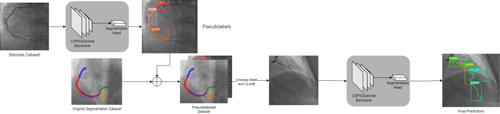
The input to our YOLO model was a combined dataset consisting of images and annotation from segmentation dataset as well as the pseudolabel images and annotations. The pseudolables are generated using a model trained earlier on the actual dataset with weak train time augmentations. To obtain better generalization for our instance segmentation model we trained the model for 120 epochs and the final model was not only the best model but an averaged model from five separate epochs close to the end of training.
3.2 Loss Function
The loss function in coronary artery segmentation is the sum of IOU loss, mask loss, class loss and Distributional focal loss with different gain coefficients adjusted to obtain better performance.
| (1) |
where, is the Multilabel classification BCE, represents Distributional Focal Loss, is the BCE loss for segmentation and is the IOU-Loss. Corresponding lambda are gain coefficient for each loss function treated as hyperparameters.The box loss is defined as:
| (2) |
The BCE loss is used for segmentation and classification, where c is the class number, n is the number of sample in the batch and is the weight of positive answer for the class c.
| (3) |
The Distributional Focal [14] forces the network to rapidly focus on the values near label , by explicity enlarging probabilities and nearest to .
| (4) |
And finally the mask loss for is calculated as:
| (5) |
We further explore the values of hyperparameters and their choice in implementation section.
4 Experiments
4.1 Dataset and Preprocessing
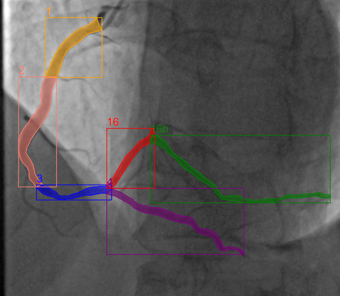
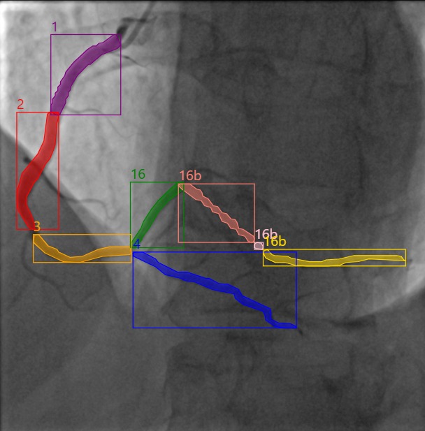
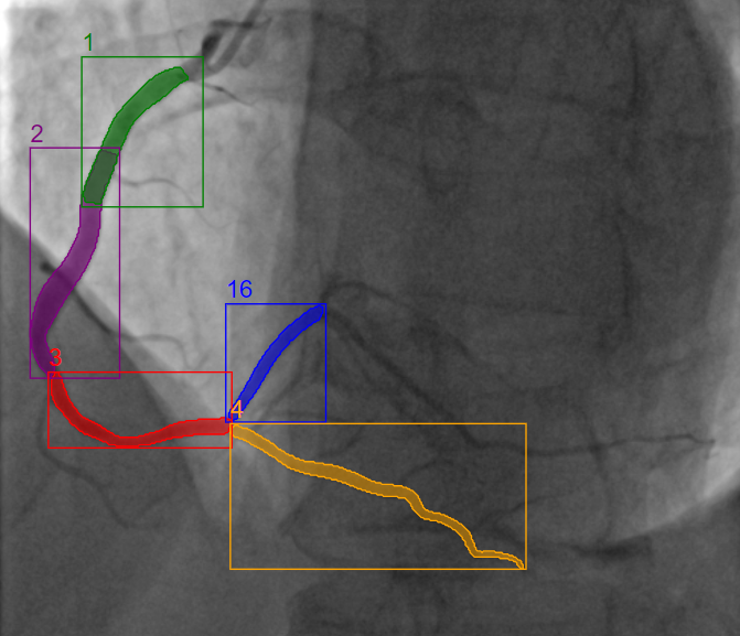
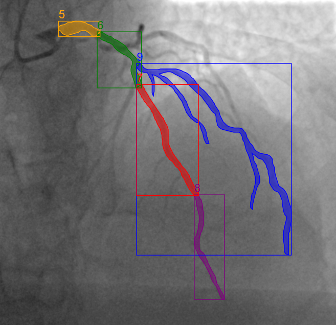
The ARCADE dataset [22] consists of 1200 in total for each task.The spatial size of the images in dataset is 512 x 512 px. We compared different instance segmentation models by testing on ARCADE dataset.
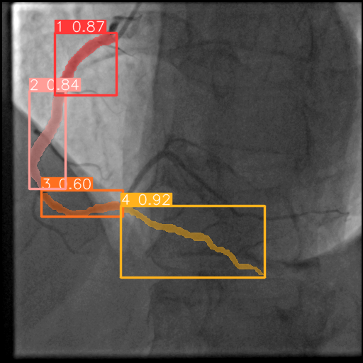
The pseudo-labels were generated from the simple training of a YOLO-v8 instance segmentation model with little augmentations(CLAHE and Median Blur). Predictions with confidence score 0.5 or greater were considered valid annotations and saved for a new training dataset consisting of pseudolabels as well as the segmentation detection dataset. The new combined dataset, including the validation set, underwent preprocessing using a series of image enhancement techniques. These techniques included the application of an Unsharp Mask filter followed by Contrast Limited Adaptive Histogram Equalization (CLAHE).This enhancement was applied to effectively improve the quality of the dataset for further analysis and model training.
4.2 Implementation details
In this study, we made use of YOLO-v8 segmentation model for vessel segmentation following a augmentated pseudolabeling pipeline. YOLO-v8 segmentation model for segmentation detection was trained on NVIDIA RTX 3090 graphic card for 120 epochs using AdamW optimizers (, ) with an initial learning rate of 1 × and a decay rate of 0.0005 per epoch with batch size of 16. And a series of random augmentations were applied to the dataset to increase the diversity in training examples which included HSV Hue Adjustment, HSV Saturation Adjustment,HSV Value Adjustment, Translation, Scaling,Vertical Flipping and Horizontal Flipping. The coefficients for gains of loss functions we used are . We settled on these values after carefully evaluating the performance of our model at different settings. The selected gains allowed the model achieve smoother training.
4.3 Quantitative Results
The quantitative result for Coronary Artery Segmentation with 25 classes is compared in Table 1. The F1scores for corresponding models on validation set as well as test set are compared as metrics. Even though MaskDino [13] performed quite well on the validation set the imbalanced nature of problem meant that the results weren’t translated very well to the test set, which seems to be the general trend for most of models under consideration. The self supervised approach of MaskDino furthermore didn’t have any improvements upon testing on the pseudolabel pipeline. The performance of most of these models hence was skewed based on classes as well. The classes with fairly good amount of examples performed much better than some of the classes which had very few examples. We oversampled some of the underrepresented classes but achieving generalizability proved difficult and the model couldn’t perform well even when classes with fewer samples were oversampled.
| Architecture | F1Score(Val) | F1Score(Test) |
|---|---|---|
| Yolov8 Ensemble | 0.34 | 0.26 |
| ConvNext | 0.21 | 0.27 |
| ConvNextv2 | 0.26 | 0.29 |
| MaskDino | 0.38 | 0.33 |
| Pseudolabels MaskDino | 0.38 | 0.33 |
| Yolov8 | 0.32 | 0.33 |
| Pseudolabels Yolo | 0.41 | 0.35 |
The quality of detection was also be tracked through MAP scores obtained by the models on validation set to verify that they will have generalizablility in the test set. The following table shows the best MAP/50 scores achieved by corresponding models on the validation set.
| Architecture | MAP/50(Val) |
|---|---|
| MaskDino | 0.54 |
| Yolov8 | 0.53 |
| Convnxtv2 | 0.46 |
| Pseudolabels Yolo | 0.59 |
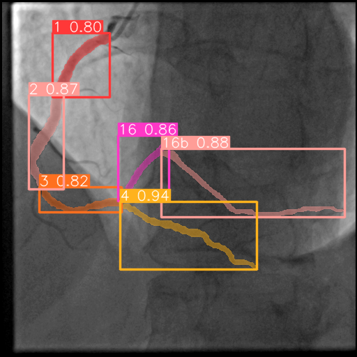
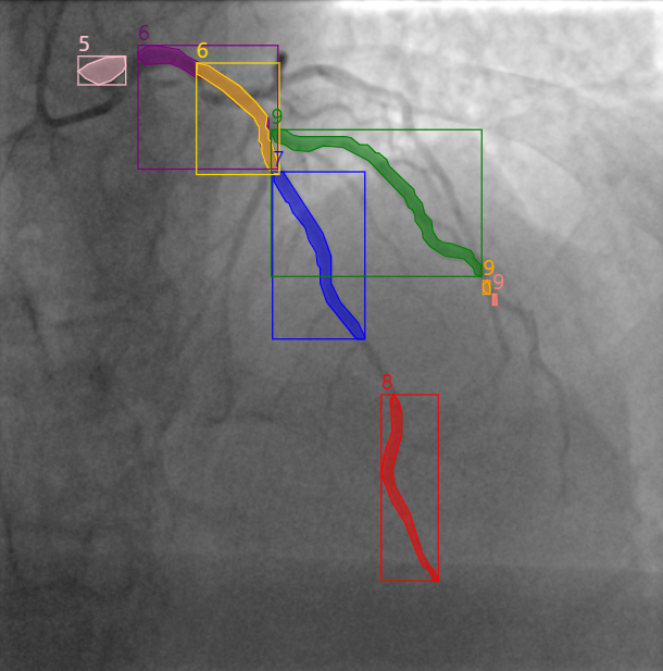
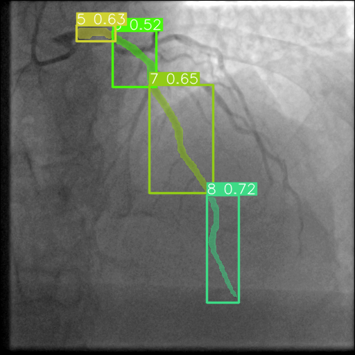
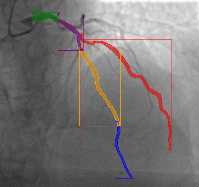
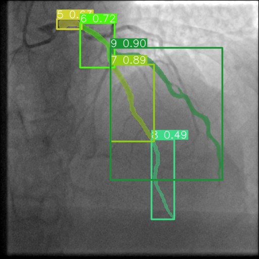
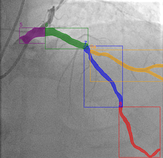
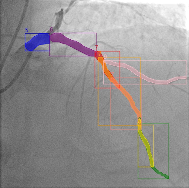
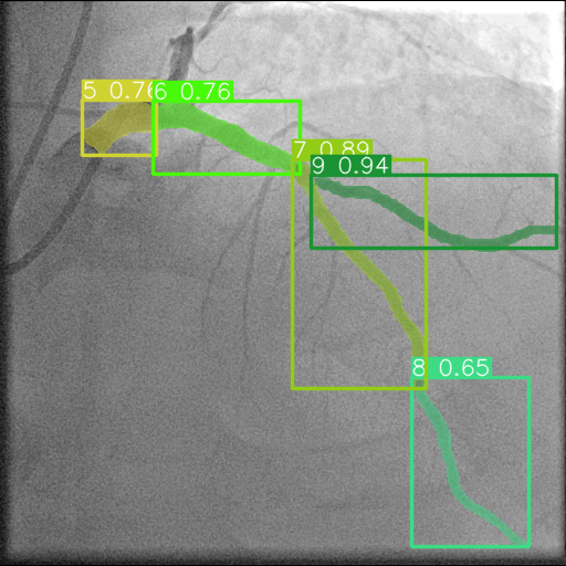
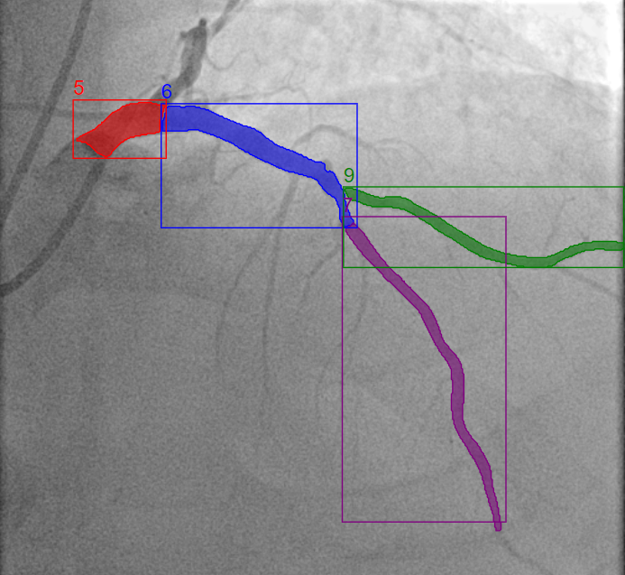
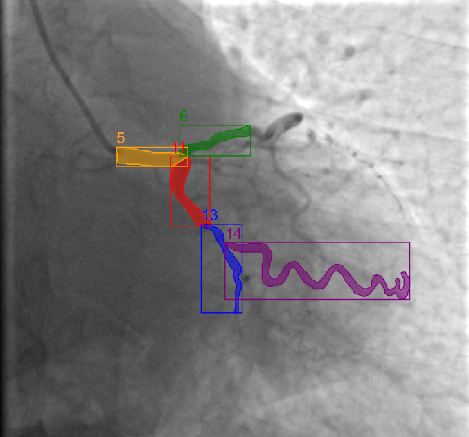
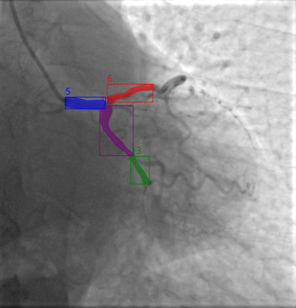
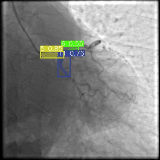
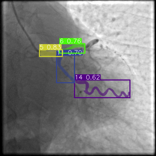
4.4 Qualitative Results
Qualitative comparison of the baseline models along with pseudolabel YOLO on the unseen test dataset indicates that the proposed model showcases finer attention to detail in different illumination conditions. For images with good contrast between vessel and background almost all of the baseline models perform well in detection as well as segmentation. However, when the contrast deters baseline models make erratic predictions or are missing predictions all together.


4.5 Ablation Studies
Starting a a model from scratch was inefficient as the dataset was relatively small. Hence, the models we tested were initialized on pretrained MS-COCO dataset. This modification was applied to all the models we tested as starting from scratch lead to a slower convergence and couldn’t achieve adequate accuracy.
Another important improvement in our model was achieved when instead of single best model we used an average of five models exported from near the end of the training phases. The averaged model achieved better performance on F1 Score when compared to the best model from previous training owing to the improved generalizability when averaged. Table 3 illustrates this fact.
| Model | F1-score() |
|---|---|
| Pseudolabel YOLO | 0.34 |
| A͡veraged Pseudolabel YOLO - | 0.35 |
5 Conclusion
Our approach tackles the problem of sparsity of data and difficulty in curating a new one by adopting augmentations via pseudo labels to improve upon the baseline models. With the extensive support of pseudo labelling and model weights averaging, YOLO-V8 extends its performance quality in segmentation task and is able to outperform well established ConvNeXt and ConvNeXtv2 models.
References
- [1] Brown, J.C., Gerhardt, T.E., Kwon, E.: Risk factors for coronary artery disease. In: StatPearls. StatPearls Publishing, Treasure Island (FL) (Jan 2023)
- [2] Cervantes-Sanchez, F., Cruz-Aceves, I., Hernandez-Aguirre, A., Hernandez-Gonzalez, M.A., Solorio-Meza, S.E.: Automatic segmentation of coronary arteries in x-ray angiograms using multiscale analysis and artificial neural networks. Applied Sciences 9(24) (2019)
- [3] Chen, L.C., Papandreou, G., Schroff, F., Adam, H.: Rethinking atrous convolution for semantic image segmentation (2017)
- [4] Fu, Y., Guo, B., Lei, Y., Wang, T., Liu, T., Curran, W., Zhang, L., Yang, X.: Mask R-CNN based coronary artery segmentation in coronary computed tomography angiography. In: Hahn, H.K., Mazurowski, M.A. (eds.) Medical Imaging 2020: Computer-Aided Diagnosis. vol. 11314, p. 113144F. International Society for Optics and Photonics, SPIE (2020)
- [5] Gharehbaghi, A., Lindén, M., Babic, A.: A decision support system for cardiac disease diagnosis based on machine learning methods. Stud Health Technol Inform 235, 43–47 (2017)
- [6] He, K., Gkioxari, G., Dollár, P., Girshick, R.B.: Mask R-CNN. CoRR abs/1703.06870 (2017)
- [7] Holste, G., Oikonomou, E.K., Mortazavi, B.J., Coppi, A., Faridi, K.F., Miller, E.J., Forrest, J.K., McNamara, R.L., Ohno-Machado, L., Yuan, N., Gupta, A., Ouyang, D., Krumholz, H.M., Wang, Z., Khera, R.: Automated severe aortic stenosis detection on single-view echocardiography: A multi-center deep learning study. medRxiv (2022)
- [8] Hu, Z., Yang, Z., Hu, X., Nevatia, R.: Simple: Similar pseudo label exploitation for semi-supervised classification. In: Proceedings of the IEEE/CVF Conference on Computer Vision and Pattern Recognition (CVPR). pp. 15099–15108 (June 2021)
- [9] Huang, W., Huang, L., Lin, Z., Huang, S., Chi, Y., Zhou, J., Zhang, J., Tan, R.S., Zhong, L.: Coronary artery segmentation by deep learning neural networks on computed tomographic coronary angiographic images. In: 2018 40th Annual International Conference of the IEEE Engineering in Medicine and Biology Society (EMBC). pp. 608–611 (2018)
- [10] Iyer, K., Najarian, C.P., Fattah, A.A., Arthurs, C.J., Soroushmehr, S.M.R., Subban, V., Sankardas, M.A., Nadakuditi, R.R., Nallamothu, B.K., Figueroa, C.A.: AngioNet: a convolutional neural network for vessel segmentation in x-ray angiography. Scientific Reports 11(1), 18066 (Sep 2021)
- [11] Jocher, G., Chaurasia, A., Qiu, J.: YOLO by Ultralytics (Jan 2023), https://github.com/ultralytics/ultralytics
- [12] Kruzhilov, I., Ikryannikov, E., Shadrin, A., Utegenov, R., Zubkova, G., Bessonov, I.: Neural network-based coronary dominance classification of rca angiograms (2023)
- [13] Li, F., Zhang, H., xu, H., Liu, S., Zhang, L., Ni, L.M., Shum, H.Y.: Mask dino: Towards a unified transformer-based framework for object detection and segmentation (2022)
- [14] Li, X., Wang, W., Wu, L., Chen, S., Hu, X., Li, J., Tang, J., Yang, J.: Generalized focal loss: Learning qualified and distributed bounding boxes for dense object detection (2020)
- [15] Li, Y., Wu, Y., He, J., Jiang, W., Wang, J., Peng, Y., Jia, Y., Xiong, T., Jia, K., Yi, Z., Chen, M.: Automatic coronary artery segmentation and diagnosis of stenosis by deep learning based on computed tomographic coronary angiography. European Radiology 32(9), 6037–6045 (Sep 2022)
- [16] Liu, Z., Mao, H., Wu, C.Y., Feichtenhofer, C., Darrell, T., Xie, S.: A convnet for the 2020s (2022)
- [17] Mastoi, Q.U.A., Wah, T.Y., Gopal Raj, R., Iqbal, U.: Automated diagnosis of coronary artery disease: A review and workflow. Cardiology Research and Practice 2018, 2016282 (Feb 2018)
- [18] Melese, A., Salau, A., Abeje, B., Enyew, B.: Detection and classification of covid-19 disease from x-ray images using convolutional neural networks and histogram of oriented gradients. Biomedical signal processing and control 74, 103530 (01 2022)
- [19] Nakamura, M.: Angiography is the gold standard and objective evidence of myocardial ischemia is mandatory if lesion severity is questionable. - indication of PCI for angiographically significant coronary artery stenosis without objective evidence of myocardial ischemia (pro)-. Circ J 75(1), 204–10; discussion 217 (2011)
- [20] Pan, L.S., Li, C.W., Su, S.F., Tay, S.Y., Tran, Q.V., Chan, W.P.: Coronary artery segmentation under class imbalance using a u-net based architecture on computed tomography angiography images. Scientific Reports 11(1), 14493 (Jul 2021)
- [21] Papandrianos, N.I., Feleki, A., Papageorgiou, E.I., Martini, C.: Deep Learning-Based automated diagnosis for coronary artery disease using SPECT-MPI images. J Clin Med 11(13) (Jul 2022)
- [22] Popov, M., Amanturdieva, A., Zhaksylyk, N., Alkanov, A., Saniyazbekov, A., Aimyshev, T., Ismailov, E., Bulegenov, A., Kolesnikov, A., Kulanbayeva, A., Kuzhukeyev, A., Sakhov, O., Kalzhanov, A., Temenov, N., Fazli1, S.: ARCADE: Automatic Region-based Coronary Artery Disease diagnostics using x-ray angiography imagEs Dataset Phase 1 (May 2023)
- [23] Qureshi, R., RAGAB, M.G., ABDULKADER, S.J., amgad muneer, ALQUSHAIB, A., SUMIEA, E.H., Alhussian, H.: A comprehensive systematic review of YOLO for medical object detection (2018 to 2023) (Jul 2023)
- [24] Redmon, J., Divvala, S., Girshick, R., Farhadi, A.: You only look once: Unified, real-time object detection (2016)
- [25] Rodrigues, D.L., Menezes, M.N., Pinto, F.J., Oliveira, A.L.: Automated detection of coronary artery stenosis in x-ray angiography using deep neural networks (2021)
- [26] Ronneberger, O., Fischer, P., Brox, T.: U-net: Convolutional networks for biomedical image segmentation (2015)
- [27] Setiawan, N.A., Venkatachalam, P.A., Hani, A.F.M.: Diagnosis of coronary artery disease using artificial intelligence based decision support system. CoRR abs/2007.02854 (2020)
- [28] Tao, X., Dang, H., Zhou, X., Xu, X., Xiong, D.: A lightweight network for accurate coronary artery segmentation using x-ray angiograms. Frontiers in Public Health 10 (2022)
- [29] Wahab Sait, A.R., Dutta, A.K.: Developing a Deep-Learning-Based coronary artery disease detection technique using computer tomography images. Diagnostics (Basel) 13(7) (Mar 2023)
- [30] Wang, L., Liang, D., Yin, X., Qiu, J., Yang, Z., Xing, J., Dong, J., Ma, Z.: Coronary artery segmentation in angiographic videos utilizing spatial-temporal information. BMC Med. Imaging 20(1), 110 (Sep 2020)
- [31] Wang, Q., Xu, L., Wang, L., Yang, X., Sun, Y., Yang, B., Greenwald, S.E.: Automatic coronary artery segmentation of CCTA images using UNet with a local contextual transformer. Front Physiol 14, 1138257 (Aug 2023)
- [32] Wolterink, J.M., Leiner, T., Išgum, I.: Graph convolutional networks for coronary artery segmentation in cardiac ct angiography (2019)
- [33] Wolterink, J.M., van Hamersvelt, R.W., Viergever, M.A., Leiner, T., Išgum, I.: Coronary artery centerline extraction in cardiac ct angiography using a cnn-based orientation classifier. Medical Image Analysis 51, 46–60 (2019)
- [34] Woo, S., Debnath, S., Hu, R., Chen, X., Liu, Z., Kweon, I.S., Xie, S.: Convnext v2: Co-designing and scaling convnets with masked autoencoders (2023)
- [35] Yan, J., Tian, J., Yang, H., Han, G., Liu, Y., He, H., Han, Q., Zhang, Y.: A clinical decision support system for predicting coronary artery stenosis in patients with suspected coronary heart disease. Computers in Biology and Medicine 151, 106300 (2022)
- [36] Yu, J., Zhang, L., Du, S., Chang, H., Lu, K., Zhang, Z., Yu, Y., Wang, L., Ling, Q.: Pseudo-label generation and various data augmentation for semi-supervised hyperspectral object detection. In: 2022 IEEE/CVF Conference on Computer Vision and Pattern Recognition Workshops (CVPRW). pp. 304–311 (June 2022)
- [37] Zhang, H., Zhong, X., Li, G., Liu, W., Liu, J., Ji, D., Li, X., Wu, J.: Bcu-net: Bridging convnext and u-net for medical image segmentation. Computers in Biology and Medicine 159, 106960 (2023)
- [38] Zhou, Z., Siddiquee, M.M.R., Tajbakhsh, N., Liang, J.: UNet++: Redesigning skip connections to exploit multiscale features in image segmentation. IEEE Trans Med Imaging 39(6), 1856–1867 (Dec 2019)
- [39] Zou, Y., Zhang, Z., Zhang, H., Li, C.L., Bian, X., Huang, J.B., Pfister, T.: Pseudoseg: Designing pseudo labels for semantic segmentation (2021)
- [40] Zreik, M., van Hamersvelt, R.W., Wolterink, J.M., Leiner, T., Viergever, M.A., Išgum, I.: A recurrent cnn for automatic detection and classification of coronary artery plaque and stenosis in coronary ct angiography. IEEE Transactions on Medical Imaging 38(7), 1588–1598 (2019)
- [41] Zunair, H., Ben Hamza, A.: Sharp U-Net: Depthwise convolutional network for biomedical image segmentation. Comput Biol Med 136, 104699 (Jul 2021)