Signum phase mask differential microscopy
Abstract
We propose and experimentally demonstrate a differential microscopy method to obtain simultaneous amplitude, phase, and quantitative polarization gradient imaging in a single experimental embodiment. A full-field optical spatial differentiator is achieved in a relatively simple setup by placing a glass cover slip as a Signum phase mask in the Fourier plane of a standard imaging system and accordingly named Signum phase mask differential microscopy. The longstanding requisite of polarized light to obtain the spatial differentiation of the field at the object plane is eliminated in our scheme and, hence, leads to the emergence of quantitative differential polarization contrast imaging by integrating polarization degree of freedom as an additional contrast agent in the framework of differential microscopy. Implementation of the proposed differential imaging scheme in high-resolution microscopy is experimentally demonstrated alongside its functionality for a broad wavelength range. Simultaneous acquisition of differential phase, amplitude, and polarization (anisotropy) gradient imaging in a rather elementary optical setup enables a low-cost multi-functional differential microscopy system that is anticipated to emerge as a revolutionary tool in label-free imaging and optical image processing.
Interaction of light with inhomogeneous natural objects provides the intrinsic mechanism of image contrast through the spatial modulation of the amplitude, phase, and polarization of the light field, which is traditionally utilized for optical imaging and microscopy for a wide range of applications in various disciplines [1, 2, 3, 4, 5]. One of the major challenges in the early days of microscopy was to visualize the so-called phase objects (nearly transparent) under a conventional bright-field microscope, as these objects influence the amplitude of the incident light marginally. In such cases, phase and polarization imaging can capture essential morphological details about the internal structure of the object, hence, have paramount importance in label-free microscopy [6, 7, 8, 9, 10]. Many of the natural objects of biological or non-biological origin possess intrinsic polarization anisotropy effects, i.e., birefringence (phase anisotropy) and dichroism (amplitude anisotropy) due to their anisotropic molecular or structural organizations [3, 11, 12, 13]. Therefore, quantitative probing of the vectorial transformation of light with desirable spatial resolution provides a wealth of morphological, structural, and functional information on the objects and has been a key tool in biomedical and clinical research [3]. On the other hand, in the realm of phase imaging, phase contrast [14, 15, 16, 17, 18] and differential interference contrast (DIC) [19, 20] are the first-generation techniques, and are still widely adopted to produce contrast images of phase objects such as biological specimen and etc. Owing to their characteristic working principles, some longstanding limitations are associated with these traditional phase and differential microscopy methods, e.g., incapability for quantitative phase imaging, complexity and bulkiness of the setup, polarization dependency, etc. [7, 19].
In recent years, there has been a renewed engagement towards the development of next-generation differential microscopy and image processing methods with an aim to achieve versatile and compact optical microscopy systems [21, 22, 23]. A wide range of optical systems, starting from reflection in glass surfaces [24, 25, 26, 27, 28, 29] to multi-functional meta-surfaces [21, 30, 31, 32, 33, 34, 35, 36], have been utilized to realize various optical spatial differentiator. On the basis of the corresponding working principles, these recently proposed differential imaging methods can be primarily divided into two types. One, where the spatial differentiation is obtained incorporating the polarization-dependent phase or beam shift and can be considered as a DIC-inspired microscopy technique [21, 30, 25, 26]. In the other approach, the spatial dispersion of the meta-surfaces is appropriately tailored to make them perform as a spatial differentiator [37, 38, 23, 32, 22, 36]. Although these recently developed spatial differentiaton methods have shown great promises in optical computing and the possible intersection with optical microscopy systems, most of these techniques utilize polarized light to achieve the spatial differentiation[21, 30, 36, 25]. This requisite of differential imaging system makes it impossible to capture simultaneous phase and polarization images of a specimen in a single experimental embodiment. In this context, it is pertinent to note that the traditional polarization microscopic approaches are often severely compromised while probing low level spatial fluctuations of polarization anisotropy effects, which is desirable for most practical applications [3]. Thus, encompassing polarization as an contrast mechanism within the framework of conventional differential microscopy (which considers only amplitude and phase) can be a revolutionary tool in the realm of label-free microscopy.
In this work, we propose and experimentally demonstrate a Signum phase mask differential (SPMD) microscopy scheme, which enables simultaneous amplitude, phase, and quantitative differential polarization imaging/polarization gradient imaging in a simple experimental framework. The proposed microscopy scheme exploits the principle of one-dimensional (1D) Hilbert transformation [39, 40], where a full-field spatial differentiator is obtained by placing a Signum phase mask in the Fourier plane of a conventional imaging setup [41]. This simple technique enables us to realize a multi-functional differential imaging system built by using just a pair of lenses and a commercially available glass cover slip. Importantly, unlike most of the existing differential microscopy methods [19, 30, 21], the proposed spatial differentiator does not require polarized light to obtain the differentiation of the electric field distribution at the object plane. This not only reduces the complexity and bulkiness of the microscopy system but also enables utilizing the polarization of light as an additional intrinsic contrast mechanism in the framework of differential microscopy. As a consequence, two new kinds of imaging systems are emerged in the domain of differential microscopy: polarization-resolved differential imaging and quantitative polarization (anisotropy) gradient imaging. It is worth mentioning that, although the Hilbert transformation has been previously used for image processing, its utilization in the realm of differential microscopy and more specifically its potential for merging the polarization and differential imaging was not touched upon [39, 42]. Usually, the polarization properties of an object are described using the polarization anisotropy parameters (birefringence and dichroism) [3, 12, 11, 13]. Being quantitative in nature, our proposed SPMD imaging system can quantify the spatial gradient of these anisotropy parameters throughout a specimen and possess the ability to probe and subsequently produce high contrast images of pure polarization objects even with weak spatial variation of polarization anisotropy effects. The polarization-resolved differential microscopy also holds certain advantages to significantly enhance the image contrast of phase-polarization objects, which will be discussed subsequently. The adaptability of the SPMD imaging scheme in high numerical aperture (NA ) microscopy is validated alongside its applicability for broad wavelength regions by imaging various biological cells. Demonstration of such a low-cost multi-functional setup that can be easily integrated with the conventional commercial bright-field microscope is expected to have a significant impact on the next generation of label-free microscopy and optical image processing techniques.
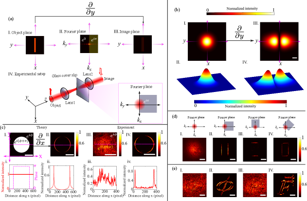
The proposed differential imaging scheme and experimental setup are illustrated in Fig. 1(a). A commercially available glass cover slip placed in the Fourier plane of a imaging setup imparts different phases ; to the negative and positive spatial frequencies on the Fourier plane. Also, due to the high optical scattering at the edge of the cover slip, the transmittance of the zero-order spatial frequencies is reduced to a great extent. In this way, a simple cover slip placed at an appropriate orientation effectively acts as a Signum mask in the Fourier plane [41]. The 1D Signum function is chosen for the sake of mathematical simplicity and corresponding experimental feasibility (see Fig. 1(a)). With a 1D Signum function (in the -direction) modulating the spatial frequencies in the Fourier plane, a full-field spatial differentiator is achieved where the output intensity distribution at the image plane can be described as (see Supporting information Sec. S.1 for details)
| (1) |
Here , describes the spatial variation of the Electric field at the object plane, which corresponds to the complex vectorial transmittance function of the input object, and are the point of spatial discontinuities/jumps in terms of either the spatial variation of the amplitude, phase, or polarization in the . As evident from Eq. (1), the spatial differentiation of the object field distribution is directly encoded in the captured intensity distribution at the image plane. It is also implied that there will be intensity localization/enhancement at the point of discontinuities in the object field, which leads to the production of high-contrast images and serves as the foundation for the SPMD imaging and microscopy scheme. More importantly, the achieved spatial differentiator considers the vectorial nature of light, which is described by the Jones vector field [3, 11]. As a consequence, alongside amplitude and phase objects, differential images of pure polarization objects can be produced in the SPMD imaging scheme by recording the polarization-resolved intensity distribution (Stokes parameters [3, 43]) at the image plane. The detailed theoretical treatment, particularly, the differential polarization imaging, is provided in the Supporting information Sec. S.1.
Differential image formation of a rectangular slit utilizing the SPMD imaging scheme is illustrated in Fig. 1(a). Owing to the amplitude gradient at the boundaries, intensity localization at the edges of the slit is observed at the output image plane. To further validate the proposed spatial differentiation scheme, we numerically simulate the experimental setup and obtain the differential image of an input Gaussian beam (Fig.1(b)I., II.), where the spatial intensity distribution at the image plane conforms to the spatial gradient of a Gaussian distribution. Next, both numerically and experimentally recorded differential images of a circular phase object are shown Fig. 1(c). The phase difference between the background and the object leads to an intensity localization at the circumference of the circle (Fig. 1(c)II., IV.). The extracted intensity profiles in each pixel of the magenta dashed line across both the bright field (Fig. 1(c)i.,iii.), and differential image (Fig. 1(c)ii.,iv.) provide compelling proof of the ability of the SPMD imaging scheme to produce high contrast images of pure phase objects. Note that, the spatial variation of the intensity distribution at the image plane (Fig. 1(c)ii.) emulates the Logarithmic behaviour as obtained in Eq. (1). Similar differential images of conventional amplitude objects, e.g., slit, and circular aperture, have also been recorded, and corresponding theoretical and experimental results are given in Supporting information Sec. S.2. The direction along which the spatial differentiation is carried out can be conveniently tuned by steering the orientation of the Signum phase mask (glass cover-slip) in the Fourier plane. A rectangular phase object (bright field image: Fig. 1(d)I.) is utilized to illustrate this, where spatial differentiation along (Fig. 1(d)II.), (Fig. 1(d)II.), (Fig. 1(d)III.) are performed, and consequent intensity localization along different edges are observed in the corresponding differential images. Bright-field and differential images of several phase objects are additionally provided to substantiate the production of high-contrast images of pure phase objects with the SPMD imaging setup (Fig. 1(e)). A spatial light modulator (SLM) is employed to prepare the phase objects, that are used as imaging targets, and the subsequent differential images are recorded using the SPMD imaging setup (Details regarding optical properties of the used SLM are provided in the method section).
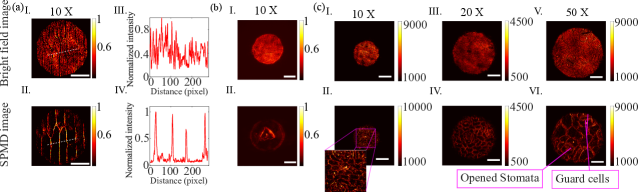
Next, we demonstrate the adaptability of the SPMD scheme in the domain of microscopy. To do so, commercial microscope objectives are integrated with the existing spatial differentiator setup and various biological cells are used as imaging specimens (Fig. 2). While the bright-field images of the unstained onion and human cheek cells show low contrast due to their pure phase nature (Fig. 2(a)I.,(b)I.), corresponding differential images exhibit high contrast with clearly visible cell structures (Fig. 2(a)II.,(b)II)(intensity profile corresponding to the white dashed line is given in (Fig. 2(a)III.,IV). Moreover, we have recorded the differential images of an Aglaonema commutatum leaf cell using three different microscope objectives with 10X (numerical aperture, NA ), 20X (NA ), and 50X (NA ) magnification (Fig.2c). The observed differential images authenticate the ability of the SPMD microscope to produce high-contrast images even with high spatial resolution. Thus, it is evident that the proposed SPMD microscopy scheme holds great potential for real-time label-free imaging of transparent biological objects. In addition to this, we also note that the spatial differentiation process in the SPMD scheme is intrinsically independent of the wavelength of the incident light, and thus, the same experimental arrangement can be utilized for a broad range of wavelengths. Indeed, high-contrast differential images of pure phase objects are obtained with laser illumination at different wavelengths, keeping the optical spatial differentiator unchanged (see Sec S.5 of SI).
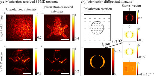
As discussed earlier, one of the biggest advantages of the SPMD imaging scheme is that it can utilize the polarization degree of freedom as an additional contrast mechanism in the domain of differential microscopy. We first demonstrate, how the integration of polarization-resolved measurements with differential imaging can significantly enhance the image contrast. For this purpose, a phase-polarization object is prepared using the SLM, and the polarization-resolved intensity patterns at the output image plane are recorded by inserting conventional polarizing optical elements, such as linear polarizers and quarter wave plates (Fig. 3(a)). Even in the case of bright-field imaging, the polarization-resolved measurement (Fig. 3(a)II.) reveals the structure of the object (by reducing the overwhelmed background signal), which is not visible in the unpolarized intensity (Fig. 3(a)I.). Consequently, the differential image corresponding to the polarization-resolved intensity pattern (Fig. 3(a)ii.) exhibits much higher contrast as compared to that with the unpolarized light (Fig. 3(a)i.). This observation highlights the utilization of polarization as an additional handle to improve the image contrast in SPMD microscopy.
Next, we describe the quantitative polarization gradient imaging capability of the proposed SPMD scheme. As Eq. (1) suggests, the proposed spatial differentiator can transfer the information of polarization anisotropy [44] gradient at the object plane to the polarization-resolved intensity distribution at the image plane. This is illustrated considering a pure polarization object, where although there is no change in the absolute amplitude or phase of the electric field transmitted through the object , the polarization of the transmitted light gets modified due to the anisotropic nature of the object, and differ with respect to the background signal. The effect of such discontinuity in the polarization anisotropy parameter, e.g., circular retardance (optical rotation) around the circumference of a circle-shaped object, is studied (Fig. 3(b)I.). As a consequence of the polarization discontinuity, differential polarization-contrast images are obtained by recording the Stokes vector elements () at the image plane (Fig. 3(b)I.). The spatial gradient of the polarization anisotropy effect (rotation) is also quantified using standard polarization algebra and presented in Fig. 3(b)i. (see Supporting information Sec. S.1 for details). In the case of natural polarization objects, that exhibit multiple anisotropy effects simultaneously, such as biological cells and tissues [3], recording the Mueller matrix (in the same experimental set up- see Supporting information Sec. S.4) and subsequent Mueller matrix decomposition analysis can provide a complete quantitative polarimetric information throughout the specimen. More importantly, this kind of polarization differential microscopy can be advantageous while investigating the morphology of an object with weak spatial variation of polarization anisotropy effects, thus showing promise in biomedical and clinical research.
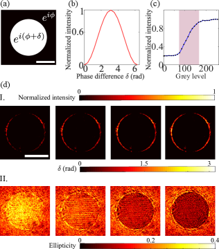
We now shift our discussion to the possible quantitative phase imaging using the SPMD scheme. Similar to the conventional differential imaging methods, in the SPMD imaging scheme, the spatial field gradient at the object plane dictates the intensity distribution at the image plane. Although the theoretical expression obtained for the output intensity pattern implies a blow-up of intensity values at the point of field discontinuity (Eq. 1), it is never the case for practical situations due to the non-ideal behaviour of real step functions, and its corresponding derivatives. Thus, the magnitude of the intensity distribution at the image plane can be incorporated to quantitatively extract the spatial phase gradient distribution, which is illustrated next. The behaviour of the localized intensity values with different phase gradients is both numerically (Fig. 4(b)) calculated and experimentally (Fig. 4(c)) studied in a circular phase object, where the phase difference between the object and the background is increased gradually. In both cases, the maximum magnitude of the localized intensity at the boundary (edge of the circle) exhibits a monotonically increasing behavior for to (Fig. 4(b), (c)). This indicates the possibility of establishing a direct relation between the phase gradient and the magnitude of the localized intensity, which can be utilized to quantify the local spatial phase gradient itself (see Supporting information Sec. S.3). To experimentally demonstrate the quantitative phase imaging capability of the SPMD scheme, circular phase objects are prepared using the SLM with varying phases (achieved by varying the grey levels). The phase step is gradually increased, which is manifested in the maxima of the localized intensities in the corresponding differential images (Fig. 4(d)I.). Now, the priorly obtained relation (calibration) between the phase gradient and the magnitude of the localized intensity (Fig. 4(c) is exploited to quantify the phase gradient at the boundaries of the objects, and consequently, the phase distributions of the circular objects are derived (see Supporting information Sec. S.3). Also, the utilized SLM in the experiment modulates the polarization properties (linear retardance) of light (hence the projected object), and quantitative polarimetric imaging of the object is also obtained by recording the polarization-resolved Stokes vectors (Fig. 4(d)II.). Through this illustration, an imaging system with the ability of simultaneous quantitative phase and polarization imaging is realized in a single experimental embodiment.
In summary, we have demonstrated a Signum phase mask differential (SPMD) imaging and microscopy protocol with the capability of simultaneous differential phase, differential amplitude, and quantitative polarization gradient imaging in a simple experimental embodiment. The longstanding requisite of using polarization degree of freedom of light to obtain a spatial differentiation of image is eliminated in our scheme, enabling us to integrate polarization as a contrast mechanism in the framework of differential imaging. Importantly, the SPMD imaging scheme leads to a novel quantitative polarization gradient imaging system with the potential to probe low levels and small spatial variations of polarization anisotropy in a sample. The utilization of the proposed SPMD scheme in the domain of microscopy is validated by producing high contrast images of biological samples with high spatial resolution by integrating high numerical aperture objectives. Simultaneous demonstration of amplitude, phase, and polarization contrast differential microscopy in a simple experimental embodiment, imaging capability with high spatial resolution, functionality over a broad wavelength region, quantitative and semi-quantitative extraction of polarization and phase gradient information, all together pave the way for new generation cost-effective multi-functional microscopy in label-free imaging and image processing.
Methods: Preparation of phase and polarization objects using a spatial light modulator.
The spatial light modulator (SLM) is generally used to modulate the amplitude, phase, and polarization of light in space and time [46]. Our work uses a twisted nematic liquid crystal-based SLM [45] to generate several phase and polarization objects that are used as imaging targets in the SPMD imaging setup. A specific grey level value in the SLM represents a defined average voltage across the liquid crystal cell. This voltage leads to a variable tilt of the anisotropy liquid crystal molecules. Due to the inherent anisotropy properties of the liquid crystal molecules, the SLM induces a particular phase according to the tilt angle of the liquid crystal layer. In the case of our twisted SLM, the net anisotropy effects are manifested as linear/circular retardance and the accumulation of dynamic phases. The complete polarization and phase modulation properties of the utilized SLM were obtained through rigorous calibration [45]. Consequently, the accumulated phase and polarization effects as the function of grey levels are obtained, which are used to prepare regulated phase and polarization objects.
References
- Mertz [2019] J. Mertz, Introduction to optical microscopy (Cambridge University Press, 2019).
- Evanko et al. [2009] D. Evanko, A. Heinrichs, and C. Rosenthal, Milestones in light microscopy, Nature Cell Biol 11, S5 (2009).
- He et al. [2021] C. He, H. He, J. Chang, B. Chen, H. Ma, and M. J. Booth, Polarisation optics for biomedical and clinical applications: a review, Light: Science & Applications 10, 194 (2021).
- Guo et al. [2020] S.-M. Guo, L.-H. Yeh, J. Folkesson, I. E. Ivanov, A. P. Krishnan, M. G. Keefe, E. Hashemi, D. Shin, B. B. Chhun, N. H. Cho, et al., Revealing architectural order with quantitative label-free imaging and deep learning, elife 9, e55502 (2020).
- Mennel et al. [2018] L. Mennel, M. M. Furchi, S. Wachter, M. Paur, D. K. Polyushkin, and T. Mueller, Optical imaging of strain in two-dimensional crystals, Nature communications 9, 516 (2018).
- Park et al. [2018] Y. Park, C. Depeursinge, and G. Popescu, Quantitative phase imaging in biomedicine, Nature photonics 12, 578 (2018).
- Yin et al. [2012] Z. Yin, T. Kanade, and M. Chen, Understanding the phase contrast optics to restore artifact-free microscopy images for segmentation, Medical image analysis 16, 1047 (2012).
- Choi et al. [2007] W. Choi, C. Fang-Yen, K. Badizadegan, S. Oh, N. Lue, R. R. Dasari, and M. S. Feld, Tomographic phase microscopy, Nature methods 4, 717 (2007).
- Kim et al. [2014] T. Kim, R. Zhou, M. Mir, S. D. Babacan, P. S. Carney, L. L. Goddard, and G. Popescu, White-light diffraction tomography of unlabelled live cells, Nature Photonics 8, 256 (2014).
- Cotte et al. [2013] Y. Cotte, F. Toy, P. Jourdain, N. Pavillon, D. Boss, P. Magistretti, P. Marquet, and C. Depeursinge, Marker-free phase nanoscopy, Nature Photonics 7, 113 (2013).
- Chipman et al. [2018] R. A. Chipman, W. S. T. Lam, and G. Young, Polarized light and optical systems (CRC press, 2018).
- Goldstein [2017] D. H. Goldstein, Polarized light (CRC press, 2017).
- Gil and Ossikovski [2022] J. J. Gil and R. Ossikovski, Polarized light and the Mueller matrix approach (CRC press, 2022).
- Zernike [1934] F. Zernike, Diffraction theory of the knife-edge test and its improved form, the phase-contrast method, Monthly Notices of the Royal Astronomical Society, Vol. 94, p. 377-384 94, 377 (1934).
- Zernike [1942a] F. Zernike, Phase contrast, a new method for the microscopic observation of transparent objects, Physica 9, 686 (1942a).
- Zernike [1942b] F. Zernike, Phase contrast, a new method for the microscopic observation of transparent objects part ii, Physica 9, 974 (1942b).
- Burch and Stock [1942] C. Burch and J. Stock, Phase-contrast microscopy, Journal of Scientific Instruments 19, 71 (1942).
- Martin [1947] L. Martin, Phase-contrast methods in microscopy, Nature 159, 827 (1947).
- Nomarski [1954] G. Nomarski, Dispositif interférentiel à polarisation pour l’étude des objets transparents ou opaques appartenant à la classe des objets de phase, Patent FR 1, 124 (1954).
- Françon [1957] M. Françon, Polarization apparatus for interference microscopy and macroscopy of isotropic transparent objects, JOSA 47, 528 (1957).
- Kwon et al. [2020] H. Kwon, E. Arbabi, S. M. Kamali, M. Faraji-Dana, and A. Faraon, Single-shot quantitative phase gradient microscopy using a system of multifunctional metasurfaces, Nature Photonics 14, 109 (2020).
- Zhou et al. [2020] Y. Zhou, H. Zheng, I. I. Kravchenko, and J. Valentine, Flat optics for image differentiation, Nature Photonics 14, 316 (2020).
- Kwon et al. [2018] H. Kwon, D. Sounas, A. Cordaro, A. Polman, and A. Alù, Nonlocal metasurfaces for optical signal processing, Physical review letters 121, 173004 (2018).
- Wang et al. [2022a] R. Wang, S. He, and H. Luo, Photonic spin-hall differential microscopy, Physical Review Applied 18, 044016 (2022a).
- Shou et al. [2023] Y. Shou, J. Liu, and H. Luo, When optical microscopy meets all-optical analog computing: A brief review, Frontiers of Physics 18, 42601 (2023).
- Zhu et al. [2019] T. Zhu, Y. Lou, Y. Zhou, J. Zhang, J. Huang, Y. Li, H. Luo, S. Wen, S. Zhu, Q. Gong, et al., Generalized spatial differentiation from the spin hall effect of light and its application in image processing of edge detection, Physical Review Applied 11, 034043 (2019).
- He et al. [2020a] S. He, J. Zhou, S. Chen, W. Shu, H. Luo, and S. Wen, Wavelength-independent optical fully differential operation based on the spin–orbit interaction of light, APL Photonics 5 (2020a).
- He et al. [2020b] S. He, J. Zhou, S. Chen, W. Shu, H. Luo, and S. Wen, Spatial differential operation and edge detection based on the geometric spin hall effect of light, Optics Letters 45, 877 (2020b).
- Wang et al. [2022b] R. Wang, S. He, S. Chen, and H. Luo, Brewster differential microscopy, Applied Physics Letters 121 (2022b).
- Zhou et al. [2022] J. Zhou, Q. Wu, J. Zhao, C. Posner, M. Lei, G. Chen, J. Zhang, and Z. Liu, Fourier optical spin splitting microscopy, Physical review letters 129, 020801 (2022).
- Zhou et al. [2021] J. Zhou, H. Qian, J. Zhao, M. Tang, Q. Wu, M. Lei, H. Luo, S. Wen, S. Chen, and Z. Liu, Two-dimensional optical spatial differentiation and high-contrast imaging, National science review 8, nwaa176 (2021).
- Cordaro et al. [2019] A. Cordaro, H. Kwon, D. Sounas, A. F. Koenderink, A. Alù, and A. Polman, High-index dielectric metasurfaces performing mathematical operations, Nano letters 19, 8418 (2019).
- Zhu et al. [2017] T. Zhu, Y. Zhou, Y. Lou, H. Ye, M. Qiu, Z. Ruan, and S. Fan, Plasmonic computing of spatial differentiation, Nature communications 8, 15391 (2017).
- Zhou et al. [2019] J. Zhou, H. Qian, C.-F. Chen, J. Zhao, G. Li, Q. Wu, H. Luo, S. Wen, and Z. Liu, Optical edge detection based on high-efficiency dielectric metasurface, Proceedings of the National Academy of Sciences 116, 11137 (2019).
- Engay et al. [2021] E. Engay, D. Huo, R. Malureanu, A.-I. Bunea, and A. Lavrinenko, Polarization-dependent all-dielectric metasurface for single-shot quantitative phase imaging, Nano Letters 21, 3820 (2021).
- Cotrufo et al. [2022] M. Cotrufo, A. Arora, S. Singh, and A. Alù, Dispersion engineered metasurfaces for broadband, high-na, high-efficiency, dual-polarization analog image processing, arXiv preprint arXiv:2212.03468 (2022).
- Silva et al. [2014] A. Silva, F. Monticone, G. Castaldi, V. Galdi, A. Alù, and N. Engheta, Performing mathematical operations with metamaterials, Science 343, 160 (2014).
- Hwang and Davis [2016] Y. Hwang and T. J. Davis, Optical metasurfaces for subwavelength difference operations, Applied Physics Letters 109 (2016).
- Davis et al. [2000] J. A. Davis, D. E. McNamara, D. M. Cottrell, and J. Campos, Image processing with the radial hilbert transform: theory and experiments, Optics Letters 25, 99 (2000).
- Davis and Nowak [2002] J. A. Davis and M. D. Nowak, Selective edge enhancement of images with an acousto-optic light modulator, Applied optics 41, 4835 (2002).
- Goodman and Sutton [1996] J. W. Goodman and P. Sutton, Introduction to fourier optics, Quantum and Semiclassical Optics-Journal of the European Optical Society Part B 8, 1095 (1996).
- Davis et al. [2001] J. A. Davis, D. A. Smith, D. E. McNamara, D. M. Cottrell, and J. Campos, Fractional derivatives—analysis and experimental implementation, Applied Optics 40, 5943 (2001).
- Gupta et al. [2015] S. D. Gupta, N. Ghosh, and A. Banerjee, Wave optics: Basic concepts and contemporary trends (CRC Press, 2015).
- Modak et al. [2021] N. Modak, B. Athira, A. K. Singh, and N. Ghosh, Generalized framework of weak-value amplification in path interference of polarized light for the enhancement of all possible polarization anisotropy effects, Physical Review A 103, 053518 (2021).
- Pal et al. [2016] M. Pal, C. Banerjee, S. Chandel, A. Bag, S. K. Majumder, and N. Ghosh, Tunable spin dependent beam shift by simultaneously tailoring geometric and dynamical phases of light in inhomogeneous anisotropic medium, Scientific reports 6, 39582 (2016).
- Efron [1994] U. Efron, Spatial light modulator technology: materials, devices, and applications, Vol. 47 (CRC press, 1994).