22email: you.zhang@utsouthwestern.edu
nnSAM: Plug-and-play Segment Anything Model Improves nnUNet Performance
Abstract
The recent developments of foundation models in computer vision, especially the Segment Anything Model (SAM), allow scalable and domain-agnostic image segmentation to serve as a general-purpose segmentation tool. In parallel, the field of medical image segmentation has benefited significantly from specialized neural networks like the nnUNet, which is trained on domain-specific datasets and can automatically configure the network to tailor to specific segmentation challenges. To combine the advantages of foundation models and domain-specific models, we present nnSAM, which synergistically integrates the SAM model with the nnUNet model to achieve more accurate and robust medical image segmentation. The nnSAM model leverages the powerful and robust feature extraction capabilities of SAM, while harnessing the automatic configuration capabilities of nnUNet to promote dataset-tailored learning. Our comprehensive evaluation of nnSAM model on different sizes of training samples shows that it allows few-shot learning, which is highly relevant for medical image segmentation where high-quality, annotated data can be scarce and costly to obtain. By melding the strengths of both its predecessors, nnSAM positions itself as a potential new benchmark in medical image segmentation, offering a tool that combines broad applicability with specialized efficiency. The code is available at https://github.com/Kent0n-Li/Medical-Image-Segmentation.
1 Introduction
Efficient and accurate segmentation of medical images is essential in the modern clinical workflow including disease diagnosis and prognosis, treatment planning and monitoring, and treatment outcome follow-up [1]. Traditionally, medical image segmentation is a very time-consuming and labor-intensive task. The advent of deep learning-based automatic segmentation techniques has significantly reduced the time and effort required from radiologists and radiation oncologists [2]. Among the many deep learning architectures that have been designed for biomedical image segmentation, U-Net stands out for its ability to capture both global and local features effectively and efficiently for better segmentation results [3]. Based on the U-Net backbone, a large number of studies developed architectures with various modifications for different tasks [4]. For example, TransUNet integrates the advantages of U-Net and Transformers, which defines a new benchmark in medical image segmentation [5]. By utilizing the global contextual understanding of Transformers and the precise localization capability of U-Net, TransUNet can capture long-range dependencies while maintaining the segmentation accuracy of local structures. Another example is UNet++ [6], which is designed to bridge the semantic gap between the encoder and decoder feature maps. It incorporates deeply supervised encoder-decoder networks interlinked with nested, dense skip pathways to enhance the segmentation accuracy. Another network, SwinUNet [7] introduces another Transformer-driven approach to medical image segmentation, leveraging the U-shaped Encoder-Decoder architecture and skip-connections for enhanced local-global semantic feature learning. This model shows superior performance over both traditional convolution-based methods and mixed transformer-convolution techniques. Many of the segmentation works, however, require substantial human effort in architecture modification and hyperparameter tuning to fit different applications or datasets. Acknowledging this challenge, the nnUNet framework [8] was proposed. The nnUNet framework, a ”no-new-Net”, takes a unique approach by abstaining from proposing new network architectures. Instead, it refocuses efforts on methodological, architectural search, and data preprocessing steps to yield optimal performance. The nnUNet strategy demonstrates that with appropriate preprocessing and postprocessing combinations, even a basic network architecture can achieve state-of-the-art performance across a wide variety of medical segmentation tasks.
Historically, deep learning models for medical image segmentation, including nnUNet, were tailor-made for specific datasets or applications, making it challenging to generalize a single model’s effectiveness to various segmentation tasks. While the emergence of nnUNet signifies a transition to more flexible approaches for medical image segmentation, the quality of segmentation results still relies on ample training data on specific segmentation tasks. Acquiring large volumes of labeled medical images for each specific segmentation task is not only costly but also challenging in data-limited scenarios. For medical image segmentation tasks with a limited amount of training data, ‘few-shot’ learning solutions, which allow new models to be trained based on a few samples, are important and more practical. The advent of the segment anything model (SAM) [9, 10], a model that was trained on 11 million images and more than a billion segmentation masks (the SA-1B training dataset), has shown a great potential to achieve ‘few-shot’ and even ‘zero-shot’ learning across a diverse array of image categories. However, recent studies on the SAM model find its accuracy limited when applied directly to medical images without additional fine-tuning [11, 12]. In addition, the SAM model requires prompts as input in addition to the image itself (bounding box, points, etc.), which hinders its seamless integration in fully automated clinical workflows. This aspect, although a boon for versatility, may pose challenges in high-throughput medical scenarios that demand real-time or uninterrupted procedures. Recently, AutoSAM was developed based on the SAM framework to directly learn prompts from input to-be-segmented images and feed the learned prompts for fully automated segmentation. However, AutoSAM needs to learn a new prompt encoder from the training dataset and can be susceptible to the scarcity of the training data in ‘few-shot’ scenarios.
Inspired by the advantages and disadvantages of nnUNet and SAM, we introduce nnSAM, a novel plug-and-play solution designed to enhance the segmentation accuracy of medical images. nnSAM synergizes the powerful feature extraction and generalization capabilities of SAM with the data-centered auto-configuration capabilities of nnUNet. By leveraging the image encoder of the SAM and seamlessly integrating it into nnUNet’s architecture, nnSAM produces an enriched latent space representation that serves as the foundation for enhanced segmentation accuracy. The fusion of SAM and nnUNet especially benefits scenarios where the training data is scant to achieve high-quality medical image segmentation.
The main contributions of this paper are summarised as follows:
-
•
We introduced nnSAM, a novel fusion of the Segment Anything Model (SAM) and nnUNet. By combining the powerful feature extraction capabilities of SAM with the auto-configurable design of nnUNet, nnSAM ensures enhanced segmentation quality, even under very limited training data.
-
•
Our comprehensive evaluation illuminates the superior performance of nnSAM over existing state-of-the-art techniques, providing a potential new baseline for medical image segmentation.
2 Method
2.1 Architecture Overview
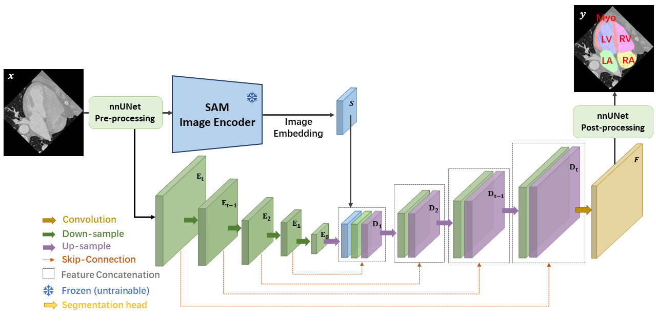
The architecture of the proposed nnSAM framework is depicted in Fig. 1. The model is designed to combine the strengths of nnUNet [8] and SAM [9]. Specifically, nnSAM consists of two parallel encoders: the nnUNet encoder and the SAM encoder. The SAM encoder is a pre-trained Vision Transformer (ViT) [13]. The embeddings from both encoders are concatenated and subsequently fed into nnUNet’s decoder to output the final segmentation map. Furthermore, the SAM encoder is used as a plug-and-play plugin whose parameters are frozen during training. Correspondingly, only the weightings of the encoder and decoder of the nnUNet are updated during the training.
2.2 Auto-configured nnUNet Architecture
Integrating nnUNet into the nnSAM allows automated network architecture and hyperparameter configuration, making it highly adaptable to the unique and specific features of each medical imaging dataset. This adaptive capability starts from a self-configuration process that automatically adjusts the nnUNet encoder’s architecture to suit training dataset characteristics including the dimensions of the medical images, the number of channels, and the number of classes involved in the segmentation task. Additionally, nnUNet uses an automated preprocessing pipeline, which includes normalizing the input data and applying data augmentation techniques such as rotations, scaling, and elastic deformations. These preprocessing and augmentation steps are crucial for improving the robustness and accuracy of the model. Beyond these, nnUNet can automatically select the most effective loss function and adjust optimizer settings based on the dataset’s inherent attributes. For example, for detected class imbalance within the dataset, nnUNet can automatically configure a weighted loss function to emphasize the minor classes. This is further supplemented by nnUNet’s hyperparameter tuning process that involves a grid search over key hyperparameters including the learning rate and the batch size. Based on each specific training dataset, nnUNet’s architecture also self-adjusts, aiming for optimal performance by dynamically modifying parameters such as the layer count and the convolutional kernel size. The comprehensive suite of auto-configurable features allows the nnUNet and correspondingly the nnSAM architecture to optimize its encoder setup for each specific medical imaging task, enhancing both its efficiency and accuracy. Since the number of layers of the nnSAM is determined by the specific dataset, in Fig. 1 we symbolize the number of encoder layers as to and the number of decoder layers as to .
Method Metrics Training Sample Size 4 8 12 16 20 UNet DICE (%) ASD (mm) SwinUNet DICE (%) 81.24 17.58 80.82 14.06 83.15 9.21 84.37 6.91 86.88 5.13 ASD (mm) 4.79 3.01 5.29 4.29 3.61 2.57 4.12 2.94 4.17 4.12 TransUNet DICE (%) 81.23 6.62 82.34 5.98 84.82 4.80 87.05 4.60 87.11 3.99 ASD (mm) 4.18 1.90 8.79 3.29 9.77 2.94 11.03 3.84 11.99 3.30 AutoSAM DICE (%) 65.10 23.62 65.89 20.58 67.63 21.77 77.55 7.55 78.53 8.30 ASD (mm) 16.55 7.83 19.60 5.78 16.98 5.96 16.73 6.02 15.92 6.36 nnUNet DICE (%) 81.77 13.68 84.45 18.72 88.36 13.00 92.35 7.55 93.15 7.86 ASD (mm) 6.97 4.87 4.90 6.08 3.15 4.74 1.56 2.26 1.40 2.20 nnSAM DICE (%) 84.67 13.52 86.36 16.19 90.74 11.89 93.20 5.53 93.75 5.35 ASD (mm) 3.87 5.04 3.29 5.15 2.18 3.97 1.43 1.69 1.23 1.64
2.3 SAM Encoder
The SAM encoder is a Vision Transformer model pre-trained on the extensive SA-1B segmentation dataset. Trained with this extremely large dataset, the SAM encoder excels at domain-agnostic feature extraction for segmentation tasks. However, its segmentation ability is highly prompt-dependent, making it unable to self-identify the segmentation target and the underlying semantics. Therefore, nnSAM only uses the SAM encoder to incorporate its feature extraction strengths, while leaving the dataset-specific task (identifying the region of interest for segmentation) to nnUNet. For an input image , where are the spatial dimensions and is the number of channels, the SAM encoder needs the input to be of size 10241024. To meet this requirement, we resize it to 10241024 using linear interpolation after the pre-processing of nnUNet. The SAM encoder produces an image embedding with dimensions 6464. We subsequently resize this embedding to match the dimensions of nnSAM’s decoder layer for concatenation (Fig. 1). To balance the inference speed of nnSAM with the segmentation accuracy, we use MobileSAM [10, 14], a lightweight SAM version that is less than 1/60 in size of the original SAM, but with comparable performance. MobileSAM is obtained by distillation from the original SAM, by which the knowledge from the original image encoder is transferred into the lightweight counterpart.
Method Sample Size DICE (%) Myo LA LV RA RV Unet 4 50.65 20.23 64.91 25.10 68.33 23.92 55.53 24.40 57.10 30.68 8 49.51 18.57 62.49 19.24 74.21 23.22 59.35 22.65 66.75 29.56 12 51.15 20.35 66.16 21.71 78.47 21.80 61.06 23.48 77.47 24.21 16 61.95 10.22 70.84 13.12 80.60 9.46 71.05 17.22 74.85 20.90 20 63.02 13.21 73.44 11.32 84.57 9.91 75.65 13.60 85.94 8.11 SwinUNet 4 67.56 21.19 83.07 20.08 90.26 9.21 79.29 22.43 86.01 17.54 8 64.56 17.34 83.43 14.34 88.02 9.57 81.18 17.75 86.92 14.92 12 65.15 17.08 85.97 10.08 88.38 7.82 85.35 8.84 90.9 5.71 16 73.19 10.44 84.55 7.78 90.88 4.51 83.27 9.72 89.94 6.05 20 76.14 9.55 87.23 6.87 92.26 3.81 87.07 6.79 91.69 4.0 TransUNet 4 67.30 9.79 86.08 11.45 89.71 4.06 81.0 9.79 82.08 6.55 8 68.16 8.19 84.53 8.04 87.56 4.59 83.52 9.80 87.94 5.85 12 70.24 8.0 85.17 9.71 89.43 4.68 88.52 6.11 90.76 3.86 16 76.61 5.95 87.51 9.34 91.63 3.38 88.08 6.65 91.42 3.16 20 77.83 5.55 85.32 10.48 92.41 3.57 88.74 4.56 91.23 2.33 AutoSAM 4 54.17 22.87 63.56 22.45 78.28 26.70 57.39 26.15 72.09 27.96 8 52.98 19.73 61.86 18.86 78.82 22.03 58.03 24.23 77.76 25.14 12 53.53 20.31 64.71 21.73 78.96 22.67 63.83 26.64 77.15 25.15 16 65.35 10.58 77.33 9.33 86.75 6.73 72.44 14.53 85.88 7.07 20 67.22 11.85 77.04 10.33 86.77 7.76 74.85 14.22 86.76 7.67 nnUNet 4 72.24 13.01 83.69 16.91 88.43 11.24 78.10 18.60 86.38 15.40 8 75.31 19.25 88.12 18.91 87.9 19.63 82.37 22.17 88.57 19.31 12 82.78 12.02 93.37 5.10 91.23 16.82 84.41 20.33 90.01 14.40 16 88.66 5.01 94.95 4.05 94.0 13.62 90.79 10.28 93.35 10.04 20 89.88 4.74 96.03 1.66 94.53 14.29 91.69 9.85 93.62 11.34 nnSAM 4 77.05 14.47 88.67 10.53 89.93 11.97 80.86 21.13 86.83 14.75 8 76.45 17.03 91.48 14.61 89.68 18.05 84.29 19.73 89.9 16.92 12 86.40 9.69 94.89 4.71 92.20 16.65 88.76 16.44 91.45 14.27 16 89.76 3.12 95.44 4.95 94.78 10.85 92.26 7.79 93.74 9.72 20 90.04 3.46 96.08 2.05 95.43 9.70 92.69 7.23 94.53 8.39
Method Sample Size ASD (mm) Myo LA LV RA RV UNet 4 16.46 6.95 17.07 7.85 20.83 8.08 27.20 11.72 16.11 9.29 8 19.33 6.43 23.09 9.35 17.35 7.02 28.35 10.84 16.0 10.98 12 13.92 5.43 20.47 8.74 12.81 5.18 28.9 12.17 13.20 9.9 16 16.52 5.73 28.38 9.26 16.67 4.24 25.13 11.38 12.25 8.91 20 16.36 5.85 22.09 9.34 13.39 5.67 26.44 12.58 12.48 11.49 SwinUNet 4 4.72 2.89 5.58 3.99 2.78 2.01 6.89 5.20 4.0 4.46 8 3.41 2.21 5.47 6.88 3.45 2.77 10.07 10.25 4.04 3.65 12 2.85 1.72 5.12 5.45 2.99 1.88 5.07 3.80 2.03 1.55 16 2.51 1.63 5.19 6.24 2.39 1.13 8.24 6.32 2.28 1.57 20 2.28 1.54 7.72 13.93 2.49 1.16 6.17 5.34 2.18 1.93 TransUNet 4 5.27 1.98 4.77 5.30 2.86 1.01 3.81 2.42 4.21 1.79 8 5.43 2.66 14.43 7.15 6.83 5.17 14.54 3.11 2.73 1.89 12 6.61 3.41 21.03 8.27 6.63 2.9 12.06 3.18 2.51 2.38 16 8.22 5.75 19.16 7.39 10.27 5.60 15.08 2.39 2.40 3.14 20 7.62 3.78 24.38 7.94 7.84 4.73 18.17 3.81 1.91 1.01 AutoSAM 4 14.18 7.62 23.89 11.01 10.33 7.67 20.66 11.12 13.68 10.81 8 19.14 6.86 26.87 9.33 15.10 5.75 25.18 11.23 11.72 7.97 12 16.31 6.92 23.55 8.9 11.91 6.02 19.42 9.73 13.72 9.98 16 14.02 5.23 26.80 9.51 11.24 5.82 22.45 9.80 9.15 7.64 20 12.24 4.69 26.81 10.99 9.11 4.43 23.80 10.81 7.62 7.49 nnUNet 4 8.19 3.39 4.26 4.77 3.98 4.11 15.38 12.83 3.06 4.50 8 3.06 4.41 5.41 9.40 3.97 5.63 8.84 11.64 3.23 5.70 12 2.09 4.13 2.36 4.40 2.43 4.19 6.56 11.39 2.33 3.66 16 1.12 1.72 1.17 2.22 1.47 2.43 2.37 3.99 1.64 2.95 20 1.01 1.60 0.78 0.27 1.50 3.31 2.08 3.27 1.60 3.53 nnSAM 4 2.73 3.37 3.43 4.46 3.47 4.22 6.61 11.05 3.13 4.48 8 2.77 3.88 2.63 6.43 3.21 4.88 5.29 8.45 2.57 4.97 12 1.71 3.22 1.07 0.84 2.34 4.21 3.65 8.38 2.16 4.31 16 1.03 1.17 1.10 2.96 1.65 2.46 1.87 2.93 1.49 2.73 20 0.9 1.00 0.78 0.34 1.25 2.20 1.77 2.86 1.48 3.22
3 Experimental Setting
We evaluated nnSAM using the MM-WHS dataset [15] for CT-based cardiac sub-structure segmentation. The preprocessed data from CFDnet [16] was utilized, which contains a collection of 212 cardiac CT images with a slice size of 240×220. The cardiac sub-structures are segmented in 2D for each slice. To evaluate nnSAM’s performance under few-shot training, we partitioned the dataset into 20 training samples, 80 validation samples, and 112 testing samples. To further evaluate nnSAM under different training dataset scarcity scenarios, we used different subsets from the training samples to train different nnSAM versions, with training sample sizes ranging from 4 to 20. This allowed us to study how the performance of nnSAM scales with the size of the available labeled data, under a simulated real-world clinical setting where labeled data might be difficult to obtain. The dataset contains labels for five different cardiac anatomy classes for a multi-class segmentation task. Specifically, the classes include the left ventricle (LV), right ventricle (RV), left atrium (LA), right atrium (RA), and myocardium of LV (Myo). Each of these classes presents its own unique challenges for segmentation, making the dataset particularly well-suited for testing the accuracy and robustness of nnSAM.
In addition to nnSAM, we also evaluated SwinUNet [7], TransUNet [5], UNet [3], the original nnUNet [8], and AutoSAM [17] for comparison. For SwinUNet, TransUNet, UNet, and nnUNet, we used publicly available codes for model training. AutoSAM has no official open-source codes, we reproduced it based on the publication descriptions. All methods were trained and tested on an NVIDIA GPU card (A100 with 80 GB memory). For the evaluation metric, we used the Average Symmetric Surface Distance (ASD) and the Dice Similarity Coefficient (DICE) [18]. ASD quantifies the average distance between the surfaces of two segmented objects and DICE evaluates their volumetric overlap.
For SwinUNet [7], TransUNet [5], UNet [3], and nnUNet [8], we have taken widely used public codes, while for AutoSAM [17], since there is no official open-source code, we have reproduced it as much as possible based on the article descriptions. All methods were trained and tested on A100 80G. For the evaluation metric, we used Average Symmetric Surface Distance (ASD) and the Dice Similarity Coefficient (DICE) [18]. The ASD is a metric that quantifies the average distance between the surfaces of two segmented objects. DICE evaluates the similarity between two segmented objects, considering the volume overlap between the two objects.
4 Results
Table 1 shows the model performance under different numbers of training data samples (4 to 20). The proposed nnSAM outperforms all other segmentation methods in terms of DICE and ASD for all training sample sizes. When trained with 20 labeled images, nnSAM achieves an average DICE score of 93.75% and an average ASD of 1.23 mm. The segmentation accuracy of nnUNet, which is recognized as one of the best segmentation models, is similarly substantially higher than the other methods, but slightly lower than that of nnSAM. The other methods, including SwinUNet, TransUNet, and AutoSAM, are of much lower accuracies, with DICEs all below 90% and ASDs all above 4 mm. The worst performance is from the vanilla UNet, which is expected as SwinUNet, TransUNet, and AutoSAM are all based on pre-trained models, while UNet is trained from scratch and is most affected by the lack of training samples. In addition, as the number of training samples gradually decreases, the advantage of nnSAM over the other methods becomes more prominent. In particular, when trained with only 4 labeled images, for DICE nnSAM outperforms the second-place nnUNet by 2.9%, and outperforms the other methods by more. Overall, nnSAM provides higher segmentation accuracy compared to the other methods, especially when the amount of training data is limited.
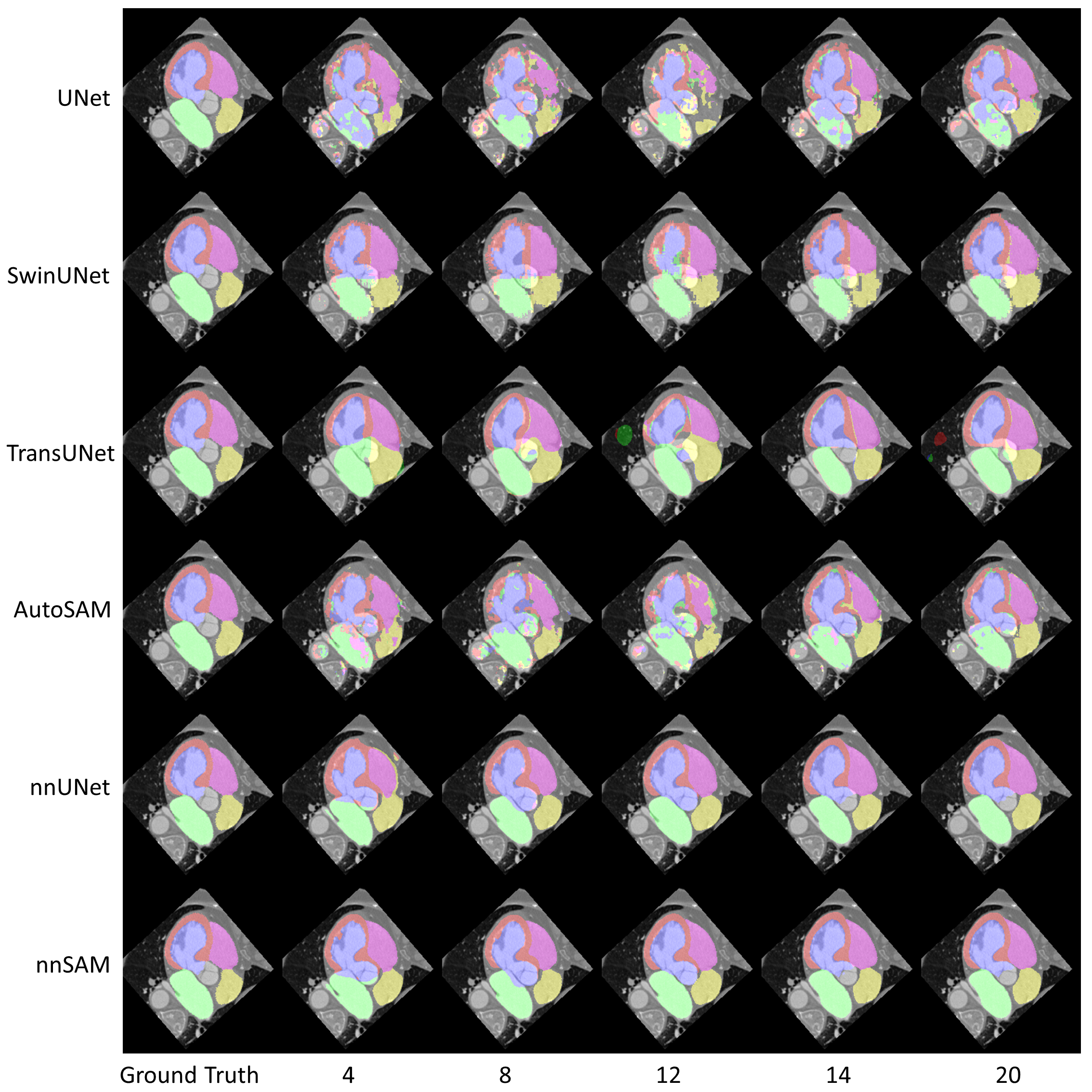
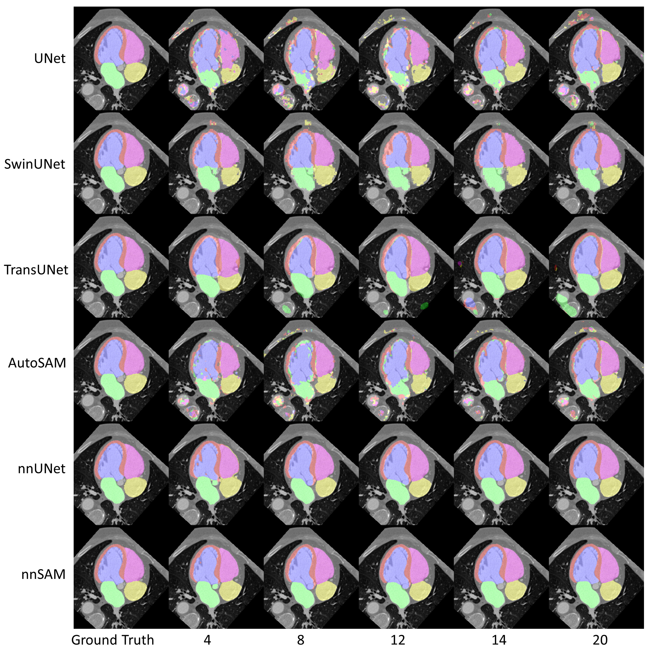
Table 2 and Table 3 show the performance of DICE and ASD under the corresponding categories of labels. In both tables, nnSAM provides the best results in most cases. As the second-ranked model, nnUNet has the closest performance to our nnSAM, however, in the visualization results of Fig. 3, nnUNet does not achieve a good segmentation for the Myo category with more false positives when the training sample size is 4. Besides, there are also some outliers with counterintuitive trends. For instance, the SwinUNet and TransUNet results on the LA label show that the ASD becomes larger when the sample data size increases. According to Fig. 2 and Fig. 3, we found that TransUNet and SwinUNet show more false positive segmentations as the sample size increases, and all these false positives are far away from the true segmentation position, leading to anomalous results in ASD. In general, UNet and AutoSAM generate poor segmentation results. The myocardium of LV (Myo) is almost unrecognizable for both methods when the training data size is limited. Since AutoSAM relies heavily on custom-trained encoders to provide automatic prompts, the ‘few-shot’ learning scenario poses challenges in learning accurate prompts for SAM segmentation, and results in poor accuracy. These results suggest that nnSAM offers superior accuracy in segmenting challenging targets with only a small number of training samples, which is attributed to the strong generality of the SAM encoder and the adaptive power of nnUNet’s auto-configurable framework.
5 Discussion
The results demonstrate the superior performance of nnSAM for medical image segmentation, especially in few-shot learning scenarios where labeled training data is limited. By integrating the pretrained SAM encoder into nnUNet’s framework, nnSAM can leverage SAM’s powerful feature extraction capabilities while simultaneously benefiting from nnUNet’s adaptive architecture configuration and hyperparater optimization. The evaluation using the MM-WHS cardiac CT dataset highlights several key advantages of nnSAM. First, nnSAM consistently achieved the highest accuracy across all sizes of training sets (4 to 20 samples), outperforming state-of-the-art models like nnUNet, SwinUNet, and TransUNet. This ability to produce accurate segmentations from very few examples could make nnSAM valuable for medical applications where acquiring labeled data is difficult and expensive. Models like SwinUNet and TransUNet showed erratic results when labeling some structures where more training samples yielded worse results, indicating they might be overfitted to the training data distribution. In contrast, nnSAM’s segmentation quality improved consistently with more training data added. Compared with nnSAM, AutoSAM uses a custom encoder to replace the prompt encoder, making it able to automatically generate and feed prompts to SAM. However, AutoSAM is not optimized for semantic segmentation of medical images as nnUNet and does not have the powerful preprocessing and auto-configuration capabilities as nnUNet either. Since the emergence of nnUNet, it has become the state-of-the-art in most medical image segmentation tasks, representing a top-of-the-line, end-to-end model for traditional task-specific semantic segmentation. SAM, on the other hand, is a prompt-based segmentation framework and a representative model with strong generalizability. Combining the best models from two different segmentation frameworks has proved effective to further improve the medical image segmentation accuracy and sets a potential new benchmark.
Our current study has some limitations that should be addressed in future work. First, we evaluated nnSAM on a single dataset of cardiac CT scans. Future studies testing it on larger and more diverse medical imaging datasets are warranted. Second, the current nnSAM framework still requires a limited amount of training data and labels, and future works are needed to explore the possibility of achieving end-to-end segmentation with only one sample (‘one-shot’ learning) or without any labeling at all (‘zero-shot’ learning). In addition, we used 2D slices for training and testing; the extension to 3D volume-based segmentation may further enhance the segmentation accuracy but is currently challenged by technical difficulties on merging 3D SAM embeddings with 3D nnUNet embeddings. Future investigations are warranted to search for potential solutions or alternatives.
6 Conclusion
We introduce nnSAM, a novel, few-shot learning solution for medical image segmentation that melds the strengths of the Segment Anything Model (SAM) and nnUNet. Our extensive evaluation across different numbers of 2D training samples sets a potential new benchmark in medical image segmentation, especially in scenarios where training data is scarce. The results also highlight the robustness and superior segmentation performance of nnSAM, making it a promising tool for future research and practical applications in medical imaging.
7 Acknowledgement
We thank Xiang Feng and Yongbo He for their helpful discussions and partial attempts at the early stages of the project.
References
- [1] Xiangbin Liu, Liping Song, Shuai Liu, and Yudong Zhang. A review of deep-learning-based medical image segmentation methods. Sustainability, 13(3):1224, 2021.
- [2] Berkman Sahiner, Aria Pezeshk, Lubomir M Hadjiiski, Xiaosong Wang, Karen Drukker, Kenny H Cha, Ronald M Summers, and Maryellen L Giger. Deep learning in medical imaging and radiation therapy. Medical physics, 46(1):e1–e36, 2019.
- [3] Olaf Ronneberger, Philipp Fischer, and Thomas Brox. U-net: Convolutional networks for biomedical image segmentation. arXiv preprint arXiv:1505.04597, 2015.
- [4] Yunxiang Li, Shuai Wang, Jun Wang, Guodong Zeng, Wenjun Liu, Qianni Zhang, Qun Jin, and Yaqi Wang. Gt u-net: A u-net like group transformer network for tooth root segmentation. In Machine Learning in Medical Imaging: 12th International Workshop, MLMI 2021, Held in Conjunction with MICCAI 2021, Strasbourg, France, September 27, 2021, Proceedings 12, pages 386–395. Springer, 2021.
- [5] Jieneng Chen, Yongyi Lu, Qihang Yu, Xiangde Luo, Ehsan Adeli, Yan Wang, Le Lu, Alan L. Yuille, and Yuyin Zhou. Transunet: Transformers make strong encoders for medical image segmentation. arXiv preprint arXiv:2102.04306, 2021.
- [6] Zongwei Zhou, Md Mahfuzur Rahman Siddiquee, Nima Tajbakhsh, and Jianming Liang. Unet++: Redesigning skip connections to exploit multiscale features in image segmentation. IEEE transactions on medical imaging, 39(6):1856–1867, 2019.
- [7] Hu Cao, Yueyue Wang, Joy Chen, Dongsheng Jiang, Xiaopeng Zhang, Qi Tian, and Manning Wang. Swin-unet: Unet-like pure transformer for medical image segmentation. arXiv preprint arXiv:2105.05537, 2021.
- [8] Fabian Isensee, Paul F Jaeger, Simon A A Kohl, Jens Petersen, and Klaus H Maier-Hein. nnu-net: a self-configuring method for deep learning-based biomedical image segmentation. Nature Methods, 17(2):203–211, 2020.
- [9] Alexander Kirillov, Eric Mintun, Nikhila Ravi, Hanzi Mao, Chloe Rolland, Laura Gustafson, Tete Xiao, Spencer Whitehead, Alexander C Berg, Wan-Yen Lo, et al. Segment anything. arXiv preprint arXiv:2304.02643, 2023.
- [10] Chaoning Zhang, Dongshen Han, Yu Qiao, Jung Uk Kim, Sung-Ho Bae, Seungkyu Lee, and Choong Seon Hong. Faster segment anything: Towards lightweight sam for mobile applications. arXiv preprint arXiv:2306.14289, 2023.
- [11] Maciej A Mazurowski, Haoyu Dong, Hanxue Gu, Jichen Yang, Nicholas Konz, and Yixin Zhang. Segment anything model for medical image analysis: an experimental study. Medical Image Analysis, 89:102918, 2023.
- [12] Jun Ma, Yuting He, Feifei Li, Lin Han, Chenyu You, and Bo Wang. Segment anything in medical images. arXiv preprint arXiv:2304.12306, 2023.
- [13] Alexey Dosovitskiy, Lucas Beyer, Alexander Kolesnikov, Dirk Weissenborn, Xiaohua Zhai, Thomas Unterthiner, Mostafa Dehghani, Matthias Minderer, Georg Heigold, Sylvain Gelly, et al. An image is worth 16x16 words: Transformers for image recognition at scale. arXiv preprint arXiv:2010.11929, 2020.
- [14] Kan Wu, Jinnian Zhang, Houwen Peng, Mengchen Liu, Bin Xiao, Jianlong Fu, and Lu Yuan. Tinyvit: Fast pretraining distillation for small vision transformers. In European Conference on Computer Vision, pages 68–85. Springer, 2022.
- [15] Xiahai Zhuang, Lei Li, Christian Payer, Darko Štern, Martin Urschler, Mattias P Heinrich, Julien Oster, Chunliang Wang, Örjan Smedby, Cheng Bian, et al. Evaluation of algorithms for multi-modality whole heart segmentation: an open-access grand challenge. Medical image analysis, 58:101537, 2019.
- [16] Fuping Wu and Xiahai Zhuang. Cf distance: a new domain discrepancy metric and application to explicit domain adaptation for cross-modality cardiac image segmentation. IEEE Transactions on Medical Imaging, 39(12):4274–4285, 2020.
- [17] Tal Shaharabany, Aviad Dahan, Raja Giryes, and Lior Wolf. Autosam: Adapting sam to medical images by overloading the prompt encoder. arXiv preprint arXiv:2306.06370, 2023.
- [18] Yunxiang Li, Guodong Zeng, Yifan Zhang, Jun Wang, Qun Jin, Lingling Sun, Qianni Zhang, Qisi Lian, Guiping Qian, Neng Xia, et al. Agmb-transformer: Anatomy-guided multi-branch transformer network for automated evaluation of root canal therapy. IEEE Journal of Biomedical and Health Informatics, 26(4):1684–1695, 2021.