Delta-alpha cross-frequency coupling for different brain regions
Abstract
Neural interactions occur on different levels and scales. It is of particular importance to understand how they are distributed among different neuroanatomical and physiological relevant brain regions. We investigated neural cross-frequency couplings between different brain regions according to the Desikan-Killiany brain parcellation. The adaptive dynamic Bayesian inference method was applied to EEG measurements of healthy resting subjects in order to reconstruct the coupling functions. It was found that even after averaging over all subjects, the mean coupling function showed a characteristic waveform, confirming the direct influence of the delta-phase on the alpha-phase dynamics in certain brain regions and that the shape of the coupling function changes for different regions. While the averaged coupling function within a region was of similar form, the region-averaged coupling function was averaged out, which implies that there is a common dependence within separate regions across the subjects. It was also found that for certain regions the influence of delta on alpha oscillations is more pronounced and that oscillations that influence other are more evenly distributed across brain regions than the influenced oscillations. When presenting the information on brain lobes, it was shown that the influence of delta emanating from the brain as a whole is greatest on the alpha oscillations of the cingulate frontal lobe, and at the same time the influence of delta from the cingulate parietal brain lobe is greatest on the alpha oscillations of the whole brain.
The delta-alpha cross-frequency coupling is proving to be a valuable descriptor in increasingly more brain states and domains. Here, by applying the adaptive dynamic Bayesian inference to EEG signals of subjects at rest we reconstructed the neural cross-frequency delta to alpha coupling functions that describe the interaction mechanisms of different regions of the brain. With this analysis framework we found a number of significant brain connections, as well as several characteristic differences between the brain regions.
I Introduction
The interactions in the brain are fundamental to the human ability to perceive and interact with the world. The brain is a heavily connected dynamical network system [1], with interactions that are very complex and involve a vast network of neurons and synapses. Such complex system can mediate a vast number of functions, from a relatively static structure. Importantly, the brain can evolve with time, and different changes and transitions can occur [2, 3]. Because not all the neurons and network processes in the brain are active at all time, and because they can exhibit collective, clustered, and synchronized behaviour [4, 5, 6], different types of changes, disruptions and transitions in the neural activity can occur [7, 8].
Since the functions of the brain are highly dependent on its structure, and different functions are probably performed by different brain regions with different architecture, it is essential to identify the different regions of the brain in order to better understand its functions. For that reason, a significant effort has been invested by the scientific community in the direction of parcellation of the brain, starting from the classic Brodmann map, through the widely used Desikan-Killiany atlas [9], all the way to the recently published human Brainnetome atlas [10] and Human Connectome Project (HCP) multimodal parcellation [11] using in vivo MRI data.
The brain connectivity is crucial to understand how the neurons and the brain dynamics evolve. A particularly accessible and useful approach has been the study of neural cross-frequency coupling, usually extracted from an electroencephalograph (EEG) recording [12, 13, 14, 15, 16]. Neural cross-frequency coupling refers to the interaction between different frequencies of neural brainwave oscillations in the brain. Cross-frequency coupling occurs when the amplitude or phase of one frequency band of oscillations is modulated by another frequency band. Thus, there are different types of cross-frequency coupling, such as amplitude-amplitude coupling, phase-phase coupling, and amplitude-phase coupling.
Neural cross-frequency coupling can be studied between different combinations of brainwave oscillations. In this work, we will focus on the delta-to-alpha neural cross-frequency coupling. Namely, it is well known that delta and alpha brainwave oscillations play an important role in the brain dynamics [17, 18, 19, 20, 21, 22]. For instance, there are differences in frequency and power during different sleep stages which appear in the separate delta and alpha brainwave dynamics [23, 24, 25, 26] and in their related delta-alpha effect [27, 28]. In another example, in a previous study about general anesthesia [29] it was found that the delta-alpha coupling function is statistically significant and strong during anaesthesia. Similarly, previous works observe a prominent delta-alpha coupling in resting state [15, 30], during the orienting response [32] and during sleep within the network physiology approach [20]. A characteristic form of the delta-alpha coupling functions was also established [29, 30, 31]. These works point out that the choice to investigate the delta-to-alpha coupling among the brain regions had a relevance for the present study of resting state.
To perform the analysis needed we used comprehensive set of methods. First, to observe the oscillatory content of the brainwave oscillations we used wavelet time-frequency analysis. Then, we used the fact that the delta and alpha brainwaves have pronounced oscillating dynamics in order to study the interactions through their reduced phase dynamics [33], thus observing phase-phase cross-frequency coupling. Here, we applied a method based on adaptive dynamical Bayesian inference for analysis of data to reconstruct a dynamical phase model describing the systems and their interactions [34, 35, 36]. The method of dynamical inference reconstructs effective connectivity [37, 1] and reveals the underlying dynamical mechanisms. Here, we reconstruct the phase coupling functions which describe how the interaction occurs and manifest, thus revealing a functional mechanism [38]. The design of powerful methods and the explicit assessment of coupling functions have led to applications in different scientific fields including chemistry [39], climate [40], secure communications [41], mechanics [42], social sciences [43], and oscillatory interaction in physiology for cardiorespiratory interactions [44, 45]. Arguably, the greatest recent interest for coupling functions is coming from neuroscience [46], where works have encompassed the theory and inference of a diversity of neural phenomena, physical regions, and physiological conditions [47, 48, 49, 50, 51, 30, 52, 53].
II Materials and Methods
II.1 Adaptive Dynamical Bayesian Inference
When investigating a complex dynamical oscillatory system, such as the oscillatory behaviour of the brain, one way to gain new insights is by modeling the system by using differential equations. Usually, by measuring some signals originating from the oscillatory time evolution of the system, one can infer the parameters of a model that describes the system under certain conditions. According to the phase reduction theory, in case when the interactions between the oscillators are sufficiently weak, the behaviour of the system can be approximated with its phase dynamics [33, 54, 55]. If the phases of the system can be considered as monotonic change of the variables, the partial dynamical process of the node as influenced by another node can be represented with the system of differential equations:
| (1) |
where is the phase of the i-th oscillator, is its angular frequency parameter, is the coupling function which describes the influence of the j-th oscillator on the i-th oscillator, and represents the noise. Usually, the noise is assumed as a white Gaussian noise given by , where the information about the correlation between the noises of the different oscillators is included in the symmetric matrix . In theory, the full model for the phase dynamics of a brain region oscillator should contain all the connections at once, in a single phase equation. However, due to the high dimensionality and computational expense, with equation (1) we infer a partial part of the full model dynamics related only to the two brain regions involved in a coupling connection. This procedure is then applied for each pair of brain regions.
Because of the periodic nature of the system, the coupling function can be represented by a Fourier decomposition:
| (2) |
For a system of two coupled oscillators, reduction to a finite number K of Fourier terms will give:
| (3) |
where, and the rest and are the most important Fourier components (in this work we use ). With the assumption for a white Gaussian noise, the task is then reduced to inference of the unknown parameters of the model:
| (4) |
When the parameters of the model are inferred, one can determine then the coupling functions which describe the underlying mechanisms of the interaction of the oscillators [38].
In this work we employ the method of adaptive dynamical Bayesian inference (aDBI) [35, 56, 57, 36] in order to gain new insights into the oscillatory behaviour of the brain regions and the brain lobes. In this method, the time series of phases of the oscillators are considered to be time sequences of blocks of samples. In each block, the samples from a certain time interval are included, and the duration of this time interval is specified by the time window . In the inference procedure, the initial assumptions for the parameters of the model are that , and therefore at least few inference blocks are required to obtain appropriate estimates of the model parameters values and the corresponding coupling functions. To obtain improved inference in every subsequent block, part of the information from the previous block is included in the following one. The so-called propagation parameter controls how much of the information of the previous block is included in the following one. In the method of aDBI both the time window and the propagation parameter are adaptively determined, based on the time variabilities present in the signal. After determination of and the final inference is performed. This final inference provides the values of the parameters and the coupling functions for each block of inference, thus observing the time evolution of the system and the inter interactions, with a temporal resolution defined by the time window . Further technical details about the parametrization, convergence and robustness of the aDBI method can be found in previous papers [56, 57, 36, 35]. Even though the aDBI was introduced for studies of coupled phase oscillators with oscillating frequencies in the cardiorespiratory range, the procedures is applicable to the frequency range of the brain waves as well. The application of the aDBI method on the subject dataset that was used in this study yielded a time window of s and a propagation parameter of .
The aDBI method represents a further improvement of the DBI method [35, 56], by minimizing the covariance matrix which is an indicator of the quality of the inference. The details of the method are given elsewhere [36] and in summary it leads to an improved inference of the model parameters without losing information about the temporal changes in the behavior of the oscillators.

II.2 Dataset
The dataset used in this study is publicly available [58] (https://osf.io/mndt8/). The data source contains the empirical region-average fMRI (functional magnetic resonance imaging), EEG source activity and structural connectomes of the parcellated cortical regions of the brain of 15 healthy human subjects, age 18-31, eight of whom are female.The data consists of a resting state time series, where the subjects were asked to just stay awake and keep their eyes closed. In this study we used the empirical EEG source activity and the structural connectomes. The time series of the source activity for each patient and each cortical region have duration of minutes with sampling frequency of Hz. The description of the parcellated cortical regions is given in the Appendix.
II.3 Wavelet transform and the phase extraction procedure
In order to check the existence of brain wave oscillations and their frequency content, the EEG time-series signals were first analyzed using a continuous wavelet transform [59, 60, 61]. The continuous wavelet transform is defined by the equation
| (5) |
where is the signal, denotes the angular frequency, is the time and
is the complex Morlet wavelet, with central frequency , , and with being the imaginary unit. The continuous wavelet transform is a time-frequency representation which contains both the phase and the amplitude dynamics of the oscillatory elements from the analyzed signal.
The initial wavelet observation of the oscillations contained in the corresponding EEG signals was carried out for several brain regions. After the initial wavelet observations, a phase extraction procedure was performed for the delta and alpha waves of the EEG signal in order to obtain the instantaneous phase time-series. These phase time-series then act as an input to the aDBI method. The oscillatory intervals were first evaluated by standard digital filtering procedure including FIR filter followed by a zero phase filtering procedure (“filtfilt") to ensure that no time or phase lags are introduced with the filtering procedure. The delta waves signal limits were from 1 to 4 Hz, while the alpha waves signal limits were from 8 to 12 Hz [62].
The phases of the filtered signals were estimated via Hilbert transformation, thus obtaining the protophases. On these protophases, the protophase-to-phase transformation was applied [42] in order to obtain the independent phases which act as input signals for the
Bayesian inference.
II.4 Surrogate data testing
When oscillatory signals are analyzed, the inferred coupling between the signals is always positive and non-zero, even if the oscillators are uncoupled or unrelated. Therefore, it is necessary to establish a significance threshold in order to determine if the obtained coupling strength indicates a genuine connection and interdependence of the phenomena. Such surrogate data is used for statistical testing of the coupling strength. A threshold is usually defined by constructing randomized surrogates of the original signals [63, 64] and calculating the coupling strength for these surrogates. The coupling strength obtained in this manner represent a baseline for the confirmation of the coupling of the oscillators.
In this work surrogates were constructed for each of the delta-alpha couplings going from, and to, each of the regions of the brain by using a procedure called cyclic phase permutation surrogates [64] based on rearrangement of the cycles within the extracted phase. The surrogate threshold taken in this work is the mean plus two standard deviations (mean + 2STD) of the surrogate couplings.
III Results
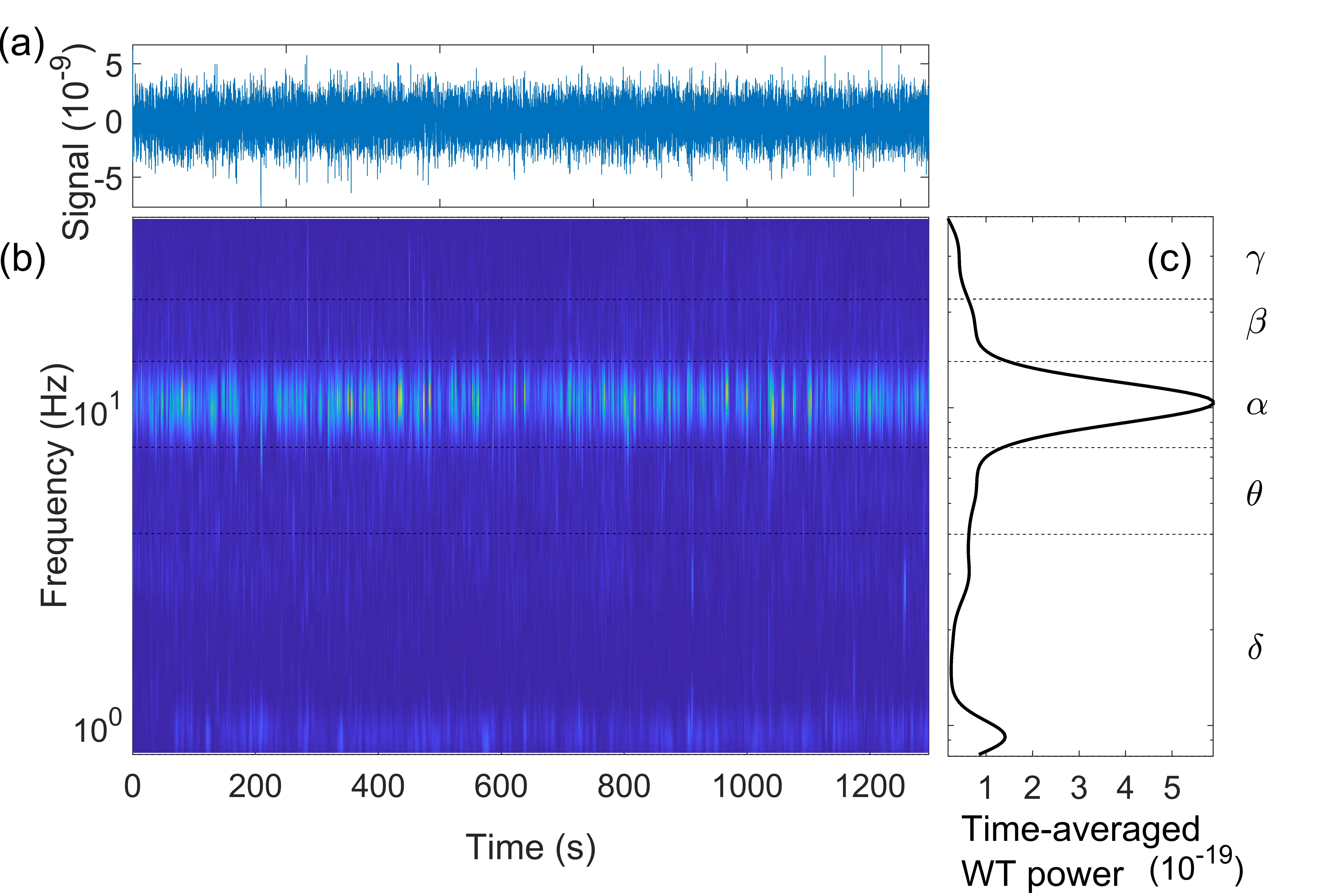
Fig. 1 shows the wavelet transform of the measured signal for one of the brain regions in one of the subjects. The signal itself is presented in Fig. 1 (a), while the corresponding time-frequency wavelet transform is shown in Fig. 1 (b). To show the oscillatory frequencies present in the signal more clearly, the time-averaged intensity of the wavelet is presented in Fig. 1 (c). The frequency intervals of the corresponding brain waves are given with the dashed lines. From the figure, one can clearly see the strong alpha wave, as well as the delta wave with a slightly lower wavelet power.
The delta-alpha coupling functions are presented in Fig. 2. The coupling functions are evaluated first on individual subjects for specific regions – Fig. 2 (a)-(c). Here, they show the characteristic functional form where the delta-alpha phase coupling function depends predominately on the delta dynamics, or in other words it reflects the direct influence that the delta phase dynamics exert on the alpha phase dynamics by accelerating or decelerating the alpha brainwave oscillations. This specific form belongs to the category of direct, among the separation of self, direct and common coupling functions [65, 67]. By comparing the three plots for the coupling functions Fig. 2 (a), (b) and (c), one can notice that this direct influence is like a wave that shifts from left to right from to along the delta axis. In general, it keeps the direct delta dependence (i.e. it still changes predominately on delta axis) but it shifts the maximum of the function along the delta axis.
When we average the coupling functions for the same region from all the subjects, as shown in Fig. 2 (d), the remaining average delta-alpha coupling function still reflects the direct delta dependence, albeit with slightly reduced amplitude due to averaging. The Appendix A shows how this coupling function is similar or different in respect to all the order regions. Furthermore, when we average all the coupling functions across regions and subjects, the average coupling function disappeared i.e. it was insignificantly low without a common form of the function. In other words the region average coupling function averaged out, because there was no specific common form between regions.
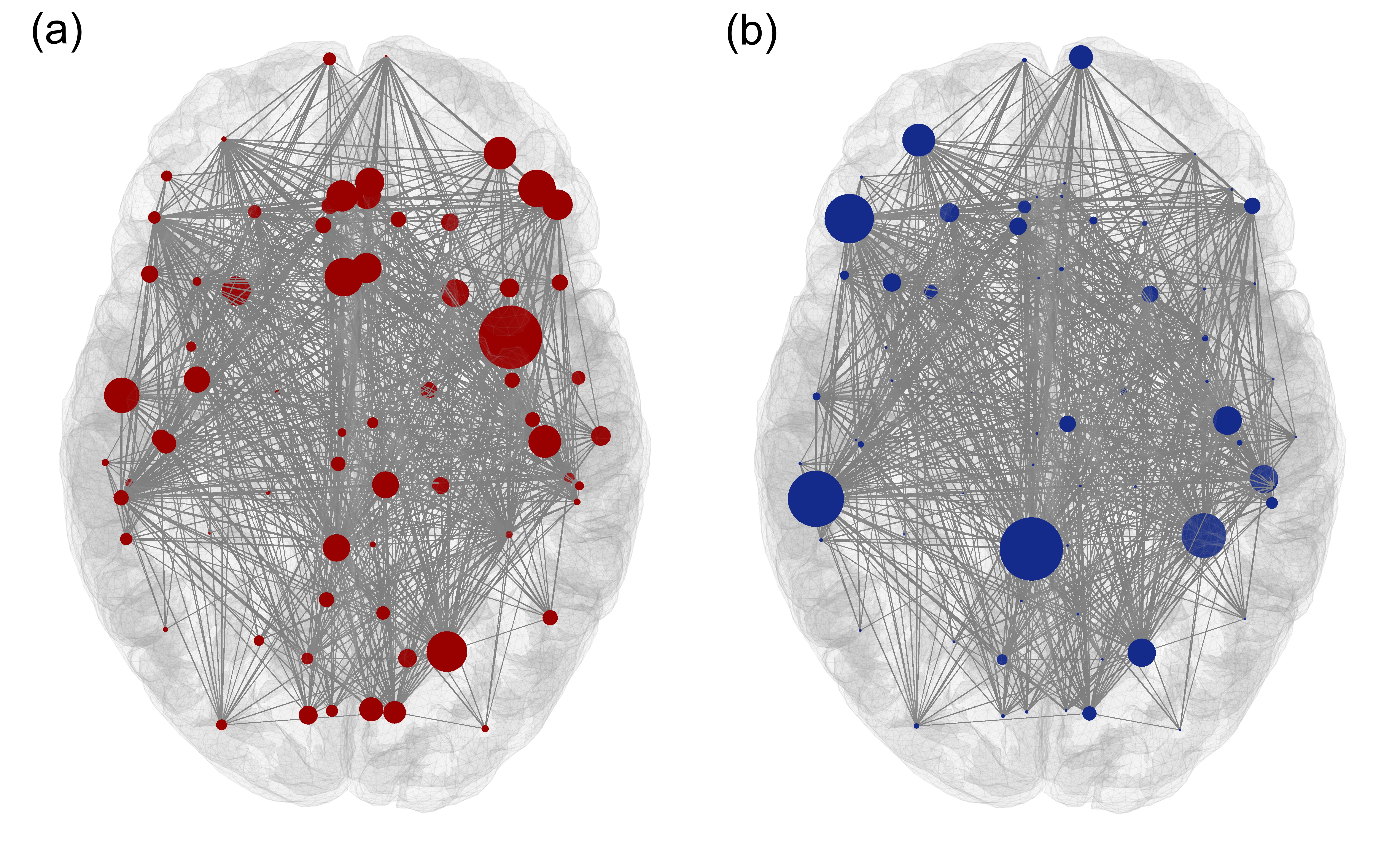
Fig. 3 shows a matrix representing the significant delta-alpha coupling functions for all the brain regions. The vertical axis shows the number of the region for the delta brainwaves, while on the horizontal axis the number of the region of the alpha brainwaves is given. The matrix is not symmetrical and the coupling indicated by the columns is different than the one in the rows. The figure indicates that for some regions of the brain (e.g. columns , etc) there is a stronger influence from the delta waves to the alpha waves. This is also shown in Fig. 4, where these regions are marked as circles on a cross-section of the brain.
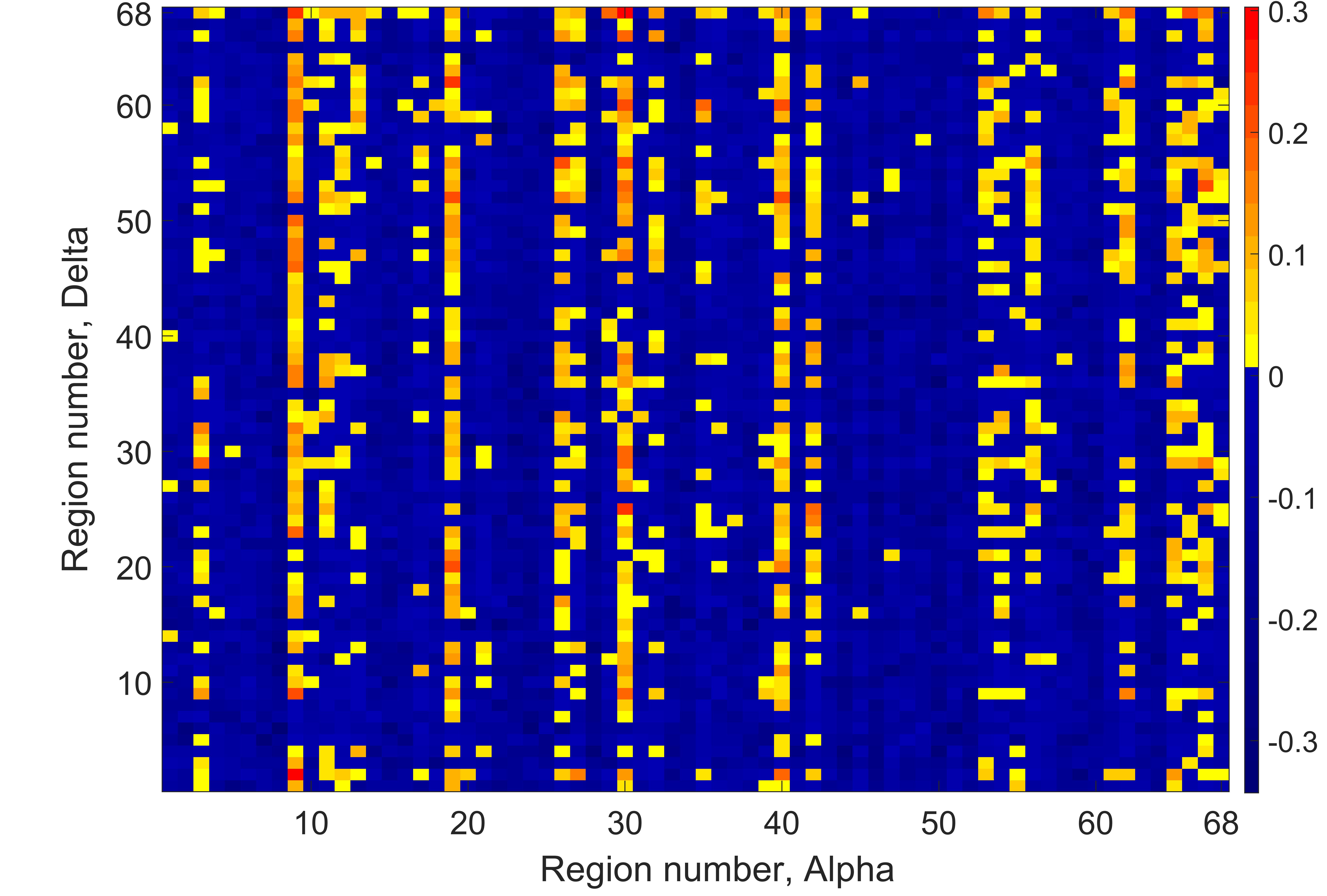
Fig. 4 (a) shows the summarized delta-alpha coupling strengths coming from a specific regions with red circles, while Fig. 4 (b) shows the sum of the delta-alpha coupling strengths for the alpha of a specific regions with blue circles. The radii of the circles are proportional to the sum of the corresponding coupling strengths. One can notice that while the significant delta-alpha couplings emanate from various different brain regions, they end up in much smaller number of the brain regions.
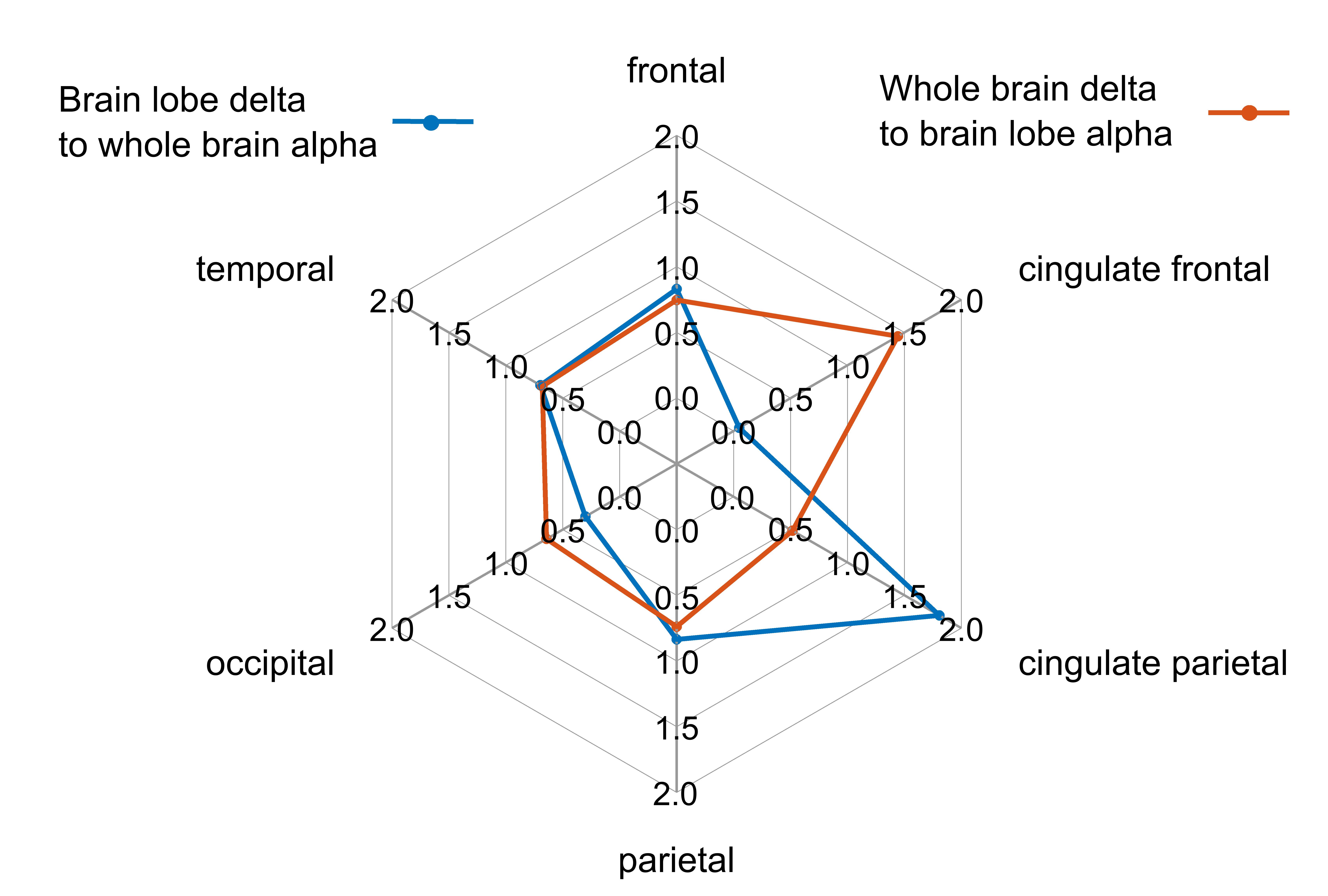
In order to obtain more tangible information about the overall interactions between the different brain lobes (frontal, cingulate frontal, cingulate parietal, parietal, occipital and temporal lobe), a summation of the significant coupling functions by brain lobes was performed. The sums obtained were normalized by the number of regions involved in each of the brain lobes. The results are presented in Fig. 5. The normalized sum of the delta-alpha couplings, where the delta is from specific brain lobe and alpha from any lobe of the brain (whole brain alpha), is shown with blue line. While the normalized sum of the delta-alpha couplings, where the delta comes from any lobe of the brain (whole brain delta) and alpha from specific brain lobe, is shown with red line. From the spider plot (Fig. 5) it can be seen that the greatest influence on the whole brain alpha has the cingulate parietal delta and at the same time the greatest influence of the whole brain delta is on the cingulate frontal alpha.
IV Discussion and Conclusions
The influence of delta brain waves on alpha brain waves for a resting subject has previously been determined at the whole brain level. In this paper we try to gain a deeper insight by investigating this delta-alpha influence for different brain regions according to the Desikan-Kiliany anatomical parcellation of the brain [30, 15, 32, 20]. As presented in the results, it can be concluded that this influence is clearly visible for different regions, because even after averaging the delta-alpha coupling functions for a particular region across all the subjects, the mean coupling function still shows the characteristic shape (Fig. 2), confirming the direct influence of the delta-phase dynamics on the alpha-phase dynamics in certain brain regions. This influence consists in acceleration or deceleration of alpha oscillations under the influence of delta oscillations.
In terms of analysis we have applied a comprehensive methodological framework for interacting brainwave oscillations. The nature of delta and alpha oscillations were observed with wavelet transform using standard parameters, with central frequency. This is a simple standard widely used procedure for time-frequency analysis. For the reconstruction of the phase model we used the adaptive Bayesian inference. It is a well established method which has been widely used and tested for robustness and convergence, where its parametrization has been systematically investigated on different numerical and biological systems [56, 57, 36, 35]. For verifying the statistical significance of the inferred delta-alpha coupling we have applied surrogate data testing [64].
The model equation (1) assumes pairwise interaction between two regions and includes only coupling function with two phase variables. This is a simplified approximation, as the brain regions form parts of a complex network, and the full model of a phase oscillator should include all the brain connections in a single equation. With equation (1) we have thus separated the inference for a partial dynamics on two-by-two basis for all the pairs of brain regions. This was possible because the Bayesian method can allow such partial dynamical filtering. While the reason to do this and separate the inference was due to the high dimensionality (68x68 regions) and the computational complexity, which otherwise could have lead to problems such as parameters overfitting. Also, we have used only pairwise coupling functions. Thus, a natural extension of this work could include also non-pairwise multivariate coupling functions [66, 65]. This is a case where the coupling function in the dynamics of one region will have more than two phase variables from phases of other regions.
The coupling function results demonstrated that there is a common waveform, predominantly due to direct influence from delta oscillations, but the wave shifts along the delta axis for different regions – Fig. 2 (a-c). We present three characteristic regions here, but the general observation from all the regions was that the wave was shifting for different regions. The quantitative analysis in Appendix A also supported this by showing relative variations of the form for different regions. The answer to why the wave for coupling function forms shifts for different regions might be because there are different lengths for the structural pathways, through which different regions interact. This most likely implies different time delays for the signal propagation [68, 69], which is known to impact the synchronization and phase arrangement between brain regions [70] and is crucial for the information transfer [71]. This time delay manifests itself as a phase shift for the oscillatory activity, i.e. within the phase coupling functions (e.g. as in ), which in turn can be the cause of the phase shift of the wave observed in the figures. Our current initial observation of the structural and time delay information in this direction can stimulate future systematic analysis for quantifying how the space-time structure of the brain regions, as defined by the weights and time-delays of the connectome [72], impacts the resulting coupling functions. Such a question is even more valid because the averaged coupling function within a region was of similar form Fig. 2 (d), while the region-averaged coupling function was averaged out. The latter implies that there is a common deterministic dependence within regions across the subjects, which is different for the separate brain regions. This kind of analysis would first require better identification of the propagation velocities and the time-delays on a personalized level, which is still not established beside promising results of MRI as a myelin biomarker [73], and proposed in vivo techniques [68]. However, our results indicate that even aggregated atlases for time-delays [69] could be useful, since some of the patterns are consistent across the subjects.
We have seen that this influence is not evenly distributed across brain regions, but for certain brain regions the influence of delta oscillations on alpha oscillations is more pronounced, as is the case for isthmus cingulate, pars triangularis (associated with verbal and non-verbal communication [74, 75, 76]) and the supramarginal region of the left hemisphere (involved in language processing [77, 78] and tool use action [79, 80]) and the fusiform region of the right hemisphere (involved in object and face recognition [81, 82, 83]). To lesser extent this is also noticeable for the lateral orbitofrontal and the rostral middle frontal region of the left hemisphere and the inferior temporal, pars triangularis, posterior cingulate, superior parietal, frontal pole, temporal pole and transverse temporal region of the right hemisphere (see Fig.3). These regions are located in different lobes of the brain, most of them in the frontal and temporal lobes, but some in the parietal and cingulate parietal as well. No clear distinctions can be made in terms of the brain hemisphere, as is expected, since the different brain centers responsible for different actions are located in the different hemispheres.
This uneven distribution of delta influence on alpha oscillations from different regions is less pronounced on the delta side of the regions, as shown in Fig. 3 and more specifically in Fig. 4 (a). Fig. 4 shows that while the delta-alpha influence is more concentrated for the alpha waves in certain regions, the distributions of significant couplings in terms of delta waves is more even across brain regions. This means that the influencing oscillations are more evenly distributed across the brain regions then the influenced oscillations, which are more concentrated in certain regions.
Additional insights into delta-alpha influences in the brain can be gained by condensing this information down to the level of brain lobes, as shown in the spider plot (Fig. 5). These results indicate that the influence of delta oscillations of all brain regions is greatest on alpha oscillations of the cingulate frontal lobe. At the same time, the influence of cingulate parietal brain lobe’s delta oscillations on the alpha oscillations is greatest among all the regions of the brain. This influence of the cingulate frontal and cingulate parietal regions of the brain on other brain regions and on the brain as a whole should be further investigated and put into the context of the functioning of the brain from a neurological point of view.
Finally, it is worth noting that we presented the methodological framework for interactions in the brain regions network for the resting state, however, the framework carries important implications and can readily be used also for other neural states, or interacting oscillatory networks more generally.
Acknowledgements.
D.L., T.S. and P.J. acknowledge support from the bilateral Macedonian - Chinese project for scientific and technological cooperation. P.J. acknowledge support from STI2030-Major Projects (2021ZD0204500), the NSFC (62076071), and Shanghai Municipal Science and Technology Major Project (2018SHZDZX01).Data Availability Statement
The data that support the findings of this study are publicly available.
V REFERENCES
References
- Park and Friston [2013] H.-J. Park and K. Friston, “Structural and functional brain networks: from connections to cognition,” Science 342, 1238411 (2013).
- Lehnertz et al. [2014] K. Lehnertz, G. Ansmann, S. Bialonski, H. Dickten, C. Geier, and S. Porz, “Evolving networks in the human epileptic brain,” Physica D 267, 7–15 (2014).
- Chai et al. [2017] L. R. Chai, A. N. Khambhati, R. Ciric, T. M. Moore, R. C. Gur, R. E. Gur, T. D. Satterthwaite, and D. S. Bassett, “Evolution of brain network dynamics in neurodevelopment,” Network Neuroscience 1, 14–30 (2017).
- Rudrauf et al. [2006] D. Rudrauf, A. Douiri, C. Kovach, J. P. Lachaux, D. Cosmelli, M. Chavez, C. Adam, B. Renault, J. Martinerie, and M. L. Van Quyen, “Frequency flows and the time-frequency dynamics of multivariate phase synchronization in brain signals,” NeuroImage 31, 209–227 (2006).
- Li et al. [2022] Q. Li, T. Peron, T. Stankovski, and P. Ji, “Effects of structural modifications on cluster synchronization patterns,” Nonlinear Dynamics 108, 3529–3541 (2022).
- Sauseng et al. [2008] P. Sauseng, W. Klimesch, W. R. Gruber, and N. Birbaumer, “Cross-frequency phase synchronization: a brain mechanism of memory matching and attention,” Neuroimage 40, 308–317 (2008).
- Suo et al. [2015] X. Suo, D. Lei, K. Li, F. Chen, F. Li, L. Li, X. Huang, S. Lui, L. Li, G. J. Kemp, et al., “Disrupted brain network topology in pediatric posttraumatic stress disorder: A resting-state fmri study,” Human brain mapping 36, 3677–3686 (2015).
- Olde Dubbelink et al. [2014] K. T. Olde Dubbelink, A. Hillebrand, D. Stoffers, J. B. Deijen, J. W. Twisk, C. J. Stam, and H. W. Berendse, “Disrupted brain network topology in parkinson’s disease: a longitudinal magnetoencephalography study,” Brain 137, 197–207 (2014).
- Desikan et al. [2006] R. S. Desikan, F. Ségonne, B. Fischl, B. T. Quinn, B. C. Dickerson, D. Blacker, R. L. Buckner, A. M. Dale, R. P. Maguire, B. T. Hyman, M. S. Albert, and R. J. Killiany, “An automated labeling system for subdividing the human cerebral cortex on mri scans into gyral based regions of interest,” NeuroImage 31, 968–980 (2006).
- Fan et al. [2016] L. Fan, H. Li, J. Zhuo, Y. Zhang, J. Wang, L. Chen, Z. Yang, C. Chu, S. Xie, A. R. Laird, et al., “The human brainnetome atlas: a new brain atlas based on connectional architecture,” Cerebral cortex 26, 3508–3526 (2016).
- Glasser et al. [2016] M. F. Glasser, T. S. Coalson, E. C. Robinson, C. D. Hacker, J. Harwell, E. Yacoub, K. Ugurbil, J. Andersson, C. F. Beckmann, M. Jenkinson, et al., “A multi-modal parcellation of human cerebral cortex,” Nature 536, 171–178 (2016).
- Canolty et al. [2006] R. T. Canolty, E. Edwards, S. S. Dalal, M. Soltani, S. S. Nagarajan, H. E. Kirsch, M. S. Berger, N. M. Barbaro, and R. T. Knight, “High gamma power is phase-locked to theta oscillations in human neocortex,” Science 313, 1626–1628 (2006).
- Jensen and Colgin [2007] O. Jensen and L. L. Colgin, “Cross-frequency coupling between neuronal oscillations,” Trends Cognit. Sci. 11, 267–269 (2007).
- Palva, Palva, and Kaila [2005] J. M. Palva, S. Palva, and K. Kaila, “Phase synchrony among neuronal oscillations in the human cortex,” J. Neurosci. 25, 3962–3972 (2005).
- Jirsa and Müller [2013] V. Jirsa and V. Müller, “Cross-frequency coupling in real and virtual brain networks,” Frontiers Comput. Neurosci. 7, 78 (2013).
- Sorrentino et al. [2022a] P. Sorrentino, M. Ambrosanio, R. Rucco, J. Cabral, L. L. Gollo, M. Breakspear, and F. Baselice, “Detection of cross-frequency coupling between brain areas: An extension of phase linearity measurement,” Frontiers in Neuroscience 16 (2022a).
- Delimayanti et al. [2020] M. K. Delimayanti, B. Purnama, N. G. Nguyen, M. R. Faisal, K. R. Mahmudah, F. Indriani, M. Kubo, and K. Satou, “Classification of brainwaves for sleep stages by high-dimensional fft features from eeg signals,” Applied Sciences 10, 1797 (2020).
- Gorgoni et al. [2020] M. Gorgoni, S. Scarpelli, F. Reda, and L. De Gennaro, “Sleep eeg oscillations in neurodevelopmental disorders without intellectual disabilities,” Sleep Medicine Reviews 49, 101224 (2020).
- Krueger et al. [2016] J. M. Krueger, M. G. Frank, J. P. Wisor, and S. Roy, “Sleep function: Toward elucidating an enigma,” Sleep medicine reviews 28, 46–54 (2016).
- Bashan et al. [2012] A. Bashan, R. P. Bartsch, J. W. Kantelhardt, S. Havlin, and P. C. Ivanov, “Network physiology reveals relations between network topology and physiological function,” Nat. Commun. 3, 702 (2012).
- Penzel et al. [2007] T. Penzel, M. Hirshkowitz, J. Harsh, R. D. Chervin, N. Butkov 5, M. Kryger, B. Malow, M. V. Vitiello, M. H. Silber, C. A. Kushida, et al., “Digital analysis and technical specifications,” Journal of clinical sleep medicine 3, 109–120 (2007).
- Palva and Palva [2007] S. Palva and J. M. Palva, “New vistas for -frequency band oscillations,” Trends Neurosci. 30, 150–158 (2007).
- Ehlers and Kupfer [1989] C. L. Ehlers and D. J. Kupfer, “Effects of age on delta and rem sleep parameters,” Electroencephalography and clinical neurophysiology 72, 118–125 (1989).
- Keshavan et al. [1998] M. S. Keshavan, C. F. Reynolds, J. M. Miewald, D. M. Montrose, J. A. Sweeney, R. C. Vasko, and D. J. Kupfer, “Delta sleep deficits in schizophrenia: evidence from automated analyses of sleep data,” Archives of general psychiatry 55, 443–448 (1998).
- Benca et al. [1999] R. M. Benca, W. H. Obermeyer, C. L. Larson, B. Yun, I. Dolski, K. D. Kleist, S. M. Weber, and R. J. Davidson, “Eeg alpha power and alpha power asymmetry in sleep and wakefulness,” Psychophysiology 36, 430–436 (1999).
- Scheuler, Stinshoff, and Kubicki [1983] W. Scheuler, D. Stinshoff, and S. Kubicki, “The alpha-sleep pattern,” Neuropsychobiology 10, 183–189 (1983).
- Hauri and Hawkins [1973] P. Hauri and D. R. Hawkins, “Alpha-delta sleep,” Electroencephalography and clinical Neurophysiology 34, 233–237 (1973).
- Vijayan et al. [2015] S. Vijayan, E. B. Klerman, G. K. Adler, and N. J. Kopell, “Thalamic mechanisms underlying alpha-delta sleep with implications for fibromyalgia,” Journal of neurophysiology 114, 1923–1930 (2015).
- Stankovski et al. [2016] T. Stankovski, S. Petkoski, J. Raeder, A. F. Smith, P. V. E. McClintock, and A. Stefanovska, “Alterations in the coupling functions between cortical and cardio-respiratory oscillations due to anaesthesia with propofol and sevoflurane.” Phil. Trans. R. Soc. A 374, 20150186 (2016).
- Stankovski et al. [2017a] T. Stankovski, V. Ticcinelli, P. V. E. McClintock, and A. Stefanovska, “Neural cross-frequency coupling functions,” Front. Syst. Neurosci. 11, 10.3389/fnsys.2017.00033 (2017a).
- [31] Manasova, D. & Stankovski, T. Neural Cross-Frequency Coupling Functions in Sleep. Neuroscience. 523 pp. 20-30 (2023)
- Isler et al. [2008] J. R. Isler, P. G. Grieve, D. Czernochowski, R. I. Stark, and D. Friedman, “Cross-frequency phase coupling of brain rhythms during the orienting response,” Brain Res. 1232, 163–172 (2008).
- Kuramoto [1984] Y. Kuramoto, Chemical Oscillations, Waves, and Turbulence (Springer-Verlag, Berlin, 1984).
- Smelyanskiy et al. [2005] V. N. Smelyanskiy, D. G. Luchinsky, A. Stefanovska, and P. V. E. McClintock, “Inference of a nonlinear stochastic model of the cardiorespiratory interaction,” Phys. Rev. Lett. 94, 098101 (2005).
- Stankovski et al. [2012] T. Stankovski, A. Duggento, P. V. E. McClintock, and A. Stefanovska, “Inference of time-evolving coupled dynamical systems in the presence of noise,” Phys. Rev. Lett. 109, 024101 (2012).
- Lukarski et al. [2020] D. Lukarski, M. Ginovska, H. Spasevska, and T. Stankovski, “Time window determination for inference of time-varying dynamics: application to cardiorespiratory interaction,” Frontiers in Physiology 11 (2020).
- Friston [2011] K. J. Friston, “Functional and effective connectivity: a review,” Brain. Connect. 1, 13–36 (2011).
- Stankovski et al. [2017b] T. Stankovski, T. Pereira, P. V. E. McClintock, and A. Stefanovska, “Coupling functions: Universal insights into dynamical interaction mechanisms,” Rev. Mod. Phys. 89, 045001 (2017b).
- Kiss et al. [2007] I. Z. Kiss, C. G. Rusin, H. Kori, and J. L. Hudson, “Engineering complex dynamical structures: Sequential patterns and desynchronization,” Science 316, 1886–1889 (2007).
- Moon and Wettlaufer [2019] W. Moon and J. S. Wettlaufer, “Coupling functions in climate,” Philosophical Transactions of the Royal Society A 377, 20190006 (2019).
- Stankovski, McClintock, and Stefanovska [2014] T. Stankovski, P. V. E. McClintock, and A. Stefanovska, “Coupling functions enable secure communications,” Phys. Rev. X 4, 011026 (2014).
- Kralemann et al. [2008] B. Kralemann, L. Cimponeriu, M. Rosenblum, A. Pikovsky, and R. Mrowka, “Phase dynamics of coupled oscillators reconstructed from data,” Phys. Rev. E 77, 066205 (2008).
- Ranganathan et al. [2014] S. Ranganathan, V. Spaiser, R. P. Mann, and D. J. T. Sumpter, “Bayesian dynamical systems modelling in the social sciences,” PLoS ONE 9, e86468 (2014).
- Kralemann et al. [2013] B. Kralemann, M. Frühwirth, A. Pikovsky, M. Rosenblum, T. Kenner, J. Schaefer, and M. Moser, “In vivo cardiac phase response curve elucidates human respiratory heart rate variability,” Nat. Commun. 4, 2418 (2013).
- Lukarski, Stavrov, and Stankovski [2022] D. Lukarski, D. Stavrov, and T. Stankovski, “Variability of cardiorespiratory interactions under different breathing patterns,” Biomedical Signal Processing and Control 71, 103152 (2022).
- Stankovski [2021] T. Stankovski, “Coupling functions in neuroscience,” in Physics of Biological Oscillators (Springer, 2021) pp. 175–189.
- Bick et al. [2020] C. Bick, M. Goodfellow, C. R. Laing, and E. A. Martens, “Understanding the dynamics of biological and neural oscillator networks through exact mean-field reductions: a review,” Journal of Mathematical Neuroscience 10, 9 (2020).
- Yeldesbay, Fink, and Daun [2019] A. Yeldesbay, G. R. Fink, and S. Daun, “Reconstruction of effective connectivity in the case of asymmetric phase distributions,” Journal of neuroscience methods (2019).
- Suzuki, Aoyagi, and Kitano [2018] K. Suzuki, T. Aoyagi, and K. Kitano, “Bayesian estimation of phase dynamics based on partially sampled spikes generated by realistic model neurons,” Frontiers in computational neuroscience 11, 116 (2018).
- Jafarian et al. [2019] A. Jafarian, P. Zeidman, V. Litvak, and K. Friston, “Structure learning in coupled dynamical systems and dynamic causal modelling,” Phil. Trans. R. Soc. A 377, 20190048 (2019).
- Su et al. [2018] H. Su, C. Huo, B. Wang, W. Li, G. Xu, Q. Liu, and Z. Li, “Alterations in the coupling functions between cerebral oxyhaemoglobin and arterial blood pressure signals in post-stroke subjects,” PloS one 13, e0195936 (2018).
- Takembo et al. [2018] C. N. Takembo, A. Mvogo, H. P. E. Fouda, and T. C. Kofané, “Effect of electromagnetic radiation on the dynamics of spatiotemporal patterns in memristor-based neuronal network,” Nonlinear Dynamics , 1–12 (2018).
- Gruszecka et al. [2021] A. Gruszecka, M. K. Nuckowska, M. Waskow, J. Kot, P. J. Winklewski, W. Guminski, A. F. Frydrychowski, J. Wtorek, A. Bujnowski, P. Lass, et al., “Coupling between blood pressure and subarachnoid space width oscillations during slow breathing,” Entropy 23, 113 (2021).
- Nakao, Yanagita, and Kawamura [2014] H. Nakao, T. Yanagita, and Y. Kawamura, “Phase-reduction approach to synchronization of spatiotemporal rhythms in reaction-diffusion systems,” Phys. Rev. X 4, 021032 (2014).
- Rodrigues et al. [2016] F. A. Rodrigues, T. K. D. M. Peron, P. Ji, and J. Kurths, “The Kuramoto model in complex networks,” Phys. Rep. 610, 1–98 (2016).
- Duggento et al. [2012] A. Duggento, T. Stankovski, P. V. E. McClintock, and A. Stefanovska, “Dynamical Bayesian inference of time-evolving interactions: From a pair of coupled oscillators to networks of oscillators,” Phys. Rev. E 86, 061126 (2012).
- Stankovski et al. [2014] T. Stankovski, A. Duggento, P. V. E. McClintock, and A. Stefanovska, “A tutorial on time-evolving dynamical Bayesian inference,” Eur. Phys. J. Special Topics 223, 2685–2703 (2014).
- Schirner et al. [2018] M. Schirner, A. R. McIntosh, V. Jirsa, G. Deco, and P. Ritter, “Inferring multi-scale neural mechanisms with brain network modelling,” eLife 7, e28927 (2018).
- Daubechies [1992] I. Daubechies, Ten lectures on wavelets (SIAM, 1992).
- Kaiser [1994] G. Kaiser, A Friendly Guide to Wavelets (Birkhäuser, Boston, 1994).
- Stefanovska, Bračič, and Kvernmo [1999] A. Stefanovska, M. Bračič, and H. D. Kvernmo, “Wavelet analysis of oscillations in the peripheral blood circulation measured by laser Doppler technique,” IEEE Trans. Bio. Med. Eng. 46, 1230–1239 (1999).
- Buzsáki and Draguhn [2004] G. Buzsáki and A. Draguhn, “Neuronal oscillations in cortical networks,” Science 304, 1926–1929 (2004).
- Schreiber and Schmitz [2000] T. Schreiber and A. Schmitz, “Surrogate time series,” Physica D 142, 346–382 (2000).
- Lancaster et al. [2018] G. Lancaster, D. Iatsenko, A. Pidde, V. Ticcinelli, and A. Stefanovska, “Surrogate data for hypothesis testing of physical systems,” Phys. Rep. 748, 1–60 (2018).
- Stankovski et al. [2015] T. Stankovski, V. Ticcinelli, P. V. E. McClintock, and A. Stefanovska, “Coupling functions in networks of oscillators,” New J. Phys. 17, 035002 (2015).
- Battiston et al. [2020] F. Battiston, G. Cencetti, I. Iacopini, V. Letora,M. Lucas, A. Patania, J. Young, and G. Petri, “Networks beyond pairwise interactions: Structure and dynamics,” Phys. Rep. 874, 1–92 (2020).
- Iatsenko et al. [2013] D. Iatsenko, A. Bernjak, T. Stankovski, Y. Shiogai, P. J. Owen-Lynch, P. B. M. Clarkson, P. V. E. McClintock, and A. Stefanovska, “Evolution of cardio-respiratory interactions with age,” Phil. Trans. R. Soc. Lond. A 371, 20110622 (2013).
- Sorrentino et al. [2022b] P. Sorrentino, S. Petkoski, M. Sparaco, E. T. Lopez, R. Rucco, E. Signoriello, F. Baselice, S. Bonavita, M. Pirozzi, M. Quarantelli, G. Sorrentino, and V. Jirsa, “Whole-brain propagation delays in multiple sclerosis, a combined tractography - magnetoencephalography study,” Journal of Neuroscience , JN–RM–0938–22 (2022b).
- Lemaréchal et al. [2021] J.-D. Lemaréchal, M. Jedynak, L. Trebaul, A. Boyer, F. Tadel, M. Bhattacharjee, P. Deman, V. Tuyisenge, L. Ayoubian, E. Hugues, et al., “A brain atlas of axonal and synaptic delays based on modelling of cortico-cortical evoked potentials,” Brain-A Journal of Neurology (2021).
- Petkoski and Jirsa [2019] S. Petkoski and V. K. Jirsa, “Transmission time delays organize the brain network synchronization,” Philosophical Transactions of the Royal Society A: Mathematical, Physical and Engineering Sciences 377, 20180132 (2019).
- Fries [2015] P. Fries, “Rhythms for Cognition: Communication through Coherence,” Neuron 88, 220–235 (2015).
- Petkoski and Jirsa [2022] S. Petkoski and V. K. Jirsa, “Normalizing the brain connectome for communication through synchronization,” Network Neuroscience 6, 722–744 (2022).
- Mancini et al. [2020] M. Mancini, A. Karakuzu, J. Cohen-Adad, M. Cercignani, T. E. Nichols, and N. Stikov, “An interactive meta-analysis of mri biomarkers of myelin,” Elife 9, e61523 (2020).
- Foundas et al. [1996] A. L. Foundas, C. M. Leonard, R. L. Gilmore, E. B. Fennell, and K. M. Heilman, “Pars triangularis asymmetry and language dominance.” Proceedings of the National Academy of Sciences of the United States of America 93 2, 719–22 (1996).
- Mai and Paxinos [2011] J. Mai and G. Paxinos, The Human Nervous System (Elsevier Science, 2011).
- Johns [2014] P. Johns, Clinical Neuroscience: An Illustrated Colour Text, An Illustrated Colour Text Series (Churchill Livingstone/Elsevier, 2014).
- Deschamps, Baum, and Gracco [2014] I. Deschamps, S. R. Baum, and V. L. Gracco, “On the role of the supramarginal gyrus in phonological processing and verbal working memory: Evidence from rtms studies,” Neuropsychologia 53, 39–46 (2014).
- Leinweber et al. [2014] J. Leinweber, S. Heim, F. Altenbach, A. Chatterjee, U. Habel, and K. Sass, “Deeper insights into semantic relations: An fmri study of part-whole and functional associations,” Brain and language 129C, 30–42 (2014).
- Ma et al. [2011] L. Ma, S. Narayana, D. Robin, P. Fox, and J. Xiong, “Changes occur in resting state network of motor system during 4 weeks of motor skill learning,” NeuroImage 58, 226–33 (2011).
- Assmus et al. [2003] A. Assmus, J. Marshall, A. Ritzl, J. Noth, K. Zilles, and G. Fink, “Left inferior parietal cortex integrates time and space during collision judgments,” NeuroImage 20 Suppl 1, S82–8 (2003).
- Zhao et al. [2018] Y. Zhao, Z. Zhen, X. Liu, Y. Song, and J. Liu, “The neural network for face recognition: Insights from an fmri study on developmental prosopagnosia,” NeuroImage 169, 151–161 (2018).
- Katanoda, Yoshikawa, and Sugishita [2000] K. Katanoda, K. Yoshikawa, and M. Sugishita, “Neural substrates for the recognition of newly learned faces: a functional mri study,” Neuropsychologia 38, 1616–1625 (2000).
- Axelrod and Yovel [2015] V. Axelrod and G. Yovel, “Successful decoding of famous faces in the fusiform face area,” PloS one 10, e0117126 (2015).
Appendix A Similarity index for the inferred coupling functions
The analysis about the coupling functions in Fig. 2 shows qualitatively that there is a similar form for different regions for some subjects and for subject average on some regions. To quantify how big is this similarity and how much there is variations and differences from the coupling functions presented, now we calculate the similarity in respect to all the other regions.
One way to quantify the form of the coupling functions is to use the so-called similarity index [44] between the coupling functions. The similarity index measure, , gives the similarity between two coupling functions and , regardless of their coupling strengths. Thus, the similarity index is a coupling function unique measure that quantifies the form of the functions. It is calculated as correlation between the coefficients from the inferred coupling parameters [44]. The index is determined as:
| (6) |
where denotes spatial averaging over a two dimensional domain , and and .
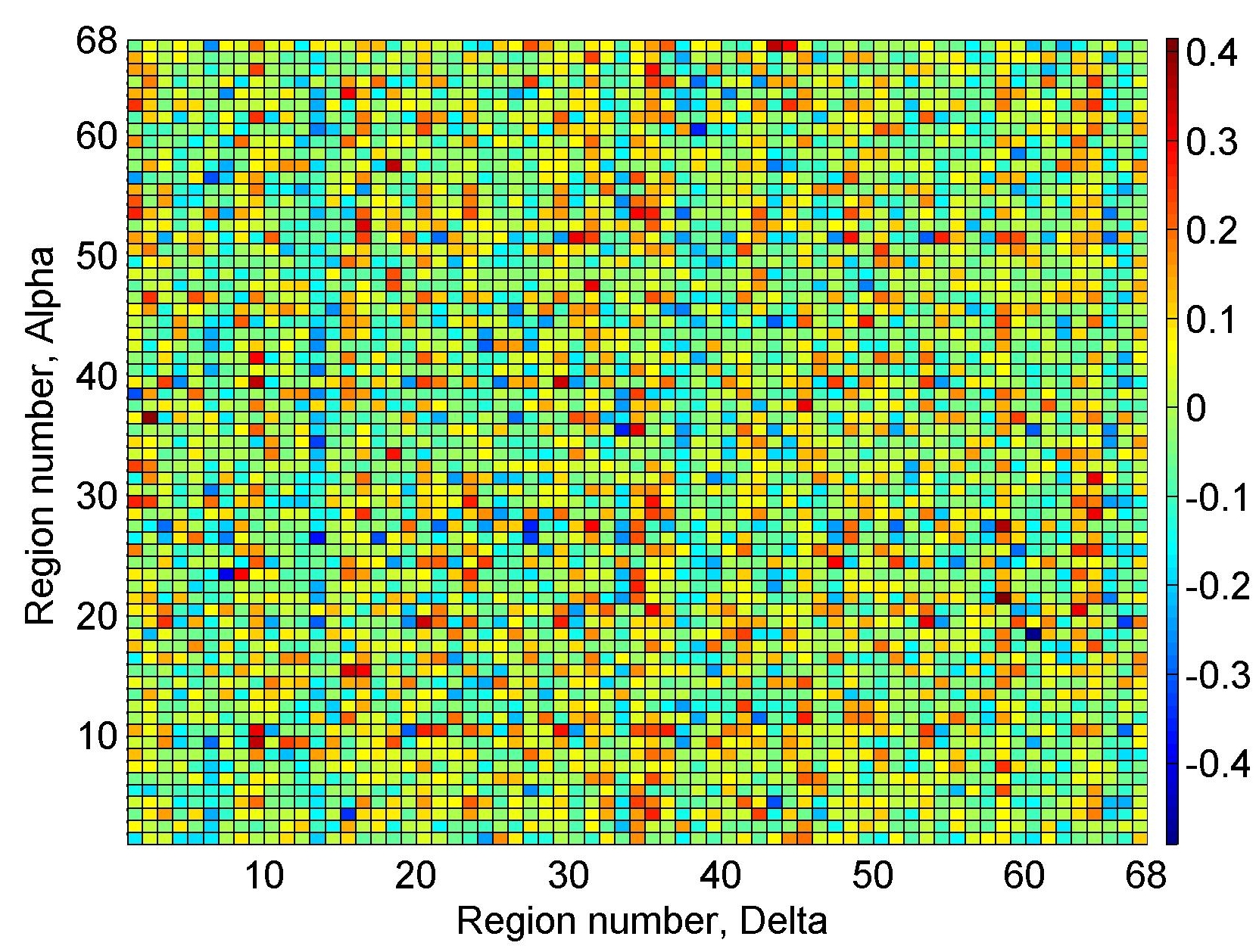
The results for the similarity index for the inferred coupling functions across regions are shown in Fig. 6. The indices presented show the average similarity for all the subjects between the coupling functions of each region and the average coupling function for all subjects for a characteristic case between delta region 58 and alpha region 21 i.e. the coupling function as presented in Fig. 2 (d). Or in other words, because we could not present visually all the 68x68=4624 coupling function combinations, in the main text we present some characteristic cases, and here in the Appendix with Fig. 6 we extend this by quantitative analysis with all the other cases. Thus, Fig. 6 shows the extent of similarity and deviations from the visually presented coupling functions. From the observations, one can notice that there are relative variations of the form, with some regions being more or less similar, and there is also negative similarity i.e. -shifted similarity.
Appendix B Sample size robustness – variance of the coupling functions in respect to number of subjects
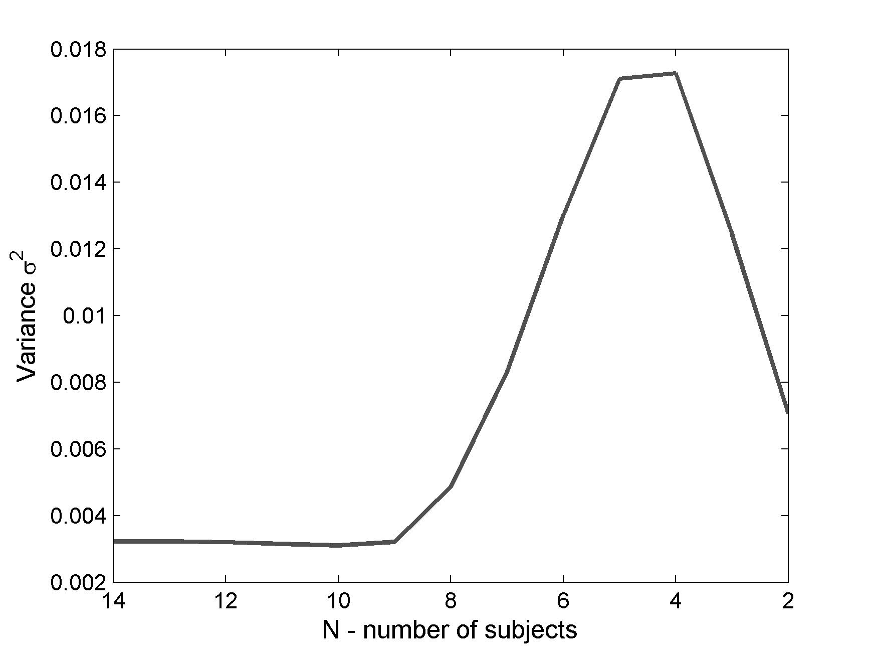
The coupling function analysis were perform on sample of N=15 subjects. In order to test if the number of subjects had an effect on the resulting average coupling function (like e.g. in Fig. 2(d)) we tested how much the coupling function varies when smaller number of subjects is averaged. This was done by systematically calculating the average coupling function from smaller number of subjects N=14, then N=13, and so on until N=2. Here, for each smaller N we calculated the average coupling function for all N-combinations. Then, we calculated the similarity index between each N average coupling function and the coupling function from all 15 subjects (as in Fig. 2(d)). Finally, we calculated the variance for all similarity indexes of each combination for one N. For example for N=14, there are 15 different combinations of N=14 subject groups; we calculated 15 average coupling functions and compared the similarity index of each in respect of the full coupling function, so as to calculate the variance of this 15 indexes.
Fig. 7 shows the variance dependence on the reduced number of subjects N. One can notice that the variance is relatively low. The dependence on N shows that the variance is low for reducing N until N=10 (perhaps N=9), after which for lower N the variance is rapidly increased. Therefore, the full number of sample size N=15 subjects is quite robust and has no big effect on the averaged coupling function. The results in Fig. 7 were calculated for two regions (delta 58 and alpha 21), but our investigation on other regions showed similar results for the variance where it was low for N=10 and then it got rapidly increased.
Appendix C Association of region numbers to appropriate brain regions
Table 1 shows the relationship between the ordinal numbers of the regions as used in this paper and the designations of the regions according to the Desikan-Kiliany anatomical parcellation.
| Region number | Brain region | Hemisphere | Brain lobe | Region number | Brain region | Hemisphere | Brain lobe |
|---|---|---|---|---|---|---|---|
| banksst | left | temporal | banksst | right | temporal | ||
| caudalanteriorcingulate | left | cingulate frontal | caudalanteriorcingulate | right | cingulate frontal | ||
| caudalmiddlefrontal | left | frontal | caudalmiddlefrontal | right | frontal | ||
| cuneus | left | occipital | cuneus | right | occipital | ||
| entorhinal | left | temporal | entorhinal | right | temporal | ||
| fusiform | left | temporal | fusiform | right | temporal | ||
| inferiorparietal | left | parietal | inferiorparietal | right | parietal | ||
| inferiortemporal | left | temporal | inferiortemporal | right | temporal | ||
| isthmuscingulate | left | cingulate parietal | isthmuscingulate | right | cingulate parietal | ||
| lateraloccipital | left | occipital | lateraloccipital | right | occipital | ||
| lateralorbitofrontal | left | frontal | lateralorbitofrontal | right | frontal | ||
| lingual | left | occipital | lingual | right | occipital | ||
| medialorbitofrontal | left | frontal | medialorbitofrontal | right | frontal | ||
| middletemporal | left | temporal | middletemporal | right | temporal | ||
| parahippocampal | left | temporal | parahippocampal | right | temporal | ||
| paracentral | left | frontal | paracentral | right | frontal | ||
| parsopercularis | left | frontal | parsopercularis | right | frontal | ||
| parsorbitalis | left | frontal | parsorbitalis | right | frontal | ||
| parstriangularis | left | frontal | parstriangularis | right | frontal | ||
| pericalcarine | left | occipital | pericalcarine | right | occipital | ||
| postcentral | left | parietal | postcentral | right | parietal | ||
| posteriorcingulate | left | cingulate parietal | posteriorcingulate | right | cingulate parietal | ||
| precentral | left | frontal | precentral | right | frontal | ||
| precuneus | left | parietal | precuneus | right | parietal | ||
| rostralanteriorcingulate | left | cingulate frontal | rostralanteriorcingulate | right | cingulate frontal | ||
| rostralmiddlefrontal | left | frontal | rostralmiddlefrontal | right | frontal | ||
| superiorfrontal | left | frontal | superiorfrontal | right | frontal | ||
| superiorparietal | left | parietal | superiorparietal | right | parietal | ||
| superiortemporal | left | temporal | superiortemporal | right | temporal | ||
| supramarginal | left | parietal | supramarginal | right | parietal | ||
| frontalpole | left | frontal | frontalpole | right | frontal | ||
| temporalpole | left | temporal | temporalpole | right | temporal | ||
| transversetemporal | left | temporal | transversetemporal | right | temporal | ||
| insula | left | temporal | insula | right | temporal |