MediViSTA-SAM: Zero-shot Medical Video Analysis with Spatio-temporal SAM Adaptation
Abstract
The Segmentation Anything Model (SAM) has attracted considerable attention as a foundational model well-known for its robust generalization capabilities across various downstream tasks. However, SAM does not exhibit satisfactory performance in the realm of medical image analysis. In this study, we introduce the first study on adapting SAM on video segmentation, called MediViSTA-SAM, a novel approach designed for medical video segmentation. Given video data, MediViSTA, spatio-temporal adapter captures long and short range temporal attention with cross-frame attention mechanism effectively constraining it to consider the immediately preceding video frame as a reference, while also considering spatial information effectively. Additionally, it incorporates multi-scale fusion by employing a U-shaped encoder and a modified mask decoder to handle objects of varying sizes. To evaluate our approach, extensive experiments were conducted using state-of-the-art (SOTA) methods, assessing its generalization abilities on multi-vendor in-house echocardiography datasets. The results highlight the accuracy and effectiveness of our network in medical video segmentation. Our code will be open sourced at: https://github.com/kimsekeun/MediViSTA-SAM
1 Introduction
In recent years, the field of Natural Language Processing (NLP) has witnessed a remarkable transformation, with the advent of large-scale models such as GPT [2], DINO [18], and SegGPT [32]. These models, which trained on extensive datasets and utilizing abundant computational resources, have facilitated new accomplishments in various tasks. Segmentation Anything Model (SAM) [12], a visual foundation model renowned for its prowess in image segmentation. SAM’s distinctive design enables it to segment any specified object of interest in an image based on user-provided prompts, offering enhanced flexibility and adaptability. SAM’s unique design allows for more precise segmentation across diverse scenarios, outperforming the capabilities of specialized models. Utilizing the power of the expansive SA-1B dataset, comprising over a billion masks and millions of natural images, SAM undergoes training to discern intricate patterns and structures within the data.
The success of SAM has attracted interest in their potential applications in more specialized fields. Particularly in medical imaging, where unique challenges such as the need for high precision and the diverse medical imaging modality are prevalent. While SAM exhibits robust performance with natural images, recent studies indicate a decline in its efficacy when applied to medical image segmentation.The decline in efficacy primarily stems from the substantial data domain gap between natural and medical images. This leads to inconsistent segmentation performance across different domain of dataset and modality. Unlike natural image datasets, medical datasets are collected from multi-center and acquired from multi-vendor sources. Furthermore, medical images have physics-based properties which significantly differ from natural images. The intrinsic idea is fully training model data, however, generating qualified data for training medical AI models requires the expertise of domain specialists, incurring substantial costs and time investments. Moreover, the Vision Transformer (ViT) typically demands vast datasets for effective training [6]. Utilizing these models as initialized models is well known to offer the potential for expedited convergence in medical image analysis tasks [11], thereby reducing the computational burden.
In order to apply SAM to medical applications, there have been two main efforts to address this challenges. The first involves the integration of several parameter-efficient Adapter modules into the base model, with modifications limited to the Adapter parameters while leaving the pre-trained parameters untouched [8]. The second strategy, known as the Adaptation model or Parameter Efficient Fine-tuning (PEFT), aims to enhance accuracy in automated medical image segmentation [33, 31]. It is worth noting that while there has been substantial research adapting 2D SAM model to 3D medical segmentation, to the best of our knowledge, the medical video segmentation models which incorporate temporal information have not been reported.
Analyzing medical video data necessitates a deep understanding of both the spatial information, which includes the varying sizes of objects, and the temporal dynamics essential for precise object boundary delineation. This task is further complicated by the presence of artifacts in ultrasound imagery, where ambiguous boundaries may not be clearly discernible in certain frames, yet become more clear in subsequent ones. To address this issue, researchers primarily rely on two established video learning approaches: 2D-based methods that emphasize temporal feature aggregation and modeling [29], and 3D CNN-based methods that interpret depth information as temporal data [28]. While 2D strategies require meticulous temporal feature aggregation, 3D approaches grapple with high parameter overheads and complexity. Interestingly, the strategies utilized in Natural Language Processing (NLP), particularly the long-range self-attention models, have proven to be highly effective not only for video modeling but also in the application of attention mechanisms for handling sequential data. Much like the textual sequential data in NLP, video medical data exhibits a unique characteristic of temporal consistency between frames. This similarity could potentially aid in addressing the challenges associated with the time-related frames in medical video data, utilizing the beneficial attention mechanism.
Expanding on the previously mentioned strategies, it is essential to further explore the complexities of analyzing medical video data, where both spatial and temporal aspects hold significant importance. Particularly, addressing the spatial characteristics of multi-scale objects in medical images is vital. Many anatomical structures or lesions in medical images are quite small, and achieving a higher resolution is often necessary to ensure improved discrimination in the context of medical imaging. The U-Net architecture [26], a cornerstone in the domain of Fully Convolutional Networks (FCN), addresses these issues with its hallmark encoder-decoder, or contraction-expansion structure.
Motivated by these observations, we propose an innovative video segmentation structure that utilizes the U-shaped convolution framework and the transformer block of SAM for medical video segmentation, adept at capturing the spatio-temporal information. To transition from 2D images to video data, we employ the TimeSformer[1] as a foundational element. We have customized the TimeSformer model to incorporate both long and short-range temporal attention, fostering temporally consistent segmentation through the development of a new cross-frame attention mechanism. This approach aims to enhance the accuracy and consistency of video segmentation tasks, demonstrating precise and reliable medical video analysis in extensive experiments.
Our contributions are summarized as three-folds:
• We propose a new spatio-temporal adapter to modify the SAM for better compatibility with medical video segmentation. Especially, we have designed it to incorporate both long-range and short-range information with cross-frame attention mechanism to dependencies between frames. This approach successfully integrates the 2D SAM model into video segmentation tasks, enhancing its functionality and efficiency.
• We modify the multi-scale fusion framework and SAM to include SAM’s generalization performance in our model, demonstrating a superior performance in echocardiography segmentation tasks compared to state-of-the-art(SOTA) methods.
• We conduct comprehensive validation of our methods using in-house data from multiple centers and vendors in the field of echocardiography. The results underscore the remarkable generalization abilities of our models, surpassing even the performance of state-of-the-art methods currently available.
2 Related Work
2.1 Foundation models in Medical
The large-scale model, key contributors such SegGPT [32], SEEM [40], CLip [25], have made significant contributions in foundation model. Their remarkable zero-shot and few-shot generalization capabilities enable prompt adaptation and extension to specified tasks or domains, predominantly through pre-training and subsequent fine-tuning methodologies.Also, change enabling rapid adaptation and extension to target tasks or domains through pre-training and fine-tuning paradigms. SegGPT [32] unifies segmentation and contextual learning using through transforming diverse segmentation data into images of a standardized format. This allows for a more comprehensive understanding of the image content. SEEM [40] proposes a interface that utilizing the various sources to effectually segment and categorize contents in images or videos concurrently. Clip [25] proposes a unified vision and language model that can be utilized for various downstream tasks, such as classification, and visual questioning task. SAM [12], distinguished for its zero-shot and few-shot capabilities, has been acknowledged for its effectiveness in specific tasks or domains, attributed to its adaptability and broad applicability. Recent research highlight SAM’s zero-shot capability within medical image segmentation. Oliveira et al. [17] explored its applicability with four distinct medical imaging modalities: X-ray, Ultrasound, Dermatoscopic, and Colonoscopy images, and observed a notable improvement in SAM’s performance when utilizing prompts. Wang et al. [30] assessed SAM on surgical instruments data. Given the similarity of this data to natural image datasets, SAM exhibited exceptional zero-shot performance in their analyses.
2.2 Parameter-Efficient Fine-tuning in SAM
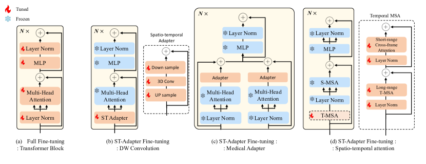
In the context of zero-shot and few-shot performance in medical image analysis, the emphasis is placed on fine-tuning Self-Attention Mechanism (SAM) for particular medical segmentation datasets. ST-Adapter [20] incorporates 3D convolution layer in each transformer block to capture spatial-temporal features for recognition tasks as in Fig 1 (b). However, it increases computational cost with increasing video clips and degrade training efficiency. Second type leverage mathematical techniques, such as the use of low-rank matrices [10], to approximate parameter updates to minimize the number of trainable parameter. SAMed [36]incorporates the Low-Rank Adaptation (LoRA) which indicates SOTA performance in various PETL tasks on image encoder of SAM.
2.3 Time Sequence Modeling
Understanding the temporal dimension of data is crucial for various computer vision tasks such as object tracking, segmentation, and detection. Time sequence modeling can be broadly categorized into two approaches. The first approach focuses on leveraging extracted features from individual frames or directly processing videos seamlessly to model temporal data. Typically, this involves extracting motion features between frames, such as optical flow, to capture temporal information. The second approach involves extracting features while considering the temporal aspect of data as depth information using 3D convolutions [24, 27] This approach offers a more adaptable prompting system for open-set segmentation tasks. Once features are extracted from video frames, standard sequence models like LSTMs [23] or Transformers can be applied to handle various tasks. These models aim to capture dependencies within sequences, either through recurrent neural networks or self-attention mechanisms. For modeling long sequences and time series, is gaining attention in sequence modeling. For end-to-end video modeling, Transformer adaptations such as Video Swin Transformer [9], and TimeSformer [1] are popular choices.
3 Methodology
In this section, we will begin with an overview of SAM models, with a particular emphasis on the model perspective. Subsequently, we will introduce the design of adaptations for SAM.
3.1 Segment anything model
We provide an initial overview of the Segment Anything Model (SAM). SAM is structured around three core components: an image encoder, a prompt encoder, and a mask decoder. The image encoder employs a conventional Vision Transformer (ViT) that has been pre-trained using MAE. The author has officially released three pre-trained models, which are vit_b, vit_l, and vit_h, corresponding to various network model sizes. The image encoder’s output embedding is downsampled by a factor of 16x from the input image. The prompt encoder can be categorized as either using sparse point points and boxes or dense mask prompts. The mask decoder consists of two convolution layers that up-sample the feature maps by 4 times. Through these two blocks, SAM enhances the image embedding’s resolution, with a subsequent transformation performed by an MLP on the resulting token.
3.2 Spatio-Temporal ViT architecture
3.2.1 Spatial and long-range temporal attention
SAM designed within a 2D framework, however, medical data frequently encompasses 2DT, 3D or even higher-dimensional details (3DT). It becomes imperative to modify the existing SAM to accommodate higher-dimensional data. Therefore, we present a refined Vision Transformer (ViT), proficient in effectively integrating spatio-temporal information. The spatio-temporal ViT (ST-ViT) block consists of a normalization layer, long/short range temporal attention, and spatial attention layer followed by an adapter as in Figure 1 (d). In order to transfer the pretrained SAM (vit_h), we freeze the pre-trained SAM parameters within its transformer block, which constitutes the core component of SAM for downstream tasks.
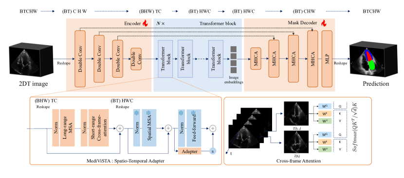
The overall model structures are depicted in Figure 2. Given a batch of video medical data as input , where B indicates batch size, H and W represents the dimension of embedding, T indicates total number of frames, and C denotes the input channels. We first need to reshape them to , after which it is passed through the encoder backbone. Within the transformer block, we designed the attention process into two separate attention sequences: spatial attention and temporal attention. The input feature matrix of dimensions into the multi-head attention mechanism within the temporal dimension which we refer to as long-range temporal attention since it spans the temporal axis. Following long-range temporal attention, we apply short-range cross-frame attention to establish inter-frame dependencies between adjacent frames, which will be discussed in the next section. Finally, by transposing the dimensions to , now we can utilize the pretrained weights of SAM model. We refer to as spatial attention since it operates within the spatial domain. Following spatial attention, we scale the embeddings using a scaling factor , as described in [4].
3.2.2 Short-range cross-frame attention (optional)
In the context of medical video segmentation, where there is limited motion between frames, we have devised a strategy for exploring short-range temporal information. Previous studies in video generation tasks have shown the effectiveness of cross-frame attention techniques in learning temporal dependencies and incorporating contextual information across video frames. et al. introduced the concept of cross-frame attention to anchor the object’s appearance and shape in the initial frame of video, which in effectively generate subsequent video frames.
However, relying solely on anchoring the first frame with cross-frame attention is inadequate when dealing with intermittent noise and image obscuration, especially in medical images such as echocardiography [16]. To address this issue, we redesigned cross-frame attention mechanism that capitalizes on the approach of using the immediately preceding video frame as a constraint. More specifically, self-attention layer receives the latent feature of , and linearly projects into query, key, and value (, , ) to produce the output of self-attention as follows:
| (1) |
The self-attention output is calculated using the following equation:
| (2) |
where t indicates frame, is step size. On the contrary, the cross-frame attention employs the key and value from the first frame, along with the query from the current frame. We integrate information from both the current frame and the frame occurring at a time step preceding the current frame. The specific value of can be determined according to the frame rate of the scanned image.
3.3 Multi-scale fusion
To leverage the advantages of both CNN and the pretrained SAM model, we have devised an image encoder and made modifications to SAM’s lightweight mask decoder. In the image encoder, we employ a CNN-based encoder to extract features while progressively downsampling input features. Within the modified SAM’s mask decoder, consisting of two layers of transpose convolution, we integrate multi-head cross attention (MHCA) [22] to interact with multi-scale encoder features. The fundamental concept behind the MHCA module is to reduce the influence of irrelevant or noisy regions within the skip connection features while emphasizing areas of significant relevance for the specific application. This approach effectively combines the strengths of both CNN and self-attention mechanisms, thereby enhancing the model’s performance in feature extraction and segmentation tasks. Our experimental evaluations have demonstrated the improved performance of our model, as illustrated in Table 4, confirming the effectiveness of our multi-scale fusion strategy.
4 Experiment settings
4.1 Datasets and implementation details
In this study, we employed the publicly available CAMUS dataset to train all the models presented. On the other hand, the multi-center in-house dataset was exclusively used as the testing set in our study.
CAMUS Dataset
CAMUS contains 1,000 patients’s two-dimensional (2D) echocardiography, comprising both apical two-chamber (2CH) and four-chamber (4CH) views of 500 patients. It provides sparse annotation along the cardiac cycle only at end-diastole (ED) and end-systole (ES). The ground truth of three structures the left ventricle , the epicardium , and the left atrium (LA) for 2CH and 4CH are provided. Half of the patients have an ejection fraction(EF) lower than 45%, 19% of the images have poor quality.
Concerning the aspect of variability, the CAMUS dataset reveals a substantial spectrum of dice similarity coefficients (Dice) in relation to both inter and intra variability, as assessed by experienced experts. In the case of the CAMUS dataset, we utilized 402 patients for training the model, while the remaining 98 patients were reserved for testing. Specifically, the testing dataset comprised full-cycle apical 4-chamber (A4C) sequences, and it included dense annotation data for and , as made available by [15].
In-house Dataset
We collected an multi-center dataset consists of B-mode echocardiography images from 100 patients consists of apical two and four-chamber view (A2/4C). It was collected from two hospitals with two different imaging vendors, including GE and Philips, utilizing their respective flagship models, the Vivid E95 and the Philips EPIQ 7C. Each manufacturer contributed equally, providing samples from 50 patients each, thereby ensuring a balanced representation in the study. The images were collected at the Massachusetts General Hospital and Brigham and Women’s Hospital between 2017 and 2022 who needs a clinical care.
The annotation process was undertaken with utmost precision by two skilled clinicians. The annotation include the boundaries of the Left Ventricle endo (), Left ventricle epicardium (), and Left Atrium (LA) during the end-diastole (ED) and end-systole (ES) phases. Given the intermittent noise and image obscuration in images, the clinicians meticulously examined adjacent frames in the video sequences to pinpoint and define accurate boundaries. following the recommendations of the American Society of Echocardiography [13]. This annotation process was accomplished using the Slicer 3D software [7], a tool well-regarded for its precision in medical imaging analysis.
Implementation details Our model was trained using the Dice loss function with an ignore index. In this context, the ignore index represents frames in the dataset that do not have any annotations. Frames that do have annotations are assigned labels, while those without annotations are designated with the ignore index. This approach helps us manage the problem of label imbalance during training by effectively ignoring frames that lack annotations. A MADGRAD optimizer [5] with a learning rate of is used for training. We employed gradient norm clipping to avoid exploding gradients and a soft constraint for the vanishing gradients problem and ensuring effective convergence, a gradient norm clipping was applied with a maximum norm of 1.0. In the pre-processing, we selected one cardiac cycle comprising 32 frames to more effectively address the SAM issue. The image intensities, initially ranging from [0, 255], were normalized to a scale between [0, 1] using min-max normalization technique. To maintain a consistent input size that includes one complete cardiac cycle, we sampled images during the period between end-diastole (ED) and end-systole (ES) phases. In cases where the desired number of frames was not obtained through this method, we conducted additional sampling after the ES frame to ensure that the required number of frames was acquired. We constructed our proposed model on the Pytorch platform, leveraging the computing power of 8 NVIDIA A100 GPUs to facilitate the process.
| CAMUS data | ||||||||
|---|---|---|---|---|---|---|---|---|
| Dice | dH(mm) | dA(mm) | L | Dice | dH(mm) | dA(mm) | L | |
| LUNet [14] | 91.3 | 7.66 | 1.73 | 0.08 | 80.0 | 11.10 | 1.84 | 0.22 |
| Deeplabv3 [3] | 92.6 | 6.23 | 1.19 | 0.07 | 84.1 | 8.42 | 1.21 | 0.14 |
| Unet++ [39] | 93.1 | 6.42 | 1.20 | 0.08 | 83.4 | 9.44 | 1.42 | 0.12 |
| Enet [21] | 89.4 | 7.54 | 1.53 | 0.10 | 79.1 | 13.26 | 1.75 | 0.16 |
| ICNet [37] | 90.8 | 9.48 | 1.84 | 0.05 | 79.4 | 10.81 | 1.43 | 0.13 |
| BiSeNetV2 [35] | 92.2 | 6.74 | 1.27 | 0.07 | 84.2 | 8.35 | 1.13 | 0.14 |
| SegFormer [34] | 90.4 | 10.51 | 1.93 | 0.04 | 80.3 | 11.16 | 1.52 | 0.11 |
| U-Transformer [22] | 94.1 | 6.81 | 0.84 | 0.06 | 88.4 | 8.31 | 1.19 | 0.12 |
| SwinUNETR [9] | 94.0 | 5.02 | 1.32 | 0.05 | 88.9 | 10.10 | 1.23 | 0.10 |
| SAM (2pts/slice) | 68.4 | 16.22 | 3.92 | 0.44 | - | - | - | - |
| SAM (1box/slice) | 85.1 | 8.43 | 1.87 | 0.21 | - | - | - | - |
| MediViSTA-SAM | 96.0 | 4.25 | 0.74 | 0.02 | 89.1 | 8.93 | 1.02 | 0.08 |
| In-house data | ||||||||||||
|---|---|---|---|---|---|---|---|---|---|---|---|---|
| Dice | dH | dA | L | Dice | dH | dA | L | Dice | dH | dA | L | |
| LUNet [14] | 87.9 | 14.21 | 6.10 | 0.16 | 75.1 | 15.63 | 7.10 | 0.18 | 84.5 | 18.48 | 4.91 | 0.09 |
| Deeplabv3 [3] | 88.1 | 13.30 | 5.44 | 0.10 | 74.2 | 13.95 | 6.90 | 0.13 | 85.7 | 16.22 | 4.88 | 0.06 |
| Unet++ [39] | 85.1 | 16.34 | 6.42 | 0.19 | 72.2 | 19.43 | 7.49 | 0.21 | 83.5 | 23.04 | 5.91 | 0.07 |
| Enet [21] | 80.4 | 20.12 | 7.02 | 0.16 | 70.6 | 23.23 | 9.92 | 0.24 | 81.2 | 26.21 | 7.11 | 0.12 |
| ICNet [37] | 85.2 | 18.22 | 6.10 | 0.15 | 71.2 | 21.26 | 7.44 | 0.21 | 82.3 | 21.53 | 5.31 | 0.09 |
| BiSeNetV2 [35] | 86.1 | 16.04 | 4.98 | 0.09 | 73.1 | 20.71 | 5.93 | 0.12 | 84.9 | 19.04 | 5.92 | 0.12 |
| SegFormer [34] | 83.5 | 19.87 | 6.22 | 0.14 | 70.9 | 25.14 | 9.34 | 0.16 | 82.1 | 18.94 | 4.11 | 0.10 |
| U-Transformer [22] | 86.2 | 14.52 | 5.11 | 0.18 | 74.2 | 12.34 | 6.85 | 0.18 | 87.1 | 13.22 | 4.02 | 0.08 |
| SwinUNETR [9] | 87.8 | 13.98 | 5.88 | 0.18 | 78.4 | 13.29 | 6.73 | 0.16 | 88.9 | 13.11 | 5.22 | 0.19 |
| SAM (2pts/slice) | 65.2 | 28.46 | 24.18 | 0.74 | - | - | - | - | 66.2 | 28.11 | 12.24 | 0.45 |
| SAM (1box/slice) | 83.2 | 18.23 | 5.47 | 0.20 | - | - | - | - | 80.4 | 16.11 | 5.97 | 0.21 |
| MediViSTA-SAM | 91.0 | 11.03 | 3.26 | 0.05 | 80.0 | 11.94 | 4.56 | 0.10 | 90.0 | 10.62 | 3.22 | 0.05 |
4.1.1 Evaluation Metric
Region-based metrics In our study, we use three region-based metrics to assess segmentation precision, including the Dice coefficient, Hausdorff distance, and Average Symmetric Surface Distance (ASSD). The Dice coefficient is a statistical tool that quantifies the overlap between the actual and predicted segmentation areas. The Hausdorff distance measures the greatest distance from a point in one set to the closest point in the other set. Additionally, we employ the Average Surface Distance (ASD) metric to determine the mean distance between binary objects found in two segmented images, providing a comprehensive view of segmentation accuracy and consistency.
Temporal metrics
To evaluate the temporal coherence in the segmentation results, we apply normalization to each temporal sequence, . To evaluate the temporal smoothness [19]of an temporal sequence data, we analyze its second-order derivative. A high derivative value suggests periods of substantial variation, while a lower derivative indicates local smoothness. Given the discrete nature of cardiac time frames, we approximate the second-order derivative numerically as follows:
| (3) |
This approximation functions act as a Laplacian filter, evaluating the temporal alignment of three consecutive values across the cardiac cycle. We then measure the difference between each data point and the average of its neighboring data points, providing us with a measurable indicator of temporal consistency, L.
Clinical metrics The Left Ventricular Ejection Fraction (LVEF) is a critical measure that indicates the percentage of blood ejected from the heart’s main pumping chamber, commonly used to evaluate cardiac function and performance. Following the clinical guidelines for echocardiography [13], we evaluate our method using the biplane simpson’s method, a common approach for estimating the volume of the left ventricle (LV) of the heart. After segmenting the left ventricle (LV) from both the 2-chamber and 4-chamber views, the ventricle is then divided into multiple disks or slices using contour information, with the volume of each disk or slice calculated individually.
| (4) |
where is the volume of the left ventricle is the length of the long axis. is the diameter of the disk. is number of disks, typically with a value of 20. The total volume of the ventricle is then calculated by summing the volumes of all the disks. The ejection fraction (EF) can be calculated as follows:
| (5) |
where is the ejection fraction, is the end-diastolic volume, and is the end-systolic volume, respectively.
5 Results and Analysis
5.1 Comparision with Statie-of-the-Art Methods
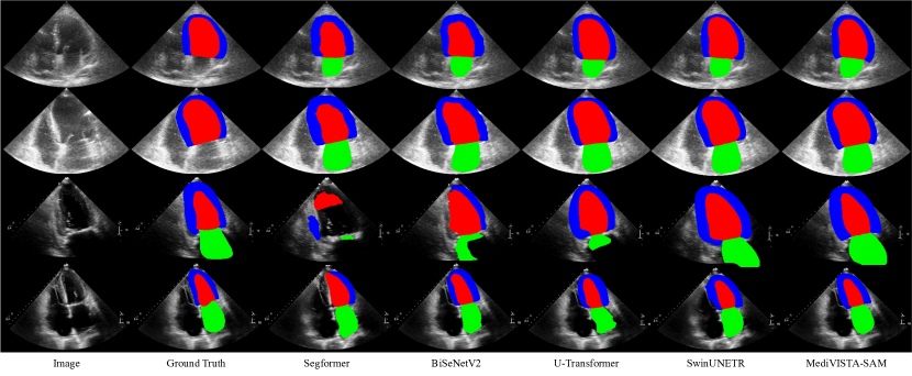
We have conducted an extensive evaluation of our methods in two datasets: camus dataset and in-house multi-center dataset. We compare state-of-the-art(SOTA) methods including six CNN-based methods: LUNet [14], Deeplabv2 [3], [39], Enet,[21] , ICNet [37], and BiSeNetV2 [35] and three transformer based methods: SegFormer [34], U-transformer [22], and SwinUNETR [9]. Table 1 presents the results for both the CAMUS and in-house datasets. While the CAMUS dataset comprises two labels, LV endo and LV epi, the in-house dataset extends to three, incorporating LA.
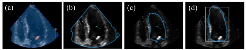
For CAMUS dataset, all the methods demonstrated comparable segmentation performance for LV endo and LV epi. As detailed in Table 1, our model outperformed the next best method by enhancing the Dice score by 1.9% and 0.2%, and improving the temporal smoothness by 0.03. It’s crucial to recognize the inherent challenges of the CAMUS dataset, which have a high inter and intra-observer variability, reaching up to 6% in the ground truth data [15]. This variability can be further observed in Figure 3 and Table 1. Furthermore, we have included our analysis both SAM-based and non-SAM-based methods. However, for SAM without prompt, and one point prompt, we find they fail to generate plausible results, e.g., w/o prompts generate segmentation of scan of region area totally different from what we want as in Figure 4. Furthermore, when it comes to segmenting for the , which surrounds the , none of the prompt methods achieve successful segmentation. Therefore, we only include the results using two points prompts and box prompts as in Table 1. We presented all results without any post-processing, enabling a direct comparison of segmentation performance.
5.2 Generalization Evaluation
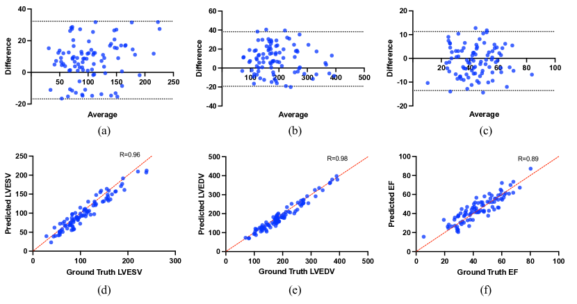
Evaluating the generalization capability of a model is of paramount significance. This is especially important in the medical field where data might come from diverse hospitals and vendors, each with their distinct scanning protocols. In our experiment, we evaluate the zero-shot capability of the proposed methods, which were trained on the CAMUS dataset, by testing our model on our in-house dataset. MediViSTA-SAM demonstrated a performance improvement over SwinUNETR, with a 3.18%, 1.6%, and 1.1% enhancement in the Dice coefficient and 2.62, 0.06, and 0.16 improvements in temporal smoothness for , , and LA, respectively. Furthermore, we conducted an additional evaluation of the consistently high-performing MediViSTA-SAM method using critical clinical metrics. The correlation were strong showing an pearson correlation coefficient of 0.96 for LVEDV, 0.98 for LVESV, and 0.89 for EF. These high correlation scores not only validate the accuracy of our model but also highlight its trustworthiness when applied to essential clinical indicators pivotal for cardiac health evaluations. The correlation and Bland–Altman plots for these indices are presented in Figure 5.
6 Ablation Studies
In this section, we conduct a comprehensive examination of the various elements that make up our proposed model. We begin our assessment by evaluating the effectiveness of the spatio-temporal design and then proceed to assess the impact of integrating multi-scale fusion. Furthermore, we evaluated the impact of pretrained SAM on model performance.
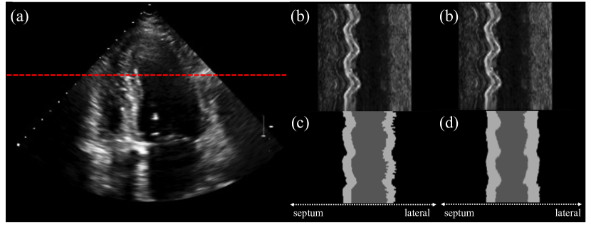
Spatio-Temporal Apater Design In this section, we investigate the impact of spatio-temporal adapter designs on the performance of our method. We conducted ablation experiments to examine the effects of cross-frame attention in our model. Our experiments demonstrate that the inclusion of cross-frame attention leads to a 0.2% improvement in Dice and a 0.03 increase in temporal smoothness, showcasing its positive impact on our method’s performance. Figure 6 provides a visual comparison of MediViSTA across frames, emphasizing the presence of ambiguous boundaries on the lateral side. Nevertheless, our model successfully captures temporal information along the time axis, resulting in segmented images with enhanced temporal smoothness. This observation highlights the interdependence between adjacent frames, where each frame significantly influences its neighboring frames.
To further investigate the impact of the order in which the spatio-temporal adapter is built, we conducted a comparison by reversing the order of spatial and temporal attention. The quantitative evaluation results, presented in Table 3, reveal that initiating with spatial attention, followed by temporal attention, leads to a decrease of 0.5% and an increase of 0.02 in the metrics. In comparison, the concurrent application of spatial and temporal attention, as Fig 1 [33], results in a reduction of 0.4% in the dice coefficient and a 0.02% increase in temporal smoothness. Based on these findings, we have chosen to structure the temporal and spatial attention in the specific sequence used in our approach.
Multi-scale fusion Design To evaluate the efficacy of the multi-scale fusion approach, we conduct an ablation study focusing on the multi-scale fusion encoder and the mask decoder components. By eliminating both the multi-scale image encoder and the mask decoder, we demonstrate its effectiveness on segmentation performance. As illustrated in Table 4 (row 4 and 5), without multi-scale fusion leads to a decline in segmentation outcomes by 1.1 %, also with an increase in temporal smoothness by 0.03. Finally, we obtain the best results when both, spatio-temporal adapter and multi-scale fusion are activated.
Effectiveness of pretrained SAM
Table 5 compares the performance of the pretrained SAM using various weight types, including vit_h, vit_l, and vit_b. The largest pretrained model, vit_h, exhibits the best accuracy across all metrics, achieving the highest dice score and exhibiting low temporal smoothness. Performance declines with decreasing model size, with vit_b recording the lowest dice score. Experiments without the pretrained SAM were conducted using the smallest model architecture comprising 12 blocks due to GPU memory constraints. Our tests showed a 4.42% decrease in dice compared to when using pretrained SAM weights. Consistent with many studies, we also demonstrate that larger network models have superior performance for downstream tasks [38].
| Attention | Dice [%] | L |
|---|---|---|
| w/o Cross-frame attention | 86.8 | 0.09 |
| w Cross-frame attention | 87.0 | 0.06 |
| Attention | Dice [%] | L |
|---|---|---|
| Spatial after temporal attention | 86.5 | 0.08 |
| Temporal after spatial attention | 87.0 | 0.06 |
| Parallel attention [33] | 86.6 | 0.08 |
7 Discussion
SAM, a large-parameter vision foundation model, is a strategy for domain generalization that allows neural networks to transfer knowledge effectively to other domains, improving performance in downstream tasks. However, SAM’s performance in directly applying to medical images is unsatisfactory. This is mainly because supervised learning relies on training data distribution, which primarily consists of natural images. Additionally, 2D SAM has limitations in medical image tasks, as most modalities in clinical practice are 3D or 2DT data. Therefore, we propose MediViSTA-SAM, a method specifically designed for medical video segmentation.
By using a pre-trained SAM in our model, our method better adapts to generalization tasks, resulting in improved segmentation performance in zero-shot analysis on echocardiography data. We also found that the introduction of spatial attention and long and short-range temporal attention improves model performance for two reasons. First, it captures spatiotemporal information. We adopted a multi-scale fusion framework in our model to easily capture various object sizes. By combining these components, we achieve more robust results, and enhanced model generalization. Compared to other state-of-the-art methods and domain-specific approaches, our proposed method demonstrates superior performance across various metrics, including region-based, temporal, and clinical metrics.
Our study’s limitation is that we tested the MediViSTA-SAM method only on hospitalized patients. In future research, we plan to apply our method to diverse patient groups, including healthy individuals and those with various medical conditions. This will help us evaluate how well our approach works across a broader range of patient populations.
| MediViSTA | pre-trained SAM | Multi-scale fusion | Dice [%] | L |
|---|---|---|---|---|
| 80.9 | 0.18 | |||
| ✓ | 82.1 | 0.14 | ||
| ✓ | ✓ | 84.8 | 0.11 | |
| ✓ | ✓ | 85.9 | 0.09 | |
| ✓ | ✓ | ✓ | 87.0 | 0.06 |
| Backbone | Dice [%] | L |
|---|---|---|
| Full-training | 82.1 | 0.17 |
| ViT_B | 85.9 | 0.13 |
| ViT_L | 86.2 | 0.11 |
| ViT_H | 87.0 | 0.06 |
8 Conclusion
To the best of our knowledge, we introduce the first study exploring the potential of SAM for medical video segmentation task, focusing on echocardiography. We propose a new framework named MediViSTA-SAM incorporating spatio-temporal apapter and multi-scale fusion. Taking advantage of both spatial multi-scale feature and long/short-range temporal information, our framework achieves highest segmentation accuracy performance. Extensive experiments show that our approach demonstrates promising results when compared to state-of-the-art methods in zero-shot echocardiography analysis.
References
- Bertasius et al. [2021] Bertasius, G., Wang, H., Torresani, L., 2021. Is space-time attention all you need for video understanding?, in: ICML, p. 4.
- Brown et al. [2020] Brown, T., Mann, B., Ryder, N., Subbiah, M., Kaplan, J.D., Dhariwal, P., Neelakantan, A., Shyam, P., Sastry, G., Askell, A., et al., 2020. Language models are few-shot learners. Advances in neural information processing systems 33, 1877–1901.
- Chen et al. [2017] Chen, L.C., Papandreou, G., Schroff, F., Adam, H., 2017. Rethinking atrous convolution for semantic image segmentation. arXiv preprint arXiv:1706.05587 .
- Chen et al. [2022] Chen, S., Ge, C., Tong, Z., Wang, J., Song, Y., Wang, J., Luo, P., 2022. Adaptformer: Adapting vision transformers for scalable visual recognition. Advances in Neural Information Processing Systems 35, 16664–16678.
- Defazio and Jelassi [2022] Defazio, A., Jelassi, S., 2022. Adaptivity without compromise: a momentumized, adaptive, dual averaged gradient method for stochastic optimization. The Journal of Machine Learning Research 23, 6429–6462.
- Dosovitskiy et al. [2020] Dosovitskiy, A., Beyer, L., Kolesnikov, A., Weissenborn, D., Zhai, X., Unterthiner, T., Dehghani, M., Minderer, M., Heigold, G., Gelly, S., et al., 2020. An image is worth 16x16 words: Transformers for image recognition at scale. arXiv preprint arXiv:2010.11929 .
- Fedorov et al. [2012] Fedorov, A., Beichel, R., Kalpathy-Cramer, J., Finet, J., Fillion-Robin, J.C., Pujol, S., Bauer, C., Jennings, D., Fennessy, F., Sonka, M., et al., 2012. 3d slicer as an image computing platform for the quantitative imaging network. Magnetic resonance imaging 30, 1323–1341.
- Gong et al. [2023] Gong, S., Zhong, Y., Ma, W., Li, J., Wang, Z., Zhang, J., Heng, P.A., Dou, Q., 2023. 3dsam-adapter:holistic adaptation of sam from 2d to 3d for promptable medical image segmentation. arXiv preprint arXiv:2306.13465 .
- Hatamizadeh et al. [2021] Hatamizadeh, A., Nath, V., Tang, Y., Yang, D., Roth, H.R., Xu, D., 2021. Swin unetr: Swin transformers for semantic segmentation of brain tumors in mri images, in: International MICCAI Brainlesion Workshop, Springer. pp. 272–284.
- Hu et al. [2021] Hu, E.J., Shen, Y., Wallis, P., Allen-Zhu, Z., Li, Y., Wang, S., Wang, L., Chen, W., 2021. Lora: Low-rank adaptation of large language models. arXiv preprint arXiv:2106.09685 .
- Kalapos and Gyires-Tóth [2022] Kalapos, A., Gyires-Tóth, B., 2022. Self-supervised pretraining for 2d medical image segmentation, in: European Conference on Computer Vision, Springer. pp. 472–484.
- Kirillov et al. [2023] Kirillov, A., Mintun, E., Ravi, N., Mao, H., Rolland, C., Gustafson, L., Xiao, T., Whitehead, S., Berg, A.C., Lo, W.Y., et al., 2023. Segment anything. arXiv preprint arXiv:2304.02643 .
- Lang et al. [2015] Lang, R.M., Badano, L.P., Mor-Avi, V., Afilalo, J., Armstrong, A., Ernande, L., Flachskampf, F.A., Foster, E., Goldstein, S.A., Kuznetsova, T., et al., 2015. Recommendations for cardiac chamber quantification by echocardiography in adults: an update from the american society of echocardiography and the european association of cardiovascular imaging. European Heart Journal-Cardiovascular Imaging 16, 233–271.
- Leclerc et al. [2020] Leclerc, S., Smistad, E., Østvik, A., Cervenansky, F., Espinosa, F., Espeland, T., Berg, E.A.R., Belhamissi, M., Israilov, S., Grenier, T., et al., 2020. Lu-net: a multistage attention network to improve the robustness of segmentation of left ventricular structures in 2-d echocardiography. IEEE Transactions on Ultrasonics, Ferroelectrics, and Frequency Control 67, 2519–2530.
- Leclerc et al. [2019] Leclerc, S., Smistad, E., Pedrosa, J., Østvik, A., Cervenansky, F., Espinosa, F., Espeland, T., Berg, E.A.R., Jodoin, P.M., Grenier, T., et al., 2019. Deep learning for segmentation using an open large-scale dataset in 2d echocardiography. IEEE transactions on medical imaging 38, 2198–2210.
- Mitchell et al. [2019] Mitchell, C., Rahko, P.S., Blauwet, L.A., Canaday, B., Finstuen, J.A., Foster, M.C., Horton, K., Ogunyankin, K.O., Palma, R.A., Velazquez, E.J., 2019. Guidelines for performing a comprehensive transthoracic echocardiographic examination in adults: recommendations from the american society of echocardiography. Journal of the American Society of Echocardiography 32, 1–64.
- de Oliveira et al. [2023] de Oliveira, C.M., de Moura, L.V., Ravazio, R.C., Kupssinskü, L.S., Parraga, O., Delucis, M.M., Barros, R.C., 2023. Zero-shot performance of the segment anything model (sam) in 2d medical imaging: A comprehensive evaluation and practical guidelines. CoRR abs/2305.00109 .
- Oquab et al. [2023] Oquab, M., Darcet, T., Moutakanni, T., Vo, H., Szafraniec, M., Khalidov, V., Fernandez, P., Haziza, D., Massa, F., El-Nouby, A., et al., 2023. Dinov2: Learning robust visual features without supervision. arXiv preprint arXiv:2304.07193 .
- Painchaud et al. [2022] Painchaud, N., Duchateau, N., Bernard, O., Jodoin, P.M., 2022. Echocardiography segmentation with enforced temporal consistency. IEEE Transactions on Medical Imaging 41, 2867–2878.
- Pan et al. [2022] Pan, J., Lin, Z., Zhu, X., Shao, J., Li, H., 2022. St-adapter: Parameter-efficient image-to-video transfer learning. Advances in Neural Information Processing Systems 35, 26462–26477.
- Paszke et al. [2016] Paszke, A., Chaurasia, A., Kim, S., Culurciello, E., 2016. Enet: A deep neural network architecture for real-time semantic segmentation. arXiv preprint arXiv:1606.02147 .
- Petit et al. [2021] Petit, O., Thome, N., Rambour, C., Themyr, L., Collins, T., Soler, L., 2021. U-net transformer: Self and cross attention for medical image segmentation, in: Machine Learning in Medical Imaging: 12th International Workshop, MLMI 2021, Held in Conjunction with MICCAI 2021, Strasbourg, France, September 27, 2021, Proceedings 12, Springer. pp. 267–276.
- Pfeuffer et al. [2019] Pfeuffer, A., Schulz, K., Dietmayer, K., 2019. Semantic segmentation of video sequences with convolutional lstms, in: 2019 IEEE intelligent vehicles symposium (IV), IEEE. pp. 1441–1447.
- Qiu et al. [2017] Qiu, Z., Yao, T., Mei, T., 2017. Learning deep spatio-temporal dependence for semantic video segmentation. IEEE Transactions on Multimedia 20, 939–949.
- Radford et al. [2021] Radford, A., Kim, J.W., Hallacy, C., Ramesh, A., Goh, G., Agarwal, S., Sastry, G., Askell, A., Mishkin, P., Clark, J., et al., 2021. Learning transferable visual models from natural language supervision, in: International conference on machine learning, PMLR. pp. 8748–8763.
- Ronneberger et al. [2015] Ronneberger, O., Fischer, P., Brox, T., 2015. U-net: Convolutional networks for biomedical image segmentation, in: Medical Image Computing and Computer-Assisted Intervention–MICCAI 2015: 18th International Conference, Munich, Germany, October 5-9, 2015, Proceedings, Part III 18, Springer. pp. 234–241.
- Song et al. [2019] Song, W., Zhang, D., Zhao, X., Yu, J., Zheng, R., Wang, A., 2019. A novel violent video detection scheme based on modified 3d convolutional neural networks. IEEE Access 7, 39172–39179.
- Tran et al. [2015] Tran, D., Bourdev, L., Fergus, R., Torresani, L., Paluri, M., 2015. Learning spatiotemporal features with 3d convolutional networks, in: Proceedings of the IEEE international conference on computer vision, pp. 4489–4497.
- Tsai et al. [2016] Tsai, Y.H., Yang, M.H., Black, M.J., 2016. Video segmentation via object flow, in: Proceedings of the IEEE conference on computer vision and pattern recognition, pp. 3899–3908.
- Wang et al. [2023a] Wang, A., Islam, M., Xu, M., Zhang, Y., Ren, H., 2023a. Sam meets robotic surgery: An empirical study on generalization, robustness and adaptation. arXiv preprint arXiv:2308.07156 .
- Wang et al. [2023b] Wang, W., Shen, J., Chen, C., Jiao, J., Zhang, Y., Song, S., Li, J., 2023b. Med-tuning: Exploring parameter-efficient transfer learning for medical volumetric segmentation. arXiv preprint arXiv:2304.10880 .
- Wang et al. [2023c] Wang, X., Zhang, X., Cao, Y., Wang, W., Shen, C., Huang, T., 2023c. Seggpt: Segmenting everything in context. arXiv preprint arXiv:2304.03284 .
- Wu et al. [2023] Wu, J., Fu, R., Fang, H., Liu, Y., Wang, Z., Xu, Y., Jin, Y., Arbel, T., 2023. Medical sam adapter: Adapting segment anything model for medical image segmentation. arXiv preprint arXiv:2304.12620 .
- Xie et al. [2021] Xie, E., Wang, W., Yu, Z., Anandkumar, A., Alvarez, J.M., Luo, P., 2021. Segformer: Simple and efficient design for semantic segmentation with transformers. Advances in Neural Information Processing Systems 34, 12077–12090.
- Yu et al. [2021] Yu, C., Gao, C., Wang, J., Yu, G., Shen, C., Sang, N., 2021. Bisenet v2: Bilateral network with guided aggregation for real-time semantic segmentation. International Journal of Computer Vision 129, 3051–3068.
- Zhang and Liu [2023] Zhang, K., Liu, D., 2023. Customized segment anything model for medical image segmentation. arXiv preprint arXiv:2304.13785 .
- Zhao et al. [2018] Zhao, H., Qi, X., Shen, X., Shi, J., Jia, J., 2018. Icnet for real-time semantic segmentation on high-resolution images, in: Proceedings of the European conference on computer vision (ECCV), pp. 405–420.
- Zhou et al. [2023] Zhou, C., Li, Q., Li, C., Yu, J., Liu, Y., Wang, G., Zhang, K., Ji, C., Yan, Q., He, L., et al., 2023. A comprehensive survey on pretrained foundation models: A history from bert to chatgpt. arXiv preprint arXiv:2302.09419 .
- Zhou et al. [2019] Zhou, Z., Siddiquee, M.M.R., Tajbakhsh, N., Liang, J., 2019. Unet++: Redesigning skip connections to exploit multiscale features in image segmentation. IEEE transactions on medical imaging 39, 1856–1867.
- Zou et al. [2023] Zou, X., Yang, J., Zhang, H., Li, F., Li, L., Gao, J., Lee, Y.J., 2023. Segment everything everywhere all at once. arXiv preprint arXiv:2304.06718 .