ResEMGNet: A Lightweight Residual Deep Learning Architecture for Neuromuscular Disorder Detection from Raw EMG Signals.
Abstract
Amyotrophic Lateral Sclerosis (ALS) and Myopathy are debilitating neuromuscular disorders that demand accurate and efficient diagnostic approaches. In this study, we harness the power of deep learning techniques to detect ALS and Myopathy. Convolutional Neural Networks (CNNs) have emerged as powerful tools in this context. We present ResEMGNet, designed to identify ALS and Myopathy directly from raw electromyography (EMG) signals. Unlike traditional methods that require intricate handcrafted feature extraction, ResEMGNet takes raw EMG data as input, reducing computational complexity and enhancing practicality. Our approach was rigorously evaluated using various metrics in comparison to existing methods. ResEMGNet exhibited exceptional subject-independent performance, achieving an impressive overall three-class accuracy of 94.43%.
Index Terms— Amyotrophic Lateral Sclerosis (ALS), Convolutional Neural Networks (CNNs), Deep Learning, Electromyography (EMG), Myopathy
1 Introduction
Amyotrophic lateral sclerosis (ALS), also known as Lou Gehrig’s disease, is a fatal motor neuron disorder characterized by the degeneration of primary motor neurons in the brain and spinal cord, leading to muscle weakness, paralysis, and various debilitating symptoms. Among these symptoms, pain is a prevalent but often overlooked and poorly managed aspect of the disease, with reports of its occurrence in nearly 70% of ALS patients. Despite its significance, there is a lack of comprehensive research and drug trials dedicated to understanding and treating pain in ALS, highlighting the urgent need for further investigations in this area [1]. However, an early diagnosis has the potential to mitigate the progression of the disorder and enhance the quality of life for individuals affected by ALS [2].
Several signal-processing techniques are available for analyzing EMG signals. These techniques are crucial for extracting relevant information from EMG signals and detecting ALS. Various methods are available for interference pattern analysis (IPA) to analyze electromyographic (EMG) signals, including time domain approaches like amplitude measurements and spike counting, as well as frequency domain analysis using power spectrum [3]. By examining time and frequency domain behaviors of EMG signals from both normal individuals and ALS patients, characteristic features like auto-correlation, cross-correlation, zero crossing rate, and Fourier transform are used for ALS detection [4, 5]. Again, [6, 7] introduces discrete wavelet transform (DWT) domain features alongside traditional time and frequency domain features for EMG signal analysis. Another way to extract features and classify normal and ALS subjects is using motor unit action potentials (MUAP) signals and using mel-frequency cepstral coefficients (MFCCs) of MUAP signals as potential features. MUAPs are extracted from EMG data through template matching, selecting the one with the maximum dynamic range, and then extracting MFCCs [8, 9]. In another study, six specific features, including spectral characteristics and signal properties, are extracted from intrinsic mode functions (IMFs) decomposed from EMG signals [10]. These features are important for detecting ALS from EMG signals.
Numerous classification techniques have been developed to classify ALS from EMG signals using these features. In [4, 6, 8, 11], Classification is performed using a K-nearest neighborhood (KNN), SVM classifier. In [10], the least square support vector machine (LS-SVM) is applied for the classification. However, efficient handcrafted features are necessary for the aforementioned classifiers to work satisfactorily. This requires both domain-specific knowledge as well as time and computational power. Hence, deep learning models are preferable for this type of classification problem. In [12], Short Time Fourier Transform (STFT) is used for time-frequency representation, and CNN is introduced for classification. Another study introduces Spectrogram, Continuous Wavelet Transform (CWT), and Smoothed Pseudo Wigner-Ville Distribution (SPWVD) methods employed for time-frequency (T-F) representation and uses CNN for classification[13]. In these Deep learning models, different transforms have been used instead of the raw data. But, for developing a lightweight ALS detection device, raw data is more feasible as it will demand less computational burden. Again, the authors of [14] proposed a one-dimensional CNN designed to identify ALS directly from raw EMG signals without pre-processing or feature extraction. However, only binary classification was performed in the study and three class classification was not explored. Additionally, subject-independent classification, a more challenging one, is not been implemented. So, the prospects of developing a lightweight architecture for solving a subject-independent three-class classification problem are yet to be explored.
In this paper, we propose ResEMGNet, a subject-independent deep-learning architecture capable of identifying ALS and Myopathy among three classes ( Myopathy, Normal, and ALS) utilizing the raw EMG data. This paper focuses on distinguishing between normal, ALS, and Myopathy based on EMG signals, categorizing them into respective classes for accurate classification. Here CNN layers with different filter sizes are used for efficiently extracting different features helpful for the classification task. As the CNN layer has prowess in extracting spatial features and the LSTM layer has expertise in temporal features, the features extracted by the CNN layers are fed to the LSTM layer as inputs for exploiting the advantages of both layers. Again, we used skip connection to tackle the vanishing gradient problem. Another, contribution of this model is that this model is very lightweight and has around sixty thousand trainable parameters only. We downsampled the data from 24000 Hz to 2000 Hz to reduce the input size of the lightweight model and this helped the model to train easily. Otherwise, it would require a larger model for such a large data. So, this lightweight model is well optimized with this small input-sized data. Hence, our model can satisfactorily differentiate ALS from Normal and Myopathy classes in a subject-independent classification. Finally, the performance of ResEMGNet is compared with different existing methods in terms of three evaluation metrics- overall accuracy, sensitivity, and specificity.
2 Dataset Preprocessing
In our experiment, we utilized the clinical EMG signals from the N2001 EMGLAB open-access Dataset.[15] This dataset comprises three distinct groups: Normal, Myopathy, and ALS. All EMG signals were recorded under these standard conditions for the analysis of MUAPs:
1. The recordings were conducted during mild voluntary muscle contractions, slightly above the minimum threshold.
2. To ensure signal quality, visual and auditory feedback was utilized for monitoring.
3. A standard concentric needle electrode was employed.
4. EMG signals were recorded from five different locations within the muscle, spanning three depths (deep, medium, and low).
5. The EMG amplifier was equipped with filters, permitting signals above 2 Hz and below 10 kHz to pass through.
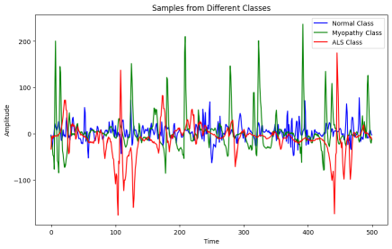
Each EMG signal was sampled at a frequency of 24 kHz and recorded for approximately 11 seconds. In the data preparation phase, our initial dataset was standardized to ensure uniformity. The original training data consisted of 294 samples, each containing 262124 data points, with a shape of (262124,1). To facilitate further analysis, we applied a windowing technique, segmenting the data into windows, each comprising 23437 data points. As a result, the training dataset was transformed into a new shape (3215, 23437, 1), while the validation dataset comprised 682 samples with the same windowed shape, (23437, 1). The test dataset also follows this windowing process, yielding a shape of (539, 23437, 1). To optimize computational efficiency and accommodate the requirements of our model, we subsequently downsampled the data to a rate of 2 KHz. This downsampling procedure reshaped our datasets, resulting in the following dimensions: the training data now had a shape of (3215, 2000, 1), the validation data was represented as (682, 2000, 1), and the test data took on the form of (539, 2000, 1). These transformations provided us with well-structured data ready for training, validation, and testing, with 3215 samples for training, 682 samples for validation, and 539 samples for testing, all uniformly shaped (2000, 1) for further analysis and model development. Figure 1 illustrates the visualization of three distinct classes.
3 Proposed Architecture
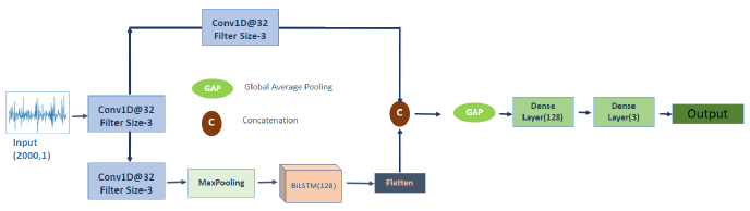
The proposed ResEMGNet, shown in 2, represents a deep learning model tailored for the classification of electromyographic (EMG) signals. This architecture is designed to effectively capture both spatial and temporal characteristics within the input EMG data, making it particularly well-suited for tasks such as gesture recognition and medical diagnostics.
The model begins with an input layer, configured to accept EMG signal data with a length of 2000 time steps and a single channel. Subsequently, two convolutional layers, conv1 and conv2, are employed with 32 filters each and a kernel size of 3. These convolutional layers utilize the Rectified Linear Unit (ReLU) activation function to extract local spatial patterns from the input EMG signals. A max-pooling layer with a pool size of 2 follows the convolutional layers, reducing the spatial dimensions of the feature maps and emphasizing essential information. Next, a bidirectional Long Short-Term Memory (LSTM) layer with 64 units is introduced to capture temporal dependencies. This bidirectional LSTM processes the input data both in the forward and backward directions, enhancing the model’s ability to recognize complex temporal patterns in EMG signals.
To further enrich feature representation, a skip connection is incorporated. This connection involves adding the output of an adjusted convolutional layer, path1, with 128 filters and a kernel size of 1, to the flattened output from the bidirectional LSTM. This mechanism preserves both low-level and high-level features throughout the network. Following this, a global average pooling layer is applied to reduce spatial dimensions, resulting in a fixed-length feature representation. This layer calculates the average value of each feature map across all positions. Lastly, two dense layers are employed for feature aggregation and classification. The first dense layer, output1, contains 16 units and utilizes the ReLU activation function to further process the global average-pooled features. The final output layer consists of three units, each representing one of the three target classes, and utilizes a softmax activation function to produce class probabilities for accurate classification.
4 Experimentaion
The ResEMGNe model was constructed using Keras and Tensorflow, in the environment of Google Colaboratory. We used Categorical Cross-entropy as the loss function during training and Adam optimizer with an initial learning rate of 0.001. During the model training process, a total of 40 epochs were executed, with each epoch consisting of batches containing 16 data samples. To enhance training, we decreased the learning rate by a factor of 10 if consecutive epochs didn’t reduce the validation loss, with a minimum learning rate set at 10∧-10. Training halted early if there was no significant improvement in validation loss over consecutive epochs.
5 Performance Matrices
In evaluating the performance of the model, we use several metrics to measure how well it makes predictions on the test data. These metrics are Accuracy, Sensitivity, and Specificity.
5.1 Accuracy
Accuracy is a crucial metric for measuring the overall correctness of our model’s prediction, including both true positives and true negatives.
| (1) |
5.2 Sensitivity
Sensitivity also known as true positive rate, measures the proportion of actual positives that our model classifies.
| (2) |
5.3 Specificity
Specificity referred to as true negative rates, measures the proportion of actual negatives that our model identifies.
| (3) |
TP, FP, TN, and FN are True Positive, False Positive, True Negative, and False Negative.
6 Result and Discussion
In our classification task involving three distinct classes (Myopathy, Normal, and ALS), we designated the test set as ’S.’ As outlined in Section 1, each segment ’s’ within this test set is processed by ResEMGNet, resulting in a probability value denoted as P(s = 1), representing the likelihood of that segment belonging to each class. To make the final classification, we select the class with the highest probability among the three classes. This approach allows us to effectively categorize the segments into one of the three classes based on the highest probability, facilitating the evaluation of our model’s performance in distinguishing between Myopathy, Normal, and ALS EMG signals. ResEMGNet accurately predicted 509 out of 539 EMG signal segments in the test set, as depicted in Figure 2’s confusion matrix.
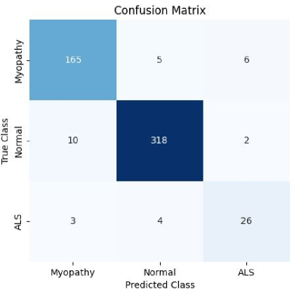
Table 1 presents the results obtained from our model’s performance evaluation. It provides a comprehensive overview of how our model performed across different classes.
| Class | Sensitivity | Specificity | Precision |
|---|---|---|---|
| Myopathy | 93.75 | 92.70 | 96.42 |
| Normal | 96.36 | 97.25 | 95.69 |
| ALS | 78.79 | 76.47 | 98.42 |
Table 2 provides a comparison of various existing approaches alongside our proposed model’s performance. Notably, none of the authors in this table tackled the three-class classification problem. Instead, they primarily focused on two-class classification tasks. In previous studies, [8, 6, 10, 13, 12, 14] primarily focused on two-class classifications involving the Normal and ALS datasets. In two-class tasks, our model excelled with accuracy, sensitivity, and specificity values reaching 98.84%, 98.54%, and 99.34%, respectively, outperforming all existing models in normal-ALS detection. Additionally, [16] made notable contributions to two separate two-class classifications, specifically in Normal-ALS and Myopathy-ALS detection tasks. Our proposed model achieved an impressive accuracy of 94.43%, demonstrating its competence in distinguishing among three classes, Myopathy, Normal, and ALS. In terms of sensitivity, our model achieved a commendable 89.63%, reflecting its ability to accurately identify positive instances across the three classes. Furthermore, the specificity of our model stands at 96.84%, indicating its proficiency in correctly recognizing negative instances. While our model excels in this three-class problem, it’s essential to acknowledge that this study marks a significant contribution to addressing the unique challenges posed by neuromuscular disorders through a multi-class classification approach.
| Techniques | Accuracy | Sensitivity | Specificity |
|---|---|---|---|
| Doulah[8] | 92.50 | 76.00 | 98.00 |
| Fattah[6] | 91.50 | 74.00 | 97.33 |
| Mishra[10] | 95.00 | 93.00 | 92.54 |
| Krishna[16] | 92.50 | 88.00 | 99.33 |
| Sengur[12] | 96.69 | 94.24 | 97.59 |
| Sengur[13] | 96.80 | 94.80 | 98.80 |
| Hassan[14] | 97.74 | 96.77 | 98.59 |
| Proposed Model | 98.84 | 98.54 | 99.34 |
| (2 Class) | |||
| Proposed Model | 94.43 | 89.63 | 96.84 |
| (3 Class) |
7 Conclusion
In this study, we introduce a deep-learning-based approach for classifying ALS and Myopathy from raw EMG signals, bypassing the need to extract features. Our method provides promising performance in terms of accuracy, sensitivity, and specificity. This approach holds potential for early neural disorder diagnosis, potentially enhancing patients’ quality of life and prolonging survival. Moreover, by eliminating the need for feature extraction, it reduces computational costs, making it highly suitable for practical implementation.
Acknowledgment
The authors would like to thank the organizers of EMGLAB[15] for their public dataset which was used to develop this research work.
References
- [1] Chalonda R. Handy, Chalonda R. Handy, Christina Krudy, Nicholas M. Boulis, and Thais Federici, “Pain in Amyotrophic Lateral Sclerosis: A Neglected Aspect of Disease,” Neurology Research International, vol. 2011, no. 2011, pp. 1–8, May 2011, MAG ID: 2042897241 S2ID: 08c06f7dcf73ae4a3b1d9f1102c39b11335ace81.
- [2] Michael Swash, “Early diagnosis of ALS/MND.,” Journal of the Neurological Sciences, vol. 160, pp. 6, Oct. 1998, MAG ID: 2067255333 S2ID: 11cc9b20c353750a44a1c4f4cfdcc6417db3ba06.
- [3] Anders Fuglsang-Frederiksen, “The utility of interference pattern analysis,” Muscle & Nerve, vol. 23, no. 1, pp. 18–36, Jan. 2000, MAG ID: 1967128670.
- [4] Anowarul Fattah, “Identifying the Motor Neuron Disease in EMG Signal Using Time and Frequency Domain Features with Comparison,” Signal & Image Processing : An International Journal, vol. 3, no. 2, pp. 99–114, Apr. 2012, MAG ID: 2334615532 S2ID: eaf6b58dfe2eba74fc1ba20c65f8f5465425c3e7.
- [5] Rohit Bose, Kaniska Samanta, and Soumya Chatterjee, “Cross-correlation based feature extraction from emg signals for classification of neuro-muscular diseases,” in 2016 International Conference on Intelligent Control Power and Instrumentation (ICICPI). IEEE, 2016, pp. 241–245.
- [6] Anowarul Fattah, A. B. M. Sayeed Ud Doulah, A. B. M. Sayeed Ud Doulah, A. B. M. Sayeed Ud Doulah, Asif Iqbal, Asif Iqbal, Celia Shahnaz, Wei-Ping Zhu, and M. Omair Ahmad, “Identification of motor neuron disease using wavelet domain features extracted from EMG signal,” pp. 1308–1311, May 2013, MAG ID: 2110943775.
- [7] Abdulhamit Subasi, Emine Yaman, Yara Somaily, Halah A Alynabawi, Fatemah Alobaidi, and Sumaiah Altheibani, “Automated emg signal classification for diagnosis of neuromuscular disorders using dwt and bagging,” Procedia Computer Science, vol. 140, pp. 230–237, 2018.
- [8] Abul Barkat Mollah Sayeed Ud Doulah and Anowarul Fattah, “Neuromuscular disease classification based on mel frequency cepstrum of motor unit action potential,” 2014 International Conference on Electrical Engineering and Information & Communication Technology, pp. 1–4, Apr. 2014, MAG ID: 2059054229 S2ID: 41782eb964be4f53889f726ee4c53f87a1aac82e.
- [9] Muhammad Irfan, Khalil Ullah, Fazal Muhammad, Salman Khan, Faisal Althobiani, Muhammad Usman, Mohammed Alshareef, Shadi Alghaffari, and Saifur Rahman, “Automatic detection of outliers in multi-channel emg signals using mfcc and svm,” INTELLIGENT AUTOMATION AND SOFT COMPUTING, vol. 36, no. 1, pp. 169–181, 2023.
- [10] V. K. Mishra, Vipin K. Mishra, Varun Bajaj, Anil Kumar, Anil Kumar, and Girish Kumar Singh, “Analysis of ALS and normal EMG signals based on empirical mode decomposition,” Iet Science Measurement & Technology, vol. 10, no. 8, pp. 963–971, Nov. 2016, MAG ID: 2504270968 S2ID: 5395aba197340d94d28e649f1e941e56fa739c9f.
- [11] Hanife Küçük and İlyas Eminoğlu, “Classification of als disease using support vector machines,” in 2015 23nd Signal Processing and Communications Applications Conference (SIU). IEEE, 2015, pp. 1664–1667.
- [12] Abdulkadir Sengur, Mehmet Gedikpinar, Yaman Akbulut, Erkan Deniz, Varun Bajaj, Amira S. Ashour, and Yanhui Guo, “DeepEMGNet: An Application for Efficient Discrimination of ALS and Normal EMG Signals,” pp. 619–625, Sept. 2017, MAG ID: 2746483198 S2ID: 7dab06c53116c188b518b37c28779ff317b59018.
- [13] Abdulkadir Sengur, Yaman Akbulut, Amira S. Ashour, Yanhui Guo, and Varun Bajaj, “Classification of amyotrophic lateral sclerosis disease based on convolutional neural network and reinforcement sample learning algorithm,” Health Information Science and Systems, vol. 5, no. 1, pp. 9–9, Oct. 2017, MAG ID: 2765859029 S2ID: ac0cb6535ed248690823a038555182c6a6581278.
- [14] K. M. Naimul Hassan, Md. Shamiul Alam Hridoy, Naima Tasnim, Atia Faria Chowdhury, Tanvir Alam Roni, Sheikh Tabrez, Arik Subhana, and Celia Shahnaz, “ALSNet: A Dilated 1-D CNN for Identifying ALS from Raw EMG Signal,” IEEE International Conference on Acoustics, Speech, and Signal Processing, May 2022, MAG ID: 4224933343 S2ID: 5dbf888a049fa9e00294c4581edeee781a63e95d.
- [15] M. Nikolic, “Detailed analysis of clinical electromyography signals: Emg decomposition, findings and firing pattern analysis in controls and patients with myopathy and amytrophic lateral sclerosis, ph.d. dissertation, faculty of health science, university of copenhagen, copenhagen, denmark, 2001. [online].url = http://www.emglab.net,” .
- [16] A. Krishna and Paul Thomas, “Classification of EMG Signals Using Spectral Features Extracted from Dominant Motor Unit Action Potential,” 2015, S2ID: b5fd05c7004c64cfda082d095e338ff3bfb08716.