A Wearable Ultra-Low-Power sEMG-triggered Ultrasound System for Long-term
Muscle Activity Monitoring
††thanks: The authors acknowledge support from the Swiss National Science Foundation (Project PEDESITE, grant agreement 193813) and from ETH Zürich (project ListenToLight, grant agreement ETH-C-01 21-2). This work was also partially supported by the ETH Future Computing Laboratory (EFCL).
Abstract
Surface electromyography (sEMG) is a well-established approach to monitor muscular activity on wearable and resource-constrained devices. However, when measuring deeper muscles, its low signal-to-noise ratio (SNR), high signal attenuation, and crosstalk degrade sensing performance. Ultrasound (US) complements sEMG effectively with its higher SNR at high penetration depths. In fact, combining US and sEMG improves the accuracy of muscle dynamic assessment, compared to using only one modality. However, the power envelope of US hardware is considerably higher than that of sEMG, thus inflating energy consumption and reducing the battery life. This work proposes a wearable solution that integrates both modalities and utilizes an EMG-driven wake-up approach to achieve ultra-low power consumption as needed for wearable long-term monitoring. We integrate two wearable state-of-the-art (SoA) US and ExG biosignal acquisition devices to acquire time-synchronized measurements of the short head of the biceps. To minimize power consumption, the US probe is kept in a sleep state when there is no muscle activity. sEMG data are processed on the probe (filtering, envelope extraction and thresholding) to identify muscle activity and generate a trigger to wake-up the US counterpart. The US acquisition starts before muscle fascicles displacement thanks to a triggering time faster than the electromechanical delay (30-100 ms) between the neuromuscular junction stimulation and the muscle contraction. Assuming a muscle contraction of 200 ms at a contraction rate of 1 Hz, the proposed approach enables more than 59% energy saving (with a full-system average power consumption of 12.2 mW) as compared to operating both sEMG and US continuously.
Index Terms:
wearable EEG, wearable healthcare, ultra-low-power design, embedded system.I Introduction
Surface electromyography (sEMG) is a well-established approach for determining muscular activity on wearable and resource-constrained devices [1]. Its popular applications include the detection of muscle activations [2] and their integration into human-machine interfaces (HMI) [3] or prosthetic [4] control systems. In sEMG, activation signals of muscles are acquired from the skin surface [5]. These collected sEMG signals reflect a total activation sum of all underlying muscle tissue [6]. As a consequence, an unambiguous correlation between surface-collected signals and the contribution of specific muscles is not always possible. Moreover, sEMG suffers from a low signal-to-noise ratio (SNR) and interference from neighbouring muscles [7]. Thus, complementing sEMG with alternative sensing modalities to enhance the quality and reliability of the measured signals is highly desired.
An alternative modality to analyze musculoskeletal activity is US [8, 9]. US has several advantages over sEMG, including a higher SNR at high penetration depths, lower susceptibility to noise and interference, and higher spatial resolution, as demonstrated by a number of different applications, such as elastography [10], motor endplates detection [11], strain analyses [12], and even gestures [13] or finger motion recognition [14]. With US, however, only the mechanical response to an electrically triggered voluntary contraction can be observed. Therefore, lacking information about the electro-chemically induced neuronal activity. In this context, the combination of sEMG and US appears as a promising approach for achieving higher accuracy in the assessment of muscle dynamics [15].
To enable continuous, long-term monitoring, there is a need for wearable solutions that are wireless, unobtrusive, and low power. Among these requirements, power consumption appears as the main challenge for the successful integration of sEMG and US into wearables. In fact, US hardware is more power-hungry than that of sEMG111considering two SoA wearable ExG and US platforms [16][17], the generation of high-voltage pulses and analog-to-digital conversion for a single-channel US consumes nearly twice the power of the analog-to-digital conversion of a single-channel ExG, and implementing concurrent, continuous sEMG and US measurements would use too much energy and drain the battery of wearable sensor systems. Therefore, to achieve low power consumption, a wearable solution that combines both modalities should use physiology knowledge to activate US measurements only when the signal is meaningful.
In this paper, we propose a wearable sensor system that combines sEMG and US for measuring muscle activity. The system is based on two SoA wearable probes for ExG [16] and US [17]. The US sub-system is kept in a sleep state when there is no muscle activity. The sEMG signal is acquired continuously, and we use an EMG-driven wake-up approach to identify muscle activity (the envelope of sEMG signals is extracted and compared to a threshold), generating a trigger to wake-up the US counterpart only when significant muscle activity is detected. The triggering is faster than the electromechanical delay () between the neuromuscular junction stimulation and the muscle contraction, thereby waking up the US sub-system before the actual displacement of muscle fascicles starts. We focus on the use case of biceps muscle contractions. By implementing the proposed wake-up approach directly on-probe, we demonstrate that the proposed methodology allows to considerably reduce the US hardware’s power consumption: assuming contractions of at a contraction rate of , the total system power is only , lower compared to operating both sEMG and US continuously leading to a battery life increase of more than 2 days with a battery.
II Material and Methods
II-A System description
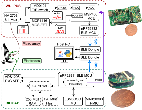
The framework proposed in this study combines two SoA ExG and US acquisition platforms, namely BioGAP [16] and WULPUS [17].
II-A1 sEMG data acquisition subsystem.
BioGAP (see Fig. 1, bottom box) is a modular biosignal acquisition and processing platform for ExG biosignals. BioGAP integrates an nRF52811 System on Chip (SoC) (Nordic Semiconductor) for Bluetooth Low Energy (BLE) connectivity and measurement control, a GAP9 Parallel Ultra Low Power (PULP) processor (GreenWaves Technologies) for online data processing, an accelerometer for movement sensing, and a dedicated power management circuit. A specialized analog front-end (AFE) (ADS1298, Texas Instruments) is employed for biopotential measurements and is configured as detailed in Tab. I. BioGAP supports both active and passive electrodes, providing flexibility for various sEMG recording setups. In this work, the BioGAP is configured in a single-channel EMG mode with active electrodes to detect the contraction of the short head of the biceps.
[b]
| Parameter | Value |
| Output data rate | |
| bandwidth | |
| PGA gain | |
| Resolution |
II-A2 US data acquisition subsystem.
WULPUS (see Fig. 1, top box) is an ultra-low-power 8-channel (time-multiplexed) wearable US probe, enabling raw data acquisition and wireless transmission to a host PC. The probe utilizes the MSP430 microcontroller unit (MCU) (Texas Instruments), specifically designed for US applications, coupled to a nRF52832 SoC (Nordic Semiconductor) for BLE connectivity. The operation of the system can be summarized as follows. First, high-frequency unipolar pulses are generated by the MSP430 US MCU. Subsequently, these pulses are amplified by a gate driver and forwarded to a transducer array via an 8:1 high voltage (HV) multiplexer. After the pulse transmission phase, the multiplexer is switched to receive mode and the backscattered US signals travel back through a transmit/receive switch and an amplifier to the MSP430 MCU. Here, the US signal is sampled at a rate of , and the data is transmitted to the nRF52832 SoC using DMA-controlled SPI. Finally, the nRF52832 MCU sends the data via BLE to a PC for data logging and visualization.
II-A3 System integration and triggering.
Time synchronization and triggering across the two subsystems is enabled by a direct connection between the nRF52811 MCU of BioGAP and the MSP430 MCU of WULPUS (purple arrow in Fig. 1).
II-B Experimental Measurement Setup
For the experimental validation of our device and algorithm we employed measurements on a biceps dynamometer. The measurement system and placement of the study participant on the testbench was as described in [18]. We acquired time-synchronized data (sEMG, US and force) of isometric voluntary contractions on the biceps brachii short head.
II-B1 Placement of sEMG electrodes and US transducer
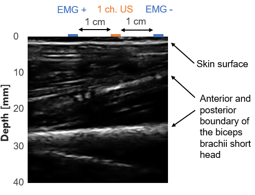
sEMG signals were acquired with one differential channel using two active, wet electrodes placed on the biceps brachii according to the SENIAM guidelines [19]. The interelectrode distance was . The bias electrode was attached to the elbow. US measurements are based on a linear array transducer with 32 channels and a central frequency of (LA-32, Vermon), which offers a favourable trade-off between penetration depth and axial resolution. The transducer was placed between the sEMG electrodes and fixed on biceps brachii using an elastic band (see Fig. 1). Short cables were used to interconnect the transducer with WULPUS. The probe was configured to drive eight central channels simultaneously in a plane-wave mode (for increased transmit energy) and to receive the echo signal from a single (the centre-most) channel. Fig. 2 shows a longitudinal B-mode image of the biceps brachii short head (acquired with an Ultrasonix Sonix 01 RP), on top of which we highlight the location of the A-mode US channel and the sEMG electrodes.
II-B2 Measurement Protocol
The measurement protocol involved three repetitions of rest and isometric voluntary contractions, corresponding to a duty cycle of .
II-C Mode of operation and firmware implementation
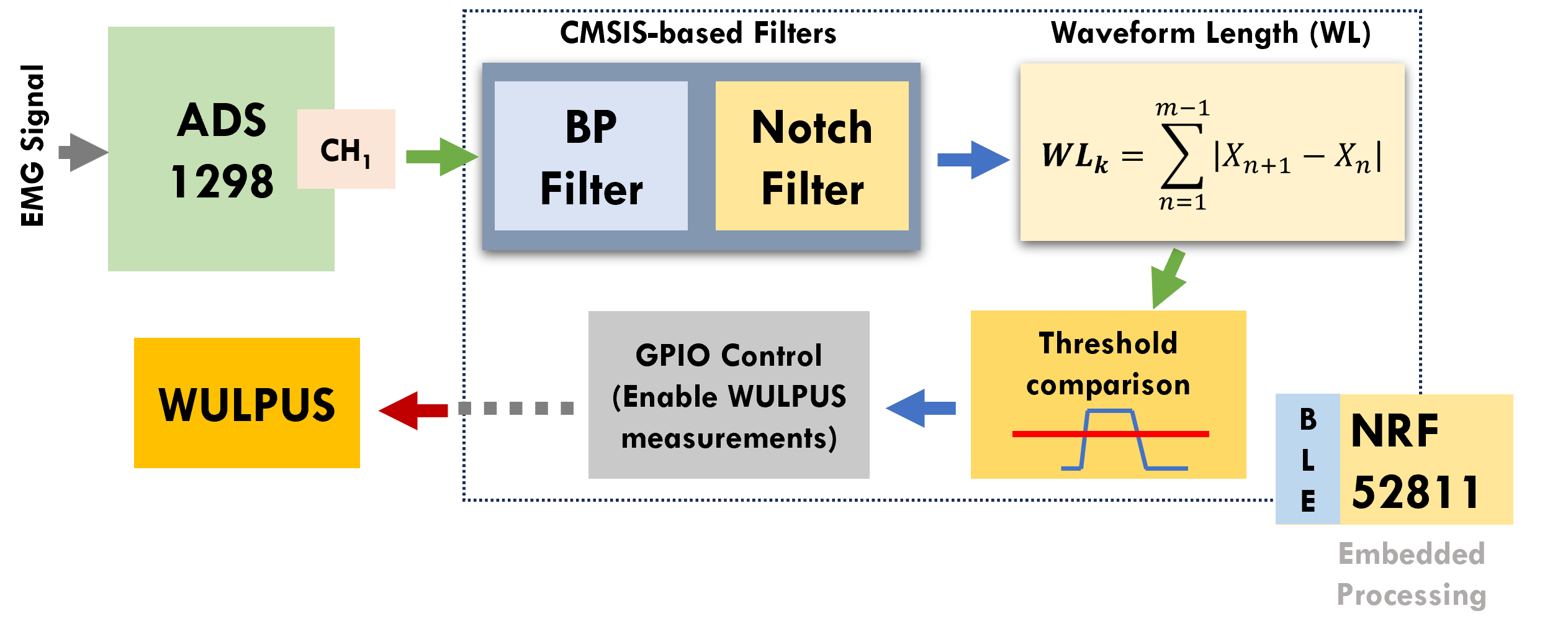
Our system operates by continuously processing sEMG data to activate the US system when muscle activity is present. The embedded processing scheme is summarized in Fig. 3 and starts with BioGAP sampling a single differential sEMG channel through the ADS1298. Sampled data are received by the nRF52811 MCU, where it undergoes Band-pass (BP) filtering (using 3-taps IIR Butterworth) and notch filtering (using 3-taps IIR Butterworth). The filtering process is implemented with ARM CMSIS libraries, with guarantees low processing time and high energy efficiency[20]. To concentrate exclusively on muscle activation-related features, we employ a Waveform Length (WL)-based envelope extraction method[21]. The WL extracts signal envelope by summing over the absolute difference between consecutive samples over a window of data (60 samples in this work). WL is also low in computational complexity (w.r.t root-mean-square or time-frequency algorithms such as wavelet transform), which renders the algorithm very suitable for low-power applications.
Once the WL signal surpasses a pre-established threshold (determined empirically as ), the SoC enables a dedicated General Purpose Input/Output (GPIO) output, connected to the MSP430 MCU of the US sub-system, which reacts by initiating a continuous A-mode US sampling operating at . The collected US data is subsequently transmitted to a PC through BLE communication. Optionally, the synchronized raw or processed EMG data can also be transmitted through a separate BLE channel. During periods of low EMG activation, indicated by values below the threshold and resulting in a low GPIO state, WULPUS enters a low-power mode.
III Results and discussion
III-A In vivo measurement
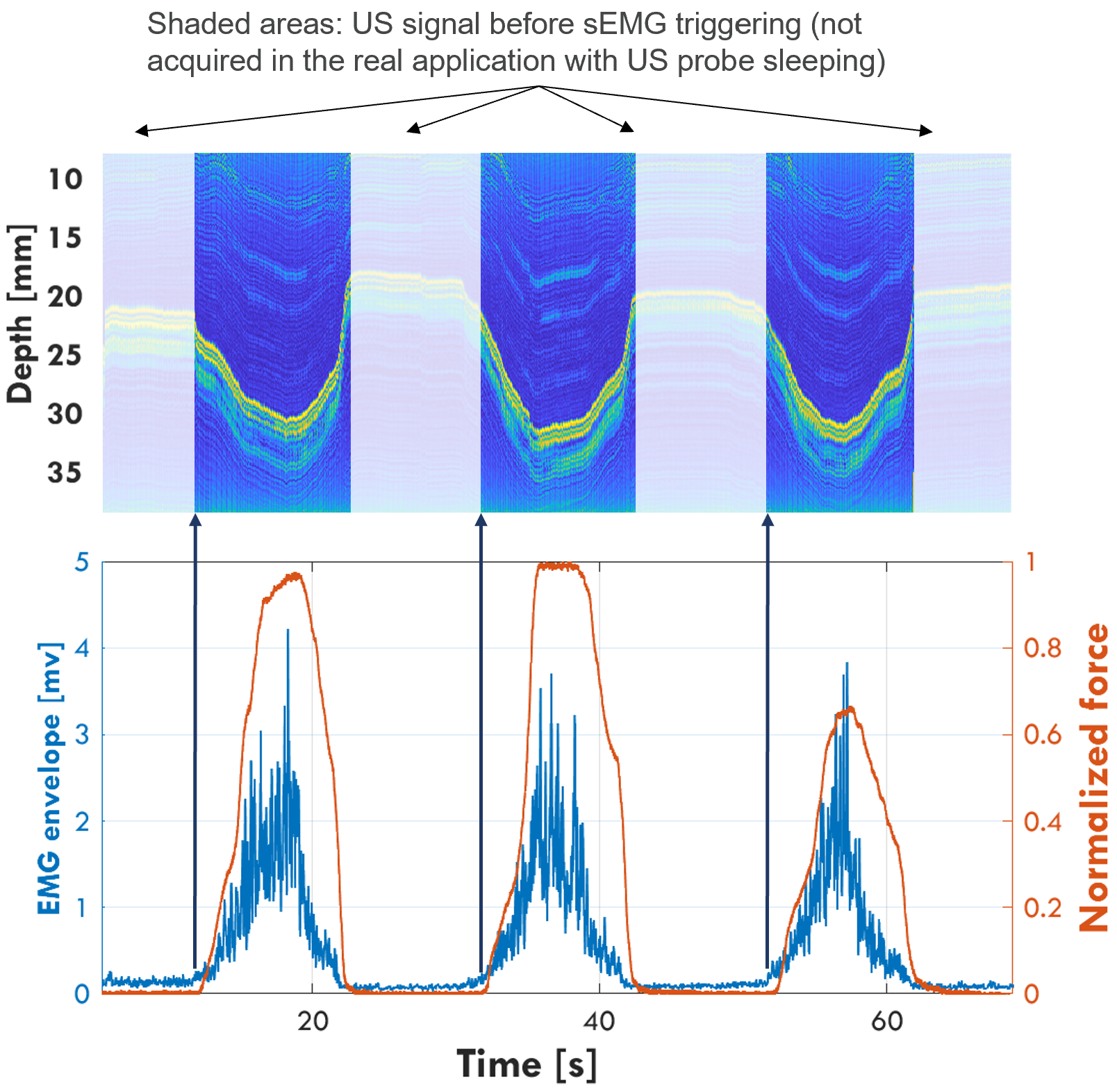
Fig. 4 (bottom) shows the force (normalized to a scale of 0 to 1 through division by the maximum value), as well as the sEMG envelope. Upon reaching the threshold of the sEMG envelope, the sEMG subsystem outputs the trigger (dark blue arrows), which activates the US measurement with a acquisition rate. Fig. 4 (top) shows the corresponding M-mode US measurement, where the three contractions can be seen clearly. The pronounced reflection shifting between approx. with each contraction corresponds to the posterior boundary of the short head of the biceps brachii. For the sake of clarity, the semi-transparent part of the figure also shows the US measurement when there is no sEMG activity. In the final application, the US subsystem is in sleep mode during periods of low sEMG activity, and such data are not collected.
III-B Power measurements
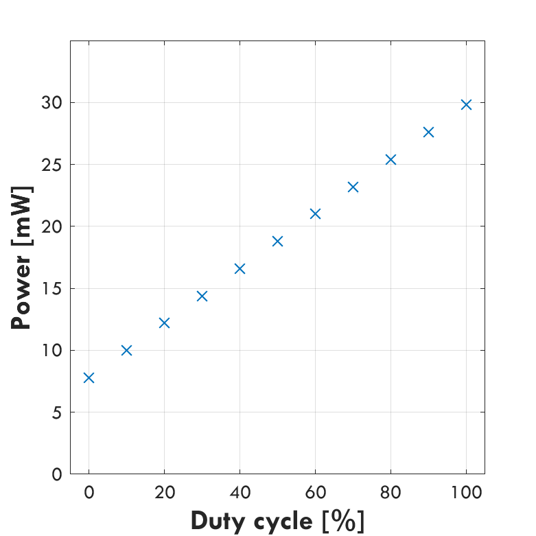
Figure 5 presents the power consumption analysis of the entire system at various muscle contraction duty cycles. At the baseline of 0% duty cycle (i.e., when BioGAP performs a single-channel sEMG measurement and onboard processing, while WULPUS is in sleep mode), the power consumption amounts to , corresponding to more than 6 days of battery life with a battery. As the duty cycle increases, the activation of the US system results in increasing power consumption. At 100% duty cycle (i.e., both systems are active all the time), the total power consumption amounts to , resulting in a battery life of more than 1.5 days.
IV Conclusion
This work demonstrates a physiologically-informed triggering approach that integrates sEMG signals with wearable US devices to improve the energy efficiency of heterogeneous biosignal acquisition systems. We demonstrate the use of sEMG signals as a trigger for initiating US measurements, as the electrical activity is responsible for the muscle contraction. The trigger generation is based on the enveloping and thresholding of sEMG signals and is faster than the electromechanical delay (), thereby enabling to initiate US acquisitions before the displacement of muscle fascicles starts. This approach allows the US system to be used only during the contraction phase, thereby boosting the battery lifetime. The system was implemented using WULPUS and BioGAP, two SoA systems for wearable acquisition of US and ExG, respectively. Selectively using the US probe ensures higher energy efficiency with respect to continuous sampling. With long contractions at contraction rate, the system consumes only , reducing the power envelope by compared to operating both sEMG and US continuously. The proposed solution paves the way for the development of energy-efficient sensor-fusion solutions for sEMG and US, where US data are collected only when significant information is available.
Acknowledgment
We thank A. Blanco Fontao and H. Gisler (ETH Zürich) for technical support.
References
- [1] G. L. Cerone, A. Botter, and M. Gazzoni, “A modular, smart, and wearable system for high density semg detection,” IEEE Transactions on Biomedical Engineering, vol. 66, no. 12, pp. 3371–3380, 2019.
- [2] C. J. De Luca, “The use of surface electromyography in biomechanics,” in J. Appl. Biomech., vol. 13, no. 2. Human Kinetics Publishers Inc., 5 1997, pp. 135–163.
- [3] S. Benatti, F. Casamassima, B. Milosevic, E. Farella, P. Schönle, S. Fateh, T. Burger, Q. Huang, and L. Benini, “A Versatile Embedded Platform for EMG Acquisition and Gesture Recognition,” IEEE Trans. Biomed. Circuits Syst., vol. 9, no. 5, pp. 620–630, 10 2015.
- [4] “Myoelectric prosthetics 101 — Ottobock US.” [Online]. Available: https://www.ottobockus.com/prosthetics/info-for-new-amputees/prosthetics-101/myoelectric-prosthetics-101/
- [5] M. B. I. Reaz, M. S. Hussain, and F. Mohd-Yasin, “Techniques of emg signal analysis: detection, processing, classification and applications,” Biological procedures online, vol. 8, pp. 11–35, 2006.
- [6] Y. Athavale and S. Krishnan, “Biosignal monitoring using wearables: Observations and opportunities,” Biomedical Signal Processing and Control, vol. 38, pp. 22–33, 2017.
- [7] E. Clancy, E. Morin, and R. Merletti, “Sampling, noise-reduction and amplitude estimation issues in surface electromyography,” Journal of Electromyography and Kinesiology, vol. 12, no. 1, pp. 1–16, 2002. [Online]. Available: https://www.sciencedirect.com/science/article/pii/S1050641101000335
- [8] B. Van Hooren et al., “Ultrasound imaging to assess skeletal muscle architecture during movements: a systematic review of methods, reliability, and challenges,” J. Appl. Physiol., vol. 128, no. 4, pp. 978–999, 2020.
- [9] G. Lichtwark, “Ultrasound Technology for Examining the Mechanics of the Muscle, Tendon, and Ligament,” in Handbook of Human Motion, Müller Bertram et al., Eds. Cham: Springer International Publishing, 2017, pp. 1–20.
- [10] M. Tanter, J. Bercoff, L. Sandrin, and M. Fink, “Ultrafast compound imaging for 2-D motion vector estimation: application to transient elastography,” IEEE Trans. Ultrason., Ferroelectr., Freq. Control, vol. 49, no. 10, pp. 1363–1374, 10 2002.
- [11] C. Leitner, S. Vostrikov, H. Penasso, P. A. Hager, A. Cossettini, L. Benini, and C. Baumgartner, “Detection of Motor Endplates in Deep and Pennate Skeletal Muscles in-vivo using Ultrafast Ultrasound,” in Proc. IEEE-IUS), 2020.
- [12] C. Leitner, S. Vostrikov, M. Tilp, P. A. Hager, A. Cossettini, L. Benini, and C. Baumgartner, “Human Fascicle Strain Behavior During Twitch using Ultrafast Ultrasound,” in Proc. IEEE-IUS, 2020.
- [13] S. Vostrikov et al., “Hand gesture recognition via wearable ultra-low power ultrasound and gradient-boosted tree classifiers,” in Proc. IEEE IUS, 2023, pp. 1–4.
- [14] X. Yang, X. Sun, D. Zhou, Y. Li, and H. Liu, “Towards wearable a-mode ultrasound sensing for real-time finger motion recognition,” IEEE Transactions on Neural Systems and Rehabilitation Engineering, vol. 26, no. 6, pp. 1199–1208, 2018.
- [15] A. Botter, T. M. M. Vieira, I. D. Loram, R. Merletti, and E. F. Hodson-Tole, “A novel system of electrodes transparent to ultrasound for simultaneous detection of myoelectric activity and b-mode ultrasound images of skeletal muscles,” Journal of Applied Physiology, vol. 115, no. 8, pp. 1203–1214, 2013.
- [16] S. Frey, M. Guermandi, S. Benatti, V. Kartsch, A. Cossettini, and L. Benini, “Biogap: a 10-core fp-capable ultra-low power iot processor, with medical-grade afe and ble connectivity for wearable biosignal processing,” in 2023 IEEE International Conference on Omni-layer Intelligent Systems (COINS), 2023, pp. 1–7.
- [17] S. Frey et al., “Wulpus: a wearable ultra low-power ultrasound probe for multi-day monitoring of carotid artery and muscle activity,” in Proc. IEEE IUS, 2022, pp. 1–4.
- [18] C. Leitner, S. Benatti, K. Keller, A. Cossettini, V. Kartsch, H. Penasso, L. Benini, F. Greco, and C. Baumgartner, “UStEMG: an Ultrasound Transparent Tattoo-based sEMG System for Unobtrusive Parallel Acquisitions of Muscle Electro-mechanics,” in 2021 43rd Annual International Conference of the IEEE Engineering in Medicine & Biology Society (EMBC), Nov. 2021, pp. 7077–7082, iSSN: 2694-0604.
- [19] “Seniam guidelines.” [Online]. Available: http://www.seniam.org/
- [20] ARM, “CMSIS Library,” 2016. [Online]. Available: https://www.arm.com/products/processors/cortex-m/cortex-microcontroller-software-interface-standard.php
- [21] V. Kartsch et al., “Using Low-Power, Low-Cost IoT Processors in Clinical Biosignal Research: An In-depth Feasibility Check,” in Proc. EMBC, vol. 2020-July, jul 2020, pp. 4008–4011.