:
\theoremsep
\jmlrvolume
\jmlryear
\jmlrsubmitted
\jmlrpublished
\jmlrworkshop
Retrieving Evidence from EHRs with LLMs:
Possibilities and Challenges
Abstract
Unstructured Electronic Health Record (EHR) data often contains critical information complementary to imaging data that would inform radiologists’ diagnoses. However, time constraints and the large volume of notes frequently associated with individual patients renders manual perusal of such data to identify relevant evidence infeasible in practice. Modern Large Language Models (LLMs) provide a flexible means of interacting with unstructured EHR data, and may provide a mechanism to efficiently retrieve and summarize unstructured evidence relevant to a given query. In this work, we propose and evaluate an LLM (Flan-T5 XXL) for this purpose. Specifically, in a zero-shot setting we task the LLM to infer whether a patient has or is at risk of a particular condition; if so, we prompt the model to summarize the supporting evidence. Enlisting radiologists for manual evaluation, we find that this LLM-based approach provides outputs consistently preferred to a standard information retrieval baseline, but we also highlight the key outstanding challenge: LLMs are prone to hallucinating evidence. However, we provide results indicating that model confidence in outputs might indicate when LLMs are hallucinating, potentially providing a means to address this.
keywords:
NLP, LLMs, radiology1 Introduction
We consider using Large Language Models (LLMs) as interfaces to unstructured data (e.g., notes) in patient Electronic Health Records (EHRs), ultimately to aid radiologists performing imaging diagnosis. The motivation here is that unstructured evidence within EHR may support (or render less likely) particular diagnostic hypotheses radiologists come to based on imaging alone, but time constraints—combined with the often lengthy records associated with individual patients—make manually finding and drawing upon such evidence infeasible in practice. Consequently, radiologists often perform diagnosis with comparatively little knowledge of patient history.
Modern LLMs offer a flexible mechanism to interface with unstructured EHR data. For example, recent work has shown that LLMs can perform “zero-shot” information extraction from clinical notes with reasonable accuracy (Agrawal et al., 2022). In this work, we propose and evaluate an approach to extract evidence from clinical notes in order to aid diagnosis. More concretely, we envision a clinician first providing an initial suspected diagnosis as a query. The model (an LLM) should then confirm whether there is unstructured (textual) evidence in the patient record that might support this diagnosis, and—if so—summarize this for the clinician. Figure 1 provides a schematic of the approach.
LLMs provide an attractive mechanism to permit such interactions given their flexibility and established dexterity working with unstructured text. We anticipate—and empirically verify in this work—that they will therefore be able to find and summarize evidence relevant to an arbitrary query more capably than “traditional” (which is to say, pre-LLM) information retrieval (IR) methods. Critically, they can also answer general questions (e.g., “Is this patient at risk of Atrial fibrillation?”) and provide summaries of supporting evidence identified; retrieval methods do not offer such functionality. However, they also bring serious challenges: Skillful as they are, it is well-known that LLMs are also prone to “hallucinating” content (Azamfirei et al., 2023; Zhang et al., 2023).
We therefore perform an empirical evaluation with practicing radiologists to assess both the potential benefits and risks of using LLMs to aid diagnosis. The expert evaluations confirm that LLMs are more capable than a standard IR system at surfacing and summarizing evidence relevant to a given diagnosis. However, such models also bring inherent challenges. How can we know, e.g., that the summary of evidence supporting a given condition is in fact faithful to the patient record at hand? We highlight several examples where the LLM fabricates plausible patient history that would support a condition of interest. This is potentially dangerous and certainly frustrates the provider, who must then read through the record carefully to ascertain that there is in fact no such evidence, eliminating both the safety and efficiency benefits hoped for from use of LLMs.
Our contributions are summarized as follows. (1) We introduce an approach in which we task an LLM (specifically, Flan-T5 XXL; Chung et al. 2022) to assess whether a patient is at risk of or has a given condition, and to produce a conditional summary of any corresponding supporting evidence. We conduct expert evaluation of this and find it considerably outperforms baseline evidence retrieval approaches. (2) We show in-depth examples that highlight key challenges to using LLMs for this task; hallucinated content. This points to future research needed to address issues currently precluding the use of LLMs as an interface to EHR.
2 Retrieving and summarizing evidence with LLMs
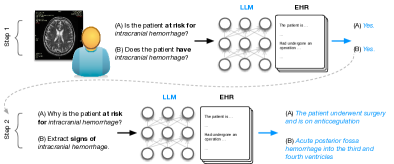
For a given query ( condition), we attempt to retrieve two distinct types of evidence from patient history: (A) Snippets that indicate a patient may be at risk of developing the condition in the future, and (B) those that suggest the patient currently has the condition. For example, a patient on anticoagulants after a recent posterior fossa surgery may be at risk of an intracranial hemorrhage (but not experiencing one currently). By contrast, observing acute posterior fossa hemorrhage indicates the patient most likely has intracranial hemorrhage.
Extracting evidence for risk informs clinicians about occurrences in the patient’s history (such as procedures, diagnoses) that make them more vulnerable to the condition. Extracting evidence for signs of a condition serves two purposes: signs that occur in the patient’s immediate history indicate the patient likely has the condition. Signs that occur earlier indicate the patient has a history of the condition which also serves as important information.
We use Flan-T5 XXL as our base LLM. While larger, proprietary models may offer superior results, we wanted to use an accessible LLM to ensure reproducibility. Moreover, protections for patient privacy mandated by the Health Insurance Portability and Accountability Act (HIPAA), and our institutional policy on use of LLM restrict us to using models that can be deployed “in-house”, precluding hosted variants (e.g., those provided by OpenAI).
Zero-shot sequential prompting
We adopt a sequential prompting approach to find and summarize evidence. First we ask the LLM whether a given note indicates that the corresponding patient is at risk for, or has a given query diagnosis—this prompts the LLM for a binary decision regarding these. When the answer is ‘Yes’, we prompt the model to provide support for its response.
Specifically, to query whether the patient is at risk for the given diagnosis, we use the prompts below.
‘Read the following clinical note of a patient: [NOTE].
Question: Is the patient at risk of [DIAGNOSIS]?
Choice -Yes -No.
Answer:
To elicit supporting evidence from the model for such risk predictions, we use the following prompt.
‘Read the following clinical note of a patient: [NOTE].
Answer step by step: based on the note, why is the patient at risk of [DIAGNOSIS]?
Answer:
Similarly, to query whether the patient has a given diagnosis, we ask the model instead “ Question: Does the patient have [DIAGNOSIS]?” (asking for a binary response). And then to obtain evidence supporting this assessment (in the case of a positive response), we prompt with: “Question: Extract signs of [DIAGNOSIS] from the note.”.
In the above prompts, [NOTE] denotes a patient note, and [DIAGNOSIS] a potential diagnosis for which we would like to retrieve supporting evidence. We then combine and present the result for the two types of evidence (risks and signs) to the end user.
Few-shot prompting
We also experimented with a single few-shot prompt to extract evidence (see Appendix A), but preliminary results were not promising so we did not pursue further.
A retrieval baseline
As a point of comparison for unsupervised evidence extraction, we use a simple ranking approach using neural embeddings.111Other baselines (e.g., BM25, TF-IDF) are possible, but the expert time needed for annotations limit our ability. Specifically, given a query [DIAGNOSIS], we retrieve associated [RISK FACTORS] using GPT-3.5 and generate an embedding of the sentence: ‘Risk factors of [DIAGNOSIS] include [RISK FACTORS]’ using ClinicalBERT (Alsentzer et al., 2019).222Note that this does not entail passing any sensitive data to OpenAI; we send only a condition name. Table 3 shows examples of risk factors provided by GPT-3.5. The intuition here is to generate -grams that are likely to indicate risk of the corresponding diagnosis so that we can match these against notes in EHR. Then, for a patient and [DIAGNOSIS], we retrieve the top sentences in the patient notes most similar to . One downside of such a retrieval-based approach is the need to pre-specify the number of evidence snippets to retrieve (here, we arbitrarily set this to 20). By contrast, our approach implicitly adjusts this threshold. We refer to this baseline as CBERT.
| Diagnosis | Notes | Evidence |
|---|---|---|
| MIMIC | ||
| intracranial hemorrhage* | ||
| stroke | ||
| small vessel disease | ||
| pneumocephalus | ||
| sinusitis | ||
| total | 188 | 67 |
| BWH | ||
| small vessel disease | ||
| chemoradiation necrosis | ||
| demyelination | ||
| brain tumor | ||
| intracranial hypotension | ||
| craniopharyngioma | ||
| infarction | ||
| sinus disease | ||
| total | 144 | 117 |
3 Data
For evaluation we worked with radiologists (specializing in neuroimaging) from the Brigham and Women’s Hospital in Boston (BWH). For experiments, we used a private dataset from this hospital and the publicly available MIMIC-III (Johnson et al., 2016) dataset, to ensure that our findings are robust and (partially) reproducible.
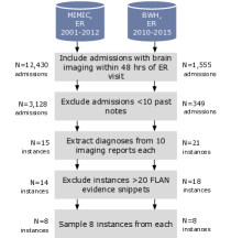
BWH dataset comprises patients admitted to the Emergency Room (ER) of BWH between 2010 and 2015 along with clinical notes including: cardiology, endoscopy, operative, pathology, pulmonary, radiology reports, and discharge summaries. We sampled patients who underwent brain imaging within 48 hours of their ER visit because they are likely to have undetermined diagnoses. We are interested in scenarios where patients are associated with a large volume of EHR data, so we included patients with 10 EHR notes.
MIMIC-III is a publicly available database of deidentified EHR from patients admitted to the Intensive Care Unit (ICU) of the Beth Israel Deaconess Medical Center between and . It contains both structured data (e.g, demographics, vital sign measurements, lab test results), and unstructured data (e.g., nurse and physician notes, ECG and radiology reports and discharge summaries). Similar to the BWH dataset, we sampled patients that underwent brain imaging within 48 hours of their ER or Urgent Care visit, whose EHR included notes.
We sampled data for individual patients, but evaluated models with respect to diagnoses. For example, if a patient report mentioned ‘stroke’ and ‘sinusitis’, the radiologist evaluated the surfaced evidence for each condition independently. To reduce annotation effort, we discarded diagnoses with more than 20 pieces of evidence and finally sampled 8 instances from each source to create our final evaluation dataset. See Figure 2 for a schematic of our data sampling procedure. Table 1 reports statistics about the set of examples used for evaluation.
4 Evaluation
For expert evaluation, one of the collaborating radiologists identified all diagnoses discussed in the Findings and Impressions sections of the radiology reports of 10 patients from each dataset (excluding MIMIC-III patients from the pilot study).333While this is a relatively small number of patients, we emphasize that manual evaluation is expensive: Radiologists on our team spent 9 hours manually assessing outputs. Then, for each diagnosis, we retrieved supporting evidence from all patient notes using the zero-shot prompting strategy from Section 2. Three collaborating radiologists then manually assessed each retrieved piece of evidence.
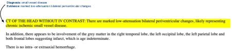
Figure 3 shows the evaluation interface that our radiologist team-members used to assess model outputs. Because the relevance of an evidence snippet inherently depends on the context, we ask radiologists to ground their assessments by assuming the following hypothetical setting: “You are a radiologist reviewing a scan of a patient in the ER. Based on the scan, you are concerned that the patient has the diagnosis stated below. Assess the relevance of the retrieved evidence to support your inference.” For each piece of evidence surfaced by a model, radiologists answered two questions:
Is the evidence present in the note? LLMs can hallucinate evidence. Therefore, we first ask radiologists to confirm whether the model generated evidence is in fact supported by the note on the basis of which it was produced. To aid the radiologists in finding the corresponding sentences, we compute ClinicalBERT (Alsentzer et al., 2019) embeddings of sentences in the notes and highlight those with a cosine similarity of with the ClinicalBERT embedding of the generated evidence. This heuristic approach realizes high precision but low recall. Therefore, if a highlighted sentence is incongruous with generated evidence, we ask radiologists to read through the entire note to try and manually identify support.
Note that the (non-generative) retrieval method to which we compare as a baseline is extractive, and so incapable of hallucinating content; we nevertheless ask this question with regards to the baseline for consistency and to ensure blinding.
Is the evidence relevant? If the generated evidence is supported by the note, we ask radiologists whether it is relevant to the query diagnosis. Specifically, we collect assessments on the following scale.
0: Not Useful The information is not useful; is irrelevant to the query condition (e.g., ‘The patient is on a ventilator’ for pneumocephalus).
1: Weak Correlation The information surfaced has a plausible but probably weak correlation with the query condition (e.g., ‘The patient is in a flutter’ for intracranial hemorrhage).
2: Useful The retrieved evidence is relevant and may inform one’s diagnostic assessment (e.g., ‘The patient has a TBI’ for intracranial hemorrhage).
3: Very Useful The retrieved evidence is clearly relevant and would likely inform diagnosis (e.g., ‘hypertension’ for small vessel disease (SVD)).
To capture whether the two models we evaluated—one an LLM, the other a retrieval approach—provide distinct evidence, we asked annotators if one model surfaced relevant evidence that the other did not.
We also ask radiologists to choose which of the two models they subjectively prefer and to offer any relevant comments. Finally, we record the time taken to evaluate each piece of evidence.
5 Results
To first assess agreement between radiologists, we had all of them annotate evidence surfaced by the LLM for one particular patient, selected at random from the BWH dataset. For this patient, the model generated 10 pieces of (potentially) relevant evidence for the query chemoradiation necrosis. On this shared set, the inter-annotator agreement score (average pairwise Cohen’s ) for relevance assessments between the three radiologists was .
To verify a given piece of evidence provided by a model it took radiologists an average of and seconds for FLAN-T5 and CBERT models, respectively. Evaluating FLAN-T5 demands more time because the highlights are approximations (heuristically matched to generations) and so do not always map correctly to the sentence corresponding to the evidence. Evaluating hallucinations takes even longer ( seconds per piece of evidence) because radiologists must peruse the note multiple times to confirm that said evidence is not present.
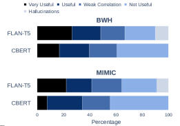
Figure 4 shows results (see Table 2 for examples). Radiologists found (MIMIC) and (BWH) of the evidence generated by FLAN-T5 to be (very) useful. In comparison, (MIMIC) and (BWH) of CBERT’s evidence was (very) useful. (MIMIC) and (BWH) of the evidence generated by FLAN-T5 showed weak correlations (discussed in Section 5.3). (MIMIC) and (BWH) of evidence was hallucinated (discussed in 5.2). ((MIMIC) and (BWH) of the evidence was deemed not useful; the latter was primarily true information about the patient’s condition which was unrelated to the diagnosis.
5.1 Binary decision recall
As discussed above, we first ask the LLM whether a given note indicates that the corresponding patient is at risk for, or has, a given query diagnosis. The precision of this LLM inference is implicitly measured by the assessment of generated evidence; if the patient does not have and/or is not at risk for a given condition, any generated evidence will necessarily be irrelevant. But this does not capture recall, i.e., how sensitive the model is with respect to identifying when a patient indeed has or is at risk of a condition.
Therefore, to additionally estimate model recall with respect to inferences based on notes, we sampled patients from the BWH data and followed prior work (McInerney et al., 2020) in terms of evaluation. Specifically, we asked radiologists to browse reports from up to one year after the patient’s reference radiology report and tag relevant diagnoses; these can then be viewed as “future” diagnoses with respect to the original reference report. The radiologists then selected past notes containing supporting evidence for these diagnoses. Out of the notes marked as containing evidence, FLAN-T5 correctly identified , corresponding to a recall of .
fig:misc
\subfigure[Cross evidence similarity]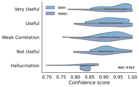 \subfigure[Language Model likelihood]
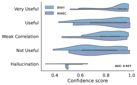
5.2 Hallucinations
Concerningly, some of the model hallucinations flagged by radiologists were about risk factors that are highly relevant to the query diagnosis. We provide a few illustrative examples:
Example 1 For a patient with demyelination as the query diagnosis, the model hallucinated the evidence ‘axonal degeneration’. Demyelination is commonly viewed as the primary factor responsible for the deterioration of axons within multiple sclerosis lesions. The model also hallucinated signs of demyelination as evidence (‘numbness and tingling in the arms and legs’). There was no evidence in the record indicating axonal degeneration or either of these symptoms.
Example 2 For a patient with chemo-radiation necrosis as the query diagnosis, the model hallucinated that ‘the patient had a history of chemo-radiation necrosis’. A history of radiation necrosis would be (very) relevant to its diagnosis, but there was no such history in the EHR.
In other instances, the model hallucinated vague evidence such as ‘The patient is taking a lot of medications that can cause small vessel disease’ for small vessel disease as the query diagnosis (a radiologist went through the note and was unable to find mention of any such medication).
Can hallucinations be identified by soft-matching the evidence back with the note?
A possible way to identify hallucinations is to try to ‘match’ generated evidence to text within patient notes. If so, this intuitively suggests the generated content is also present in the underlying note. Practically, we do this by generating embeddings of generated evidence texts and sentences in notes and scoring their similarities; we can then consider a generation supported if this similarity exceeds some threshold.
Note, however, that this approach may construe accurate evidence as ‘hallucinated’ if the the generation is comparatively abstractive. For example, the model output ‘The patient has had multiple heart surgeries’ is true, but has a low ClinicalBERT embedding similarity score with the corresponding sentence ‘This is a 64 year-old gentelman with known coronary artery disease status post an inferoposterior myocardial infarction in [**2116**] who is status post redo-CABG times six and bovine AVR’ in the note.
Because abstraction—the ability to render relevant patient history concisely—is a major benefit of LLMs, using heuristic matching to noisily filter outputs (limiting to ostensibly ‘faithful’ cases) does not seem a particularly promising approach.
How certain is the model about the hallucinations?
We evaluate the degree to which model uncertainty, quantified both via normalized output likelihoods under the LM and using ‘self-consistency’ (Huang et al., 2022; Si et al., 2022), can be used to infer when the model is ‘hallucinating’ content. The latter method entails eliciting ‘Chain-of-Thought’ (CoT) reasoning (Wei et al., 2022) from models repeatedly, sampling tokens at each step to yield multiple reasoning paths. The fraction of these that resolve to the same final output (i.e., which are consistent) can then be taken as a proxy for model certainty in that output. Outputs here are evidence summaries, so we take the average similarity of sampled outputs as a proxy for certainty (details in Appendix C).
We find that both methods provide confidence scores that are highly indicative of hallucinations; this can be seen in Figure 5. This is promising, because it suggests we might be able to simply abstain from providing outputs in such cases.
5.3 Weakly correlating evidence
One factor complicating our evaluation is that the LLM often surfaced evidence which might have a relatively weak correlation with the query condition. One could argue that the model was ‘correct’ in retrieving such evidence from a population epidemiology perspective, but incorrect from an individual patient clinical perspective. In other words, the evidence is so weakly correlated with the condition that it is minimally useful. (see Appendix E.1 for examples).
5.4 Preferences
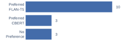
Figure 6 shows how often radiologists preferred each model. FLAN-T5 provided comparatively precise and concise output. Abstractive evidence was considered better than the extractive snippets from CBERT, which often chunked useful evidence with neighboring irrelevant sentences (notes are usually poorly formatted, making sentence-parsing difficult). See Appendix E.1 for examples. While radiologists largely preferred FLAN-T5 (XXL), CBERT was preferred in three cases:
(1) Poor precision For pneumocephalus, of the evidence surfaced by FLAN-T5 was not useful.
(2) Poor recall: In the case of chemoradiation necrosis, FLAN-T5 had a poor recall and failed to retrieve essential evidence related to the patient’s radiation therapy. This may be because of the term ‘chemoradiation’, which is more commonly referred to as ‘radiation necrosis’. Changing the diagnosis to ‘radiation necrosis’, resulted in the model retreiving evidence related to the patient’s radiation therapy.
(3) Supported alternate diagnosis: Interestingly, our radiologist preferred CBERT for the case of demyelination because it helped confirm that the patient did have demyelination, but in fact had a glioma (tumor). Demyelinating lesions and glioma present similar imaging characteristics and can be difficult to diagnose based on conventional MR imaging (Toh et al., 2012). A brain biopsy is often conducted to differentiate between the two. All the evidence evaluated as (very) useful were snippets from the pathology report discussing the tests and related results that indicated that demyelination was less likely and that the findings were most consistent with glioma.
| Evaluation | Diagnosis | Evidence | Explanation |
|---|---|---|---|
| Very Useful | intracranial hemorrhage | Recent fossa surgery and now on anticoagulants | Surgery in the brain inevitably leaves some hemorrhage. Anticoagulants increase the risk of hemorrhage. ‘Recent surgery’ and ‘anticoagulants’ make hemorrhage highly likely. |
| Useful | infarction | There is calcified thrombus obstructing the origins of the M2 branches | ‘Thrombus’ is diagnostic of infarction, which is very useful information. But ‘calcified thrombus’ implies chronicity, so the thrombus could have been present for a long time and there may not be an acute infarction at this time. |
| Weak Correlation | pneumocephalus | patient was involved in a motorcycle accident | A traumatic head injury is an important risk factor of pneumocephalus. A motorcycle accident increases the likelihood of a head injury. |
| Not Useful | small vessel disease (SVD) | patient is at risk of endocarditis | Not helpful in diagnosing SVD. |
| Hallucination | intracranial hemorrhage | patient has a brain tumor | Not present in the note (and relevant to the diagnosis). |
5.5 Robustness to query variation
As mentioned in Section 5.4, FLAN-T5 performed better when the diagnosis ‘chemoradiation necrosis’ was revised to the more commonly used ‘radiation necrosis’. Another such case was ‘sinus disease’, more commonly known as sinusitis. The model did well in this case; of the evidence was useful. CBERT, however, surfaced evidence for cardiac sinus disease (sinus bradycardia, sinus tachycardia, etc). Much of the evidence was related to the patient’s cardiac history which would be relevant if they had cardiac sinus disease. of the evidence was hence not useful.
6 Related Work
NLP for EHR.
Navigating EHRs is cumbersome, motivating several efforts in summarization of and information extraction from EHR (Pivovarov and Elhadad, 2015). For example, in recent related work, Jiang et al. (2023) created a proactive note retrieval system based on the current clinical context to aid note-writing. Adams et al. (2021) considered “hospital-course summarization”, aiming to condense the notes of a patient visit into a paragraph, while Liang et al. (2019) proposed to create disease-specific extractive summaries from clinical notes.
NLP in Radiology.
Previous works regarding NLP in radiology primarily focus on processing radiology reports. Some work has sought to generate the the clinical Impression section based on the Findings section of reports (Van Veen et al., 2023; Zhang et al., 2019; Sotudeh et al., 2020). Other efforts have focussed on extracting specific observations from radiology reports (Smit et al., 2020; Jaiswal et al., 2021), and modeled disease progression using radiology reports (Di Noto et al., 2021; Khanna et al., 2023).
The prior works most relevant to this effort concern aiding radiologists in diagnosing conditions. (McInerney et al., 2020) propose using a distantly supervised model (trained to predict ICD codes) to perform extractive summarization conditioned on a diagnoses. Our work addresses the problem in a zero-shot setting. Tang et al. (2023) address diagnostic uncertainty by suggesting less likely diagnosis to radiologists by learning to differentiate between likely and less likely diagnoses via contrastive learning.
More broadly, recent work has shown the potential of ML and NLP to assist radiologists across a range of tasks, including breast cancer screening (Wu et al., 2020), malignant lung nodules detection (Sim et al., 2020), and chest X-ray interpretation (Seah et al., 2021). Suggesting diagnosis (Tang et al., 2023) based on findings and supporting it with evidence (McInerney et al., 2020) also fall in this area.
7 Discussion and Limitations
We have proposed and evaluated a method for using LLMs to retrieve and summarize evidence from patient records which might be relevant to a particular condition or diagnosis of interest, with the ultimate aim of aiding radiologists performing imaging diagnosis. Expert evaluations of model outputs performed by radiologists suggest that this is a promising approach, in that annotators tended to prefer LLM outputs to simple retrieval results.
But there are important limitations to the approach and to our evaluation. For example, we found that LLMs are prone to hallucinating (plausible) evidence, potentially hindering their utility for the envisioned use. However, our results also suggest that confidence scores might allow one to pro-actively identify hallucinations, and abstain in such cases. Interestingly, confidence also seems to correlate with perceived usefulness. This suggests an interesting direction to explore in future work.
Our evaluation was limited in a few key ways, which might reduce the generalizability of our findings. First, we enlisted a small set of radiologists to perform in-depth evaluation of a small number of instances. This is because evaluation is time consuming: We re-emphasize that this exercise required substantial allocation (9 hours) of scarce expert time. Another limitation here is that we considered only one LLM (specifically, FLAN-T5): Other LLMs might, naturally, perform better or worse. Finally, we did not extensively iterate on the specific prompts used, and this too could substantially affect results.
References
- Adams et al. (2021) Griffin Adams, Emily Alsentzer, Mert Ketenci, Jason Zucker, and Noémie Elhadad. What’s in a summary? laying the groundwork for advances in hospital-course summarization. In Proceedings of the conference. Association for Computational Linguistics. North American Chapter. Meeting, volume 2021, page 4794. NIH Public Access, 2021.
- Agrawal et al. (2022) Monica Agrawal, Stefan Hegselmann, Hunter Lang, Yoon Kim, and David Sontag. Large language models are zero-shot clinical information extractors. arXiv preprint arXiv:2205.12689, 2022.
- Alsentzer et al. (2019) Emily Alsentzer, John Murphy, William Boag, Wei-Hung Weng, Di Jindi, Tristan Naumann, and Matthew McDermott. Publicly available clinical BERT embeddings. In Proceedings of the 2nd Clinical Natural Language Processing Workshop, pages 72–78, Minneapolis, Minnesota, USA, June 2019. Association for Computational Linguistics. 10.18653/v1/W19-1909. URL https://aclanthology.org/W19-1909.
- Azamfirei et al. (2023) Razvan Azamfirei, Sapna R Kudchadkar, and James Fackler. Large language models and the perils of their hallucinations. Critical Care, 27(1):1–2, 2023.
- Chung et al. (2022) Hyung Won Chung, Le Hou, Shayne Longpre, Barret Zoph, Yi Tay, William Fedus, Eric Li, Xuezhi Wang, Mostafa Dehghani, Siddhartha Brahma, et al. Scaling instruction-finetuned language models. arXiv preprint arXiv:2210.11416, 2022.
- Di Noto et al. (2021) Tommaso Di Noto, Chirine Atat, Eduardo Gamito Teiga, Monika Hegi, Andreas Hottinger, Meritxell Bach Cuadra, Patric Hagmann, and Jonas Richiardi. Diagnostic surveillance of high-grade gliomas: towards automated change detection using radiology report classification. In Joint European Conference on Machine Learning and Knowledge Discovery in Databases, pages 423–436. Springer, 2021.
- Honnibal and Montani (2017) Matthew Honnibal and Ines Montani. spaCy 2: Natural language understanding with Bloom embeddings, convolutional neural networks and incremental parsing. To appear, 2017.
- Huang et al. (2022) Jiaxin Huang, Shixiang Shane Gu, Le Hou, Yuexin Wu, Xuezhi Wang, Hongkun Yu, and Jiawei Han. Large language models can self-improve. arXiv preprint arXiv:2210.11610, 2022.
- Jaiswal et al. (2021) Ajay Jaiswal, Liyan Tang, Meheli Ghosh, Justin F Rousseau, Yifan Peng, and Ying Ding. Radbert-cl: Factually-aware contrastive learning for radiology report classification. In Machine Learning for Health, pages 196–208. PMLR, 2021.
- Jiang et al. (2023) Sharon Jiang, Shannon Shen, Monica Agrawal, Barbara Lam, Nicholas Kurtzman, Steven Horng, David Karger, and David Sontag. Conceptualizing machine learning for dynamic information retrieval of electronic health record notes. arXiv preprint arXiv:2308.08494, 2023.
- Johnson et al. (2016) Alistair EW Johnson, Tom J Pollard, Lu Shen, Li-wei H Lehman, Mengling Feng, Mohammad Ghassemi, Benjamin Moody, Peter Szolovits, Leo Anthony Celi, and Roger G Mark. Mimic-iii, a freely accessible critical care database. Scientific data, 3(1):1–9, 2016.
- Khanna et al. (2023) Sameer Khanna, Adam Dejl, Kibo Yoon, Quoc Hung Truong, Hanh Duong, Agustina Saenz, and Pranav Rajpurkar. Radgraph2: Modeling disease progression in radiology reports via hierarchical information extraction. arXiv preprint arXiv:2308.05046, 2023.
- Liang et al. (2019) Jennifer Liang, Ching-Huei Tsou, and Ananya Poddar. A novel system for extractive clinical note summarization using ehr data. In Proceedings of the Clinical Natural Language Processing Workshop, pages 46–54, 2019.
- McInerney et al. (2020) Denis Jered McInerney, Borna Dabiri, Anne-Sophie Touret, Geoffrey Young, Jan-Willem Meent, and Byron C Wallace. Query-focused ehr summarization to aid imaging diagnosis. In Machine Learning for Healthcare Conference, pages 632–659. PMLR, 2020.
- Pivovarov and Elhadad (2015) Rimma Pivovarov and Noémie Elhadad. Automated methods for the summarization of electronic health records. Journal of the American Medical Informatics Association, 22(5):938–947, 2015.
- Seah et al. (2021) Jarrel CY Seah, Cyril HM Tang, Quinlan D Buchlak, Xavier G Holt, Jeffrey B Wardman, Anuar Aimoldin, Nazanin Esmaili, Hassan Ahmad, Hung Pham, John F Lambert, et al. Effect of a comprehensive deep-learning model on the accuracy of chest x-ray interpretation by radiologists: a retrospective, multireader multicase study. The Lancet Digital Health, 3(8):e496–e506, 2021.
- Si et al. (2022) Chenglei Si, Zhe Gan, Zhengyuan Yang, Shuohang Wang, Jianfeng Wang, Jordan Boyd-Graber, and Lijuan Wang. Prompting gpt-3 to be reliable. arXiv preprint arXiv:2210.09150, 2022.
- Sim et al. (2020) Yongsik Sim, Myung Jin Chung, Elmar Kotter, Sehyo Yune, Myeongchan Kim, Synho Do, Kyunghwa Han, Hanmyoung Kim, Seungwook Yang, Dong-Jae Lee, et al. Deep convolutional neural network–based software improves radiologist detection of malignant lung nodules on chest radiographs. Radiology, 294(1):199–209, 2020.
- Smit et al. (2020) Akshay Smit, Saahil Jain, Pranav Rajpurkar, Anuj Pareek, Andrew Y Ng, and Matthew P Lungren. Chexbert: combining automatic labelers and expert annotations for accurate radiology report labeling using bert. arXiv preprint arXiv:2004.09167, 2020.
- Sotudeh et al. (2020) Sajad Sotudeh, Nazli Goharian, and Ross W Filice. Attend to medical ontologies: Content selection for clinical abstractive summarization. arXiv preprint arXiv:2005.00163, 2020.
- Tang et al. (2023) Liyan Tang, Yifan Peng, Yanshan Wang, Ying Ding, Greg Durrett, and Justin F Rousseau. Less likely brainstorming: Using language models to generate alternative hypotheses. arXiv preprint arXiv:2305.19339, 2023.
- Toh et al. (2012) CH Toh, K-C Wei, S-H Ng, Y-L Wan, M Castillo, and C-P Lin. Differentiation of tumefactive demyelinating lesions from high-grade gliomas with the use of diffusion tensor imaging. American journal of neuroradiology, 33(5):846–851, 2012.
- Turpin et al. (2023) Miles Turpin, Julian Michael, Ethan Perez, and Samuel R Bowman. Language models don’t always say what they think: Unfaithful explanations in chain-of-thought prompting. arXiv preprint arXiv:2305.04388, 2023.
- Van Veen et al. (2023) Dave Van Veen, Cara Van Uden, Maayane Attias, Anuj Pareek, Christian Bluethgen, Malgorzata Polacin, Wah Chiu, Jean-Benoit Delbrouck, Juan Manuel Zambrano Chaves, Curtis P Langlotz, et al. Radadapt: Radiology report summarization via lightweight domain adaptation of large language models. arXiv preprint arXiv:2305.01146, 2023.
- Wei et al. (2022) Jason Wei, Xuezhi Wang, Dale Schuurmans, Maarten Bosma, Fei Xia, Ed Chi, Quoc V Le, Denny Zhou, et al. Chain-of-thought prompting elicits reasoning in large language models. Advances in Neural Information Processing Systems, 35:24824–24837, 2022.
- Wolf et al. (2020) Thomas Wolf, Lysandre Debut, Victor Sanh, Julien Chaumond, Clement Delangue, Anthony Moi, Pierric Cistac, Tim Rault, Remi Louf, Morgan Funtowicz, Joe Davison, Sam Shleifer, Patrick von Platen, Clara Ma, Yacine Jernite, Julien Plu, Canwen Xu, Teven Le Scao, Sylvain Gugger, Mariama Drame, Quentin Lhoest, and Alexander Rush. Transformers: State-of-the-art natural language processing. In Proceedings of the 2020 Conference on Empirical Methods in Natural Language Processing: System Demonstrations, pages 38–45, Online, October 2020. Association for Computational Linguistics. 10.18653/v1/2020.emnlp-demos.6. URL https://aclanthology.org/2020.emnlp-demos.6.
- Wu et al. (2020) Nan Wu, Jason Phang, Jungkyu Park, Yiqiu Shen, Zhe Huang, Masha Zorin, Stanisław Jastrzębski, Thibault Févry, Joe Katsnelson, Eric Kim, Stacey Wolfson, Ujas Parikh, Sushma Gaddam, Leng Leng Young Lin, Kara Ho, Joshua D. Weinstein, Beatriu Reig, Yiming Gao, Hildegard Toth, Kristine Pysarenko, Alana Lewin, Jiyon Lee, Krystal Airola, Eralda Mema, Stephanie Chung, Esther Hwang, Naziya Samreen, S. Gene Kim, Laura Heacock, Linda Moy, Kyunghyun Cho, and Krzysztof J. Geras. Deep neural networks improve radiologists’ performance in breast cancer screening. IEEE Transactions on Medical Imaging, 39(4):1184–1194, 2020. 10.1109/TMI.2019.2945514.
- Zhang et al. (2023) Muru Zhang, Ofir Press, William Merrill, Alisa Liu, and Noah A Smith. How language model hallucinations can snowball. arXiv preprint arXiv:2305.13534, 2023.
- Zhang et al. (2019) Yuhao Zhang, Derek Merck, Emily Bao Tsai, Christopher D Manning, and Curtis P Langlotz. Optimizing the factual correctness of a summary: A study of summarizing radiology reports. arXiv preprint arXiv:1911.02541, 2019.
Appendix A Few-shot Prompting
We provide additional details on our preliminary experiments with few-shot prompting. We randomly sampled patients from the MIMIC dataset and followed McInerney et al. (2020)’s approach of using ‘future’ ICD code as query diagnosis. We also randomly sampled ICD codes (excluding the patient’s future diagnoses) as negative query diagnosis. We used the following prompt: {quoting} Read the following clinical note of a patient: [RANDOM NOTE SNIPPET].
Answer step by step: can the patient possibly have cardioembolic strokes in the future?
Answer: There is no evidence. Final answer: No.
Read the following clinical note of a patient: patient stopped taking a blood thinning medication required for a heart valve due to side effects.
Answer step by step: can the patient possibly have cardioembolic strokes in the future?
Answer: The patient stopped taking a blood thinning medication required for a heart valve. The medication thins the blood and prevents blood clots. Blood clots can lead to strokes. Final answer: Yes.
Read the following clinical note of a patient: [NOTE].
Answer step by step: based on the note, why is the patient at risk of [DIAGNOSIS]?
Answer:
We observed that with few-shot prompting the model surfaced evidence for almost every diagnosis. However, some pieces of evidence were clearly wrong. For a patient with ‘with g/j tube in place for gastroparesis’, the model’s output for the negative diagnosis, encephalitis, was ‘The patient has a jejunostomy tube in place. The jejunostomy tube can be pulled out. The jejunostomy tube can be pulled out of the body. The jejunostomy tube can be pulled out of the body and into the brain. Final answer: Yes’. We suspect the prompt biases the model to support the query diagnosis which then makes the model generate incorrect explanations as evidence (Turpin et al., 2023). We also experimented with prompts such as ‘Extract evidence for [DIAGNOSIS]. Output N/A if no evidence exists’ but the model then mostly generated ‘N/A’. Given these results, we carried the rest of the evaluation with the zero-shot prompting approach.
Appendix B Retrieval Baseline
Table 3 shows examples of risk factors provided by GPT-3.5.
| Diagnosis | Risk Factors |
|---|---|
| pneumocephalus | head injury, skull fracture, neurosurgical procedures, sinus or mastoid surgery, meningitis, cerebrospinal fluid leak, barotrauma, diving or scuba diving accidents, iatrogenic causes, such as lumbar puncture or spinal anesthesia |
| stroke | hypertension, smoking, diabetes, obesity, sedentary lifestyle, high cholesterol levels, atrial fibrillation, family history of stroke, previous history of stroke, excessive alcohol consumption, drug abuse. |
| intracranial hemorrhage | hypertension, aneurysms, arteriovenous malformations, blood clotting disorders, trauma, drug abuse, liver disease, brain tumor, stroke, coagulopathy |
| brain tumor | progression genetics, exposure to ionizing radiation, family history of brain tumors, certain hereditary conditions, weakened immune system, previous history of brain tumor. |
| infarction | smoking, high blood pressure, high cholesterol, diabetes, obesity, sedentary lifestyle, family history of heart disease, excessive alcohol consumption, and drug abuse. |
| intracranial hypotension | obesity, connective tissue disorders, previous spinal or cranial surgery, leaking cerebrospinal fluid, spinal epidural anesthesia, lumbar puncture or spinal tap |
| Data | Model | Hallucinations | Not Useful | Weak Correlations | Useful | Very Useful |
|---|---|---|---|---|---|---|
| MIMIC | FLAN | |||||
| CBERT | ||||||
| BWH | FLAN | |||||
| CBERT |
| Diagnosis | Evidence | Explanation |
|---|---|---|
| intracranial hemorrhage | patient had multiple cardiac surgeries | Multiple cardiac surgeries may suggest anticoagulation or underlying cardiac dysfunction which could in turn predispose the patient to intracranial hemorrhage. |
| intracranial hypotension | The patient has a ventriculoperitoneal shunt. | A ventriculoperitoneal shunt (VPS) is a surgical device used to relieve intracranial pressure by draining excessive cerebrospinal fluid. Having a VPS catheter may increase the risk of intracranial hypotension due to over drainage. |
| craniopharyngioma | s/p resection X2, s/p VPS and panhypopitutiarism with second resection | Panhypopituitarism and the fact that something was removed through surgery suggests there was a tumor involving the sella which may or may not have been craniopharyngioma. |
Appendix C Self-Consistency
In general, self-consistency calculates the “confidence” the model has in a given output as the fraction of sampled reasoning paths that resolve to it (out of all such paths; Si et al. 2022). Computing self-consistency is straightforward when the final answer is from a fixed answer set or when an exact string match serves as a good metric of consistency. Neither of the two conditions, however, apply to diagnosis evidence extraction. Evidence generation is open-ended; we therefore use cross-evidence similarity as a confidence score.
Specifically, given a note and diagnosis , we sample decoding paths, with temperature0 after prompting for evidence for in . We then compute cross-evidence similarity of as:
| (1) |
Where denotes cosine similarity and are feature representations of evidence texts; we use ClinicalBERT to derive these. A low similarity score suggests that the generations are diverse and therefore dissimilar.
We sample decoding paths for each evidence using temperature sampling () and top- sampling (). Figure 5 reports cross-evidence similarity distributions associated with pieces of evidence marked as having varying utility. The model is inconsistent and generates a variety of unrelated evidence when hallucinating. Table 6 provides examples of consistent and inconsistent evidence generation.
| Consistent Evidence | Inconsistent Evidence |
|---|---|
| The patient has vascular calcifications | The patient has a history of ischemic heart disease. |
| The patient has vascular calcifications. | The patient is having heart rhythm problems. |
| vascular calcifications in the left upper quadrant of the abdomen | The heart is at risk of ischemia. |
| vascular calcifications | The patient is a non-smoker. |
| vascular calcifications in the left upper quadrant of the abdomen | The patient has a pulmonary embolism. |
| The patient has vascular calcifications. | The patient has a heart murmur. |
| The patient has a history of atrial fibrillation | numbness and tingling in the arms and legs |
| The patient developed rapid atrial fibrillation. | The patient is a 63 year old male who presents with a complaint of numbness and tingling in his left arm |
| The patient has developed rapid atrial fibrillation. | JC/jp |
| The patient developed rapid atrial fibrillation. | The patient had a white matter lesion of the brain |
| The patient has a history of atrial fibrillation. | axonal loss |
| The patient developed rapid atrial fibrillation this morning. | marrow swelling |
Appendix D Implementation Details
We used the HuggingFace (Wolf et al., 2020) library to run inference using FLAN-T5 XXL and ClinicalBERT. We split notes into sentences using the spaCy (en_core_web_sm) (Honnibal and Montani, 2017) library. We processed notes in chunks of size tokens (including the prompt text) for FLAN-T5 and ran 8-bit inference. When generating evidence, we set max_tokens to . When computing cross-evidence similarity, we set max_tokens to as this was sufficient to measure similarity across evidence.
Appendix E Results
E.1 Weak correlations
Table 5 shows examples of evidence evaluated to be weak correlations.
E.2 Evidence conciseness
| FLAN-T5 | CBERT |
|---|---|
| CAD (s/p stents x 2, MIs, on Coumadin INR=1.9) hx of TIAs in past 2.5 yrs multiple AAAs (largest last measured at 5.5 cm, surg intervention held 2/2 cardiac status | IMMUNIZATIONS: INFLUENZA VACCINE (INACTIVATED) IM Given [DATE] ALLERGY: AMOXICILLIN ADMIT DIAGNOSIS: Stroke PRINCIPAL DISCHARGE DIAGNOSIS ;Responsible After Study for Causing Admission) same OTHER DIAGNOSIS;Conditions,Infections,Complications,affecting Treatment/Stay CAD (s/p stents x 2, MIs, on Coumadin INR=1.9) hx of TIAs in past 2.5 yrs multiple AAAs (largest last measured at 5.5 cm, surg intervention held 2/2 cardiac status OPERATIONS AND PROCEDURES: None.OTHER TREATMENTS/PROCEDURES (NOT IN O.R.) |
| The patient has a high ICP and low CPP | AT THIS TIME, NO FAMILY PRESENT NEURO- EYES DO NOT OPEN, PERTLA @ 2.0 VERY SLUGGISHLY, WITHDRAWS FROM PAIN WITH ALL 4 EXTREMITIES, MOVES EXTREMITIES UNPURPOSEFULLY ON BED, NO SEDATION GIVEN, OETT, VENTRICULOSTOMY OPEN TO CONTINUOUS DRAIN, ICP AS HIGH AS 29, CPP RUNNING LOW- BUT STABILIZING, GOALS- |
| The patient has a TBI | A/P- S/P REPAIR [**Doctor Last Name **] & LL ORTHOPEDIC INJURIES STABLE TBI W/CLOSE MONITORING FOR CHANGES STABLE LIVER LAC AT PRESENT SUCCESSFULL WEAN/EXTUBATION POST-OP PAIN CONT TO MONITOR PER ORDERS- Q2/HR NEURO & PERIPHERAL VASCULAR CHECKS…? |
Table 7 shows demonstrative examples of when FLAN-T5 was more concise than CBERT.