Melting, bubble-like expansion and explosion of superheated plasmonic nanoparticles
Abstract
We report on time-resolved coherent diffraction imaging of gas-phase silver nanoparticles, strongly heated via their plasmon resonance. The x-ray diffraction images reveal a broad range of phenomena for different excitation strengths, from simple melting over strong cavitation to explosive disintegration. Molecular dynamics simulations fully reproduce this behavior and show that the heating induces rather similar trajectories through the phase diagram in all cases, with the very different outcomes being due only to whether and where the stability limit of the metastable superheated liquid is crossed.
Condensed matter, when heated slowly, undergoes the familiar phase transitions from solid to liquid to gas. Extremely fast heating, on the other hand, results in additional phenomena, such as strong overheating of solids or liquids or even a change in the chemical bonding itself due to strongly excited electrons [1, 2, 3, 4]. Many aspects of matter under such extreme conditions, like the coupling between a very hot electron gas and the ionic system, are not yet fully understood [5, 6]. Isolated nanoscale particles have been identified as well-controlled test objects for the study of highly excited matter [7, 8, 9], especially in combination with single-shot coherent diffraction imaging (CDI) using intense x-ray free-electron laser pulses [10, 11, 12, 13, 14], which permits to follow the dynamics with high spatiotemporal resolution [15]. Up to now, most time-resolved studies concentrated on rare gas particles in free flight, excited by strong laser fields [15, 16, 9, 17, 18], and a few other systems like silicon dioxide particles [19]. Time-resolved diffraction studies on metal nanoparticles were mainly performed using particles supported on surfaces, leading to the observation of vibrational excitation, melting, or disintegration [20, 21, 22, 23, 24, 25, 26, 27]. However, an unambiguous analysis of the particle dynamics free from the spurious and mostly unknown influence of the support is only possible in the gas phase.
In this work, we study silver nanoparticles heated in free flight by excitation of their plasmon resonance with moderately intense picosecond laser pulses. Such particles have been shown to form well-defined faceted crystalline structures at lower temperatures [28, 29, 30], distinctly different from the round shape of a liquid droplet, which should facilitate the observation of melting. Furthermore, they exhibit a strong Mie plasmon resonance in the near UV [31, 32], which permits to excite them using fairly weak laser fields [32, 33, 34, 35, 36]. This excitation leads to a rather uniform heating of the particle via very fast formation of a hot electron gas and a subsequent transfer of the energy to the nuclear degrees of freedom on a time scale of several picoseconds [36, 37, 38, 39, 40, 41]. Employing pump-probe CDI, we determine the morphology of the particles as a function of delay time. Accompanying molecular dynamics simulations of the process provide comprehensive insight into the origin of the observed phenomena. The strength of a decompression wave following the almost isochoric heating of the particles turns out to be a decisive parameter for the dynamics.
The experiment was performed at the free-electron laser (FEL) FLASH [42] using the CAMP endstation [43]. Silver nanoparticles with diameters in the range of were produced by a magnetron sputter gas aggregation source [44, 45], operated with a mixture of xenon and argon. The beam of neutral nanoparticles traversed a differential pumping stage before entering the main chamber. Here, the particles were intercepted by FEL pulses with about pulse duration at wavelength, focused to a spot size of . Photons scattered from the particles were collected by a pnCCD detector [46] at a distance of from the interaction region at a repetition rate of . Heating of the particles was achieved by laser pulses synchronized with the FEL pulses and overlaid in a near-collinear geometry. They were produced by a frequency doubled Ti:Sapphire femtosecond laser system and stretched to a duration of about by propagating through 100 mm of fused silica. We note that due to the time scale of the electron-phonon heat transfer of several picoseconds, shorter pulses would not increase the temporal resolution, but would just lead to unwanted strong fields effects. The pulses were weakly focused to a spot size of roughly diameter in the interaction region. Inhomogeneities in the laser beam profile did not permit a precise UV laser intensity determination; we estimate a maximal value of .
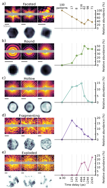
In Fig. 1 (a)-(e) examples of scattering images recorded with the pump pulse present are shown, along with real-space reconstructions obtained by iterative phase retrieval reconstruction of the small angle part of the images [30] (see the Supplemental Material [47] for details). Five different classes can be identified, based on the real-space features of the samples. The faceted class in Fig. 1(a) contains polyhedra with well-defined facets, which appear not to be strongly affected by the pump laser, either because the images were recorded at negative or small positive delay times, or because the particles have interacted only with a low-intensity part of the pump laser profile. Fig. 1(b) represents examples of the round class - these are mostly spherical particles with homogeneous density, most probably liquid droplets melted due to the heating. Fig. 1(c) shows the most spectacular class, hollow particles with a rounded outer surface and a large, close to spherical cavity inside. In some cases the remaining shell has a thickness of only about of its diameter. The fragmenting class (Fig. 1(d)) refers to particles with a very inhomogenous density, indicating disintegration probably due to fairly strong excitation. The exploded class (Fig. 1(e)) includes all patterns that correspond to unconnected fragments of the original particle, which leads to speckle-like diffraction patterns. These obviously are particles which completely disintegrated after the interaction with the pump laser. Interestingly, rather symmetric speckle patterns are sometimes observed (like in the third image in this row), corresponding to a small number of large fragments in a rather regular arrangement.
The relative abundances of these five classes as a function of the time delay between the pump laser and the FEL pulse are plotted in the right column of Fig. 1. The total number of identifiable diffraction images recorded at each time delay is given on the top, showing the overall low hitrate in this experiment. For negative and small positive time delays up to (here grouped together in a single data point) only faceted structures were observed. Other morphologies start to appear at a delay of . The abundance of faceted samples drops to a level of about within the first few hundreds of picoseconds. The abundance of round specimen exhibits the opposite behavior, rising up to in the first few hundreds of picoseconds. Exploded patterns appear even later, at around , and tend to become more and more abundant towards longer time delays. The other two classes appear only transiently. The contribution of the fragmenting class is significant only between and ; a similar behavior is seen for the hollow class, with the maximum abundance apparently shifted to slightly longer delays.
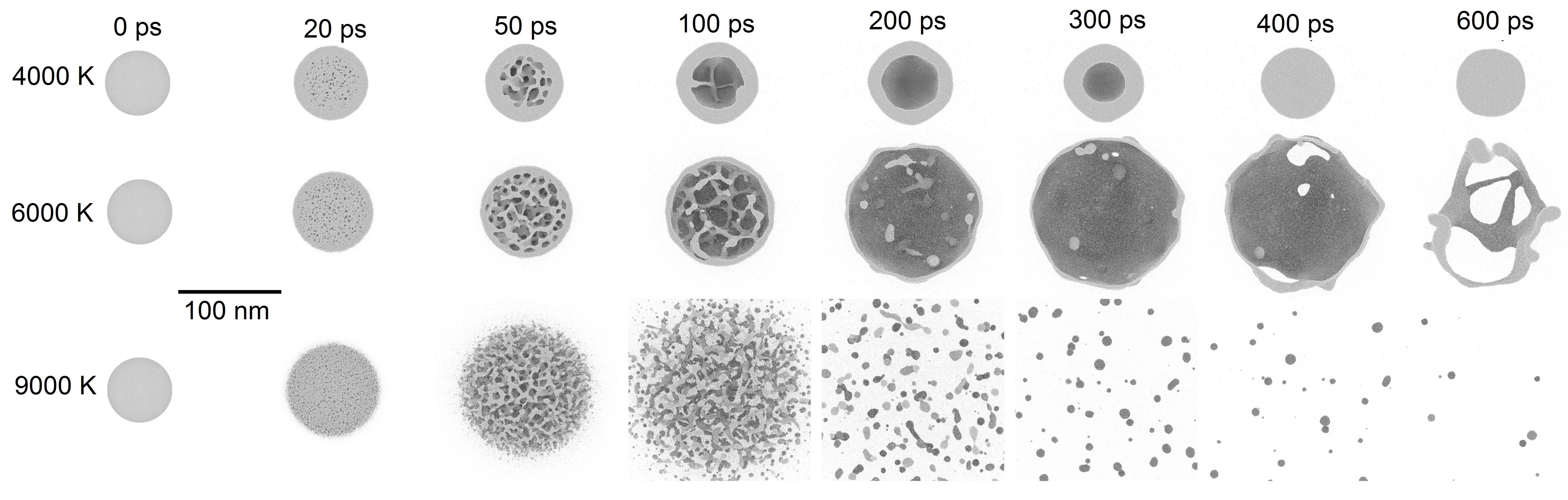
In order to better understand the observed dynamics, the temporal evolution of strongly heated silver particles was simulated by classical molecular dynamics (MD), employing the embedded-atom-method interaction potential of Sheng et al. [48]. Particles with atoms were simulated starting from initially spherical particles (R=), a shape which can be most easily compared to analytical models. In order to mimic the heating process by electron-phonon-coupling, the particles were thermalized for a duration of using a Langevin thermostat with a given temperature in a range between and , using a relaxation time of (see the Supplemental Material [47] for details). This would in principle allow the atomic system to reach a temperature of of the heat bath temperature. However, the actual temperatures at the end of the heating interval are lower due to cooling by evaporation, melting and expansion during the heating. Results of the MD simulations for three different heat bath temperatures are shown in Fig. 2. Snapshots of the nanoparticles are depicted at different times after the start of the heating process. In the Supplemental Material [47], videos of the dynamics are provided. In general, the simulation results are in line with those of earlier studies [26, 49, 50, 51]. In the case of a heat bath temperature of (first row), small voids start to form in the inner part of the sphere briefly before . These voids grow and coalesce into a single large cavity in the center of the particle within . This central cavity then contracts again, leading to a round liquid droplet at a delay time of 400 ps. For the case of (second row), the initial behaviour is similar, but the void formation now happens even closer to the particle surface. As before, the voids merge to form a single central cavity within , but with a substantially thinner outer shell. In this case, the surface tension is not strong enough to reverse the expansion of the bubble; it further expands and bursts in less than a nanosecond, forming rather large, initially tubular and eventually round fragments. The last case represents the dynamics of the system for a heat bath temperature of . Here, the formation of voids happens in the whole volume, even at the surface of the sample. This leads to a direct and violent disintegration of the sample within the first , with fragments spreading at significant velocities.
These results are in good agreement with the experimentally observed time evolution (Fig. 1). As mentioned, the faceted particles surviving up to long time delays (Fig. 1(a)) most probably have only weakly interacted with the pump laser. The appearance of round droplets around is compatible with still rather weakly excited particles, which just melt, or with slightly more strongly excited ones, where a central cavity appears and vanishes again, like in the simulation for heat bath temperature (Fig. 2). The observation of large bubbles only between and is congruent with the simulations at both and , where the bubble vanishes again at larger times, either by contraction or by fragmentation. The rapid increase of fragmenting samples already at can be assigned to early, fairly violent void formation, somewhere between the behavior simulated for and . The still significant presence of these samples up to around hints at processes more in line with the simulation, indicating bursting bubbles. Finally, the exploded samples can stem as well either from early violent fragmentation or a late bursting of a bubble - especially the third example in Fig. 1(e) hints at such a case, because of the small number of larger fragments detected. When comparing measured and simulated time dependencies, one should keep in mind that the size of the particles in the simulation is on the lower end of the size distribution of particles observed in the experiment; for the larger particles the dynamics can be expected to be slower than in the simulation.
The question arises why the single large cavity is always formed at the center of the particle. This can be understood with the help of the simulation results, as shown in Fig. 3. Rapid uniform heating of a particle leads to the buildup of a high pressure, which will eventually lead to an overall expansion of the particle. This expansion, however, does not occur via a simple breathing mode motion, but instead in form of a decompression wave: as an acceleration of the material requires a pressure gradient, the motion starts at the particle surface, with the boundary between moving material and material at rest propagating into the particle with the velocity of sound. [52, 26, 53, 49]. Fig. 3(a) shows the pressure within the particle during the heating phase and the following as a function of radial position for the case of a heat bath. One can see that a pressure of more than is reached in the particle center after of heating. The motion of the material sets in immediately when the heating is started, with the boundary between moving and non-moving layers propagating towards the particle center. The position of this boundary is indicated by the black line in Fig. 3(a) (see the Supplemental Material for radial profiles of the velocity distributions [47]). It reaches the particle center at about , which gives a velocity of sound of about under these conditions, slightly higher than the room temperature value of [54]. When this happens, all of the atoms are moving outwards, which leads to a strong pressure drop within the particle. This pressure drop is slightly less abrupt than might be expected due to the heating continuing until ; nevertheless, after the start of the heating, a negative pressure of about is reached (Fig. 3(a)). As further discussed below, at such a negative pressure cavitation sets in immediately, leading to the many voids as seen in Fig. 2 at for and heat bath temperature. The voids grow and merge into a larger cavity, surrounded by material which continues to move outward as it keeps the momentum gained in the first phase of the expansion. We want to stress that it is not the vapor pressure which drives the growing of this bubble, but rather the outward movement of its shell. Therefore, if this motion is not too fast, it can be reversed by surface tension, like in the case where the large central void starts to shrink again at about and has completely vanished at . For higher bath temperatures, the initial shell velocity is so high that the surface tension could reverse the motion only at much later stages of the expansion. Before that happens, however, any small hole formed in the thinning liquid layer leads to the rupture of the bubble [55, 56].
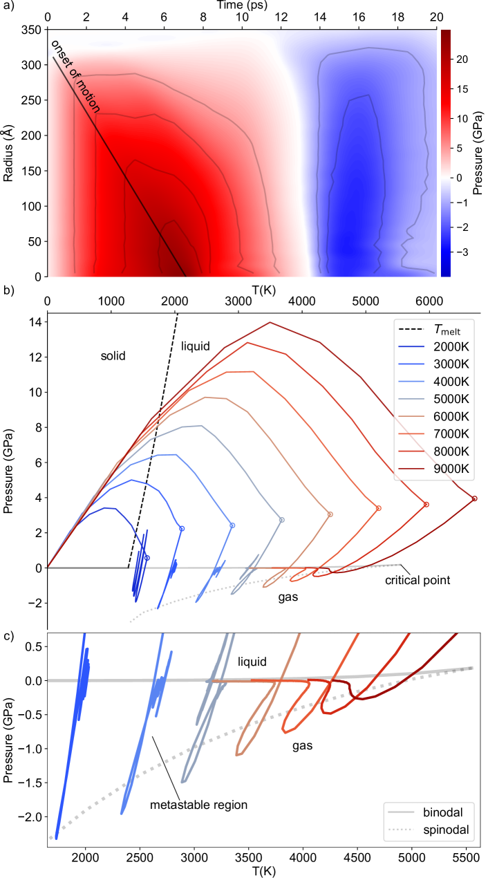
At even higher heat bath temperature the particle rips apart immediately. Nevertheless, the underlying mechanism is quite similar to the case of the cavitation process discussed before, as can be best seen by comparing the system trajectories through the phase diagram of silver as shown in Fig. 3(b) and 3(c). In Fig. 3(b) the state of silver is shown as a function of pressure and temperature; in Fig. 3(c) the region most relevant for the experiment is magnified. This phase diagram has been obtained from bulk silver MD simulations using the same interaction potential as for the silver particle simulations (see the Supplemental Material [47]). In it, the liquid and gaseous region are separated by the so-called binodal, which towards higher temperatures ends in the critical point. Below the binodal, matter should always be in the gaseous state. Nevertheless, the phase transition here is kinetically hindered by seed bubble formation; there is a critical bubble size, which has to be overcome, and which increases the closer the system is to the binodal. Only at a second boundary, the so-called spinodal, the liquid becomes unstable and homogeneous evaporation sets in [52, 57, 26]. Thus, in the region between the binodal and the spinodal, the liquid can be overheated, staying in a metastable liquid state, while this is not possible anymore beyond the spinodal. The evolution of the average temperature and pressure within the particles with time is indicated for eight different heat bath temperatures. One can observe a strong initial increase of temperature and pressure upon heating. This in fact moves the system away from the liquid-gas boundary, therefore somewhat stabilizing the liquid. Due to the particle expansion, the pressure drops again despite ongoing heating. When the heating ends, which is indicated by the circles, the temperature starts to drop as well and the systems dive through the binodal. Cavitation, however, sets in only when the systems cross the spinodal, which for the lower temperatures happens at significantly negative pressures. This process is very similar for all heating temperatures; what makes a difference for the highest temperatures is that here the spinodal is crossed at only slightly negative pressures. Therefore, even after expansion, the system stays close to the spinodal and rupturing continues until total disintegration is reached. One can also observe that for the lowest simulated heat bath temperature () the system does not reach the spinodal; in this case no void formation sets in. Instead the now liquid droplet exhibits a significant breathing mode vibration, a motion strongly damped by the void formation for the next three higher temperature cases which also end up in compact liquid droplets. Here, however, some breathing mode vibration is excited by the collapse of the cavity, most notably in the case.
In conclusion, we have demonstrated that the dynamics of strongly heated silver nanoparticles is dominated by a decompression wave caused by the near-isochoric heating, leading to distinct temperature and time dependent effects.
Our results show that time-resolved x-ray diffraction experiments on nanoparticles in the gas phase are ideally suited to study superheated matter with high spatial and temporal resolution, thus providing access to material properties at very high temperatures and pressures.
I Acknowledgements
We acknowledge DESY (Hamburg, Germany), a member of the Helmholtz Association HGF, for the provision of experimental facilities. Parts of this research were carried out at FLASH. Beamtime was allocated for proposal F-20170541. This research was supported in part through the Maxwell computational resources operated at Deutsches Elektronen-Synchrotron DESY, Hamburg, Germany. We acknowledge the Max Planck Society for funding the development and the initial operation of the CAMP end-station within the Max Planck Advanced Study Group at CFEL and for providing this equipment for CAMP@FLASH. The installation of CAMP@FLASH was partially funded by the BMBF grants 05K10KT2, 05K13KT2, 05K16KT3 and 05K10KTB from FSP-302. TM and BvI acknowledge funding by a DFG Koselleck Project MO 719/13 and IS61/14. AC, LH, MS, and DR acknowledge funding form Leibniz society, Germany, via grant no. SAW/2017/MBI4 and from SNF, Switzerland, via grant no. 200021E/193642. TR and MM acknowledge the Gauss Centre for Supercomputing e.V. for providing computing time on the GCS Supercomputer JUWELS [58] at Jülich Supercomputing Centre. Additional computing time was granted by the state of Baden-Württemberg through bwHPC and the DFG through grant no. INST39/961-1FUGG (bwForCluster NEMO).
References
- Recoules et al. [2006] V. Recoules, J. Clérouin, G. Zérah, P. M. Anglade, and S. Mazevet, Phys. Rev. Lett. 96, 055503 (2006).
- Lindenberg et al. [2005] A. M. Lindenberg, J. Larsson, K. Sokolowski-Tinten, K. J. Gaffney, C. Blome, O. Synnergren, J. Sheppard, C. Caleman, A. G. MacPhee, D. Weinstein, D. P. Lowney, T. K. Allison, T. Matthews, R. W. Falcone, A. L. Cavalieri, D. M. Fritz, S. H. Lee, P. H. Bucksbaum, D. A. Reis, J. Rudati, P. H. Fuoss, C. C. Kao, D. P. Siddons, R. Pahl, J. Als-Nielsen, S. Duesterer, R. Ischebeck, H. Schlarb, H. Schulte-Schrepping, T. Tschentscher, J. Schneider, D. von der Linde, O. Hignette, F. Sette, H. N. Chapman, R. W. Lee, T. N. Hansen, S. Techert, J. S. Wark, M. Bergh, G. Huldt, D. van der Spoel, N. Timneanu, J. Hajdu, R. A. Akre, E. Bong, P. Krejcik, J. Arthur, S. Brennan, K. Luening, and J. B. Hastings, Science 308, 392 (2005).
- Ernstorfer et al. [2009] R. Ernstorfer, M. Harb, C. T. Hebeisen, G. Sciaini, T. Dartigalongue, and R. D. Miller, Science 323, 1033 (2009).
- Medvedev and Milov [2020] N. Medvedev and I. Milov, Sci. Rep. 10, 12775 (2020).
- Mo et al. [2021] M. Mo, Z. Chen, and S. Glenzer, MRS Bull. 46, 694 (2021).
- Lee et al. [2021] J.-W. Lee, M. Kim, G. Kang, S. M. Vinko, L. Bae, M. S. Cho, H.-K. Chung, M. Kim, S. Kwon, G. Lee, C. H. Nam, S. H. Park, J. H. Sohn, S. H. Yang, U. Zastrau, and B. I. Cho, Phys. Rev. Lett. 127, 175003 (2021).
- Saalmann et al. [2006] U. Saalmann, C. Siedschlag, and J. M. Rost, J. Phys. B: At. Mol. Opt. Phys. 39, R39 (2006).
- Fennel et al. [2010] T. Fennel, K.-H. Meiwes-Broer, J. Tiggesbäumker, P.-G. Reinhard, P. M. Dinh, and E. Suraud, Rev. Mod. Phys. 82, 1793 (2010).
- Nishiyama et al. [2019] T. Nishiyama, Y. Kumagai, A. Niozu, H. Fukuzawa, K. Motomura, M. Bucher, Y. Ito, T. Takanashi, K. Asa, Y. Sato, D. You, Y. Li, T. Ono, E. Kukk, C. Miron, L. Neagu, C. Callegari, M. Di Fraia, G. Rossi, D. E. Galli, T. Pincelli, A. Colombo, T. Kameshima, Y. Joti, T. Hatsui, S. Owada, T. Katayama, T. Togashi, K. Tono, M. Yabashi, K. Matsuda, C. Bostedt, K. Nagaya, and K. Ueda, Phys. Rev. Lett. 123, 123201 (2019).
- Neutze and Moffat [2012] R. Neutze and K. Moffat, Curr. Opin. Struct. Biol. 22, 651 (2012).
- Marchesini et al. [2003] S. Marchesini, H. N. Chapman, S. P. Hau-Riege, R. A. London, A. Szoke, H. He, M. R. Howells, H. Padmore, R. Rosen, J. C. H. Spence, and U. Weierstall, Opt. Express 11, 2344 (2003).
- Raines et al. [2010] K. S. Raines, S. Salha, R. L. Sandberg, H. Jiang, J. A. Rodríguez, B. P. Fahimian, H. C. Kapteyn, J. Du, and J. Miao, Nature 463, 214 (2010).
- Loh et al. [2012] N. D. Loh, C. Y. Hampton, A. V. Martin, D. Starodub, R. G. Sierra, A. Barty, A. Aquila, J. Schulz, L. Lomb, J. Steinbrener, R. L. Shoeman, S. Kassemeyer, C. Bostedt, J. Bozek, S. W. Epp, B. Erk, R. Hartmann, D. Rolles, A. Rudenko, B. Rudek, L. Foucar, N. Kimmel, G. Weidenspointner, G. Hauser, P. Holl, E. Pedersoli, M. Liang, M. M. Hunter, L. Gumprecht, N. Coppola, C. Wunderer, H. Graafsma, F. R. N. C. Maia, T. Ekeberg, M. Hantke, H. Fleckenstein, H. Hirsemann, K. Nass, T. A. White, H. J. Tobias, G. R. Farquar, W. H. Benner, S. P. Hau-Riege, C. Reich, A. Hartmann, H. Soltau, S. Marchesini, S. Bajt, M. Barthelmess, P. Bucksbaum, K. O. Hodgson, L. Strüder, J. Ullrich, M. Frank, I. Schlichting, H. N. Chapman, and M. J. Bogan, Nature 486, 513 (2012).
- Bostedt et al. [2012] C. Bostedt, E. Eremina, D. Rupp, M. Adolph, H. Thomas, M. Hoener, A. R. B. de Castro, J. Tiggesbäumker, K.-H. Meiwes-Broer, T. Laarmann, H. Wabnitz, E. Plönjes, R. Treusch, J. R. Schneider, and T. Möller, Phys. Rev. Lett. 108, 093401 (2012).
- Gorkhover et al. [2016] T. Gorkhover, S. Schorb, R. Coffee, M. Adolph, L. Foucar, D. Rupp, A. Aquila, J. D. Bozek, S. W. Epp, B. Erk, L. Gumprecht, L. Holmegaard, A. Hartmann, R. Hartmann, G. Hauser, P. Holl, A. Hömke, P. Johnsson, N. Kimmel, K.-U. Kühnel, M. Messerschmidt, C. Reich, A. Rouzée, B. Rudek, C. Schmidt, J. Schulz, H. Soltau, S. Stern, G. Weidenspointner, B. White, J. Küpper, L. Strüder, I. Schlichting, J. Ullrich, D. Rolles, A. Rudenko, T. Möller, and C. Bostedt, Nat. Photonics 10, 93 (2016).
- Flückiger et al. [2016] L. Flückiger, D. Rupp, M. Adolph, T. Gorkhover, M. Krikunova, M. Müller, T. Oelze, Y. Ovcharenko, M. Sauppe, S. Schorb, C. Bostedt, S. Düsterer, M. Harmand, H. Redlin, R. Treusch, and T. Möller, New J. Phys. 18, 043017 (2016).
- Bacellar et al. [2022] C. Bacellar, A. S. Chatterley, F. Lackner, C. D. Pemmaraju, R. M. P. Tanyag, D. Verma, C. Bernando, S. M. O. O’Connell, M. Bucher, K. R. Ferguson, T. Gorkhover, R. N. Coffee, G. Coslovich, D. Ray, T. Osipov, D. M. Neumark, C. Bostedt, A. F. Vilesov, and O. Gessner, Phys. Rev. Lett. 129, 073201 (2022).
- Langbehn et al. [2022] B. Langbehn, Y. Ovcharenko, A. Clark, M. Coreno, R. Cucini, A. Demidovich, M. Drabbels, P. Finetti, M. Di Fraia, L. Giannessi, C. Grazioli, D. Iablonskyi, A. C. LaForge, T. Nishiyama, V. O. Á. de Lara, C. Peltz, P. Piseri, O. Plekan, K. Sander, K. Ueda, T. Fennel, K. C. Prince, F. Stienkemeier, C. Callegari, T. Möller, and D. Rupp, New J. Phys. 24, 113043 (2022).
- Peltz et al. [2022] C. Peltz, J. A. Powell, P. Rupp, A. Summers, T. Gorkhover, M. Gallei, I. Halfpap, E. Antonsson, B. Langer, C. Trallero-Herrero, C. Graf, D. Ray, Q. Liu, T. Osipov, M. Bucher, K. Ferguson, S. Möller, S. Zherebtsov, D. Rolles, E. Rühl, G. Coslovich, R. N. Coffee, C. Bostedt, A. Rudenko, M. F. Kling, and T. Fennel, New J. Phys. 24, 043024 (2022).
- Ihm et al. [2019] Y. Ihm, D. H. Cho, D. Sung, D. Nam, C. Jung, T. Sato, S. Kim, J. Park, S. Kim, M. Gallagher-Jones, Y. Kim, R. Xu, S. Owada, J. H. Shim, K. Tono, M. Yabashi, T. Ishikawa, J. Miao, D. Y. Noh, and C. Song, Nat. Commun. 10, 2411 (2019).
- Clark et al. [2015] J. N. Clark, L. Beitra, G. Xiong, D. M. Fritz, H. T. Lemke, D. Zhu, M. Chollet, G. J. Williams, M. M. Messerschmidt, B. Abbey, R. J. Harder, A. M. Korsunsky, J. S. Wark, D. A. Reis, and I. K. Robinson, Proc. Natl. Acad. Sci. U.S.A. 112, 7444 (2015).
- Clark et al. [2013] J. N. Clark, L. Beitra, G. Xiong, A. Higginbotham, D. M. Fritz, H. T. Lemke, D. Zhu, M. Chollet, G. J. Williams, M. Messerschmidt, B. Abbey, R. J. Harder, A. M. Korsunsky, J. S. Wark, and I. K. Robinson, Science 341, 56 (2013).
- Von Reppert et al. [2016] A. Von Reppert, R. Sarhan, F. Stete, J. Pudell, N. Del Fatti, A. Crut, J. Koetz, F. Liebig, C. Prietzel, and M. Bargheer, J. Phys. Chem. C 120, 28894 (2016).
- Jung et al. [2021] C. Jung, Y. Ihm, D. H. Cho, H. Lee, D. Nam, S. Kim, I.-T. Eom, J. Park, C. Kim, Y. Kim, J. Fan, N. Ji, J. R. Morris, S. Owada, K. Tono, J. H. Shim, H. Jiang, M. Yabashi, T. Ishikawa, D. Y. Noh, and C. Song, Sci. Adv. 7, eabj8552 (2021).
- Sung et al. [2021] D. Sung, D. Nam, M. jin Kim, S. Kim, K. S. Kim, S.-Y. Park, S. M. Hwang, C. Jung, H. Lee, D. H. Cho, M. Kim, I. Eom, S. Y. Lee, C. Song, and S. Kim, Appl. Sci. 11, 5082 (2021).
- Wu and Zhigilei [2014] C. Wu and L. V. Zhigilei, Appl. Phys. A 114, 11 (2014).
- Shin et al. [2023] J. Shin, C. Jung, Y. Ihm, S.-P. Heo, D. Nam, S. Kim, M. Kim, I. Eom, J. H. Shim, D. Y. Noh, and C. Song, Nano Lett. 23, 1481 (2023).
- Reinhard et al. [1997] D. Reinhard, B. D. Hall, D. Ugarte, and R. Monot, Phys. Rev. B 55, 7868 (1997).
- Barke et al. [2015] I. Barke, H. Hartmann, D. Rupp, L. Flückiger, M. Sauppe, M. Adolph, S. Schorb, C. Bostedt, R. Treusch, C. Peltz, S. Bartling, T. Fennel, K.-H. Meiwes-Broer, and T. Möller, Nat. Commun. 6, 6187 (2015).
- Colombo et al. [2023] A. Colombo, S. Dold, P. Kolb, N. Bernhardt, P. Behrens, J. Correa, S. Düsterer, B. Erk, L. Hecht, A. Heilrath, R. Irsig, N. Iwe, J. Jordan, B. Kruse, B. Langbehn, B. Manschwetus, F. Martinez, K.-H. Meiwes-Broer, K. Oldenburg, C. Passow, C. Peltz, M. Sauppe, F. Seel, R. M. P. Tanyag, R. Treusch, A. Ulmer, S. Walz, T. Fennel, I. Barke, T. Möller, B. von Issendorff, and D. Rupp, Sci. Adv. 9, eade5839 (2023).
- Kreibig and Genzel [1985] U. Kreibig and L. Genzel, Surf. Sci. 156, 678 (1985).
- El-Sayed [2001] M. A. El-Sayed, Acc. Chem. Res. 34, 257 (2001).
- Klar et al. [1998] T. Klar, M. Perner, S. Grosse, G. von Plessen, W. Spirkl, and J. Feldmann, Phys. Rev. Lett. 80, 4249 (1998).
- Link et al. [2000] S. Link, C. Burda, B. Nikoobakht, and M. A. El-Sayed, J. Phys. Chem. B 104, 6152 (2000).
- Petrova et al. [2007] H. Petrova, C.-H. Lin, S. de Liejer, M. Hu, J. M. McLellan, A. R. Siekkinen, B. J. Wiley, M. Marquez, Y. Xia, J. E. Sader, and G. V. Hartland, J. Chem. Phys. 126, 094709 (2007).
- Hartland [2011] G. V. Hartland, Chem. Rev. 111, 3858 (2011).
- Link and El-Sayed [2003] S. Link and M. A. El-Sayed, Annu. Rev. Phys. Chem. 54, 331 (2003).
- Hartland et al. [2017] G. V. Hartland, L. V. Besteiro, P. Johns, and A. O. Govorov, ACS Energy Lett. 2, 1641 (2017).
- Voisin et al. [2001] C. Voisin, N. Del Fatti, D. Christofilos, and F. Vallée, J. Phys. Chem. B 105, 2264 (2001).
- Beane et al. [2018] G. Beane, T. Devkota, B. S. Brown, and G. V. Hartland, Rep. Prog. Phys. 82, 016401 (2018).
- Hoeing et al. [2023] D. Hoeing, R. Salzwedel, L. Worbs, Y. Zhuang, A. K. Samanta, J. Lübke, A. D. Estillore, K. Dlugolecki, C. Passow, B. Erk, N. Ekanayake, D. Ramm, J. Correa, C. C. Papadopoulou, A. T. Noor, F. Schulz, M. Selig, A. Knorr, K. Ayyer, J. Küpper, and H. Lange, Nano Lett. 23, 5943 (2023).
- Ackermann et al. [2007] W. a. Ackermann, G. Asova, V. Ayvazyan, A. Azima, N. Baboi, J. Bähr, V. Balandin, B. Beutner, A. Brandt, A. Bolzmann, et al., Nature photonics 1, 336 (2007).
- Erk et al. [2018] B. Erk, J. P. Müller, C. Bomme, R. Boll, G. Brenner, H. N. Chapman, J. Correa, S. Düsterer, S. Dziarzhytski, S. Eisebitt, H. Graafsma, S. Grunewald, L. Gumprecht, R. Hartmann, G. Hauser, B. Keitel, C. von Korff Schmising, M. Kuhlmann, B. Manschwetus, L. Mercadier, E. Müller, C. Passow, E. Plönjes, D. Ramm, D. Rompotis, A. Rudenko, D. Rupp, M. Sauppe, F. Siewert, D. Schlosser, L. Strüder, A. Swiderski, S. Techert, K. Tiedtke, T. Tilp, R. Treusch, I. Schlichting, J. Ullrich, R. Moshammer, T. Möller, and D. Rolles, J. Synchrotron Radiat. 25 (2018).
- Haberland et al. [1991] H. Haberland, M. Karrais, and M. Mall, Z. Phys. D Atom. Mol. Cl. 20, 413 (1991).
- Haberland et al. [1994] H. Haberland, M. Mall, M. Moseler, Y. Qiang, T. Reiners, and Y. Thurner, J. Vac. Sci. Technol. A 12, 2925 (1994).
- Strüder et al. [2010] L. Strüder, S. Epp, D. Rolles, R. Hartmann, P. Holl, G. Lutz, H. Soltau, R. Eckart, C. Reich, K. Heinzinger, et al., Nucl. Instrum. Methods Phys. Res. 614, 483 (2010).
- [47] See Supplemental Material at [URL will be inserted by publisher] for additional figures, videos and methods, which includes Refs. [48, 59, 60, 61, 62, 63, 64, 65, 43, 66, 30].
- Sheng et al. [2011] H. W. Sheng, M. J. Kramer, A. Cadien, T. Fujita, and M. W. Chen, Phys. Rev. B 83, 134118 (2011).
- Jiang et al. [2021] C. Jiang, Y. Mo, H. Wang, R. Li, M. Huang, and S. Jiang, Comput. Mater. Sci. 196, 110545 (2021).
- Gan et al. [2022] Y. Gan, H. Cai, and C. Niu, ACS Omega 7, 39287 (2022).
- Castro-Palacio et al. [2020] J. C. Castro-Palacio, K. Ladutenko, A. Prada, G. González-Rubio, P. Díaz-Núñez, A. Guerrero-Martínez, P. Fernández de Córdoba, J. Kohanoff, J. M. Perlado, O. Peña-Rodríguez, and A. Rivera, J. Phys. Chem. Lett. 11, 5108 (2020).
- Paltauf and Dyer [2003] G. Paltauf and P. E. Dyer, Chem. Rev. 103, 487 (2003).
- Fahdiran et al. [2018] R. Fahdiran, E. Handoko, I. Sugihartono, and H. M. Urbassek, MATEC Web Conf. 197, 04004 (2018).
- Haynes [2017] W. Haynes, Handbook of Chemistry and Physics, 97th Ed. (CRC Press: Boca Raton, FL, 2017).
- Taylor [1959] G. I. Taylor, Proc. R. Soc. London A 253, 313 (1959).
- Lhuissier and Villermaux [2012] H. Lhuissier and E. Villermaux, J. Fluid Mech. 696, 5 (2012).
- Vogel and Venugopalan [2003] A. Vogel and V. Venugopalan, Chem. Rev. 103, 577 (2003).
- Jülich Supercomputing Centre [2021] Jülich Supercomputing Centre, JLSRF 7, A183 (2021).
- Thompson et al. [2022] A. P. Thompson, H. M. Aktulga, R. Berger, D. S. Bolintineanu, W. M. Brown, P. S. Crozier, P. J. in ’t Veld, A. Kohlmeyer, S. G. Moore, T. D. Nguyen, R. Shan, M. J. Stevens, J. Tranchida, C. Trott, and S. J. Plimpton, Comput. Phys. Commun. 271, 108171 (2022).
- Larsen et al. [2017] A. H. Larsen, J. J. Mortensen, J. Blomqvist, I. E. Castelli, R. Christensen, M. Dułak, J. Friis, M. N. Groves, B. Hammer, C. Hargus, E. D. Hermes, P. C. Jennings, P. B. Jensen, J. Kermode, J. R. Kitchin, E. L. Kolsbjerg, J. Kubal, K. Kaasbjerg, S. Lysgaard, J. B. Maronsson, T. Maxson, T. Olsen, L. Pastewka, A. Peterson, C. Rostgaard, J. Schiøtz, O. Schütt, M. Strange, K. S. Thygesen, T. Vegge, L. Vilhelmsen, M. Walter, Z. Zeng, and K. W. Jacobsen, J. Phys. Condens. Matter 29, 273002 (2017).
- [61] P. Grigorev, L. Frérot, F. Birks, A. Gola, J. Golebiowski, J. Grießer, J. L. Hörmann, A. Klemenz, G. Moras, W. G. Nöhring, J. A. Oldenstaedt, P. Patel, T. Reichenbach, L. Shenoy, M. Walter, S. Wengert, J. R. Kermode, and L. Pastewka, matscipy: materials science at the atomic scale with python https://github.com/libAtoms/matscipy.
- Stukowski [2009] A. Stukowski, Model. Simul. Mater. Sci. Eng. 18, 015012 (2009).
- Parrinello and Rahman [1981] M. Parrinello and A. Rahman, J. Appl. Phys. 52, 7182 (1981).
- Bögels and Caracas [2022] T. F. J. Bögels and R. Caracas, Phys. Rev. B 105, 064105 (2022).
- Sorokin et al. [2019] A. A. Sorokin, Y. Bican, S. Bonfigt, M. Brachmanski, M. Braune, U. F. Jastrow, A. Gottwald, H. Kaser, M. Richter, and K. Tiedtke, J. Synchrotron Radiat. 26, 1092 (2019).
- Kirian and Chapman [2016] R. A. Kirian and H. N. Chapman, in Synchrotron Light Sources and Free-Electron Lasers (Springer International Publishing, 2016) pp. 1135–1195.