Denture reinforcement via topology optimization
Abstract
We present a computational design method that optimizes the reinforcement of dental prostheses and increases the durability and fracture resistance of dentures. Our approach optimally places reinforcement, which could be implemented by modern multi-material, three-dimensional printers. The study focuses on reducing deformation by identifying regions within the structure that require reinforcement (E-glass material). Our method is applied to a three-dimensional removable lower jaw dental prosthesis and aims to improve the living quality of denture patients and pretend fracture of dental reinforcement in clinical studies. To do this, we compare the deformation results of a non-reinforced denture and a reinforced denture that has two materials. The results indicate the maximum deformation is lower and node-based displacement distribution demonstrates that the average displacement distribution is much better in the reinforced denture.
keywords:
Dental prostheses, finite element analysis, reinforcement, structural analysis, topology optimization[LUT] organization=School of Engineering Sciences, Lappeenranta-Lahti University of Technology LUT, addressline=Yliopistonkatu 34, city=FI-53850 Lappeenranta, country=Finland
[TUAS] organization=Computational Engineering and Analysis Research Group, Turku University of Applied Sciences, addressline=Joukahaisenkatu 3, city=FI-20520 Turku, country=Finland
Abbreviations
We abbreviate finite element analysis by FEA, polymethylmethacrylate by PMMA, solid isotropic material with penalization by SIMP, partial differential equation by PDE, and computer-aided design by CAD.
1 Introduction
Removable acrylic dentures with fiber increase their strength and prevent the prosthesis from breaking in clinical use [1, 2]. There are two ways to strengthen acrylic dentures: support the entire denture base plate with fiber reinforcement (total reinforcement) [3] or place fiber reinforcement in the weakest part of the denture only (partial reinforcement) [4, 5]. The first approach is mainly made with a fiber reinforcement net (bidirectional fiber sheet), and it is generally used for upper dentures, where the denture base plate is as big as the palate. Partial fiber reinforcement is a more popular type of reinforcement, and it can be used with both upper and lower dentures. The effectiveness of partial reinforcement in dentures is clinically proven [2]. Consequently, effective use of partial fiber reinforcement requires reliable information about existing or presumed paths of fracture lines.
Cobolt-chromium alloy metal wires, aramid fiber, polyethylene fiber, carbon fiber, and glass fiber are used for reinforcing prostheses [6, 7]. Currently, denture reinforcement is most often made of E-glass. Fibers with unidirectional orientation are tailor-made for dentistry to fit inside the denture. Moreover, as unidirectional dental fiber reinforcements are often small in diameter, the reinforcement must be positioned correctly. Hence we use E-glass as reinforcement in our study with Young´s modulus and Poisson´s ratio as parameters.
Different materials are used in dental prostheses, such as ceramics, metals, and, most commonly, acrylic resin [8, 9, 10, 11]. The material’s properties are a critical factor in the performance of dental prostheses. Several approaches are introduced to increase the strength of dental prostheses made of polymethylmethacrylate (PMMA) [12, 13] and decrease the stress distribution in the prosthetic structure [14, 15]. Their results indicate that the fiber reinforcement’s type and thickness significantly affect the denture structure’s resistance [16]. We use PMMA since it is commonly used clinically in dental prostheses due to its biocompatibility, and ease of fabrication. It is well-tolerated by the human body, lasts and resits well in the moist oral environment, and can be quickly processed into various shapes and sizes. Additionally, PMMA offers esthetic benefits and has a long history of use in dental research and clinical practice.
In dentistry and oral health, finite element analysis (FEA) is used to model dental implants and to analyze fiber reinforcement of fixed partial dentures [17] and bridges [18]. Considerable research is undertaken to improve and optimize the conventional design of fiber reinforcement. For example, [19] uses traditional shape optimization methods to optimize the cavity shape and minimize interfacial stresses between the restoration and the tooth, thus reducing the likelihood of debonding. Two optimization methods are used: Stress-induced material transformation and stress-induced volume transformation. Beyond that, [18] optimizes the design of the fiber reinforcement substructure of a three-unit fixed partial denture to reduce stresses that can cause failure. The work uses structural optimization methods and FEA to gradually reinforce areas of fixed partial dentures with fiber reinforcement where high tensile stresses are identified via stress-induced material transformation. Here, the direction of the maximum principal stress determines the orientation of the fiber reinforcement. Generally, fiber reinforcement optimization studies traditionally using FEA mainly examine case-by-case studies, which limits their clinical applicability [17, 18, 19].
For this reason, we propose a novel holistic topology optimization method that has a numerical optimizer scheme called solid isotropic material with penalization (SIMP) to optimize the dentures that places E-glass reinforcement such that the longevity of the denture is maximized while, at the same time, keeping material costs for the reinforcements low. To this end, we heavily rely on FEA. Using multi-material three-dimensional printers holds promise for implementing advanced tools like reinforced dentures in practical applications. This study primarily focuses on a computational design approach for dental prostheses and comes up with a new reinforcement optimization method in dentures, with limited emphasis on the feasibility of manufacturing or the physical production of the prostheses. Challenges encountered in the study relate to the complexity of the model, which involves two different materials: PMMA (a weak material) and E-glass (a strong material). To address this issue, an approach is devised in which the optimization process begins with one material, and the results are validated with a mesh sensitivity study.
Objectives include:
-
1.
We strengthen denture durability with the help of the proposed approach.
-
2.
We consider the position of the reinforcement with enhanced mechanical properties and fracture resistance.
-
3.
We minimize compliance and decrease the deformation of the denture by optimizing reinforcement topologically.
2 Methodology
Partial differential equations (PDE) might have exact [20, 21, 22] or numerical solutions [23, 24, 25]. For complex problems, numerical methods are indispensable tools for obtaining meaningful solutions. To do so, we focus on investigating numerical solutions. We take a scanned three-dimensional facet model of the denture’s geometry and transform it into a three-dimensional solid model with SpaceClaim 2021 R1 [26]. Afterward, we fill the whole denture with strong material, which will turn out to be the reinforcement, and we set the boundary conditions (Dirichlet for the fixed boundary and Neumann boundary for the load we employ) in ANSYS Mechanical 2021 R1 and use its finite element implementation to discretize the underlying PDE for linear elasticity; see [27, 28] for further details. Next, we optimize the discretized PDE with ANSYS Topology Optimization 2021 R1’s SIMP method, which will be discussed in Section 2.1. During optimization, we remove a predefined portion of the strong material, such that the deformation remains minimal. At last, we fill up the removed strong material with the weak material generating our final result. The whole procedure is summarized in Figure 1.
2.1 Topology optimization of reinforcement
To obtain an optimal distribution, we use the density-based SIMP method, which minimizes the energy of the structure compliance (maximizes the stiffness) in the given design space [29]. To this end, it uses a sequential convex programming approach and optimally distributes a predefined amount of material within the denture [30, 31].
2.2 Comparison reinforced denture against non-reinforced denture
To check whether we achieve our goals, we simulate the deformation of the given boundary conditions on the reinforced denture and the denture’s geometry that has been filled with only weak material. To make this comparison fair, we use the exact discretization in both simulations. This is not part of the methodology but helps to validate our results.
2.3 Material properties
The PMMA and E-glass material properties are given in Table 1. Both materials are assigned isotropic elastic coefficients in the models [32]. Note that unidirectional fiber reinforcement composites (unidirectional bundle of fibers) behave with anisotropic properties, however, we consider E-glass reinforcement as only a reinforcement material without anisotropic unidirectional shape to observe the optimal regions to reinforce/strengthen the denture [33, 34], thus isotropic properties of E-glass are used for reinforcement of dentures.
| Material | PMMA | E-glass |
|---|---|---|
| Young´s modulus (MPa) | 2550 Vallittu et al. [35] | 72000 Safwat et al. [36] |
| Poisson´s ratio (–) | 0.3 Ates et al. [37] | 0.2 Christiansson et al. [38] |
| Density (g/cm³) | 1.19 Vallittu et al. [35] | 2.54 Vallittu et al. [35] |
3 Results of optimized reinforcement in denture
We investigate the deformation of a dental prosthesis using the method of Section 1. A mesh convergence study is carried out to analyze how the mesh size impacts the accuracy and computational efficiency of the FEA simulations for the dental prosthesis.
3.1 Non-reinforced denture
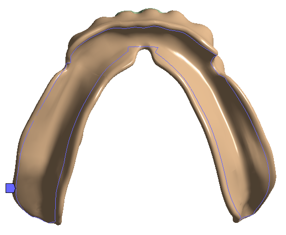
At first, we transform the scanned facet model of a dental prosthesis (see the supplementary model in Figure 2) into a three-dimensional solid model using SpaceClaim 2021 R1 with an accuracy of transformation of 0.5 mm selected since the aim is to optimize the denture base; the prosthesis is split into two bodies the denture base and the teeth. Small faces are created on top of each tooth where the tooth forces can be applied.
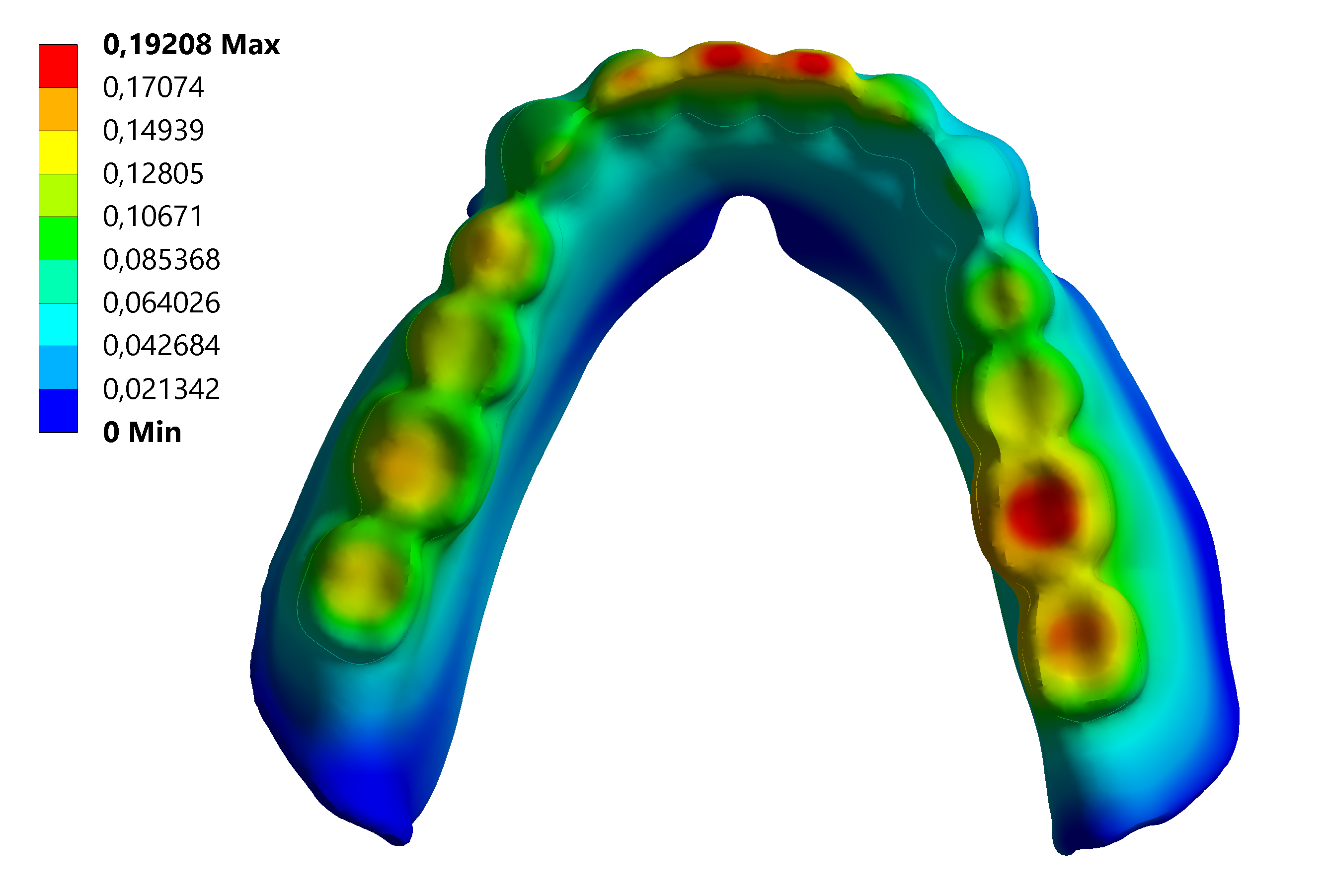
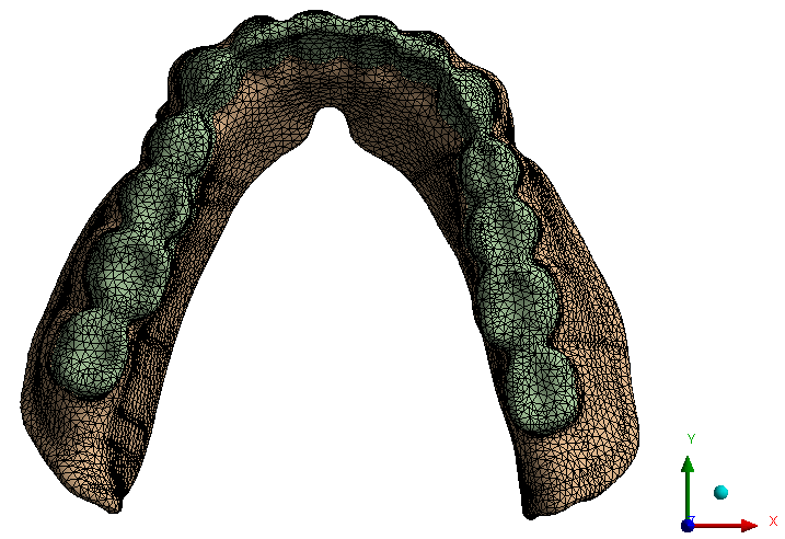
Then, for comparison purposes, we apply the whole model with PMMA, which is a weak material (has lower mechanical properties). Moreover, we mesh the solid bodies with tetrahedral quadratic elements with ANSYS, of our model of 104168 nodes and 61108 volume elements. The teeth connect to the denture base via common nodes. A fixed (Dirichlet) boundary is an edge boundary (from nodes) on the lower surface of the denture assigned, where the denture typically contacts the mouth tissue. Forces are applied vertically on the teeth. In the anterior area, the forces are 150 N, in the premolars area, are 450 N, and in the posterior area 900 N. Forces are chosen to be maximum occlusal bite forces [39]. The forces and the boundary are shown in Figure 2. The static structural analysis demonstrates that the maximum deformation of the dental prosthesis is 0.192 mm. The average deformation is 0.063 mm.
3.2 Reinforced denture
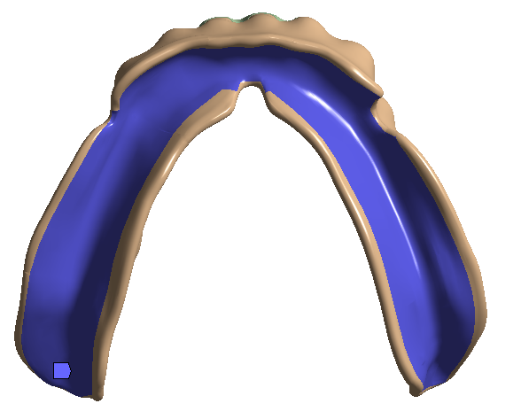
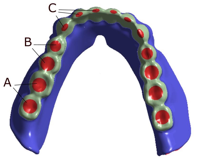
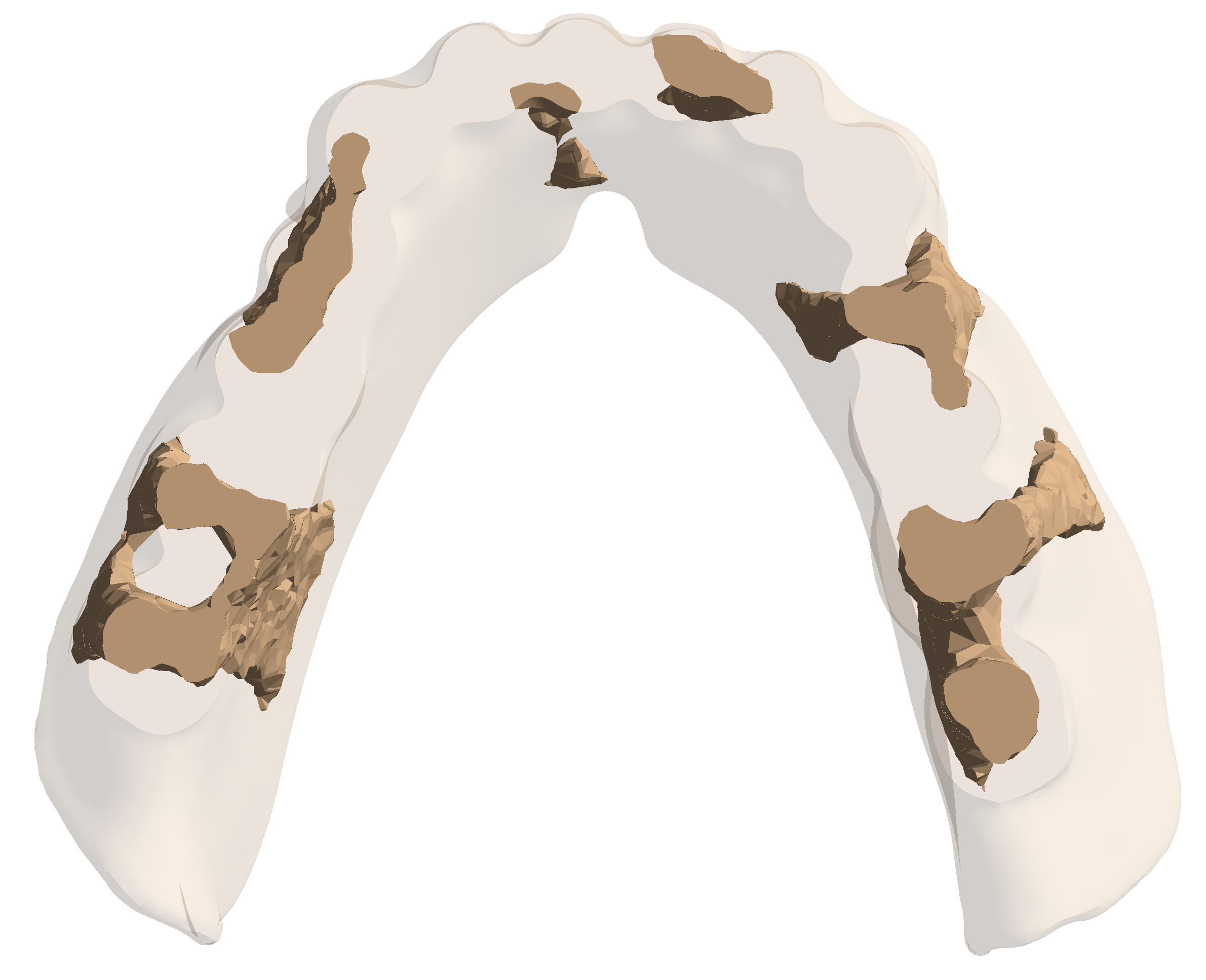
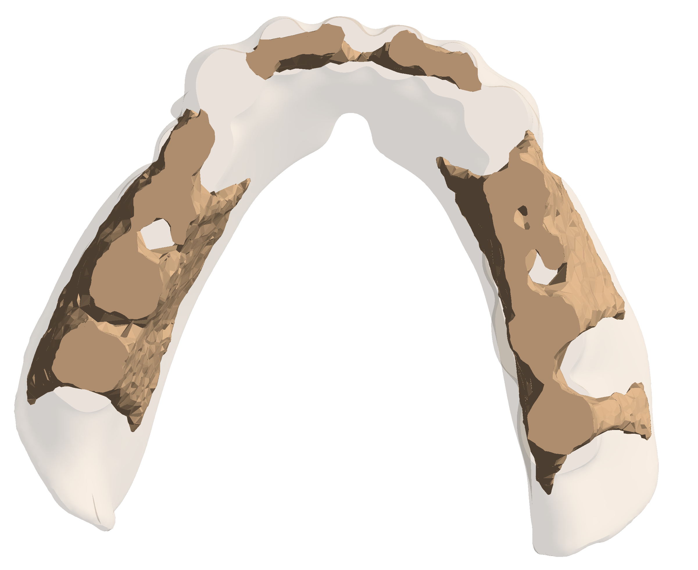
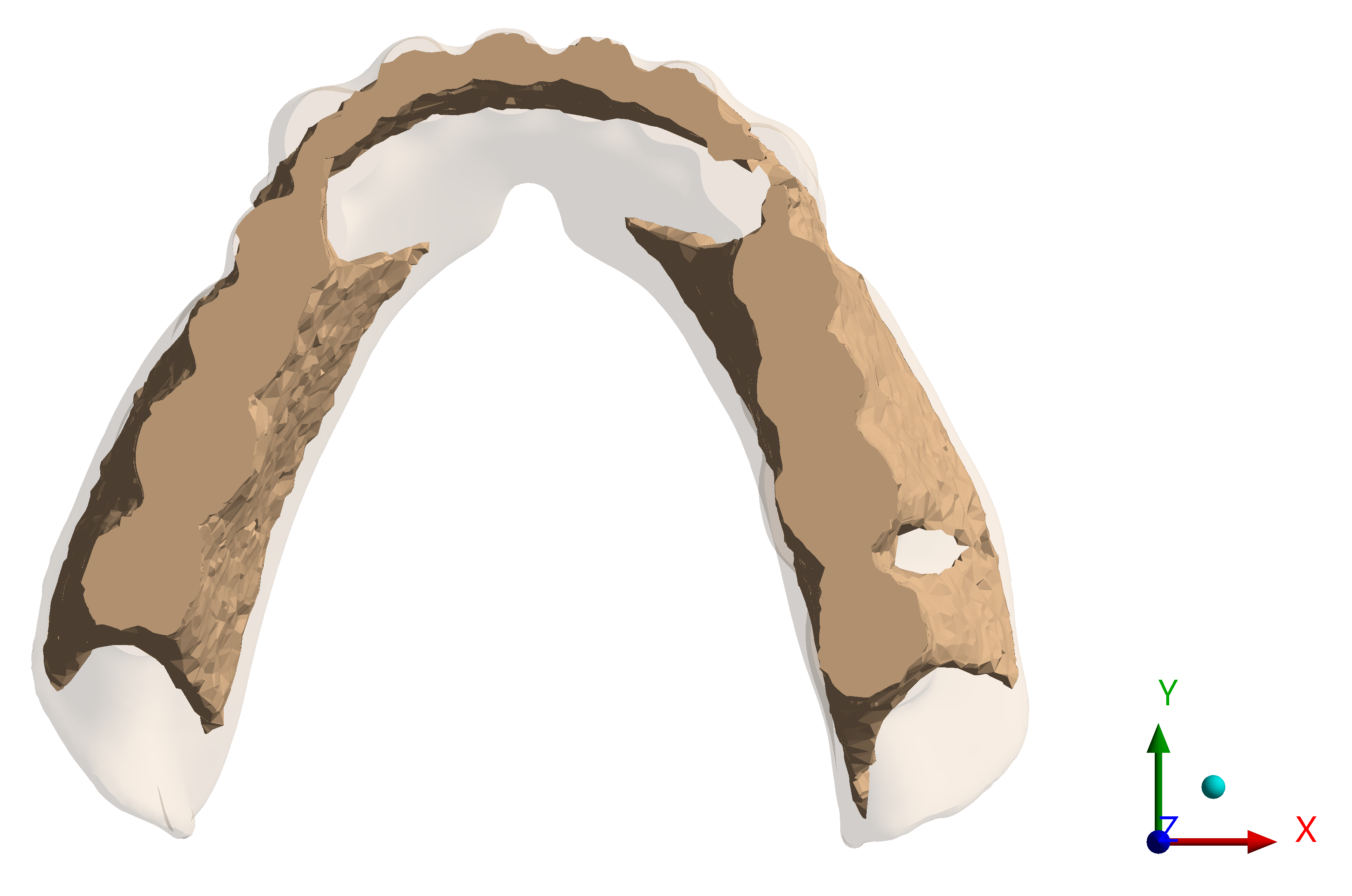
Next, we use the same denture model, but apply it with E-glass instead of PMMA. Then, we proceed as described in Section 1, but the tooth body is excluded from the optimization region, the denture base. We reduce the mass of the strong material (E-glass) by 80%, 60%, 43% and the final shape configuration of the reinforcement region as in Figure 4 respectively.
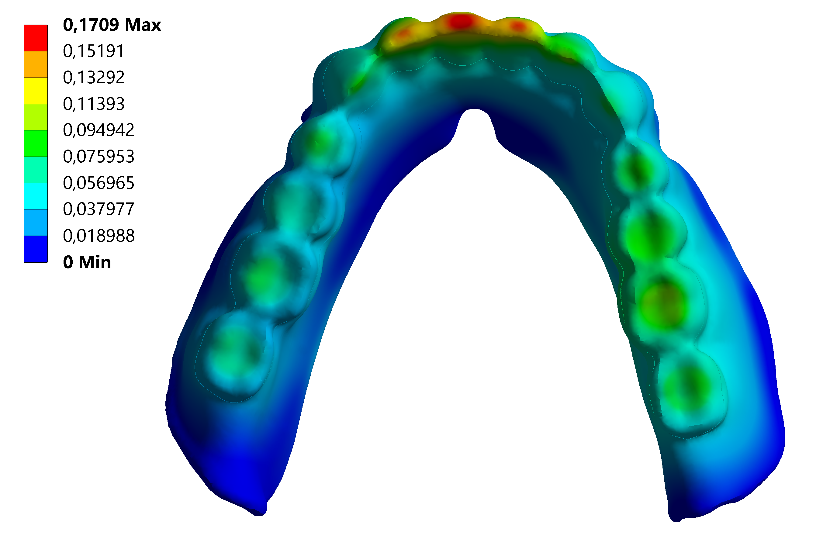
As described in Section 1, we add PMMA to the optimized reinforcement region (contacts are bonded) for the case of 4(a) in Figure 4. The deformed shape of the reinforced denture is shown in 3(b) in Figure 3. The maximum deformation of the reinforced denture is 0.171 mm. The average deformation is 0.035 mm.
3.3 Mesh convergence study
We investigate the effect of the mesh size on the accuracy and computational efficiency of the simulation of a dental prosthesis using FEA. A face-fixed boundary is applied in the mesh convergence study (see supplementary boundary 2(b) in Figure 3). An edge-fixed boundary is applied to the dental prosthesis to minimize the effect of boundary conditions on the topology optimization results (see the boundary 2(a) in Figure 3).
| Mesh size (mm) | 2 | 1 | 0.5 | 0.25 |
| Maximum principal stress (MPa) | 20,781 | 20,639 | 24,364 | 34,8 |
| Total deformation (mm) | 0,00531 | 0,00533 | 0,0053 | 0,00541 |
| Nodes | 16592 | 35396 | 112670 | 400940 |
| Elements | 9153 | 20194 | 65903 | 239684 |
The mesh convergence study on the E-glass material demonstrates a promising trend. The incremental rise in stress values as is refined the mesh indicates progress toward convergence, signaling that the simulations are stabilizing effectively. The relatively minor fluctuations in stress across varying mesh sizes underscore the robustness of the simulation setup, suggesting that the results are not overly influenced by changes in mesh resolution. We use the 0.5 mm mesh size in our study. Validation and sensitivity analysis are vital to ensuring result accuracy. Overall, it can be concluded that the FEA results for the dental prosthesis are reliable, independent of mesh size, and representative of denture behavior.
4 Discussion
After topology optimization, the retained mass (reinforcement region) is regularly distributed in the optimization region (denture base), (see the supplementary image as 4(a) in Figure 4). Interestingly, once the mass is reduced by 60%, the reinforcement is not needed in the anterior teeth (see the supplementary image as 4(b) in Figure 4). Reinforcement is connected in 4(c) in Figure 4. Whenever the yield of mass extraction is below 43%, the resulting shape is consistently a horseshoe shape, as determined by the proposed optimization method.
Once subtracting 80% of mass and applying the proposed method, comparing the non-reinforced denture and the reinforced denture demonstrates that maximum deformation decreases from 0.192 mm (non-reinforced denture) to 0.171 mm (reinforced denture). Average deformation decreases from 0.063 mm to 0.035 mm. Average deformation is approximately 44% decreasing, maximum deformation is approximately 11% decreasing.
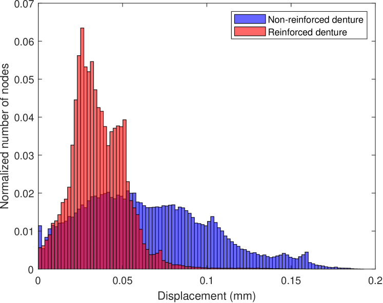
Besides, we can point out that the other optimized places of the reinforcement (see supplementary illustrations 4(b) and 4(c) in Figure 4) can give less deformation than in the case of 4(a) in Figure 4. On the other hand, node-based displacement distribution is shown for the non-reinforced denture and the reinforced denture in Figure 5. The average displacement distribution is much better in the reinforced denture. This demonstrates the deformation for each node in the denture diminishes after optimization.
The deformation of dental prostheses in FEA is influenced by many factors, including the boundary conditions applied, the material properties of the prosthesis, and its geometry and shape. Dental restorative devices and prosthodontic treatments must be designed to withstand forces similar to those experienced by natural teeth. The maximum unilateral biting force can reach 900 N, although chewing forces typically range from 50-80 N [40]. As the number of natural teeth decreases, the maximal bite force also decreases. For instance, removable partial denture wearers have a maximal bite force of 300 N, while complete denture wearers have forces of 180 N [41]. Although the simplified model in this study utilizes the same bite force value for each tooth sector, in clinical conditions the maximum bite force gradually decreases when moving from the posterior to the anterior area. Nevertheless, models with fixed bite force are commonly used in dental science literature and this study is based on its values.
FEA has been used in previous studies to simulate fiber-reinforced dentures’ behavior and optimize their mechanical properties and resistance. The density-based method proposed in this paper extends the applicability of FEA in this area and can provide valuable insights into stress distribution and deformation of the materials used, allowing for more precise and efficient design. Reinforcement of removable dentures has not previously been studied using topology optimization methods. In this study, a density-based topology optimization method is tested for an FEA model of a reinforced removable denture. From the computational perspective, the proposed approach’s advantage is that it enables predefined parameter reinforcement optimization. Moreover, computer-aided design (CAD) can design dentures, and elements of topology optimization are added to the design. Although our approach to strengthening reinforced prostheses is manual, rapid development in manufacturing technology, particularly in the realm of three-dimensional printers, raises the possibility of automated manufacture of multiple materials topology-optimized reinforced dentures, allowing for the integration of diverse material properties within a single object. This advancement presents an opportunity to print by optimizing the design of such structures, leveraging the distinct advantages of different materials to achieve enhanced performance and functionality.
5 Conclusions
A new practical method was proposed with predefined parameters optimization of the layout and topology of structures for reinforced dentures. The density-based topology optimization method and FEA enable the identification of optimal reinforcement places to minimize deformation and enhance stiffness. The study emphasizes the importance of the position of the reinforcement layer in designing dentures with improved mechanical properties and fracture resistance. Since the complexity of the model, involving as it does two different materials, poses challenges, the results were validated with a mesh sensitivity study. Thus, we provided insights into optimizing reinforcement in dentures, showcasing the potential for achieving optimal performance while reducing material usage. By leveraging FEA, we can enable accurate simulation and analysis of mechanical behavior, and the predefined topology optimization allows the determination of the optimal reinforcement distribution. This combination of methodologies provides an efficient means of improving the design of dental prostheses and enhancing their strength and durability. In future research, we will explore cost-effective manufacturing processes to make this technology accessible to patients.
Acknowledgements
We want to acknowledge ANSYS Inc. for its software which has contributed to the success of this research. The licenses have been sponsored under the Academic Partnership program of ANSYS Inc. We are grateful for the technical support and resources ANSYS offers. We are thankful to Dr. Mohamed Rabah for helping with the flow chart in the paper.
Funding
This research was supported by funding from the AMBioPharma project whose project code is A77805 and it was funded by the European regional development fund (ERDF).
Replication of results
The files used in the simulation can be sent by request.
Conflict of interest
The authors state that there is no conflict of interest.
Ethical approval
This article does not contain any studies with human participants or animals.
References
- [1] P. K. Vallittu, V. P. Lassila, R. Lappalainen, Evaluation of damage to removable dentures in two cities in Finland, Acta Odontologica Scandinavica 51 (6) (1993) 363–369.
- [2] K. K. Narva, P. K. Vallittu, H. Helenius, A. Yli-Urpo, Clinical survey of acrylic resin removable denture repairs with glass-fiber reinforcement, International Journal of Prosthodontics 14 (3) (2001).
- [3] N. Ladizesky, T. Chow, I. Ward, The effect of highly drawn polyethylene fibres on the mechanical properties of denture ease resins, Clinical Materials 6 (3) (1990) 209–225.
- [4] P. Vallittu, V. Lassila, Effect of metal strengthener’s surface roughness on fracture resistance of acrylic denture base material, Journal of Oral Rehabilitation 19 (4) (1992) 385–391.
- [5] P. Vallittu, V. Lassila, Reinforcement of acrylic resin denture base material with metal or fiber strengtheners, Journal of Oral Rehabilitation 19 (3) (1992) 225–230.
- [6] E. S. Güzelce, E. Tokar, Ö. Karacer, Evaluation of implant location on fiber-reinforced maxillary overdentures with finite element method., International Journal of Oral & Maxillofacial Implants 38 (3) (2023).
- [7] D. L. Dixon, L. C. Breeding, The transverse strengths of three denture base resins reinforced with polyethylene fibers, The Journal of Prosthetic Dentistry 67 (3) (1992) 417–419.
- [8] J. P. Matinlinna, P. K. Vallittu, Silane-based concepts on bonding resin composite to metals, The Journal of Contemporary Dental Practice 8 (2) (2007) 1–8.
- [9] P. Vallittu, M. Özcan, Clinical guide to principles of fiber-reinforced composites in dentistry, Woodhead Publishing, 2017.
- [10] Z. Raszewski, T. Nowakowska, Agnieszka, D. Nowakowska, W. Więckiewicz, Update on acrylic resins used in dentistry, Mini Reviews in Medicinal Chemistry 21 (15) (2021) 2130–2137.
- [11] N. Estrin, K. Nam, G. E. Romanos, J. Saragossi, V. J. Iacono, S. H. Bassir, Clinical outcomes of metal-ceramic vs metal–acrylic resin implant-supported fixed complete dental prostheses: A systematic review and meta-analysis., International Journal of Prosthodontics 36 (3) (2023).
- [12] M. S. Zafar, Prosthodontic applications of polymethylmethacrylate (PMMA): An update, Polymers 12 (10) (2020) 2299.
- [13] M. C. Arenas-Arrocena, L. Argueta-Figueroa, R. García-Contreras, O. Martínez-Arenas, B. Camacho-Flores, M. del Pilar Rodriguez-Torres, J. De la Fuente-Hernández, L. S. Acosta-Torres, New trends for the processing of poly(methyl methacrylate) biomaterial for dental prosthodontics, Acrylic Polymers in Healthcare (2017) 43–74.
- [14] K. Jomjunyong, P. Rungsiyakull, C. Rungsiyakull, W. Aunmeungtong, M. Chantaramungkorn, P. Khongkhunthian, Stress distribution of various designs of prostheses on short implants or standard implants in posterior maxilla: a three-dimensional finite element analysis, Oral & Implantology 10 (4) (2017) 369.
- [15] L. Minatel, F. R. Verri, G. A. H. Kudo, D. A. de Faria Almeida, V. E. de Souza Batista, C. A. A. Lemos, E. P. Pellizzer, J. F. S. Junior, Effect of different types of prosthetic platforms on stress-distribution in dental implant-supported prostheses, Materials Science and Engineering: C 71 (2017) 35–42.
- [16] M. Mousa, N. Jamayet, E. Lynch, A. Husein, Biomechanical stress in removable complete dental prostheses: a narrative review of finite element studies, Journal of International Oral Health 12 (5) (2020) 413.
- [17] A. Shinya, D. Yokoyama, Finite Element Analysis for Dental Prosthetic Design, INTECH Open Access Publisher, Sciyo, 2010.
- [18] L. Shi, A. Fok, Structural optimization of the fiber-reinforced composite substructure in a three-unit dental bridge, Dental Materials 25 (6) (2009) 791–801. doi:10.1016/j.dental.2009.01.001.
- [19] L. Shi, A. Fok, A. Qualtrough, A two-stage shape optimization process for cavity preparation, Dental Materials 24 (11) (2008) 1444–1453. doi:10.1016/j.dental.2008.03.016.
- [20] S. San, R. Altunay, Application of the generalized kudryashov method to various physical models, Applied Mathematics I& Information Sciences Letters 8 (2) (2021) 7–13.
- [21] S. Sait, R. Altunay, Abundant travelling wave solutions of 3+1 dimensional boussinesq equation with dual dispersion, Revista Mexicana de Física E 19 (2 Jul-Dec) (2022) 020203–1.
- [22] S. Sait, A. Rabia, Extended jacobi elliptic function solutions for general boussinesq systems, Revista Mexicana de Física 69 (2 Mar-Apr) (2023) 021401–1.
- [23] S. Qian, J. Weiss, Wavelets and the numerical solution of partial differential equations, Journal of Computational Physics 106 (1) (1993) 155–175.
- [24] Y. Chen, Q. Wang, Convergence and stability of galerkin finite element method for a hyperbolic partial differential equation with piecewise continuous arguments, Numerical Methods for Partial Differential Equations (2023).
- [25] Z. Zheng, M. Valdebenito, M. Beer, U. Nackenhorst, A stochastic finite element scheme for solving partial differential equations defined on random domains, Computer Methods in Applied Mechanics and Engineering 405 (2023) 115860.
- [26] ANSYS, Discovery SpaceClaim Release 2021 R1, ANSYS, Inc, Canonsburg, PA 15317, 2021.
- [27] W. S. Slaughter, The linearized theory of elasticity, Springer Science & Business Media, 2012.
- [28] M. E. Gurtin, The linear theory of elasticity, Linear Theories of Elasticity and Thermoelasticity: Linear and Nonlinear Theories of Rods, Plates, and Shells (1973) 1–295.
- [29] ANSYS, ANSYS Inc Theory Reference Release 11.0, ANSYS, Inc, 2007.
- [30] ANSYS, ANSYS Inc Mechanical User’s Guide, ANSYS, Inc, U.S.A, 2019.
- [31] C. Zillober, Global convergence of a nonlinear programming method using convex approximations, Numerical Algorithms 27 (2001) 265–289.
- [32] D. Mounier, C. Poilâne, C. Bûcher, P. Picart, Evaluation of transverse elastic properties of fibers used in composite materials by laser resonant ultrasound spectroscopy, in: Acoustics 2012, 2012, pp. 1246–1250.
- [33] M. Zhang, J. P. Matinlinna, E-glass fiber reinforced composites in dental applications, Silicon 4 (2012) 73–78.
- [34] C. A. M. Soares, C. M. M. Soares, M. J. Freitas, Mechanics of composite materials and structures, Vol. 361, Springer Science & Business Media, 2013.
- [35] P. K. Vallittu, Flexural properties of acrylic resin polymers reinforced with unidirectional and woven glass fibers, The Journal of Prosthetic Dentistry 81 (3) (1999) 318–326.
- [36] E. M. Safwat, A. G. Khater, A. G. Abd-Elsatar, G. A. Khater, Glass fiber-reinforced composites in dentistry, Bulletin of the National Research Centre 45 (2021) 1–9.
- [37] M. Ateş, A. Cilingir, T. Sülün, E. Sünbüloğlu, E. Bozdağ, The effect of occlusal contact localization on the stress distribution in complete maxillary denture, Journal of Oral Rehabilitation 33 (7) (2006) 509–513.
- [38] H. Christiansson, J. Helsing, Poisson’s ratio of fiber-reinforced composites, Journal of Applied Physics 79 (10) (1996) 7582–7585.
- [39] A. Waltimo, M. Könönen, Bite force on single as opposed to all maxillary front teeth, European Journal of Oral Sciences 102 (6) (1994) 372–375.
- [40] A. Waltimo, M. Könönen, Maximal bite force and its association with signs and symptoms of craniomandibular disorders in young Finnish non-patients, Acta Odontologica Scandinavica 53 (4) (1995) 254–258.
- [41] V. Lassila, I. Holmlund, K. K. Koivumaa, Bite force and its correlations in different denture types, Acta Odontologica Scandinavica 43 (3) (1985) 127–132.