AI in Thyroid Cancer Diagnosis: Techniques, Trends, and Future Directions
Abstract
There has been a growing interest in creating intelligent diagnostic systems to assist medical professionals in analyzing and processing big data for the treatment of incurable diseases. One of the key challenges in this field is detecting thyroid cancer, where advancements have been made using machine learning (ML) and big data analytics to evaluate thyroid cancer prognosis and determine a patient’s risk of malignancy. This review paper summarizes a large collection of articles related to artificial intelligence (AI)-based techniques used in the diagnosis of thyroid cancer. Accordingly, a new classification was introduced to classify these techniques based on the AI algorithms used, the purpose of the framework, and the computing platforms used. Additionally, this study compares existing thyroid cancer datasets based on their features. The focus of this study is on how AI-based tools can support the diagnosis and treatment of thyroid cancer, through supervised, unsupervised, or hybrid techniques. It also highlights the progress made and the unresolved challenges in this field. Finally, the future trends and areas of focus in this field are discussed.
Index Terms:
Thyroid carcinoma detection, Machine learning, Deep learning, Convolutional neural networks.I Introduction
I-A Background
The adoption of Artificial intelligence (AI) in healthcare has become a pivotal development, profoundly reshaping the landscape of medical diagnosis, treatment, and patient care. AI’s exceptional capabilities, including pattern recognition, predictive analytics, and decision-making skills, enable the development of systems that can analyze complex medical data at a scale and precision beyond human capacity [1]. This, in turn, augments early disease detection, facilitates accurate diagnoses, and aids personalized treatment planning. Moreover, AI-driven predictive models can forecast disease outbreaks, enhance the efficiency of hospital operations, and significantly improve patient outcomes [2]. Additionally, AI has the potential to democratize healthcare by bridging the gap between rural and urban health services and making high-quality care more accessible. Hence, the importance of AI in healthcare is profound and will continue to grow as technology advances, leading to more sophisticated applications and better health outcomes for patients worldwide [3, 4].
Abbreviations
- AI
- Artificial intelligence
- ATC
- Anaplastic thyroid carcinoma
- ACR TI-RADS
- American college of radiology has formulated a thyroid imaging, reporting, and data system
- ANN
- Artificial neural network
- APP
- Application
- AC
- Active contour
- ACC
- Accuracy
- AI-TS
- AI in thyroid surgery
- Bi-LSTM
- Bi-Long Short Term Memory
- BN
- Bayesian networks
- B
- Bagging
- BA
- Bootstrap aggregation
- BPSO
- Binary particle swarm optimization
- CAD
- Computer-aided diagnosis
- CNN
- Convolutional neural network
- C
- Clustering
- CFS
- Correlation-based feature selection
- CORR
- Correlation
- CT
- Computed tomography
- DL
- Deep learning
- DNN
- Deep neural network
- DAE
- Denoising autoencoder
- DT
- Decision trees
- DR
- Dimensionality reduction
- DTCW
- Double-tree complex wavelet transform
- DD
- Detected diseas
- DDTI
- Digital database thyroind image
- DA
- Dataset access
- DCG
- Discounted cumulative gain
- ELM
- Extreme learning machine
- EB
- Entropy-based
- EM
- Ensemble methods
- ER
- Error Rate
- EFC-AI
- Edge, fog, and cloud networks based on AI
- FTC
- Follicular thyroid carcinoma
- FNAB
- Fine-needle aspiration biopsy
- FKNN
- Fuzzy K-nearest neighborhood
- FB
- Feature bagging
- FS
- Feature selection methods
- F
- Female
- FE
- Feature extraction methods
- F1
- F1 Score
- FPR
- Fallout or false positive rate
- FN
- False negative
- FP
- False positive
- FL
- Federated learning
- GAN
- Generative adversarial network
- GWAS
- Genome-wide association analysis
- GEO
- Gene expression omnibus
- GLCM
- Gray-level co-occurrence matrix
- JSI
- Jaccard similarity index
- HTC
- Hürthle thyroid carcinoma
- HOG
- Histogram of oriented gradient
- IG
- Information gain
- IT
- Image types
- IF
- Image format
- ICA
- Independent component analysis
- IoMIT
- Internet of medical imaging thing
- KNN
- k-nearest neighbors
- KM
- K-means
- LSTM
- long-short-term-memory
- LTC
- Lymphoma thyroid carcinoma
- LR
- Logistic regression
- LBP
- Local binary patterns
- LMSE
- Laplacian mean square error
- MLP
- Multilayer perceptron
- ML
- Machine learning
- MTC
- Medullary thyroid carcinoma
- M
- Male
- MAE
- Mean absolute error
- MSE
- Mean squared error
- MRR
- Mean reciprocal rank
- MRI
- Magnetic resonance imaging
- NP
- Number of patients
- NM
- Number of males
- NF
- Number of females
- NN
- Number of nodules
- NBN
- Number of benign nodules
- NMN
- Number of malignant Nodule
- NCDR
- National cancer data repository
- NCI
- National cancer institute
- NPV
- Negative predictive value
- NCC
- Normalized cross-correlation
- NVF
- Noise visibility function
- NAE
- Normalized absolute error
- OB
- Objective
- PTC
- Papillary carcinoma
- PM
- Probabilistic models
- PCA
- Principal component analysis
- PD
- Private data
- PLCO
- Prostate, lung, colorectal, and ovarian
- P
- Precision
- PSNR
- Peak signal to noise ratio
- PS
- Panoptic segmentation
- RNN
- Recurrent neural network
- RBM
- Restricted boltzmann machine
- RBF
- Radial basis function
- RF
- Random forest
- R
- Relief
- RMSE
- Root mean square error
- RL
- Reinforcement learning
- RS
- Recommender systems
- SVM
- Support vector machine
- SL
- Supervised learning
- STC
- Sarcoma thyroid carcinoma
- SV
- Subjects for validation
- SEER
- Surveillance, epidemiology, and end results
- SPE
- Specificity
- SEN
- Sensitivity
- SD
- Standard deviation
- SSIM
- Structural similarity index
- SC
- Structural content
- SIFT
- Sscale invariant feature transform
- TN
- Thyroid nodules
- TG
- Thyroid gland
- TD
- Thyroid disease
- TCL
- Traditional classification
- TMC
- Thyroid microcarcinoma
- TDDS
- Thyroid disease data set
- TCD
- Thyroid cancer datase
- TCGA
- Cancer genome atlas
- TD
- Texture description
- TI
- Thyroid inputs
- TT
- Thyroid Targets
- TS
- Thyroid segmentation
- TL
- Transfer learning
- TN
- True negative
- TP
- True positive
- TIRADS
- Thyroid imaging reporting and data system
- US
- Ultrasound
- USL
- Unsupervised learning
- WEKA
- Waikato environment for knowledge analysis
- WBCD
- Wisconsin breast cancer dataset
- WP-SNR
- Weight peak signal to noise ratio
- WSNR
- Weighted signal-to-noise ratio
- WSI
- Whole slide images
- VOE
- Volumetric overlap error
- VIF
- Visual information fidelity
- VSNR
- Visual signal to noise ratio
- XAI
- Explainable artificial intelligence
- 3D-TCD
- 3D thyroid cancer detection
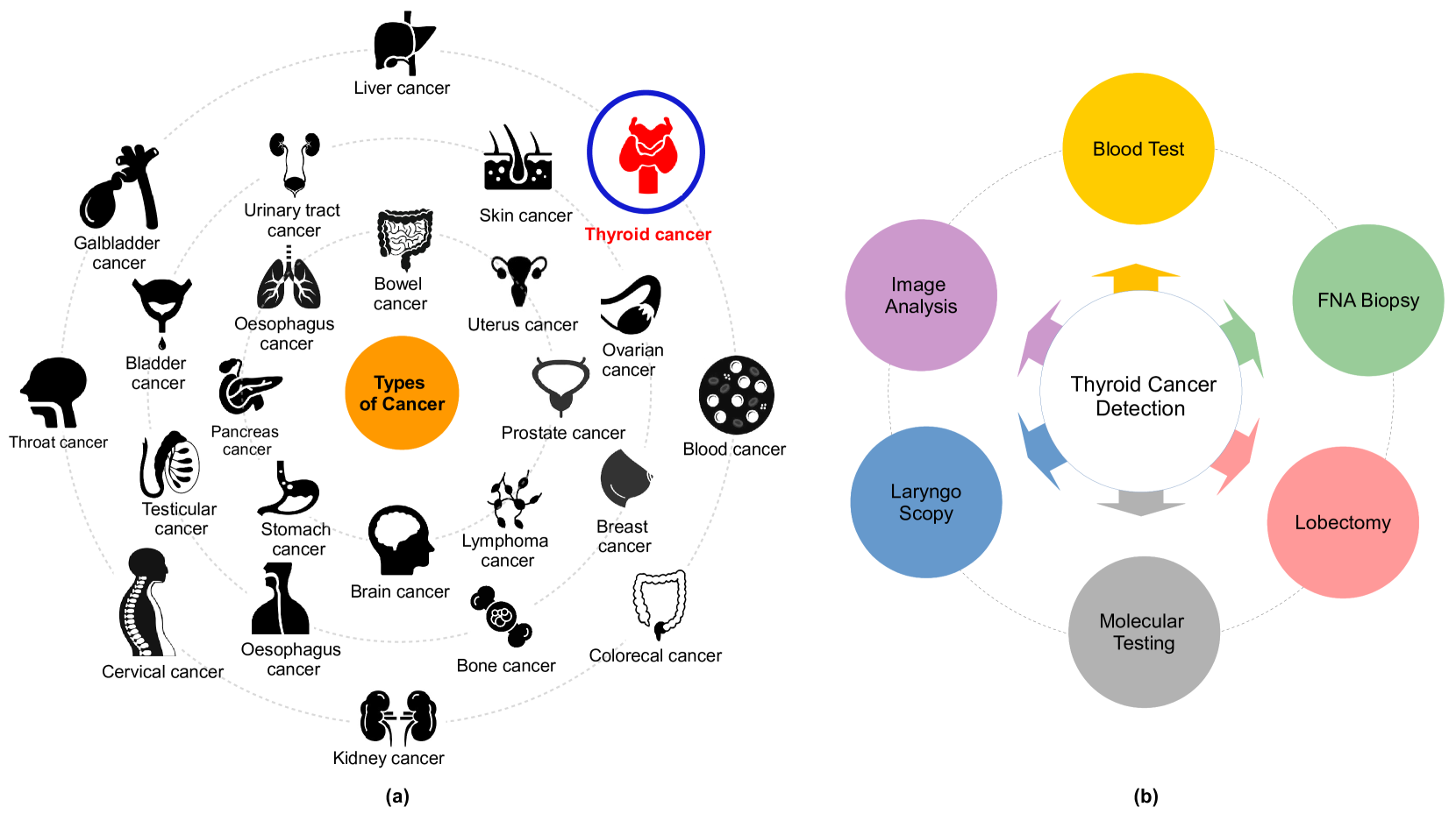
Cancer, a leading cause of death, affects various parts of the body as depicted in Fig. 1 (a). Among various types, thyroid carcinoma stands out as one of the most commonly occurring endocrine cancers globally [5, 6]. Concerns are mounting over the escalating incidence of thyroid cancer and associated mortality. Research indicates that thyroid cancer incidence is higher in women aged 15-49 years (ranked fifth globally) compared to men aged 50-69 years [7, 8, 9].
According to existing global epidemiological data, the rapid growth of abnormal thyroid nodules (TN) is driven by an accelerated increase in genetic cell activity. This condition can be categorized into four primary subtypes: Papillary carcinoma (PTC) [10], follicular carcinoma (FTC) [11], anaplastic carcinoma (ATC) [12], and medullary carcinoma (MTC) [13]. Influential factors such as high radiation exposure, Hashimoto’s thyroiditis, psychological and genetic predispositions, as well as advancements in detection technology, can contribute to the onset of these cancer types. These conditions might subsequently lead to chronic health issues, including diabetes, irregular heart rhythms, and blood pressure fluctuations [14, 15, 16]. Although the quantity of cancer cells is a significant indicator of thyroid carcinoma, obtaining results is often time-consuming due to the requirement to observe cell appearance. Thus, the detection and quantification of cell nuclei are considered crucial biomarkers for assessing cancer cell proliferation.
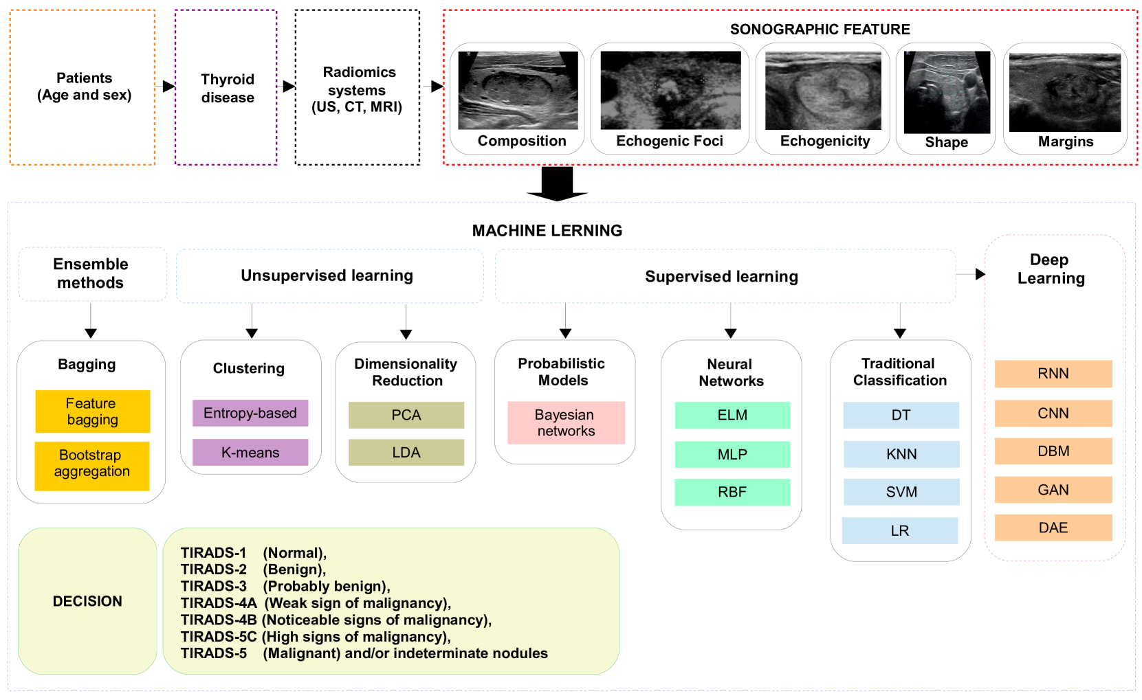
The utilization of computer-aided diagnosis (CAD) systems for analyzing thyroid cancer images has seen a significant increase in popularity in recent years. These systems, renowned for enhancing diagnostic accuracy and reducing interpretation time, have become an invaluable tools in the field. Among these technologies, radionics, when used in conjunction with ultrasonography imaging, has become widely accepted as a cost-effective, safe, simple, and practical diagnostic method in clinical practice. Endocrinologists frequently conduct US scans in the 7-15 MHz range to identify thyroid cancer and evaluate its anatomical characteristics. The American College of Radiology has formulated a Thyroid Imaging, Reporting, and Data System (ACR TI-RADS) that classifies thyroid nodules into six categories based on attributes like composition, echogenicity, shape, size, margins, and echogenic foci. These classifications range from normal (TIRADS-1) to malignant (TIRADS-6) [17, 18, 19]. Several open-source applications are available for assessing these thyroid cancer features [20, 21]. However, the identification and differentiation of nodules continue to present a challenge, largely reliant on the personal experience and cognitive abilities of radiologists. This is due to the subjective nature of human eye-based image recognition, the poor quality of captured images, and the similarities among US images of benign thyroid nodules, malignant thyroid nodules, and lymph nodes.
Moreover, ultrasonography imaging is often a time-intensive and stressful procedure, which can result in inaccurate diagnoses. Misclassifications among normal, benign, malignant, and indeterminate cases are common [22, 23, 24, 25, 26, 27]. For a more precise diagnosis, a fine-needle aspiration biopsy (FNAB) is typically conducted. However, FNAB can be an uncomfortable experience for patients, and a specialist’s lack of experience can potentially convert benign nodules into malignant ones, not to mention the additional financial burden [28, 29] (refer to Fig.1 (b)). The primary challenge in distinguishing between benign and malignant nodules resides in the selection of their characteristics. Numerous studies have explored the characterization of conventional US imaging for various types of cancers, including retina [30, 31], breast cancer [32, 33], blood cancer [34, 35], and thyroid cancer [36, 37]. However, these methods still fall short when it comes to the accurate classification of thyroid nodules.
The incorporation of AI technology plays a pivotal role in reducing subjectivity and enhancing the accuracy of pathological diagnoses for various intractable diseases, including those affecting the thyroid gland [38, 39]. This enhancement is achieved through improved interpretation of ultrasonography images and faster processing times. Machine learning (ML) and deep learning (DL) have surfaced as potential solutions for automating the classification of thyroid nodules in applications such as US, fine-needle aspiration (FNA), and thyroid surgery [40, 41]. This potential has been underscored in numerous studies, such as [42, 43, 44, 39, 45, 46]. Furthermore, there are ongoing studies examining the use of this innovative technology for cancer detection, where its effectiveness hinges on the volume of data and the precision of the classification process.
The motivation to write a review on "Artificial Intelligence for Detecting Thyroid Carcinoma" stems from the increasing prevalence of thyroid cancer, a significant endocrine malignancy where early and accurate detection is pivotal for patient outcomes. As technological advancements in AI and machine learning burgeon, their integration into medical diagnostics—spanning imaging, pathology, and genomics—offers potential improvements in detection accuracy and efficiency. Traditional thyroid carcinoma diagnostic methods, like fine-needle aspiration biopsies, sometimes present inconclusive results; AI promises less invasive alternatives with possibly superior precision. Such a review would amalgamate insights from the intersection of computer science, radiology, pathology, and endocrinology, propelling multidisciplinary collaboration. It would also spotlight AI’s clinical implications, guiding clinicians in leveraging its capabilities for patient care, while delineating future research directions. Furthermore, this review would underscore the economic and healthcare benefits, from cost savings to reduced waiting times. At the same time, it is imperative to address AI’s inherent challenges, including data privacy and ethical considerations, ensuring its balanced integration into healthcare. In essence, the review would offer a comprehensive panorama of AI’s current and potential role in thyroid carcinoma detection, benefitting both researchers and medical practitioners.
I-B Our contribution
This review provides a comprehensive examination of the application of Artificial Intelligence (AI) methods in detecting thyroid cancer. The objective of AI-based analysis in the medical field is increasingly shifting towards enhancing diagnostic accuracy, and this review aims to illustrate this trend, particularly in thyroid cancer detection. We first provide an overview of the existing frameworks and delve into the specifics of various AI techniques. These include supervised learning methods, like DL, artificial neural networks, traditional classification, and probabilistic models, as well as unsupervised learning methods, such as clustering and dimensionality reduction. We also explore ensemble methods, including bagging and boosting. Recognizing the importance of quality datasets in AI applications, we scrutinize several thyroid cancer datasets, addressing their features, as well as feature selection and extraction methods used in various studies. We then outline the standard assessment criteria used to evaluate the performance of AI-based thyroid cancer detection methods. These range from classification and regression metrics to statistical metrics, computer vision metrics, and ranking metrics. Finally, we discuss future research directions, emphasizing areas that require more attention to overcome existing barriers and improve the use and deployment of thyroid cancer detection solutions. In conclusion, we underscore the potential of AI in advancing thyroid cancer detection while also noting the need for continuous critical evaluation to ensure its responsible and effective use.
All in all, the principal contributions of our paper are as follows
-
•
Overview of existing frameworks and specifics of various AI techniques, including supervised learning (DL, artificial neural networks, traditional classification, and probabilistic models) and unsupervised learning (clustering and dimensionality reduction) methods, as well as ensemble methods (bagging and boosting).
-
•
Scrutiny of several thyroid cancer datasets, addressing their features, feature selection, and extraction methods used in various studies.
-
•
Outline of standard assessment criteria used to evaluate the performance of AI-based thyroid cancer detection methods, encompassing classification and regression metrics, statistical metrics, computer vision metrics, and ranking metrics.
-
•
Critical analysis and discussion highlighting limitations, hurdles, current trends, and open challenges in the field.
-
•
Discussion of future research directions, emphasizing areas requiring more attention to overcome existing barriers and improve thyroid cancer detection solutions.
-
•
Emphasis on the potential of AI in advancing thyroid cancer detection while advocating continuous critical evaluation for responsible and effective use.
Additionally, the principal contributions of the proposed review compared to other existing surveys are summarized in Table I.
| Ref | Year | PPY | TCDS | AIA |
|
|
|||||||||||||
| [47] | 2021 | ✓ | ✓ | ✗ |
|
|
|||||||||||||
| [48] | 2021 | ✓ | ✓ | ✗ |
|
|
|||||||||||||
| [49] | 2021 | ✓ | ✓ | ✗ |
|
|
|||||||||||||
| [50] | 2021 | ✓ | ✓ | ✗ |
|
|
|||||||||||||
| [51] | 2021 | ✓ | ✓ | ✗ |
|
|
|||||||||||||
| [52] | 2022 | ✓ | ✓ | ✗ |
|
|
|||||||||||||
| [53] | 2022 | ✓ | ✓ | ✗ |
|
|
|||||||||||||
| [54] | 2022 | ✓ | ✓ | ✗ |
|
|
|||||||||||||
| [55] | 2022 | ✓ | ✓ | ✗ |
|
|
|||||||||||||
| Our | - | ✓ | ✓ | ✓ |
|
|
I-C Roadmap
The rest of this paper is organized as follows. Section II follows, providing an overview of existing frameworks utilized in this field, and discussing their respective advantages and limitations. Section III presents various thyroid cancer datasets used in AI-based analyses, explaining their relevance and uniqueness. In Section IV, the paper delves into the vital aspect of ’Features’, discussing feature extraction and selection methods in AI models used for thyroid cancer detection. Section V outlines the standard assessment criteria used to evaluate the performance of these models. An actual instance of AI-based thyroid cancer detection is presented in Section VI to provide a real-world context to the theoretical aspects discussed earlier. The paper then proceeds to critical analysis and discussion in Section VII, where challenges, limitations, and areas for improvement in the current approaches are discussed. In Section VIII, potential future research directions are proposed, highlighting areas where further exploration and innovation can lead to advancements in AI-based thyroid cancer detection. The paper concludes with Section IX, summarizing the main findings and discussions, thereby providing a comprehensive conclusion to the discussions presented in the earlier sections.
II Overview of existing frameworks
This section showcases the various AI-based methods utilized for diagnosing thyroid gland (TG) cancers. In the illustration, Fig. 2 presents a proposed categorization of the thyroid cancer diagnosis techniques relying on AI.
II-A Objective of AI-based analysis (O)
This article focuses on the application of AI in thyroid cancer detection. In order to better understand the purpose behind each framework, it is crucial to identify the objective of each approach. O1. Classification: Thyroid carcinoma classification refers to the categorization of thyroid cancers based on their histopathological features, clinical behavior, and prognosis. There are several types of thyroid carcinomas, each of which has distinct characteristics. The primary categories include: (i) Papillary Thyroid Carcinoma (PTC): The most common type, accounting for about 80% of all thyroid cancers. PTC tends to grow very slowly, but it often spreads to lymph nodes in the neck. Despite this, it is usually curable with treatment; (ii) Follicular Thyroid Carcinoma (FTC): The second most common type, FTC can invade blood vessels and spread to distant parts of the body, but it is less likely to spread to lymph nodes; (iii) Medullary Thyroid Carcinoma (MTC): This type of thyroid cancer starts in the thyroid’s parafollicular cells, also called C cells, which produce the hormone calcitonin. Elevated levels of calcitonin in the blood can indicate MTC; and (iv) Anaplastic Thyroid Carcinoma (ATC): A very aggressive and rare form of thyroid cancer, ATC often spreads quickly to other parts of the neck and body. It is difficult to treat.
The classification of thyroid carcinomas is crucial in determining the most effective course of treatment for each patient. Various factors such as tumor size, location, and the patient’s age and overall health are also taken into consideration when forming a treatment plan. Advances in AI and machine learning are helping to automate and improve the accuracy of thyroid carcinoma classification, with many models trained to classify tumors based on medical images or genetic data. As reported by Liu et al. [56], incorporates Support vector machine (SVM) for cancer detection. Similarly, Zhang et al. [57, 58] propose Deep neural network (DNN) based strategies for segregating and categorizing benign and malignant thyroid nodules in ultrasound imagery. Furthermore, the Bi-Long Short Term Memory (Bi-LSTM) model, as presented by Chen et al. [59], demonstrates notable accuracy in classifying thyroid nodules. These classification systems constitute structured hierarchies instrumental in organizing knowledge and workflows in the specific domain of thyroid cancer.
O2. Segmentation: segmentation of thyroid carcinoma refers to the process of identifying and delineating the region of an image that corresponds to a thyroid tumor. The goal of segmentation is to separate the areas of interest, in this case, the thyroid tumor, from the surrounding tissues in the medical images. This can be done manually by an expert radiologist, or it can be automated using machine learning algorithms [60, 61]. Segmentation is a crucial step in medical image analysis because it helps to accurately determine the location, size, and shape of the tumor, which are vital parameters for diagnosis, treatment planning, and prognosis prediction. A variety of methods can be used to perform image segmentation, including thresholding, edge detection, region-growing methods, and more complex machine learning and DL techniques.
In the case of thyroid carcinoma, the segmentation can be challenging due to the high variability in the appearance and shape of the tumors, their proximity to other structures in the neck, and the presence of noise or artifacts in the images. Therefore, robust and reliable segmentation algorithms are needed to ensure accurate and consistent results. AI methods, including Convolutional neural network (CNN) and U-Net architecture, are being increasingly used for thyroid carcinoma segmentation because of their ability to learn and generalize from large amounts of data, thus improving the accuracy and reliability of the segmentation process.
O3. Prediction: The prediction of thyroid carcinoma involves the use of various diagnostic tools, tests, and techniques - often employing machine learning models - to anticipate the probability of a patient developing thyroid cancer. This predictive analysis can be based on several factors, including but not limited to (i) Genetic predisposition: Individuals with a family history of thyroid cancer are at a higher risk; (ii) Gender and age: Thyroid cancer is more common in women and people aged between 25 and 65; (iii) Radiation exposure: Exposure to high levels of radiation, especially during childhood, increases the risk of developing thyroid cancer; (iv) Diet and lifestyle: Lack of iodine in the diet and certain lifestyle factors may contribute to an increased risk. In a medical context, prediction does not necessarily mean a certain future outcome, but rather it points to an increased risk or likelihood based on current data and predictive models. For thyroid carcinoma, predictive tools and tests are typically used in conjunction with each other to achieve more accurate results. For instance, machine learning algorithms can be trained on historical medical data to predict the likelihood of a nodule being benign or malignant, aiding in early detection and more effective treatment planning. Various studies have been proposed to predict thyroid cancer. For instance, in [62], the authors employed the use of Artificial Neural Network (ANN) and Logistic Regression to make predictions. Another study [63] details the creation of a predictive machine using Convolution Neural Networking (CNN) to analyze 10068 microscopic thyroid cancer images from South Asian populations.
II-B Supervised learning (SL)
Supervised learning is a method of machine learning where an algorithm is trained to classify or predict the condition based on labeled data, which in this case is medical data related to thyroid cancer. The aim of supervised learning is to differentiate between the different forms of thyroid cancer through the use of annotated data and examples. For example, this data can include ultrasound images, radiomic features, genetic markers, patient demographics, or any other information that may be relevant to the diagnosis or prognosis of thyroid cancer. The labeled data would indicate whether each instance corresponds to a case of thyroid cancer or not, or it may provide more detailed labels such as the stage of the cancer or the type of thyroid carcinoma.
In a classification setting, the supervised learning algorithm could be trained to distinguish between benign and malignant thyroid nodules based on certain characteristics extracted from medical imaging data. The labels in the training data would specify whether each nodule is benign or malignant. After training, the algorithm can then be used to classify new, unlabeled nodules. Similarly, a regression-based supervised learning algorithm might be trained to predict the progression or the prognosis of thyroid cancer based on various patient-specific features. The labels here would correspond to a continuous outcome variable, such as the survival time of the patient or a measure of disease progression. It is important to note that the performance of these methods heavily relies on the quality and quantity of the available data. The more accurate and comprehensive the data, the better the algorithm will perform in predicting or classifying new instances. Additionally, supervised learning models in healthcare, including thyroid carcinoma detection, need to be validated on separate test datasets and in real-world clinical settings to ensure their robustness and reliability [64, 65].
II-B1 Deep learning (DL)
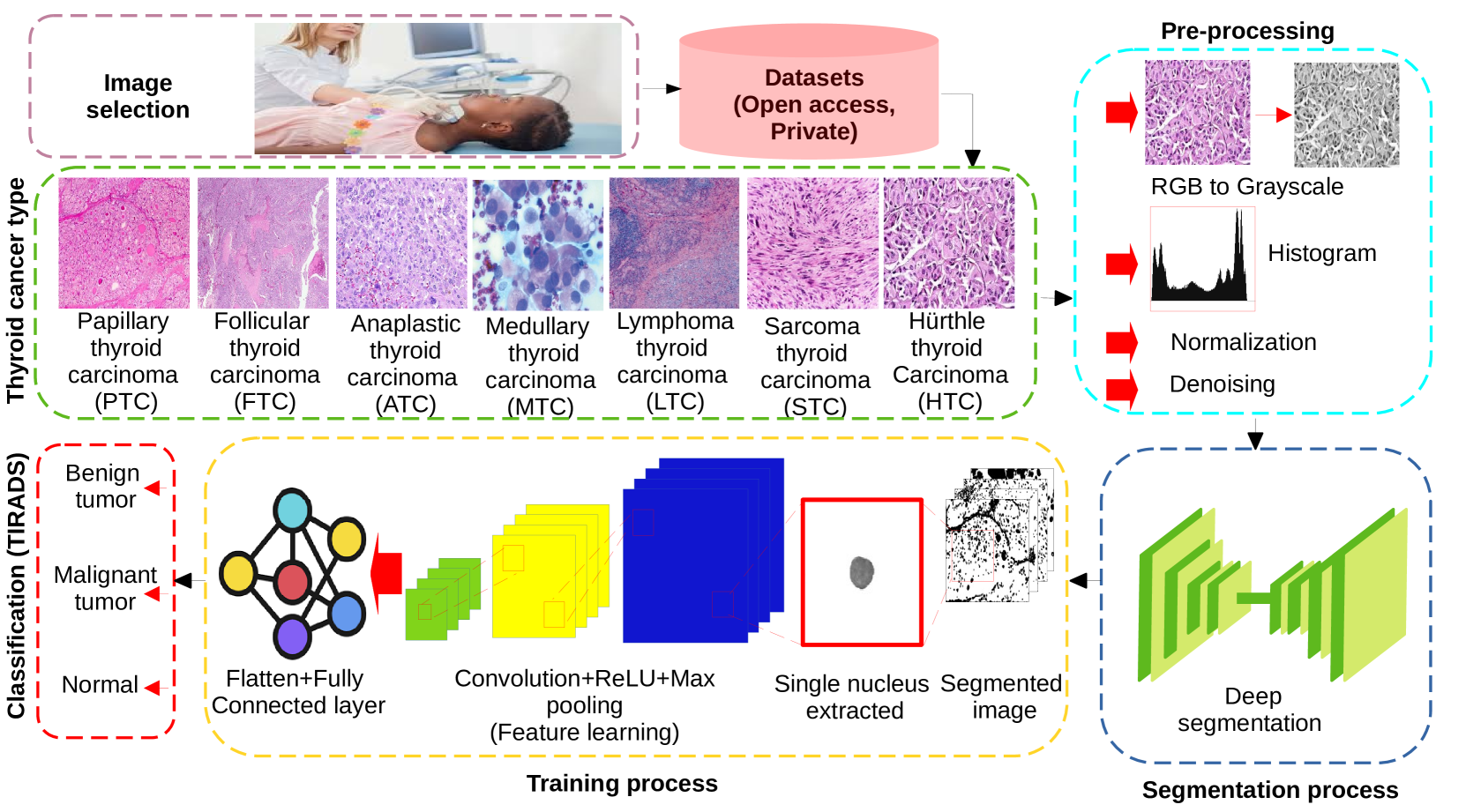
DL is a subset of machine learning and artificial intelligence that’s based on artificial neural networks with representation learning. It can automatically learn, generate, and improve representations of data by employing large neural networks with many layers—hence the term "deep" learning. In thyroid cancer, DL has been deployed to perform different tasks, including (i) Image Classification – DL algorithms like Convolutional Neural Networks (CNNs) can be trained to classify thyroid ultrasound images. For instance, they can differentiate between benign and malignant nodules based on their shape, texture, and other characteristics [66, 67, 68]. This approach can significantly reduce the time and effort required for manual interpretation, thus aiding in the early detection and treatment of thyroid cancer; (ii) Pathological Analysis – DL can also be utilized to analyze histopathological or cytopathological slide images, helping in the detection and classification of cancerous cells; (iii) Genomic Data Analysis – With the advent of genomic medicine, DL models can be employed to analyze genetic variations that may predispose individuals to thyroid cancer; (iv) Radiomics – DL models can be used to extract high-dimensional data from radiographic images, allowing for more precise and personalized treatment planning; and (v) Predictive Analysis – Using electronic health records and other patient data, DL models can be used to predict the likelihood of a patient developing thyroid carcinoma, allowing for preventive measures to be taken if necessary. Fig. 3 illustrates the different classifications of thyroid cancer using DNN.
D1. Denoising autoencoder (DAE): Denoising autoencoders (DAEs) can be beneficial for thyroid carcinoma classification by effectively learning representations from ultrasound or histopathological images. A DAE is a specific type of artificial neural network trained to reconstruct input data, often used for the purposes of dimensionality reduction or feature learning. The process for utilizing DAEs for thyroid carcinoma classification generally follows these steps (i) preprocessing, (ii) noisy input creation, (iii) DAE training, (iv) feature extraction and (v) classification. In [69], the authors implemented six autoencoder algorithms in the training process for papillary thyroid carcinoma (PTC) classification, including fixing weights and fine-tuning the network. The encoding layers and the complete auto-encoder were used to embed the network. Another study [70] employed denoising autoencoders (DAE) and stacked denoising autoencoders to extract features and identify informative genes in thyroid cancer.
D2. CNN: CNNs are a class of DL models that have shown extraordinary performance in various image processing and analysis tasks, including the classification of medical images. CNNs are especially adept at processing grid-like data, such as an image, where spatial relationships between the pixels are crucial to understanding the image content. The past few years have seen considerable effort invested in developing CNN-based methodologies for detecting thyroid cancer, especially for the automated identification and classification of nodules in ultrasound imagery [71]. The ConvNet model, a widely adopted framework within the neural network realm, emphasizes the use of convolution operations over matrix multiplications [72]. Various CNN architectures such as LeNet [73], AlexNet [74], VGG [68], ResNet [75], GoogLeNet [76], Squeeze Net [77], and DenseNet [78], are distinguished by their incorporation of key components including convolutional, pooling, and fully connected layers.
In a study conducted by [79], the potential of CNN models to prognosticate thyroid cancer was explored using 131,731 ultrasound images taken from 17,627 thyroid cancer patients. Another research effort [80] employed VGG16, Inception, and Inception-Resnet models to differentiate malignant tissues within a set of 451 thyroid images from the DDTI dataset. To mitigate the challenge of data scarcity, the images were augmented before classification. A comparison of DCNN diagnostic performance with expert radiologists in distinguishing thyroid nodules (TN) within ultrasound images was carried out by [81], involving a test set of 15,375 TN ultrasound images. They utilized CNNE1 and CNNE2 models, derived from DCNN, for differentiating between malignant and benign TNs. The study [82] proposed a CNN-based DL technique for detecting and classifying TN and breast nodules, with the results contrasted against those from ultrasound imaging. Table II presents a summary of recent CNN-based thyroid cancer classification contributions.
| Ref. | Year | Country | NP | NM | NF | NN | NBN | NMN | TP | TN | FP | FN |
|---|---|---|---|---|---|---|---|---|---|---|---|---|
| [83] | 2021 | China | 102 | 00 | 102 | 104 | 57 | 47 | 38 | 07 | 07 | 50 |
| [84] | 2021 | China | 102 | 25 | 77 | 103 | 73 | 33 | 27 | 12 | 06 | 61 |
| [85] | 2021 | Koria | 325 | 61 | 264 | 325 | 257 | 68 | 48 | 52 | 20 | 205 |
| [86] | 2021 | China | 2775 | 726 | 2049 | 2775 | 2472 | 303 | 271 | 363 | 32 | 2109 |
| [86] | 2021 | China | 163 | 48 | 115 | 175 | 67 | 108 | 86 | 09 | 22 | 58 |
| [87] | 2020 | China | 2489 | 614 | 1875 | 2489 | 1021 | 1468 | 1280 | 258 | 188 | 763 |
| [88] | 2020 | USA | 571 | 234 | 337 | 651 | 500 | 151 | 133 | 214 | 18 | 287 |
| [89] | 2020 | China | 166 | 46 | 100 | 209 | 109 | 100 | 87 | 16 | 13 | 93 |
| [81] | 2020 | Korea | 200 | 49 | 151 | 200 | 102 | 98 | 90 | 41 | 08 | 61 |
| [90] | 2020 | Korea | 340 | 79 | 261 | 348 | 252 | 96 | 31 | 25 | 65 | 227 |
| [91] | 2019 | Korea | 106 | 29 | 77 | 2018 | 132 | 86 | 69 | 23 | 17 | 109 |
| [92] | 2019 | China | 171 | 32 | 139 | 180 | 85 | 95 | 86 | 50 | 09 | 35 |
-
•
Abbreviations: Number of patients (NP); Number of males (NM); Number of females (NF); Number of nodules (NN); Number of benign nodules (NBN); Number of malignant Nodules (NMN).
D3. Recurrent neural network (RNN): Recurrent Neural Networks (RNNs) are a class of artificial neural networks where connections between nodes form a directed graph along a sequence, thus enabling them to use their internal state (memory) to process variable-length sequences of inputs. This unique feature makes RNNs particularly suitable for tasks where temporal dependencies are essential, such as time-series analysis, language translation, and speech recognition. In the context of thyroid carcinoma classification, RNNs can be utilized to analyze sequential or time-dependent data, such as the development of a patient’s clinical signs over time, the evolution of a tumor seen in a series of medical images, or changes in the gene expression related to the progression of thyroid cancer. For instance, in the study by Chen et al. (2017) [93], the authors propose a hierarchical Recurrent Neural Network (RNN) approach for classifying thyroid nodules (TN) based on historical ultrasound reports. This hierarchical RNN is composed of three layers, with each layer incorporating an individually trained Long Short-Term Memory (LSTM) network. The study’s findings indicate that the hierarchical RNN model surpasses basic models in terms of computational efficiency, control accuracy, and robustness, making it an effective tool for diagnosing TN. These advantageous attributes stem from the inherent memory mechanisms of RNNs, which allow them to remember previous states through feedback loops. This memory capability renders RNNs a popular choice for applications in cancer detection.
D4. Restricted Boltzmann Machine (RBM): A Restricted Boltzmann Machine (RBM) is a type of artificial neural network and a generative stochastic model. It was first introduced by Paul Smolensky [94] in 1986 under the name "Harmonium," but the concept of a "restricted" Boltzmann Machine was developed by Geoffrey Hinton and his students in the mid-2000s. RBMs have a layer of visible units and a layer of hidden units, but no connections within layers - this is the restriction in their name. Each node in the layer is connected to every node in the other layer. The lack of intra-layer connections simplifies the learning process. The work by Vairale et al. [95] presents an application of Restricted Boltzmann Machines (RBMs) to develop a personalized fitness recommendation system tailored for individuals diagnosed with thyroid conditions. RBMs are a particular class of generative artificial neural networks characterized by a bi-directional architecture, which operates in an unsupervised manner. This structure comprises a visible layer containing binary variables and a hidden layer, also populated with interconnected binary variables. The learning process within RBMs is primarily conducted through statistical analysis.
D5. Generative adversarial network (GAN): This type of ML network is composed of two distinct models: a generator and a discriminator. The generator maps a random input vector to an output in the data space, while the discriminator serves as a binary classifier that evaluates both input data from the training set and output data from the generator. The GAN has gained widespread use in the diagnosis of diseases, including thyroid nodules (TN) [96, 97].
II-B2 Artificial Neural networks (ANN)
ANNs are defined as a class of information processing systems comprised of interconnected non-linear elements known as neurons. These networks have proven to be effective in addressing complex issues, as they have the ability to store and retrieve information. The various types of ANNs can be divided into various classifications.
A1. Extreme learning machine (ELM): The ELM model features a layer of hidden nodes with randomized weight distribution. The weights between the hidden node inputs and outputs are learned in a single step, resulting in a more efficient learning process compared to other models. The ELM has been proven to be an effective method in the diagnosis of thyroid disease (TD), as evidenced in several studies such as [98], [99], [100] and [101].
A2. Multilayer perceptron (MLP): MLP represents a category of feedforward networks where data is processed from the input layer through to the output layer. Each layer in this network comprises a varying number of neurons. Rao et al. [102] introduced an innovative approach for thyroid nodule classification, utilizing MLP with a backpropagation learning algorithm. In their model, the MLP included four neurons in the input layer, three neurons in each of the ten hidden layers, and a single neuron in the output layer. Hosseinzadeh et al. [103] conducted a separate study with the objective of improving the accuracy of Thyroid Disease (TD) diagnosis through MLP networks. The research compared their findings with existing literature on thyroid cancer classification and found MLP networks to be superior. Isa et al. [104] delved into the exploration of activation functions within MLP networks. Their goal was to identify the optimal activation function for accurate classification of incurable diseases such as TD and breast cancer. The study evaluated multiple activation functions, including logarithmic, sigmoid, neural, sinusoidal, hyperbolic tangent, and exponential functions. The research found the neural function to be the most effective for TD classification, using the Back Propagation algorithm as the training algorithm. This result was further corroborated by Mourad et al. [105].
A3. Radial basis function (RBF): In [106], ML is applied to the classification of TN, where the MLP and RBF activation functions are utilized. The RBF activation function is found to outperform the MLP in terms of the structural classification of thyroid nodules. This approach highlights the effectiveness of activation functions in approximating functions, classifying, and predicting time series data, especially in the diagnosis of thyroid cancer.
II-B3 Traditional classification (TCL)
T1. k-nearest neighbors (KNN): The nearest k-neighbor (KNN) algorithm is a type of non-parametric supervised machine learning method used for regression and classification. The method relies on the utilization of k-training samples for predictions. In a study conducted by Chandel et al. in [107], the KNN method was applied to classify thyroid disease based on TSH, T4, and goiter parameters. Liu et al. [108] also employed the Fuzzy K-nearest Neighborhood (FKNN) approach to differentiate between hyperthyroidism, hypothyroidism, and normal cases. There is a growing interest in larger datasets for future research, as noted in [109].
T2. Support vector machines (SVM): The Support Vector Machine (SVM) is a machine learning method used for classification and regression tasks. In a study published in [110], an SVM approach was proposed for differentiating benign from malignant thyroid nodules (TN) by utilizing 98 TN samples (82 benign and 16 malignant). Another study in [111] employed six SVMs to classify nodular thyroid lesions by selecting the most important textural characteristics. The authors reported that the proposed method achieved the correct classification. In [112], a Generalized Discriminant Analysis and Wavelet Carrier Vector Machine system (GDA-WSVM) was introduced for diagnosing TN, consisting of feature extraction, classification, and testing phases.
T3. Decision trees (DT): DT learning is a method for data mining that uses a predictive model for decision-making, where the output values are represented by the leaves and the input variables are represented by branches. This approach has been applied to uncover underlying thyroid diseases as demonstrated in various studies such as [113], [114], [115], and [116].
T4. Logistic regression (LR): In [117], the Logistic Regression (LR) model was used to determine the specific characteristics of thyroid microcarcinoma (TMC) in 63 patients, based on the combination of contrast-enhanced ultrasound (CEUS) and conventional US values. Another study, conducted in northern Iran and reported in [118], applied LR to analyze 33530 cases of thyroid cancer. LR is a widely used binomial regression model in machine learning.
II-B4 Probabilistic models (PM)
II-C Unsupervised learning (USL)
In AI and computer science, unsupervised learning involves analyzing data without pre-existing labels or annotations. It aims to uncover the underlying structures in the unlabeled data. Unlike supervised learning, which uses labeled data to calculate a success score, unsupervised learning lacks this labeling, making it difficult to assess the accuracy of the results. While unsupervised learning algorithms can perform more complex tasks compared to supervised ones, they can also be more unpredictable, adding unintended categories and introducing noise instead of structure. Despite these challenges, unsupervised learning remains a valuable tool for exploring AI, as it enables the discovery of patterns and relationships in data that might not be immediately apparent [122, 123].
II-C1 Clustering (C)
The purpose of this method is to segment a set of thyroid cancer data into various homogeneous groups that possess similar characteristics, making it easier to classify the unlabeled datasets into benign and malignant. This detection approach has gained significant attention in various medical studies for its simplicity, including in the detection of DNA copy number changes [124], breast cancer recognition [125], cancer gene detection [126], skin cancer diagnosis [127], and brain tumor detection [128]. Additionally, clustering can also help identify cancer without precise definitions [129]. The clustering technique was used in [130] to identify factors that impact the normal functioning of TG, and DBSCAN and PCA were applied to manage the clusters and reduce dimensionality. An automated clustering system for thyroid diagnosis was developed in [131] to prescribe the appropriate drug datasets for hyperthyroid, hypothyroid, and normal cases. The efficiency of fuzzy clustering for thyroid and liver datasets from the UCI repository was analyzed in [132], where the FPCM and PFCM algorithms were applied and compared.
C1. K-means (KM): kM method is a technique for data partitioning and a combinatorial optimization challenge. It is commonly utilized in unsupervised learning, in which observations are separated into k groups. In [133], the authors explore the utilization of Artificial Neural Networks (ANN) and improvised k-Means for normalizing raw data. The study used thyroid data from the UCI dataset containing 215 instances.
C2. Entropy-based (EB): In [134], a parameter-free calculation framework named DeMine was developed to predict MRMs. DeMine is a three-step method based on information entropy. Firstly, the miRNA regulation network is transformed into a synergistic miRNA-miRNA network. Then, miRNA clusters are detected by maximizing the entropy density of the target cluster. Finally, the co-regulated mRNAs are integrated into the corresponding clusters to form the final MRMs. The proposed method not only provides improved accuracy but also identifies more miRNAs as potential tumor markers for tumor diagnosis.
II-C2 Dimensionality reduction (DR)
DR is a machine-learning method that transforms data from a high-dimensional space into a lower-dimensional space. This technique is popular for classification due to its cost-effectiveness and ability to eliminate unnecessary data patterns and minimize redundancy. For instance, DR was used to diagnose Thyroid Disease (TD) using cytological images [135].
R1. Principal component analysis (PCA): PCA is a multivariate statistical method that transforms variables into a reduced set of uncorrelated variables. This approach reduces the number of variables and minimizes redundant information while preserving the relationships between the data as much as possible. PCA has been widely used in cancer detection and classification of benign and malignant thyroid cells. For example, in [136], PCA was utilized to select the optimal set of wavelet coefficients from the application of Double-Tree Complex Wavelet Transform (DTCW) on noisy thyroid images, which were then classified using Random Forest (RF). In [137], PCA was applied to data from 399 patients with three types of thyroid carcinoma (papillary, follicular, and undifferentiated) in Morocco, enabling classification based on factors such as sex, age, type of carcinoma, and region.
II-D Ensemble methods (EM)
To address the complexity of cancer data and achieve higher accuracy in detection, the use of ensemble methods is commonly employed in the field. This method involves dividing the data into subgroups and applying multiple machine learning techniques to each subgroup simultaneously, then synthesizing the results to make a final diagnosis. By combining multiple models, the ensemble method aims to produce an optimal predictive model for thyroid cancer detection. This approach has been shown to be effective in various studies, such as [138], where the authors emphasize the importance of ensemble methods in achieving a more comprehensive understanding of the data and improving the accuracy of the diagnosis.
II-D1 Bagging (B)
In the realm of thyroid cancer screening, Bagging is an ensemble learning technique utilized to improve the accuracy and stability of ML algorithms. This algorithm operates by reducing variance and avoiding overfitting and can be applied to a variety of methods, particularly decision trees. The purpose of Bagging is to enhance the performance of weak classifiers in the field of thyroid cancer screening applications.
B1. Bootstrap aggregation (BA): The Bootstrap Aggregating technique is a widely utilized ensemble method aimed at improving the accuracy of Machine Learning algorithms, particularly for the purposes of classification, regression, and variance reduction. In [139], this approach was employed for diagnosing thyroid abnormalities.
B2. Feature bagging (FB): In [140], Feature Bagging (FB) is introduced as a method of ensemble learning with the goal of minimizing the correlation between the individual models in the ensemble. FB achieves this by training the models on a randomly selected subset of features, instead of all features in the dataset. The method is applied to distinguish between benign and malignant thyroid cancer cases [141].
II-D2 Boosting (O)
Meta-algorithms are often used in unsupervised learning to mitigate the variance and enhance the performance of weak classifiers by transforming them into strong classifiers.
O1. Adaboost In the study by Pan et al. [142], a new method called AdaBoost was utilized to diagnose thyroid nodules using the standard UCI dataset. The random forest and PCA techniques were employed for classification purposes and to maintain data variability, respectively.
O2. Gradient tree boosting (XGBoost) In [143], the XGBoost algorithm was introduced as a fast and efficient implementation of gradient-boosted decision trees (GTB). Since its introduction, the XGBoost algorithm has been applied to a range of research topics, including civil engineering [144], time-series classification [145], sport and health monitoring [146], and ischemic stroke readmission [147].
For thyroid cancer detection, the authors in [148] used XGBoost to diagnose benign and malignant thyroid nodules, as a solution to the challenge of obtaining accurate diagnoses with DL models when a large-scale dataset is unavailable.
Table III provides a summary of research frameworks for the detection of benign and malignant thyroid cancer, including the category, classifier, detected disease, dataset, objective, and used quantifiable metrics. This table helps to categorize AI methods used for thyroid cancer detection and highlights the current key applications.
| Ref. | Category | Classifier | DD | Dataset | O | SV | APP |
|---|---|---|---|---|---|---|---|
| [69] | DL | DAE | PTC | TCGA | O1 | 18985 features | US |
| [70] | DL | DAE | PTC | TCGA | O1 | 510 samples | Omics |
| [63] | DL | CNN | TC | PD | O1 | 10068 images | Omics |
| [149] | DL | CNN | TC | PD | O1 | 482 images | Omics |
| [150] | DL | CNN | PTC, FTC | NA | NA | NA | FNAB |
| [151] | DL | CNN | PTC | PD | O1 | 370 microphotographs | FNAB |
| [152] | DL | CNN | PTC | PD | O3 | 469 patients | FNAB |
| [153] | DL | CNN | TC | DDTI | O1 | 298 patients | US |
| [154] | DL | CNN | TC | PD | O1 | 1037 images | US |
| [155] | DL | CNN | TN | PD | O2 | 80 patients | US |
| [156] | DL | CNN | TN | PD | O2 | 300 images | US |
| [157] | DL | CNN | TC | PD | O1 | 459 labeled | US |
| [158] | DL | CNN | TD | ImageNet | O1 | 2888 samples | US |
| [79] | DL | CNN | TC | PD | O1 | 17627 patients | US |
| [80] | DL | CNN | TC | PD | O1 | 1110 images | US |
| [82] | DL | CNN | TN | PD | O1, S1 | 537 images | US |
| [93] | DL | RNN | TN | PD | O1 | 13592 patients | US |
| [95] | DL | DBM | TD | PD | O1 | 94 users | Fitness |
| [97] | DL | GAN | TC | PD | O3 | 109 images | Surgery |
| [159] | DL | NA | TC | NA | NA | NA | US |
| [160] | DL | NA | TC | PD | O1 | 1358 images | US |
| [161] | AI | NA | TC | PD | O1 | 50 patients | Surgery |
| [162] | AI | NA | TC | PD | O1 | 89 patients | US |
| [98] | ANN | ELM | TD | UCI | O1 | 215 patients | US |
| [99] | ANN | ELM | TD | UCI | O1 | 215 patients | US |
| [100] | ANN | ELM | TD | PD | O1 | 187 patients | US |
| [102] | ANN | MLP | TD | PD | O1 | 7200 samples | US |
| [103] | ANN | MLP | TD | UCI | O1 | 7200 patients | US |
| [106] | ANN | RBF | TD | PD | O1 | 487 patients | US |
| [163] | ANN | RBF | TD | PD | O1 | 447 patients | Cytopathological |
| [164] | ANN | NA | FTC | PD | O1 | 57 smears | FNAB |
| [165] | ANN | NA | FTC | NA | NA | NA | FNAB |
| [166] | ANN | NA | TC | TCGA | O3 | 482 samples | Histopathological |
| [167] | ANN | NA | TC | PD | O1 | 1264 patients | FNAB |
| [168] | ANN | NA | TN | PD | O1 | 276 patients | US |
| [107] | TCL | KNN | TD | PD | O1 | 7200 instances | US |
| [169] | TCL | KNN | FTC | PD | O1, O2 | 94 patients | Histopathological |
| [170] | TCL | SVM | FTC | PD | O1 | 43 nuclei | Histopathological |
| [171] | TCL | SVM | TN | PD | O1 | 467 TN | US |
| [110] | TCL | SVM | TC | PD | O1 | 92 subjects | US |
| [172] | TCL | SVM | PTC | TCGA | O1 | 500 patients | Omics |
| [173] | DL | DL | PTC | TCGA | O3 | 115 slides | Omics |
| [174] | ML | ML | TN | PD | O1 | 121 patients | Omics |
| [113] | TCL | DT | TC | UCI | O1 | 3739 patients | US |
| [115] | TCL | DT | TC | NA | O1 | NA | US |
| [116] | TCL | DT | TC | UCI | O1 | 499 patients | US |
| [117] | TCL | LR | TC | PD | O1 | 63 patients | US |
| [118] | TCL | LR | TN | PD | O1 | 33,530 patients | US |
| [119] | PM | BN | TD | UCI | O1 | 93 adult patients | US |
| [120] | PM | BN | TC | NA | O1 | 37 patients | US |
| [133] | C | KM | TC | UCI | O1 | 215 instances | US |
| [134] | C | EB | TC | Private data | O1 | 734 cases | US |
| [137] | DR | PCA | TC | PD | O1 | NA | NA |
| [139] | B | BA | TD | WEKA | O3 | NA | US |
| [140] | B | FB | TN | PD | O1 | 1480 patients | US |
III Thyroid cancer datasets
In the field of thyroid carcinoma research, a number of datasets have been developed to facilitate the validation of ML algorithms and models. This is especially important because the creation of such datasets is a major challenge in the area of endocrine ML. In this section, we present an overview of the most significant thyroid databases, which offer a set of standards for evaluating the performance of learning methods and assist in the diagnosis and monitoring of complicated diseases.
-
•
Waikato Environment for Knowledge Analysis (WEKA): The WEKA software, which was created at the University of Waikato using JAVA, is an open-source tool intended for pattern recognition and data analysis tasks such as preprocessing, classification, clustering, correlation, regression, feature selection, and data visualization.
-
•
ThyroidOmics: This is a dataset developed by the Thyroid Working Group of the CHARGE Consortium that aims to examine the underlying factors and consequences of TD using various omics techniques such as genomics, epigenomics, transcriptomics, proteomics, and metabolomics. The dataset consists of the results of the discovery stage of the genome-wide association analysis (GWAS) meta-analysis for thyrotropin (TSH), free thyroxine (FT4), increased TSH (hypothyroidism), and decreased TSH (hyperthyroidism) as reported in [175] and [176].
-
•
Thyroid Disease Data Set (TDDS): The dataset utilized for classifying using artificial neural networks (ANN) is referred to as the (dataset name) and features 3772 training instances and 3428 testing instances, with a combination of 15 categorical and 6 real attributes. The three defined classes in this dataset include normal (not hypothyroid), hyperfunction, and subnormal functioning [177].
-
•
KEEL Thyroid Dataset: The KEEL dataset provides a set of benchmarks to evaluate the effectiveness of various learning methods. This dataset includes several types of classification, such as standard, multi-instance, imbalanced data, semi-supervised classification, regression, time series, and unsupervised learning, which can be used as reference points for performance analysis [178].
-
•
Carcinoma of the thyroid (TNM8): A dataset was created for the purpose of reporting pathologies of thyroid resection specimens associated with carcinoma. The data does not include core needle biopsy specimens or metastasis to the thyroid gland. The dataset also does not encompass NIFTP (Non-invasive Follicular Thyroid Neoplasm with Papillary-like Nuclear Features), tumors of uncertain malignancy (UMP), thyroid carcinomas originating from struma ovarii, carcinomas originating in thyroglossal duct cysts, sarcomas, or lymphomas.
-
•
Gene Expression Omnibus (GEO): The GEO database is a genomics repository that follows the guidelines of the Minimum Information About a Microarray Experiment (MIAME). This database is designed to store gene expression datasets, arrays, and sequences, and provides researchers with access to a vast collection of experiment results, gene profiles, and platform records in GEO [179].
-
•
The Surveillance, epidemiology, and end results database (SEER): The creators of this dataset aim to supply a collection of clinical characteristics from thyroid carcinoma patients, which includes 34 details such as age, gender, lymph nodes, etc.
-
•
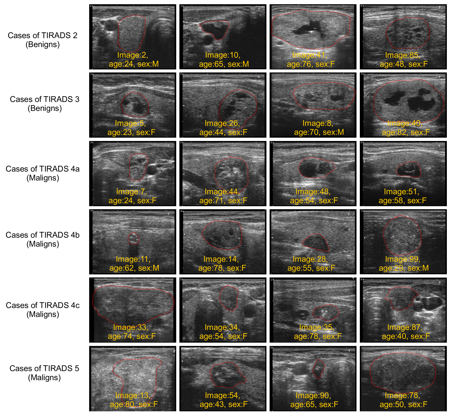
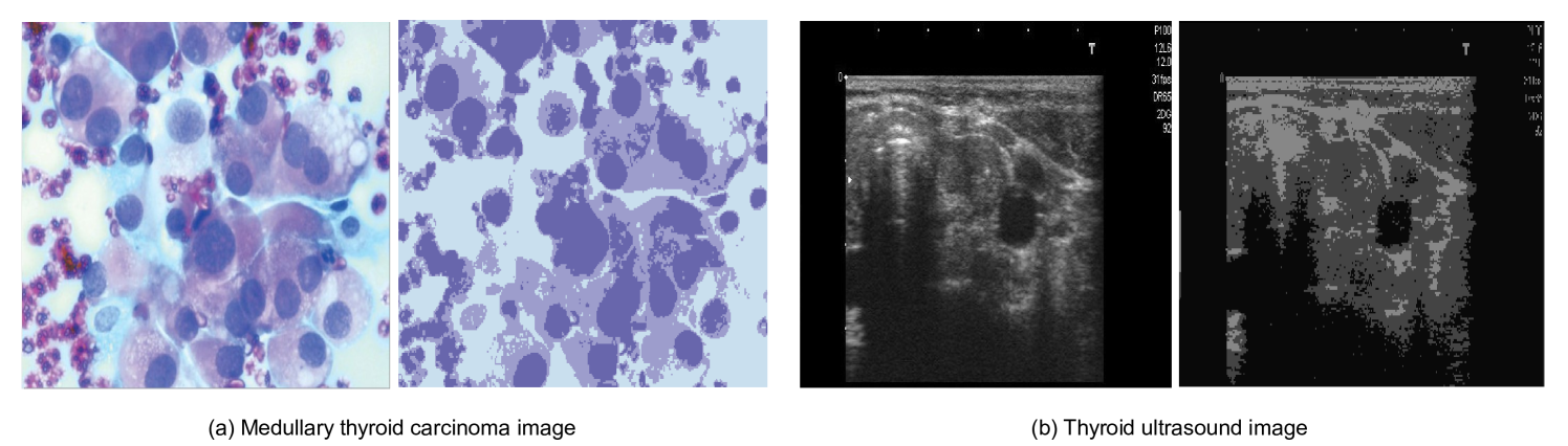
Digital Database Thyroind Image (DDTI): The DDTI dataset was developed with the support of Universidad Nacional de Colombia, CIM@LAB, and Instituto de Diagnostico Medico (IDIME). It serves as a valuable resource for researchers and new radiologists looking to develop algorithm-based computer-aided diagnosis systems for thyroid nodule analysis. The dataset comprises 99 cases and 134 images, with each patient’s data stored in an XML file format [180]. Fig. 4 provides an illustration of six samples from each of the thyroid carcinoma tissue types in the DDTI dataset.
-
•
The Cancer Genome Atlas (TCGA) data: The TCGA is a comprehensive collection of data gathered from 11,000 patients diagnosed with various types of cancer over a period of 12 years. The data consists of detailed genomic, epigenomic, transcriptomic, and proteomic information, amounting to a total of 2.5 petabytes. This extensive dataset has been instrumental in advancing the research, diagnosis, and treatment of cancer.
-
•
The National Cancer Data Repository (NCDR): The NCDR serves as a resource for healthcare and research with the goal of capturing all recorded cases of cancer in England. This data is sourced from the Office for National Statistics [181].
-
•
Prostate, Lung, Colorectal, and Ovarian (PLCO) dataset: The National Cancer Institute (NCI) supports the Prostate, Lung, Colorectal, and Ovarian (PLCO) Cancer Screening Trial, aimed at examining the direct factors that contribute to cancer in both men and women. The trial has records of 155,000 participants, and all studies regarding thyroid cancer incidence and mortality can be found within it [182].
In Table IV, we present examples of public and private thyroid cancer datasets used in thyroid cancer detection.
| Ref | Year | TCD | IT | IF | Instance | M/F | DA |
|---|---|---|---|---|---|---|---|
| [40] | 2018 | BMU | Sonographic | PNG | 1077 | 4309 | Public |
| [168] | 2019 | TCCC | US | PNG | 370 | 370 | Public |
| [183] | 2019 | Clinical | US | JPEG | 117 | 2108 | Public |
| [184] | 2019 | Hospital | US | JPEG | 62 | 12/60 | Public |
| [185] | 2020 | Tirads | US | JPEG | 5278 | NA | Public |
| [186] | 2018 | Peking Union | US | JPEG | 4309 | 1179 | Private |
| [79] | 2019 | Medical Center | US | PNG | 1425 | 2064 | Private |
| [187] | 2020 | PubMed | CT scans | JPEG | 2108 | 54/253 | Private |
| [152] | 2021 | ACR | DICOM | DICOM | 1629 | 83/289 | Private |
| [85] | 2021 | Clinical | US | PNG | 40 | 407 | Private |
-
•
Abbreviations: Thyroid cancer dataset (TCD); Image Types (IT); Image format (IF); Dataset access (DA); Male (M); Female (F).
The trengths and weaknesses of thyroid cancer detection techniques AI-based are summarized in Table V.
| Ref. | AI methods | Advantages | Drawbacks |
|---|---|---|---|
| [188] | DAE | No need labels for thyroid cancer | Insufficient training data and need relevant data |
| [189] | CNN | High thyroid cancer detection | Insufficient of labels for thyroid cancer and weak in interpretability |
| [190] | RNN | No need labels for thyroid cancer | Slow computation and difficulty in training |
| [191] | MLP | Adaptive learning for thyroid cancer | Limited in its results |
| [192] | RBF | Faster training | Slow classification |
| [193] | KNN | High sensitivity to thyroid cancer detection | Insufficient of labels for thyroid cancer |
| [194] | SVM | High sensitivity to thyroid cancer detection | Weak in interpretability and long training time |
| [195] | DT | No require scaling and normalization of data | Unstable |
| [196] | LR | Low-cost training and easier implementation | Difficulty to labels data |
| [197] | PCA | Low noise sensitivity | Loss in information |
| [198] | B | High detection of thyroid cancer | Loss of interpretability and high computational cost |
IV Features
In this section, the focus is on showcasing the crucial techniques utilized in the classification process for characteristic extraction and selection. This primarily involves identifying a subset of relevant features that positively impact the classification accuracy, and eliminating irrelevant variables.
IV-A Feature selection methods (FS)
FS1. Information Gain (IG): In this section, the focus is on showcasing the most widely-used techniques in the process of classification, with the main objective being to identify and select relevant characteristics that can positively impact the accuracy of classification while eliminating unimportant variables.
Information Gain (IG) is a straightforward method for classifying thyroid cancer features. This method evaluates the likelihood of having cancer by comparing the entropy before and after the examination. Typically, a higher gain value corresponds to lower entropy. IG has been used extensively in several applications for the diagnosis of cancerous diseases, such as in filtering informative genes for precise cancer classification [199], selecting breast cancer treatment factors based on the entropy formula [200], analyzing and classifying medical data of breast cancer [201], reducing the dimensionality of genes in multi-class cancer microarray gene expression datasets [202], and filtering irrelevant and redundant genes of cancer [199]. In [203], IG is utilized as a feature selection technique to eliminate redundant and irrelevant symptoms in datasets related to diabetes, breast cancer, and heart disease. Additionally, the IG-SVM approach, combining Information Gain and Support Vector Machine, has been employed and its results served as input for the LIBSVM classifier [199].
FS2. Correlation-based feature selection (CFS): The CFS is a technique frequently used for evaluating the correlation between different cancer features. In various studies, the CFS algorithm has been integrated into attribute selection methods for improved classification, such as in [204] where it was applied to thyroid, hepatitis, and breast cancer data from the UCI ML repository. In [121], the authors proposed a hybrid method that combined learning algorithm tools and feature selection techniques for disease diagnosis. The CFS was utilized in [205] for feature selection in microarray datasets to minimize the data’s dimensionality and identify discriminatory genes. A hybrid model incorporating the CFS and Binary Particle Swarm Optimization (BPSO) was proposed in [206] to classify cancer types and was applied to 11 benchmark microarray datasets. The CSVM-RFE, which involves the CFS, was used in [207] to reduce the number of cancer features and eliminate irrelevant ones. In [172], the authors utilized CFS-based feature selection techniques to identify key RNA expression features.
FS3. Relief (R): The Relief algorithm, commonly known as RA, is an effective method used in selecting important features by assessing their differentiation quality by assigning scores. This technique calculates the weight of various features based on the correlation between cancer attributes. In a study published in [208], a feature selection method based on the Relief algorithm was proposed as a means of improving efficiency.
FS4. Consistency-Based Subset Evaluation (CSE): The study in [209] presents a hybrid classification model for breast cancer, which is based on dividing cancer data into single-class subsets. The effectiveness of the model is evaluated using the Wisconsin Breast Cancer Dataset (WBCD).
IV-B Feature extraction methods (FE)
FE1. Principal Components Analysis (PCA): The use of Principal Component Analysis (PCA) has been highlighted in several studies as a method to reduce the dimensionality of data and uncorrelated the attributes of cancer features. For instance, in [136], PCA was applied to the dual-tree complex wavelet (DTCW) transform to select the optimum features of Thyroid Cancer. In [137], PCA was proposed as a tool for classifying different thyroid cancer subtypes such as papillary, follicular, and undifferentiated. The implementation of PCA and Linear Discriminant Analysis was also explored in [210] for classifying Raman spectra of different thyroid cancer subtypes. Finally, in [211], the authors utilized PCA on cDNA microarray data to uncover the biological basis of breast cancers.
FE2. Texture description (TD): Texture analysis is a commonly used method for extracting relevant information in the classification, segmentation, and prediction of Thyroid Cancer. There are numerous texture analysis techniques in the literature, including wavelet transform, binary descriptors, and statistical descriptors. The discrete wavelet transform, in particular, has received significant attention for its ability to perfectly decorrelate data. Many studies have utilized wavelets for thyroid cancer detection, such as in [212], where wavelet techniques were employed to identify cancer regions in thyroid, breast, ovarian, and prostate tumors. In [213], texture information was used to diagnose TN malignancy through a 2-level 2D wavelet transform. Other works exploring this area can be found in [214] and [215].
FE3. Active contour (AC): The active contour, first introduced by Kass and Witkin in 1987, is a dynamic structure primarily used in image processing. There are several approaches for solving the problem of contour segmentation using a deformable curve model, which has seen numerous applications in the field of Thyroid Cancer detection, as demonstrated in [216], [217], and [218].
FE4. Local binary patterns (LBP): The Local Binary Patterns (LBP) are features employed in computer vision to recognize textures or objects in digital images. LBP has been utilized to detect Thyroid Cancer in [214]. The combination of LBP and DL has also been proposed to classify benign and malignant thyroid nodules in [219] and [220].
FE5. Gray-level co-occurrence matrix (GLCM): The Gray-Level Co-occurrence Matrix (GLCM) is a matrix that represents the distribution of values of pixels that occur together at a specified offset in an image. In [221], GLCM is used to extract features to differentiate between different types of Thyroid Cancer. In [222], the differences between an individual with Hashimoto’s thyroiditis-associated papillary thyroid carcinoma and one with Hashimoto’s thyroiditis alone were investigated based on GLCM comparison.
FE6. Independent component analysis (ICA)): In Independent Component Analysis (ICA), information is gathered into a set of contributing features for the purpose of feature extraction. ICA is utilized to separate multivariate signals into their individual components. In [223], ICA is used to extract 29 attributes as independent and useful features for classifying data into either hypothyroid or hyperthyroid using a Support Vector Machine (SVM).
| Ref. | Year | Classifier | Features | Contributions |
|---|---|---|---|---|
| [224] | 2017 | KNN | FC/IG | - Avoid data redundancy and reduce computation time. The kNN deals with the missing dataset, and the ANFIS is provided with the resultant data as input. |
| [225] | 2017 | SVM | FC/CFS | - Extract the geometric and moment features while some kernels of the SVM classifier classify the extracted features. |
| [105] | 2020 | CNN | FC/R | - Combine ML and feature selection algorithms (namely, Fisher’s discriminant ratio, Kruskal-Wallis’ analysis, and Relief-F) to analyse the SEER database. |
| [226] | 2022 | CNN | FE/PCA | - The influence of unbalanced serum Raman data on the prediction results was minimized by using an over-sampling algorithm in this study. PCA then reduced the data’s dimension before classifying data using RF and the Adaptive Boosting. |
| [227] | 2012 | O | FE/TD | - Combine CAD and DWT and texture feature extraction. The AdaBoost classifier uses the extracted features to classify images into benign or malignant thyroid images. |
| [228] | 2021 | CNN | FE/AC | - Image enhancement, segmentation and multi-feature extraction, encompassing both geometric and texture features. Each characteristic is then classified using MLP and SVM, resulting in a determination of either benign or malignant. |
| [185] | 2020 | SVM | FE/LBP | - Deep features are extracted by CNN and are combined with the hand-crafted feat- ures, including HOG, LBC, and SIFT to create fused features. These fused features are then used for classification by an SVM. |
| [229] | 2019 | SVM | FE/GLCM | - Use a median filter to reduce noises, and delineate the contours before extracting features from thyroid regions, including GLCM texture features. SVM, random forest, and bootstrap aggregating (bagging) are then used to identify the benign and malignant nodules. |
| [223] | 2019 | SVM | FE/ICA | Multi-kernel-based is used as a classifier to distinguish the thyroid disease. |
A summary of features methods based on DL conducted in the diagnosis of thyroid cancer are illustrated in Table VI.
V Standard assessment criteria
In this section, we examine the most commonly utilized standard parameters for evaluating the identification of Thyroid Diseases (TD). These criteria serve as a measure of the effectiveness of the methods used. Selecting the right metric is crucial when evaluating the performance of machine learning models. Numerous metrics have been proposed to evaluate machine learning models in various applications. Here, we present a summary of popular metrics that are considered suitable for assessing the performance of AI algorithms applied in the detection of Thyroid Cancer. (See Table VII, VIII and IX)
V-A Classification and Regression Metrics
Table VII presents an outline of classification and regression metrics used in evaluating AI-based thyroid cancer detection frameworks.
| Metric | Mathematical formula | Description |
|---|---|---|
| Accuracy (ACC) | Give the correct percent of the total number of positive and negative predictions. | |
| Specificity (SPE) | It is the ratio of correctly predicted negative samples to the total negative samples. | |
| Sensitivity (SEN) | It is a quantifiable measure metric of real positive cases that got predicted as true positive cases. | |
| Precision (P) | Measure the proportion of true positive predictions made by the model, out of all the positive predictions made by the model. | |
| F1 Score (F1) | It is the harmonic mean of precision and sensitivity of the classification. | |
| Error Rate (ER) | It is equivalent to 1 minus Accuracy. | |
| Root mean square error (RMSE) | It is the standard deviation of the predicted error between the training and testing dataset, its lower value means that the classifier is an excellent one. | |
| The negative predictive value (NPV) | is the proportions of negative results in diagnostic tests, its higher value means the accuracy in diagnosis. | |
| Jaccard similarity index (JSI) | It has been proposed by Paul Jaccard to gauge the similarity and variety in samples. | |
| Fallout or false positive rate (FPR) | Measure the proportion of negative samples that are incorrectly classified as positive by the model. | |
| Volumetric Overlap Error (VOE) | Evaluate the similarity between the segmented region and the ground truth region. VOE measures the amount of overlap between the two regions and is defined as the ratio of the volume of the union of the segmented region and the ground truth region to the volume of their intersection. | |
| Mean Absolute Error (MAE) | It is the average of the difference between the original values and the predicted values. | |
| Mean Squared Error (MSE) | It is the average of the square of the difference between the original values and the predicted values. |
V-B Statistical Metrics
Table VIII depicts a summary of statistical metrics used in assessing AI-based thyroid cancer detection schemes.
| Metric | Mathematical formula | Description |
|---|---|---|
| Standard deviation (SD) | It is a measure of the amount of variation or dispersion in a set of data. | |
| Correlation (Corr) | It describes the degree of association or relationship between two or more variables. | |
| Kappa de Cohen | It measures the degree of concordance between two evaluators, relative to chance. |
V-C Computer Vision Metrics
Table IX portrays a summary of computer vision metrics used in assessing AI-based thyroid cancer detection schemes.
| Metric | Mathematical formula | Description |
|---|---|---|
| Peak Signal to Noise Ratio (PSNR) | It measures the ratio of the maximum possible power of a signal to the power of the noise that affects the fidelity of its representation. | |
| Structural Similarity Index (SSIM) | It evaluates the similarity between two images or videos by comparing their luminance, contrast, and structural information. | |
| Visual Information Fidelity (VIF) | It evaluates the quality of a reconstructed or compressed image or video compared to the original signal. It measures the amount of visual information preserved in the processed image or video, taking into account the spatial and frequency characteristics of the image. | |
| Normalized Cross-Correlation (NCC) | Measure the similarity between two images (or videos) by subtracting the mean value of each signal from the signal itself. Then, the signals are normalized by dividing them by their standard deviation. Finally, the cross-correlation between the two normalized signals is calculated. | |
| Structural Content (SC) | A higher value of SC (Structural Content) shows that the image is of poor quality. | |
| Weight Peak Signal to Noise Ratio (WPSNR) | It takes into account the image texture [230]. | |
| Noise Visibility Function (NVF) |
where is the luminance variance. |
It estimates the texture content in the image. |
| Visual Signal to Noise Ratio (VSNR) |
Where is the RMS contrast of the original image and is visual distortion [231]. |
It is based on the specified thresholds of distortions in the image based on the computing of contrast thresholds and wavelet transform. If the distortions are lower than the threshold, the VSNR is perfect. |
| Weighted signal-to-noise ratio (WSNR) |
where , , and represent the Discrete Fourier Transforms (2D TFD) [232]. |
It is based on the contrast sensitivity function (CSF). |
| Normalized Absolute Error (NAE): | It evaluates the accuracy of an ML model’s predictions. It measures the difference between the predicted values and the actual values, as a proportion of the range of the actual values. | |
| Laplacian Mean Square Error (LMSE) |
where is the Laplacian operator. |
It is a variant of the Mean Square Error (MSE) that uses the Laplacian distribution instead of the Gaussian distribution. |
V-D Ranking Metrics
M1. The mean reciprocal rank (MRR): The MRR is a statistic measure for evaluating the mean reciprocal rank of results for a sample of queries[233].
| (1) |
Where ranki refers to the rank position of the first relevant document for the i-th query.
M2. The Discounted cumulative gain (DCG): the DCG is used to measure the ranking quality [234].
VI Example of thyroid cancer detection using AI
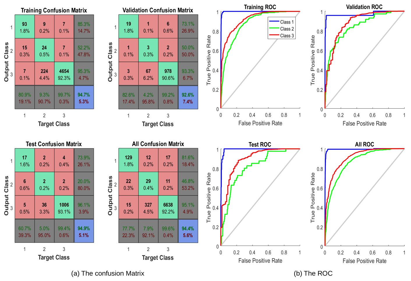
To explain how thyroid cancer has been considered in the literature and how AI can be used to detect types of cancers. In the following, we present a simple example to classify TD. It has been known that pattern recognition is the process of training a neural network to assign the correct target classes to a set of input patterns. Once trained the network can be used to classify patterns. In this section, we present an example of thyroid cancer classification as benign, malignant, and normal based on a set of features specified according to the TIRADS. In this example, the dataset (7200 samples) is selected from the UCI Machine Learning Repository [235]. This dataset can be used to create a neural network that classifies patients referred to a clinic as normal, hyperfunction, or subnormal functioning. The Thyroid Inputs (TI) and Thyroid Targets (TT) are defined as: (i) TI: a 21x7200 matrix consisting of 7200 patients characterized by 15 binary and 6 continuous patient attributes. (ii) TT: a 3x7200 matrix of 7200 associated class vectors defining which of three classes each input is assigned to. Classes are represented by a 1 in rows 1, 2, or 3. (1) Normal, not hyperthyroid. (2) Hyperfunction. (3) Subnormal functioning.
In this network, the data is divided into 5040 samples, 1080 samples, and 1080 samples used for training, validation, and testing respectively. The network is trained to reduce the error between thyroid inputs and thyroid targets or until it reaches the target goal. If the error rate does not decrease and the training does not improve, the training data is halted with data of validation. The data testing is used to deduce the values of targets. Thus, it determines the percentage of learning. For this example, the 10 hidden layer neurons are used in this model for 21 input and 3 output. After the simulation of the model, the Percent Error was been 5.337%, 7.407%, and 5.092% for training, validation, and testing respectively. Thus in the total, it recognized 94.4% and the overall error rate was 5.6%. The confusion matrix and the ROC metric are illustrated in Fig. 5.
Fig. 6, illustrates an example of thyroid segmentation (TS) in ultrasound images using k-means (3 clusters have been chosen for this example) which is one of the most commonly used clustering techniques.
VII Critical analysis and discussion
As we delve into the core of this paper, it is essential to critically assess and discuss the multitude of facets associated with the application of AI in thyroid carcinoma detection. While the promise of AI has been well-articulated in existing literature, a more nuanced perspective is needed to fully understand its impact on healthcare, both positive and negative. In this section, we undertake a critical analysis of the effectiveness of AI models for thyroid carcinoma detection. Moving beyond the optimistic numbers, we will question the robustness of these models in real-world clinical settings and discuss their role in the broader context of clinical decision-making. Furthermore, we explore the potential biases in AI models, understanding how they might inadvertently perpetuate existing inequities in healthcare. A comparative assessment of AI-based and traditional diagnostic methods will provide deeper insights into their relative effectiveness. Moving on, acknowledging the challenges to the implementation of AI tools in healthcare, we delve into the infrastructural, regulatory, and cultural barriers that might hinder their widespread adoption. Lastly, we underscore the crucial role of interdisciplinary collaboration in ensuring the successful integration of AI into healthcare.
A summary of features methods based on DL conducted in the diagnosis of thyroid cancer are detailed in Table X.
The Effectiveness of AI Models: The reported accuracy, sensitivity, and specificity of AI models in the literature may vary widely based on the dataset used, the quality of the data, and the methodology employed. AI models’ effectiveness in a controlled experimental environment may not reflect their performance in a real-world clinical setting. Factors like noise in the data, incomplete data, and changing clinical conditions can dramatically influence the outcome. Therefore, it is crucial to scrutinize the model’s robustness and reliability under various conditions.
| Ref. | AI Model | Dataset | ACC | SPE | SEN | PPV | F1 | NPV | AUC |
|---|---|---|---|---|---|---|---|---|---|
| [236] | CNN | PD | 88.00 | 79.10 | 98.10 | - | - | - | - |
| [100] | ELM | PD | 87.72 | 94.55 | 78.89 | - | - | - | - |
| [105] | MLP | PD | 87.16 | 87.05 | 91.18 | 0.162 | 0.275 | 0.997 | - |
| [138] | SVM | PD | 63.27 | 71.85 | 38.46 | 32.43 | - | 76.87 | - |
| [237] | RF | PD | 86.8 | 87.9 | 85.2 | - | - | - | 0.92 |
| [238] | LR | PD | 77.8 | 79.8 | 70.6 | - | - | - | 0.75 |
| [229] | B | PD | 84.69 | 0.8696 | 0.8269 | 0.8776 | - | 0.8163 | 0.8852 |
| [239] | Ensemble DL | Cytological images | 99.71 | - | - | - | - | - | - |
| [68] | VGG-16 | Cytological images | 97.66 | - | - | - | - | - | |
| [50] | VGG-16 | 99 | 86 | 94 | - | 88 | - | - | |
| [39] | RF | Ultrasound | - | - | - | - | - | - | 0.94 |
| [183] | k-SVM | Ultrasound | - | - | - | - | - | - | 0.95 |
| [90] | ANN | Ultrasound | - | - | - | - | - | - | 0.69 |
| [84] | SVM RF | Ultrasound | - | - | - | - | - | - | 0.951 |
| [240] | ANN SVM | Ultrasound | 0.96 | - | - | - | - | - | - |
| [241] | RF | Ultrasound | - | - | - | - | - | - | 0.75 |
| [242] | CNN | DICOM | 83.00 | 85.00 | 82.40 | - | - | - | - |
| [27] | CNN | DICOM | - | 91.50 | - | - | - | - | - |
| [76] | Fine-Tuning DCNN | PD | 99.10 | - | - | - | - | - | - |
| [243] | ResNet18-based network | PD | 93.80 | - | - | - | - | - | - |
| [244] | multiple-scale CNN | PD | 82.20 | - | - | - | - | - | - |
| [67] | ThyNet | PD | - | - | - | - | - | - | 0.921 |
| [245] | Alexnet CNN | PD | 86.00 | - | - | - | - | - | - |
| [171] | DNN | ACR TIRADS | 87.20 | - | - | - | - | - | - |
| [83] | CNN (BETNET) | Ultrasound | 98.30 | - | - | - | - | - | - |
| [246] | ResNet | TIRADS | 75.00 | - | - | - | - | - | - |
| [247] | Xception | CT images | 89.00 | 92.00 | 86.00 | - | - | - | - |
| [79] | DCNN | Sono graphic images | 89.00 | 86.00 | 93.00 | - | - | - | - |
| [248] | Google inception v3 | Histo pathology images | 95.00 | - | - | - | - | - | - |
| [249] | Cascade MaskR-CNN | Ultrasound | 94.00 | 95.00 | 93.00 | - | - | - | - |
| [250] | VGG16 | Ultrasound | - | 92.00 | 70.00 | - | - | - | - |
| [251] | VGG19 | Ultrasound | 77.60 | 81.40 | 72.50 | - | - | - | - |
| [36] | VGG16 | Ultrasound | 74.00 | 80.00 | 63.00 | - | - | - | - |
| [185] | SVM CNN | Ultrasound | 92.50 | 83.10 | 96.40 | - | - | - | - |
| [252] | CNN | TIRADS | 85.10 | 86.10 | 81.80 | - | - | - | - |
| [253] | CNN | TIRADS | 82.10 | 85.00 | 78.00 | - | - | - | - |
| [254] | CNN | TIRADS | 80.30 | 80.10 | 80.60 | - | - | - | - |
| [255] | CNN | US | 83.00 | 47.00 | 65.00 | - | - | - | - |
| [256] | CNN | MRI | 79.00 | 80.00 | 65.00 | - | - | - | - |
| [257] | CNN | US | 97.00 | 84.10 | 89.50 | - | - | - | - |
| [258] | CNN | CT image | 84.00 | 73.00 | 93.00 | - | - | - | - |
| [259] | CNN | US | 77.00 | - | - | - | - | - | - |
VII-A Limitations and open challenges
Despite the success of AI tools in thyroid cancer diagnosis, their limitations hinder the development of effective solutions, make their application costly, and limit their diffusion. For instance,to achieve precise thyroid cancer detection, it is crucial to gather and store all relevant data in one place. Then, algorithms must be developed to identify all forms of thyroid cancer. Every thyroid cancer dataset includes a set of training images, test images, nodule plans, and classifications of nodule characteristics of diverse sizes [260]. The datasets must be regularly updated using MRI, CT scans, X-rays, and clinically obtained scans to assess thyroid conditions, and they should also include demographic information such as gender and age. Additionally, it is important to establish a unified and centralized database accessible to all medical centers to test, validate, and apply AI algorithms to existing data [261]. Moving, the rest of the limitations and open challenges can be summarized as follows:
-
•
Insufficient clean data and accuracy: The lack of comprehensive and annotated data sets regarding the incidence and spread of cancer, specifically thyroid cancer, is a major hindrance to accurate cancer diagnoses and efficient treatment. Medical statistics often do not properly record the number of deaths caused by thyroid cancer, making data collection and validation challenging [262]. This results in a limited amount of data typically collected from one center, due to the absence of a dedicated thyroid cancer clinical database shared among institutions. The accuracy of AI algorithms in diagnosing thyroid cancer is also limited by the limited number of available labeled cases for clinical outcomes [263]. Researchers acknowledge that a large amount of data is necessary for the neural network to yield accurate results, but caution must be taken in regard to the data added during the learning phase as it can introduce noise.
-
•
Thyroid gland imaging: In the diagnostic evaluation of thyroid cancer, computed tomography (CT) and magnetic resonance imaging (MRI) are available options but they are not considered the preferred methods due to their high cost and unavailability in certain cases [51]. Instead, ultrasound is commonly used as an alternative to physical exams, radioisotope scans, or fine-needle aspiration biopsies. During a ultrasound examination, the doctor is able to assess the activity of the gland by observing the echo of the node and determining its echogenicity, size, limits, and the presence of calcifications. However, the results obtained from ultrasound tests are not always accurate enough to differentiate between benign and malignant nodes and the images obtained can be more prone to noise [264].
-
•
The number of DL layers: Choosing the right DL algorithm is crucial in addressing various issues, particularly those related to thyroid cancer diagnosis. Due to the close similarities between benign and malignant tumors, as well as between tumors and other types of lymphocytes, it is challenging to differentiate between them accurately [265]. To achieve this, a significant increase in the number of layers for feature extraction may be required. However, this results in a longer processing time, especially when dealing with large amounts of data, which can impact the timeliness of the diagnosis for cancer patients [50].
-
•
The computation cost and space: In the field of algorithms, time computing is a metric that assesses the computational complexity of an algorithm, which predicts the time it will take to run the algorithm by calculating the number of basic operations it performs, as well as its dependence on the size of the input. Typically, time computing is expressed as , where represents the size of the input, measured in terms of the number of bits required to represent it [266]. Researchers in the AI field, especially those working on thyroid cancer or other types of cancer diagnosis, face the challenge of finding algorithms that are both highly accurate and efficient in terms of processing time. They aim to develop algorithms that can analyze vast amounts of data quickly while still providing accurate results. Moreover, the volume of data used in these algorithms can sometimes exceed the available storage space [50].
-
•
Imbalanced dataset: The distribution of cancer elements within categories related to thyroid tissue cells is often uneven, as these cells often make up a minority of the total tissue cell dataset. As a result, the data set is highly imbalanced, consisting of both cancer cells and normal cells. This unbalanced distribution of features in cancer cell detection datasets often results in the suboptimal performance of AI algorithms used for the detection [267].
-
•
Sparse labels: Labeling is a crucial aspect of CT detection, specifically for distinguishing between normal and abnormal cancer cells. However, the process can be time-consuming and costly due to the limited number of available labels. This scarcity results in inconsistent decisions and can negatively impact the accuracy of AI algorithms, which heavily rely on labeled data. This can eventually undermine the trust and credibility of this type of application [267].
-
•
The volume of data: At present, with the advancement in technology, especially in the field of thyroid cancer diagnosis and the growing volume of medical and patient data, researchers are facing challenges in suggesting algorithms that can effectively handle a limited number of samples, noisy samples, unannotated samples, sparse samples, incomplete samples, and high-dimensional samples. This requires AI algorithms that are highly efficient and capable of processing vast amounts of data exchanged between healthcare providers and patients or among specialist physicians [268]
-
•
The error-susceptibility: Despite AI being self-sufficient, it is still susceptible to errors. For instance, when training an algorithm with thyroid cancer datasets to diagnose cancerous regions, it can result in biased predictions if the training sets are biased. This can lead to a series of incorrect results that may go unnoticed for an extended period. If detected, identifying and correcting the source of the problem can be a time-consuming process [269].
-
•
The data form: Despite the numerous advancements in the use of AI for thyroid cancer detection, several limitations persist and pose a challenge to its progress. With the growing demand for various medical imaging technologies that result in vast amounts of data needed for AI algorithms, coordinating and organizing this information has become a daunting task. This can largely be attributed to the absence of proper labeling, annotation, or segmentation of the data, making it difficult to manage effectively [270].
-
•
Unexplainable AI: The utilization of AI in the medical field can sometimes yield results that are unclear and lack proper justification, known as a "black box". This leaves doctors unsure about the accuracy of the results and may lead to erroneous decisions and treatments for patients with thyroid cancer. Essentially, AI can behave like a black box and fail to provide understandable explanations for its outputs [271].
-
•
Lack of cancer detection platform: One of the major barriers to detecting various cancers, particularly thyroid cancer, is the limited availability of platforms for reproducing and examining previous results. This shortage represents a significant weakness and hinders the comparison of AI algorithm performance, making it challenging to improve their efficacy [155]. The presence of online platforms with comprehensive data sets, cutting-edge algorithms, and expert recommendations is vital in aiding doctors, researchers, developers, and specialists to make informed decisions with a low margin of error. Such platforms also provide a crucial supplement to clinical diagnoses by allowing for more comprehensive experimentation and comparison [272].
-
•
The digitization and loss data: Digitization of medical records has become a necessity, particularly in the realm of cancer diagnosis, due to the widespread adoption of various technologies such as Whole Slide Images (WSIs). WSIs, which serve as digital versions of glass slides, facilitate the application of AI techniques for pathological analysis [273]. Despite its benefits, digitization in the medical field is confronted with certain limitations, such as the risk of significant information loss during quantification and inaccuracies that may arise from data compression utilized in autoencoder algorithms. Hence, it is crucial to be mindful in selecting the right digitization technology to preserve the information and maintain the originality of the data [274, 275].
-
•
The Contrast: The absence of sufficient contrast in the tissues neighboring the thyroid gland (TG) complicates the process of accurately analyzing and diagnosing thyroid cancer.
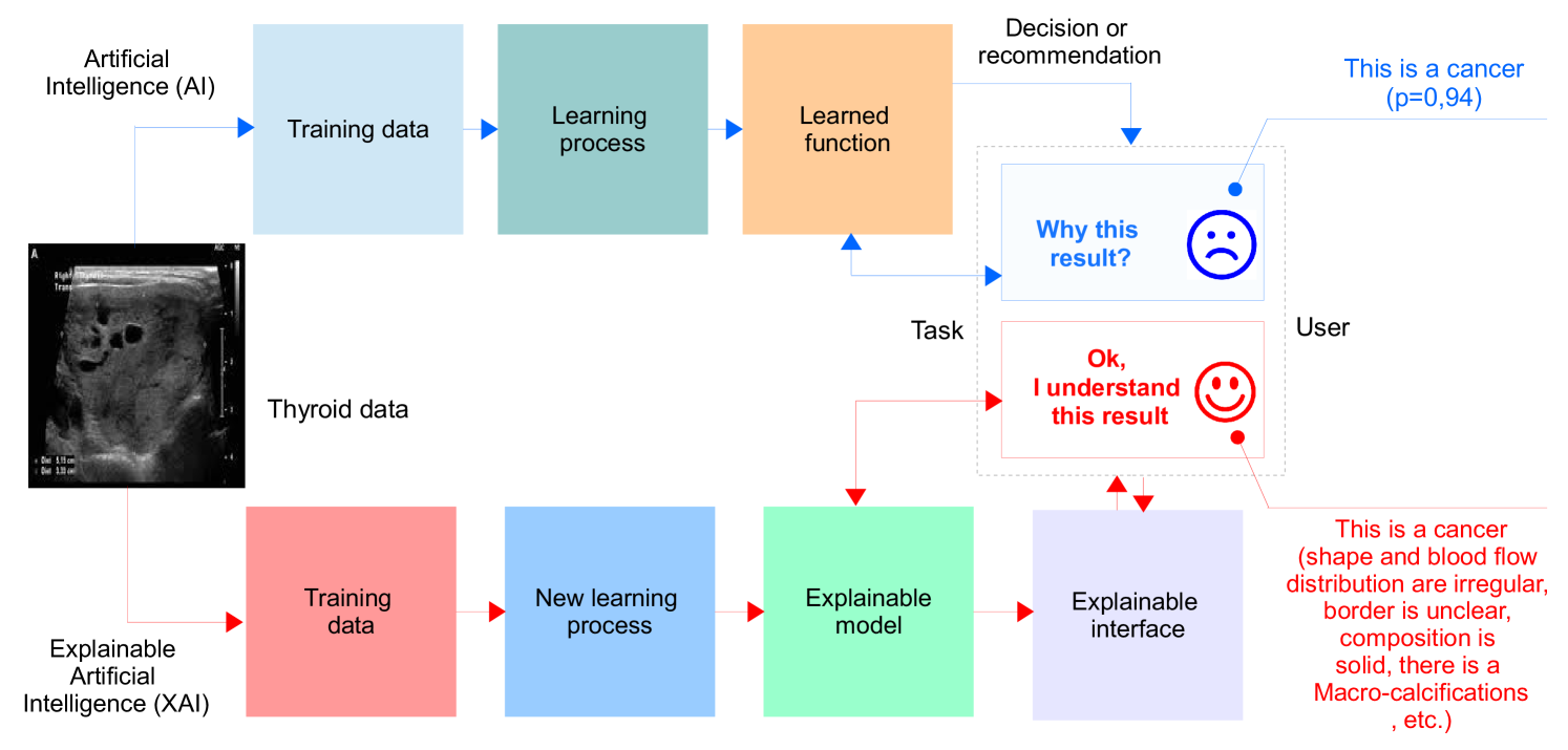
VIII Future Research directions
We also highlight the future trajectory of AI in thyroid carcinoma detection, discussing emerging trends and technologies while considering their ethical implications. The ethical considerations do not end there, as we further examine issues related to data privacy, accountability, and equity. This section highlights promising research trends that will have a major effect on enhancing thyroid cancer detection in the future.
VIII-A Explainable Artificial Intelligence (XAI)
The use of artificial intelligence (AI) systems in decision-making is crucial, but they can be complex and difficult to understand. To address this issue, the field of explainable AI (XAI) has emerged, which aims to provide transparency in AI models. The need for XAI is especially important in health applications where the interpretation of results is crucial. The use of XAI has been demonstrated in the analysis of incurable diseases affecting the TG, as seen in several studies such as [276, 277, 278, 279, 280]. The difference between AI and XAI is illustrated in Fig. 7. In [167], the authors present an XAI model for the detection of thyroid cancer, which improves the confidence of medical practitioners in the predictions. Unlike traditional AI algorithms, XAI models provide evidence to support their conclusions and avoid the limitations of "black box" algorithms. By using XAI, clinicians can make more informed decisions with greater confidence.
VIII-B Edge, fog, and cloud computing for implemetation
The edge network is a combination of edge computing and AI that processes algorithms based on AI near the source of data [281]. This allows for better performance and lower costs for applications that require heavy information processing, as well as reduces the need for long-distance communication between the patient and the doctor. The proximity of the information and storage capabilities to the end-user in the health sector allows for direct and immediate access [282]. To further enhance performance, the detection of thyroid cancer in edge networks relies on the use of fog computing, which is a decentralized computing architecture located between the cloud and the data-producing devices. This architecture allows for the flexible placement of computing and storage resources in logical locations, improving performance [283]. To ensure the proper operation of the AI-based thyroid cancer detection system, it utilizes cloud computing as an access point. This guarantees that the stored data, servers, databases, networks, and programs are accessible and shared among specialized doctors, as long as it is connected to the Internet. Such a hybrid system has proven to be effective for medical applications, including detection of thyroid cancer, as seen in various studies including [284, 285, 286, 287, 288, 289, 290, 291, 292, 293, 294].
VIII-C Reinforcement learning (RL)
RL, a subfield of ML, allows agents to make decisions in interactive environments through trial and error, observation, and learning (as depicted in Fig. 8). In recent years, there has been significant interest in using RL in detecting incurable diseases and providing explanations to aid medical decision-making. For example, reinforcement learning is used in [295] to classify cancer data, and deep reinforcement learning is used in [296] to segment lymph node sets. The authors generate pseudo-ground truths using RECIST-slices and achieve simultaneous optimization of lymph node bounding boxes through the interaction between a segmentation network and a policy network.
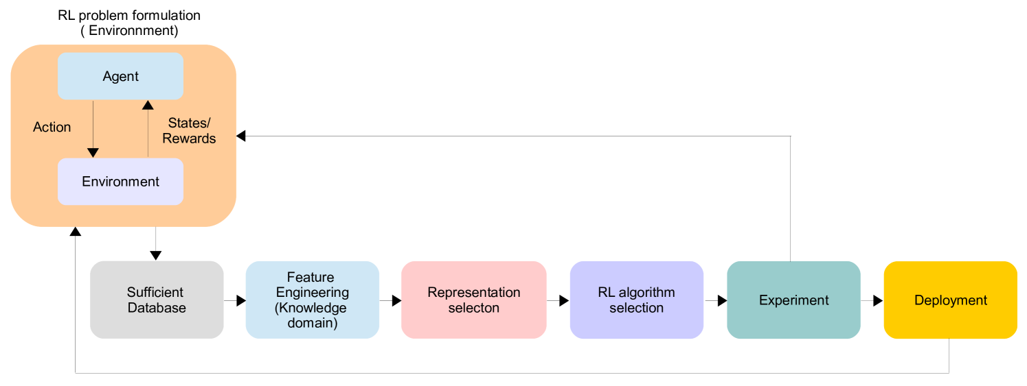
VIII-D Transfer learning (TL)
TL is a valuable solution to the overfitting and precision challenges faced by diagnosis systems [297, 298, 299]. This technique leverages stored knowledge from a specific problem to address other issues such as reducing training time and data volume [298, 268]. Its use in the diagnosis of the thyroid gland (TG) is demonstrated in Fig. 9. Moving on, in [154], the authors tackle the challenge of capturing appropriate features of benign and malignant nodules using CNNs. They transfer the knowledge learned from natural data to an ultrasound (US) image dataset to produce hybrid semantic deep features. The transfer learning technique has also been successfully applied to classify thyroid nodule (TN) images in [160]. Other related works can be found in [300, 250, 301, 158, 302].
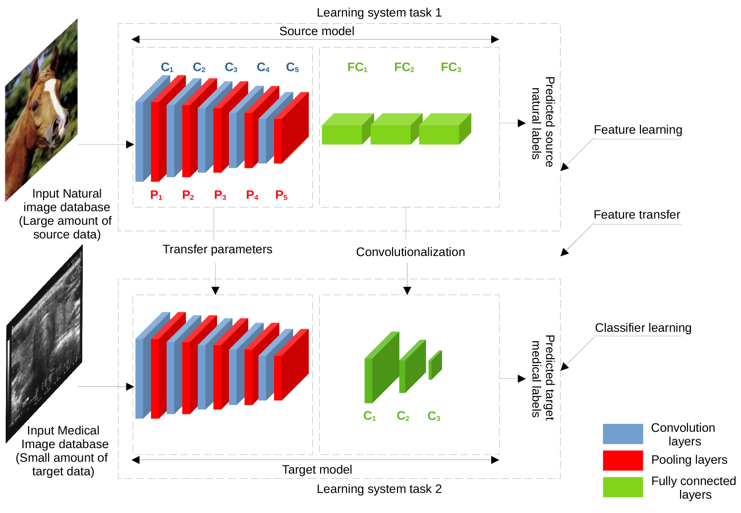
VIII-E Panoptic segmentation (PS)
The challenge of accurately separating and dividing objects with diverse and overlapping appearances remains an issue, particularly in the medical field. To address this, many researchers have put forth proposals for a comprehensive and cohesive segmentation of various details [303, 304]. The focus has been on PS, which combines both instance and semantic segmentation to identify and separate objects. In semantic segmentation, the goal is to classify each pixel into specific classes, while in instance segmentation, the focus is on segmenting individual object instances. AI has been incorporated into this model through supervised or unsupervised instance segmentation learning, making it well-suited for medical applications (Fig. 10). This has been demonstrated in works such as [305, 306].
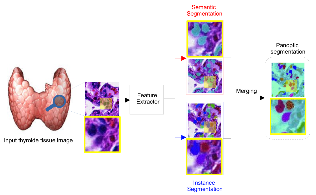
VIII-F Internet of medical imaging thing (IoMIT)
The Internet of Medical Things (IoMT) has recently gained widespread attention in the medical field, as it seeks to enhance healthcare delivery and reduce treatment costs through the exchange of health data between patients and doctors using connected devices with wireless communication (Fig. 11). One example of this integration can be found in [307], which proposes an AI-based solution for early detection of thyroid cancer in the IoMT, utilizing CNN to improve differentiation between benign and malignant nodules, ultimately saving lives. Other relevant studies related to the IoMIT have also been conducted, such as [308] and [309].
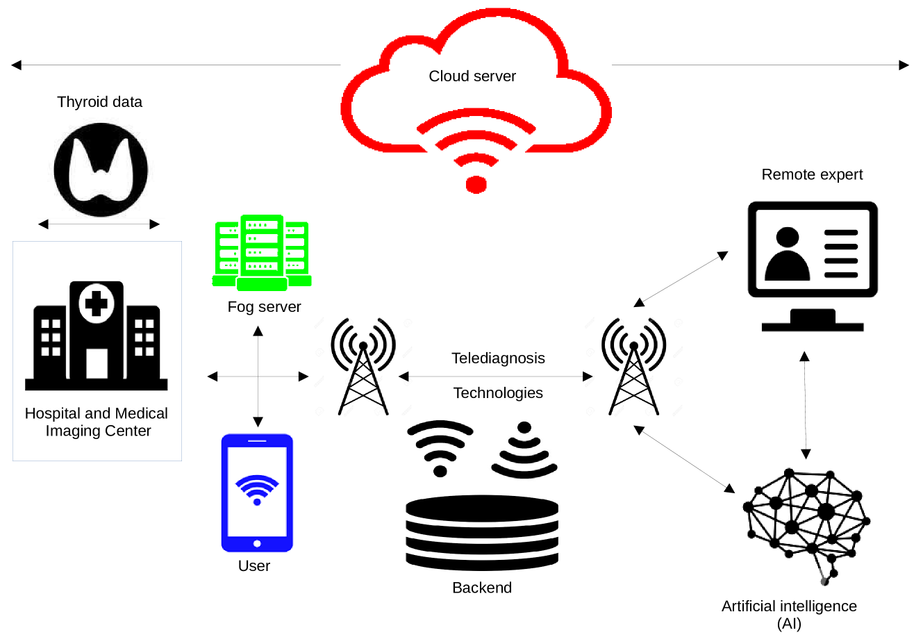
VIII-G 3D thyroid cancer detection (3D-TCD)
The conventional 2D ultrasound is widely used for diagnosing thyroid nodules, but its static images may not accurately reflect the nodule’s structures. Hence, the use of 3D ultrasound has gained attention as it provides a more comprehensive view of the lesion by reconstructing its features and enabling better differentiation between different diagnoses [310]. With the ability to examine complex growth patterns, and margins, and to shape from multiple angles and levels, 3D ultrasound can provide a more accurate evaluation of the morphological features of thyroid nodules in comparison to 2D images. This has been confirmed through comparative studies between 3D and 2D ultrasound images [311, 312, 313].
VIII-H AI in Thyroid Surgery (AI-TS)
In light of the challenges faced in surgical procedures, the use of AI-powered robots in surgical practices is becoming increasingly essential. AI has the potential to address numerous clinical issues by analyzing and sharing massive amounts of data to support decisions with a level of accuracy comparable to that of healthcare professionals [314]. Companies are incorporating AI into surgical practices by training AI-based systems and providing robots that assist surgeons in operating rooms, supply surgical materials, handle contaminated materials and medical waste, remotely monitor patients, and collect and organize patient data such as electronic medical records, vital signs, laboratory results, and video footage [315]. As such, it is important for surgeons to have a strong understanding of AI in order to grasp its impact on healthcare. While AI-powered robotic surgery may still be some time away, collaboration across various fields can accelerate AI’s capabilities and improve surgical care [316, 317, 318, 319] [320, 321, 322, 323].
VIII-I Wavelet-based AI
Recently, wavelets transform, specifically first and second-generation, has gained recognition for its ability to detect various forms of cancer, especially when integrated with AI. This combination has become crucial in the medical field, providing doctors and surgeons with a tool to accurately diagnose diseases more efficiently and quickly [324, 325]. The proposed method is based on pre-processing the dataset through discrete wavelet transfer (DWT) and then evaluating the performance of AI in classifying different types of tumors that can impact organs in the body (as explained in Fig. 12). This model holds great potential for the detection of thyroid cancer and researchers are encouraged to test different wavelets available in the literature to further improve its effectiveness [326].
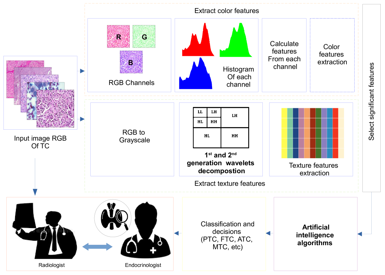
VIII-J Learning with reduced data
One of the hurdles in implementing AI in the medical sector is acquiring adequate data and annotations. AI’s capability to minimize the need for labeled data in making an accurate diagnosis is crucial [327]. This can be achieved through various learning methods such as semi-supervised learning, supervised learning, unsupervised learning, or alternative approaches that necessitate a smaller quantity of annotated data (Fig. 13)[328].
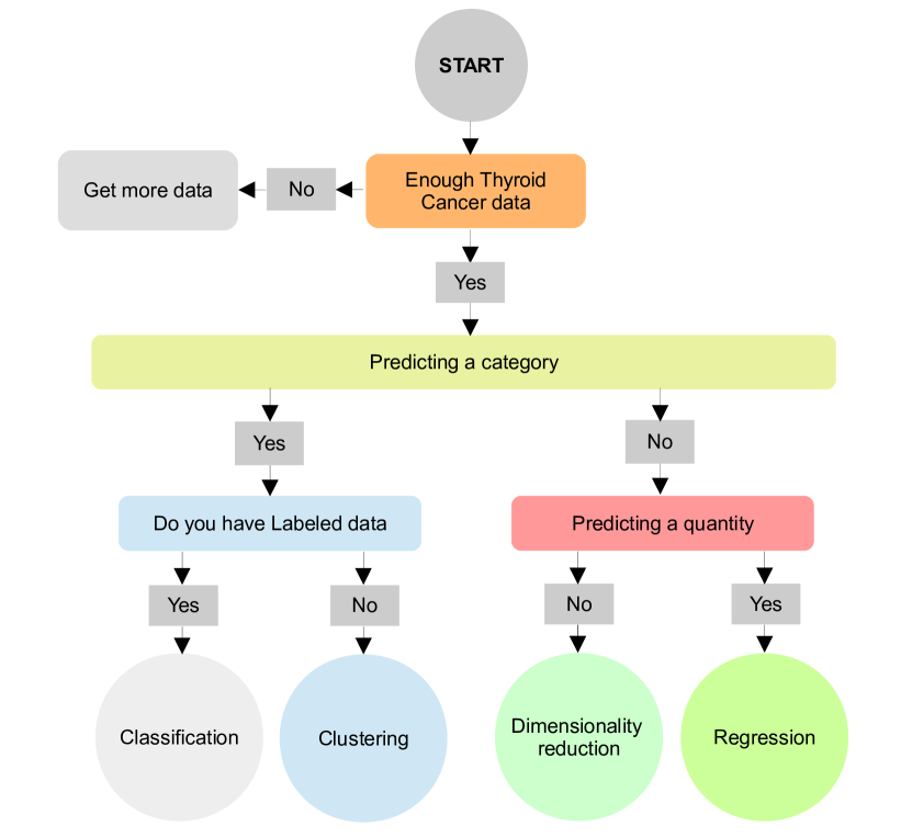
VIII-K Recommender systems (RSs)
The abundance of data collected from online medical platforms and electronic health records can make it challenging for thyroid cancer patients to access relevant and accurate information [329]. The high cost of healthcare data also poses difficulties for doctors to track patients and manage a large patient volume with various treatment options. Given these challenges, the implementation of RSs has been proposed to improve decision-making in healthcare and ease the workload for both patients and oncologists [330, 331]. The use of RS in digital health provides personalized recommendations, accurate analysis of big data, and stronger privacy protection through integration with artificial intelligence and machine learning technologies [332] as depicted in Fig. 14.
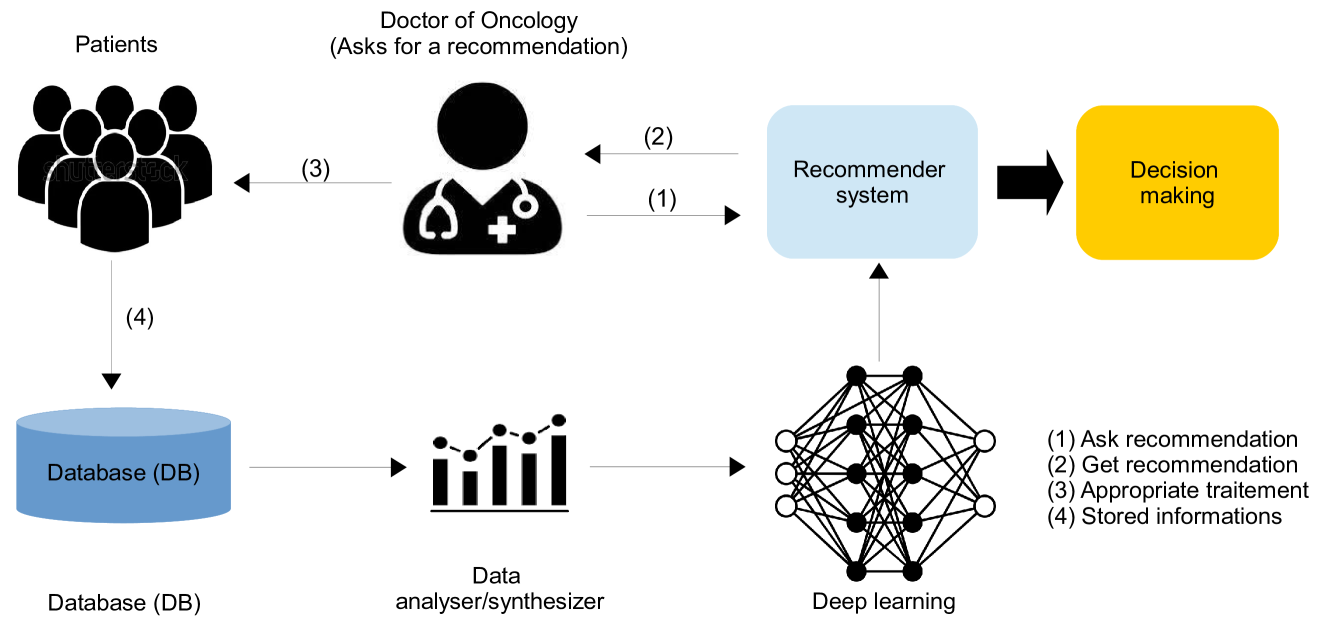
VIII-L Federated learning (FL):
The FL has become very popular in the field of healthcare applications [333]. The surrounding conditions greatly affect human health and cause negative effects on the economy. Diseases of the thyroid gland are among the most common health problems that have become noticeable among various groups of society in recent times. ML can play a vital role in such medical conditions, as the collected data can be exploited to train an ML model that can predict critical conditions. Emphasizing that patient data across different medical centers should be handled privately, the FL setup is the natural choice for such applications, as depicted in Fig.15. Therefore, In [334], the authors compared the performance of FL against the five conventional deep learning (VGG19, ResNet50, ResNext50, SE-ResNet50, and SE-ResNext50) for analysing and detect thyroid cancer datasets.
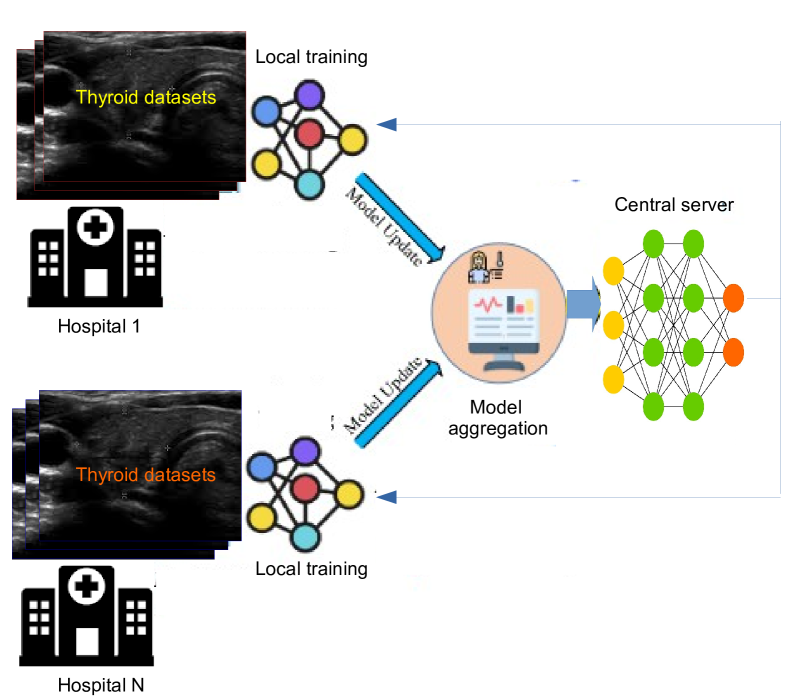
VIII-M Generative chatbots
Most recently, the realm of artificial intelligence has witnessed significant advancements, particularly in the development of generative chatbots and large language models like GPT variants [335]. These state-of-the-art models, trained on vast amounts of data, are adept at generating human-like text and engaging in coherent conversations, going beyond mere predefined responses. As their capability has expanded, so too has their potential for application across various domains, healthcare being one of the prominent ones. In the healthcare sector, these sophisticated models are being explored for patient engagement, preliminary symptom checks, providing health-related information, and even assisting professionals with medical research and data analysis [3]. The integration of such technology holds the promise of streamlining healthcare processes, enhancing patient experience, and augmenting the capabilities of healthcare professionals, albeit with the necessary precautions and ethical considerations in place [336].
Using generative chatbots or models like ChatGPT to diagnose thyroid cancer (or any medical condition) directly would be inappropriate and potentially dangerous. However, they can be incorporated into healthcare settings in auxiliary roles [337]. Typically, Chatbots can gather preliminary information from patients, including their symptoms, family history, and lifestyle habits. This data can provide a better understanding of the patient’s concerns before they meet a healthcare professional. Moving on, they can be programmed to provide information about thyroid cancer, such as risk factors, symptoms, and preventive measures [338]. Patients can learn about the disease and its potential signs, allowing them to approach healthcare providers if they find any matching symptoms. Besides, while they cannot replace professional diagnostic tools, they can be designed to guide users through a series of questions that could highlight potential risk factors or symptoms, encouraging them to consult a medical professional for a more comprehensive evaluation [339].
On the other hand, once a diagnosis has been made, chatbots can provide patients with information on treatment options, side effects, diet recommendations, and answer frequently asked questions. Additionally, they can (i) remind patients to take their medications, attend follow-up appointments, or perform regular self-examinations or monitoring, (ii) offer support in terms of relaxation techniques, provide resources for further psychological support, or even just offer a non-judgmental "listening ear", and (iii) assist doctors and other healthcare professionals by providing instant information about thyroid cancer, recent research, or treatment options, acting as a dynamic reference tool [340].
IX Conclusion
In this research, a comprehensive overview of Deep Neural Networks (DNNs) has been presented, highlighting their growing trend in recent years due to their high accuracy in results compared to other methods. A range of algorithms and training structures have been described, including their advantages and limitations. DNNs have been shown to play a critical role in various real-world applications, characterized by their generalizability and noise tolerance.
However, there are still challenges to be addressed for the widespread use of DNNs in the detection of Thyroid Cancer. One such challenge is the lack of clean datasets and platforms. It is crucial to consider these data to develop efficient and robust cancer detection models that can identify more advanced types of cancer. In the future, more research effort should be put into overcoming these problems and improving the quality of thyroid cancer detection.
The study also highlights the need for further research in the area of thyroid cancer detection and identification, especially considering the accuracy that health specialists desire. Researchers are increasingly exploring the detection of various cancers in two or three dimensions, but the lack of mastery of different geometric transformations and a two- or three-dimensional database hinders the process of diagnosing incurable diseases. The development of new methods to recognize different volumes of cancerous nodules is crucial to achieving speed in treatment and accuracy in diagnosis, as well as enabling early epidemiological surveillance and reducing the death rate.
New technologies such as Explainable AI, Edge Computing, Reinforcement learning, Panoptic Segmentation, and Recommender Systems, have opened up new avenues for research in the field of thyroid cancer detection and have greatly assisted clinicians in the early diagnosis process, reducing the time for detection, and preserving patient privacy. Future work will focus on further exploring the contributions of these technologies to drive a paradigm shift in the field of cancer detection, by developing advanced and secure technologies for the preservation of privacy and detection of thyroid cancer patients, such as telehealth.
References
- [1] Y. Himeur, S. Al-Maadeed, I. Varlamis, N. Al-Maadeed, K. Abualsaud, and A. Mohamed, “Face mask detection in smart cities using deep and transfer learning: lessons learned from the covid-19 pandemic,” Systems, vol. 11, no. 2, p. 107, 2023.
- [2] Y. Himeur, S. Al-Maadeed, N. Almaadeed, K. Abualsaud, A. Mohamed, T. Khattab, and O. Elharrouss, “Deep visual social distancing monitoring to combat covid-19: A comprehensive survey,” Sustainable cities and society, vol. 85, p. 104064, 2022.
- [3] S. S. Sohail, F. Farhat, Y. Himeur, M. Nadeem, D. Ø. Madsen, Y. Singh, S. Atalla, and W. Mansoor, “Decoding chatgpt: A taxonomy of existing research, current challenges, and possible future directions,” Journal of King Saud University-Computer and Information Sciences, p. 101675, 2023.
- [4] Y. Himeur, M. Elnour, F. Fadli, N. Meskin, I. Petri, Y. Rezgui, F. Bensaali, and A. Amira, “Ai-big data analytics for building automation and management systems: a survey, actual challenges and future perspectives,” Artificial Intelligence Review, vol. 56, no. 6, pp. 4929–5021, 2023.
- [5] Y. Deng, H. Li, M. Wang, N. Li, T. Tian, Y. Wu, P. Xu, S. Yang, Z. Zhai, L. Zhou et al., “Global burden of thyroid cancer from 1990 to 2017,” JAMA network open, vol. 3, no. 6, pp. e208 759–e208 759, 2020.
- [6] D. Hammouda, M. Aoun, K. Bouzerar et al., “Registre des tumeurs d’alger,” 2006.
- [7] L. Abid, “Le cancer de la thyroide en algérie,” Guide de la santé en Algérie: Actualité pathologie, 2008.
- [8] “NIPH,” Available online: http://www.insp.dz/index.php/Non-categorise/registre-des-tumeurs-d-alger.html, accessed: 2021-01-17.
- [9] R. L. Siegel, K. D. Miller, and A. Jemal, “Cancer statistics, 2019,” CA: a cancer journal for clinicians, vol. 69, no. 1, pp. 7–34, 2019.
- [10] L. Hitu, K. Gabora, E.-A. Bonci, A. Piciu, A.-C. Hitu, P.-A. \textcommabelowStefan, and D. Piciu, “Microrna in papillary thyroid carcinoma: A systematic review from 2018 to june 2020,” Cancers, vol. 12, no. 11, p. 3118, 2020.
- [11] M. Castellana, A. Piccardo, C. Virili, L. Scappaticcio, G. Grani, C. Durante, L. Giovanella, and P. Trimboli, “Can ultrasound systems for risk stratification of thyroid nodules identify follicular carcinoma?” Cancer cytopathology, vol. 128, no. 4, pp. 250–259, 2020.
- [12] S. M. Ferrari, G. Elia, F. Ragusa, I. Ruffilli, C. La Motta, S. R. Paparo, A. Patrizio, R. Vita, S. Benvenga, G. Materazzi et al., “Novel treatments for anaplastic thyroid carcinoma,” Gland surgery, vol. 9, no. Suppl 1, p. S28, 2020.
- [13] L. Giovanella, G. Treglia, I. Iakovou, J. Mihailovic, F. A. Verburg, and M. Luster, “Eanm practice guideline for pet/ct imaging in medullary thyroid carcinoma,” European journal of nuclear medicine and molecular imaging, vol. 47, no. 1, pp. 61–77, 2020.
- [14] T. Carling and R. Udelsman, “Thyroid cancer,” Annual review of medicine, vol. 65, pp. 125–137, 2014.
- [15] R. Yang, X. Zou, H. Zeng, Y. Zhao, and X. Ma, “Comparison of diagnostic performance of five different ultrasound ti-rads classification guidelines for thyroid nodules,” Frontiers in Oncology, vol. 10, 2020.
- [16] K. Kobayashi, T. Fujimoto, H. Ota, M. Hirokawa, T. Yabuta, H. Masuoka, M. Fukushima, T. Higashiyama, M. Kihara, Y. Ito et al., “Calcifications in thyroid tumors on ultrasonography: calcification types and relationship with histopathological type,” Ultrasound international open, vol. 4, no. 2, p. E45, 2018.
- [17] F. N. Tessler, W. D. Middleton, E. G. Grant, J. K. Hoang, L. L. Berland, S. A. Teefey, J. J. Cronan, M. D. Beland, T. S. Desser, M. C. Frates et al., “Acr thyroid imaging, reporting and data system (ti-rads): white paper of the acr ti-rads committee,” Journal of the American college of radiology, vol. 14, no. 5, pp. 587–595, 2017.
- [18] F. N. Tessler, W. D. Middleton, and E. G. Grant, “Thyroid imaging reporting and data system (ti-rads): a user’s guide,” Radiology, vol. 287, no. 1, pp. 29–36, 2018.
- [19] “Genomic Data Commons Data Portal,” Available online: https://portal.gdc.cancer.gov/, accessed: 2021-01-10.
- [20] “TI-RADS Calculator,” Available online: http://tiradscalculator.com/, accessed: 2021-01-10.
- [21] “AI TI-RADS Calculator,” Available online: https://deckard.duhs.duke.edu/~ai-ti-rads/, accessed: 2021-01-10.
- [22] M. Schlumberger, M. Tahara, L. J. Wirth, B. Robinson, M. S. Brose, R. Elisei, M. A. Habra, K. Newbold, M. H. Shah, A. O. Hoff et al., “Lenvatinib versus placebo in radioiodine-refractory thyroid cancer,” New England Journal of Medicine, vol. 372, no. 7, pp. 621–630, 2015.
- [23] M. C. Wettasinghe, S. Rosairo, N. Ratnatunga, and N. D. Wickramasinghe, “Diagnostic accuracy of ultrasound characteristics in the identification of malignant thyroid nodules,” BMC research notes, vol. 12, no. 1, p. 193, 2019.
- [24] R. Nayak, N. Nawar, J. Webb, M. Fatemi, and A. Alizad, “Impact of imaging cross-section on visualization of thyroid microvessels using ultrasound: Pilot study,” Scientific reports, vol. 10, no. 1, pp. 1–9, 2020.
- [25] N. Singh Ospina, S. Maraka, A. Espinosa DeYcaza, D. O’Keeffe, J. P. Brito, M. R. Gionfriddo, M. R. Castro, J. C. Morris, P. Erwin, and V. M. Montori, “Diagnostic accuracy of thyroid nodule growth to predict malignancy in thyroid nodules with benign cytology: systematic review and meta-analysis,” Clinical endocrinology, vol. 85, no. 1, pp. 122–131, 2016.
- [26] V. Kumar, J. Webb, A. Gregory, D. D. Meixner, J. M. Knudsen, M. Callstrom, M. Fatemi, and A. Alizad, “Automated segmentation of thyroid nodule, gland, and cystic components from ultrasound images using deep learning,” IEEE Access, vol. 8, pp. 63 482–63 496, 2020.
- [27] J. Ma, F. Wu, T. Jiang, Q. Zhao, and D. Kong, “Ultrasound image-based thyroid nodule automatic segmentation using convolutional neural networks,” International journal of computer assisted radiology and surgery, vol. 12, no. 11, pp. 1895–1910, 2017.
- [28] J. S. A. Song and R. D. Hart, “Fine-needle aspiration biopsy of thyroid nodules: Determining when it is necessary,” Canadian Family Physician, vol. 64, no. 2, p. 127, 2018.
- [29] S. Y. Hahn, J. H. Shin, Y. L. Oh, K. W. Park, and Y. Lim, “comparison between fine needle aspiration and core needle biopsy for the diagnosis of thyroid nodules: Effective indications according to us findings,” Scientific reports, vol. 10, no. 1, pp. 1–7, 2020.
- [30] H. Ullah, T. Saba, N. Islam, N. Abbas, A. Rehman, Z. Mehmood, and A. Anjum, “An ensemble classification of exudates in color fundus images using an evolutionary algorithm based optimal features selection,” Microscopy research and technique, vol. 82, no. 4, pp. 361–372, 2019.
- [31] T. Saba, S. T. F. Bokhari, M. Sharif, M. Yasmin, and M. Raza, “Fundus image classification methods for the detection of glaucoma: A review,” Microscopy research and technique, vol. 81, no. 10, pp. 1105–1121, 2018.
- [32] B. Mughal, N. Muhammad, M. Sharif, A. Rehman, and T. Saba, “Removal of pectoral muscle based on topographic map and shape-shifting silhouette,” BMC cancer, vol. 18, no. 1, pp. 1–14, 2018.
- [33] B. Mughal, M. Sharif, N. Muhammad, and T. Saba, “A novel classification scheme to decline the mortality rate among women due to breast tumor,” Microscopy research and technique, vol. 81, no. 2, pp. 171–180, 2018.
- [34] N. Abbas, T. Saba, Z. Mehmood, A. Rehman, N. Islam, and K. T. Ahmed, “An automated nuclei segmentation of leukocytes from microscopic digital images.” Pakistan journal of pharmaceutical sciences, vol. 32, no. 5, 2019.
- [35] N. Abbas, T. Saba, A. Rehman, Z. Mehmood, N. Javaid, M. Tahir, N. U. Khan, K. T. Ahmed, and R. Shah, “Plasmodium species aware based quantification of malaria parasitemia in light microscopy thin blood smear,” Microscopy research and technique, vol. 82, no. 7, pp. 1198–1214, 2019.
- [36] Y. Wang, W. Yue, X. Li, S. Liu, L. Guo, H. Xu, H. Zhang, and G. Yang, “Comparison study of radiomics and deep learning-based methods for thyroid nodules classification using ultrasound images,” Ieee Access, vol. 8, pp. 52 010–52 017, 2020.
- [37] P. Qin, K. Wu, Y. Hu, J. Zeng, and X. Chai, “Diagnosis of benign and malignant thyroid nodules using combined conventional ultrasound and ultrasound elasticity imaging,” IEEE journal of biomedical and health informatics, vol. 24, no. 4, pp. 1028–1036, 2019.
- [38] H. Wu, Z. Deng, B. Zhang, Q. Liu, and J. Chen, “Classifier model based on machine learning algorithms: application to differential diagnosis of suspicious thyroid nodules via sonography,” American Journal of Roentgenology, vol. 207, no. 4, pp. 859–864, 2016.
- [39] B. Zhang, J. Tian, S. Pei, Y. Chen, X. He, Y. Dong, L. Zhang, X. Mo, W. Huang, S. Cong et al., “Machine learning–assisted system for thyroid nodule diagnosis,” Thyroid, vol. 29, no. 6, pp. 858–867, 2019.
- [40] M. Sollini, L. Cozzi, A. Chiti, and M. Kirienko, “Texture analysis and machine learning to characterize suspected thyroid nodules and differentiated thyroid cancer: Where do we stand?” European journal of radiology, vol. 99, pp. 1–8, 2018.
- [41] C. Q. Yang, L. Gardiner, H. Wang, M. T. Hueman, and D. Chen, “Creating prognostic systems for well-differentiated thyroid cancer using machine learning,” Frontiers in endocrinology, vol. 10, p. 288, 2019.
- [42] H. Abbad Ur Rehman, C.-Y. Lin, and Z. Mushtaq, “Effective k-nearest neighbor algorithms performance analysis of thyroid disease,” Journal of the Chinese Institute of Engineers, vol. 44, no. 1, pp. 77–87, 2021.
- [43] J. N. Taylor, K. Mochizuki, K. Hashimoto, Y. Kumamoto, Y. Harada, K. Fujita, and T. Komatsuzaki, “High-resolution raman microscopic detection of follicular thyroid cancer cells with unsupervised machine learning,” The Journal of Physical Chemistry B, vol. 123, no. 20, pp. 4358–4372, 2019.
- [44] J. A. Chandio, G. A. Mallah, and N. A. Shaikh, “Decision support system for classification medullary thyroid cancer,” IEEE Access, vol. 8, pp. 145 216–145 226, 2020.
- [45] J.-H. Lee and Y. J. Chai, “A deep-learning model to assist thyroid nodule diagnosis and management,” The Lancet Digital Health, 2021.
- [46] M. Buda, B. Wildman-Tobriner, J. K. Hoang, D. Thayer, F. N. Tessler, W. D. Middleton, and M. A. Mazurowski, “Management of thyroid nodules seen on us images: deep learning may match performance of radiologists,” Radiology, vol. 292, no. 3, pp. 695–701, 2019.
- [47] Y. Liu, J. Liang, S. Peng, W. Wang, and H. Xiao, “A deep-learning model to assist thyroid nodule diagnosis and management–authors’ reply,” The Lancet Digital Health, vol. 3, no. 7, pp. e411–e412, 2021.
- [48] A. Iesato and C. Nucera, “Role of regulatory non-coding rnas in aggressive thyroid cancer: Prospective applications of neural network analysis,” Molecules, vol. 26, no. 10, p. 3022, 2021.
- [49] Y. Sharifi, M. A. Bakhshali, T. Dehghani, M. DanaiAshgzari, M. Sargolzaei, and S. Eslami, “Deep learning on ultrasound images of thyroid nodules,” Biocybernetics and Biomedical Engineering, vol. 41, no. 2, pp. 636–655, 2021.
- [50] Y.-J. Lin, T.-K. Chao, M.-A. Khalil, Y.-C. Lee, D.-Z. Hong, J.-J. Wu, and C.-W. Wang, “Deep learning fast screening approach on cytological whole slides for thyroid cancer diagnosis,” Cancers, vol. 13, no. 15, p. 3891, 2021.
- [51] E. J. Ha and J. H. Baek, “Applications of machine learning and deep learning to thyroid imaging: where do we stand?” Ultrasonography, vol. 40, no. 1, p. 23, 2021.
- [52] X. L. Wu, M. Li, X.-w. Cui, and G. Xu, “Deep multimodal learning for lymph node metastasis prediction of primary thyroid cancer,” Physics in Medicine & Biology, 2022.
- [53] S. Pavithra, G. Yamuna, and R. Arunkumar, “Deep learning method for classifying thyroid nodules using ultrasound images,” in 2022 International Conference on Smart Technologies and Systems for Next Generation Computing (ICSTSN). IEEE, 2022, pp. 1–6.
- [54] R. Paul, A. Juliano, W. Faquin, and A. W. Chan, “An artificial intelligence ultrasound platform for screening and staging of thyroid cancer,” International Journal of Radiation Oncology, Biology, Physics, vol. 112, no. 5, p. e8, 2022.
- [55] M. Ilyas, H. Malik, M. Adnan, U. Bashir, W. A. Bukhari, M. I. A. Khan, and A. Ahmad, “Deep learning based classification of thyroid cancer using different medical imaging modalities: A systematic review,” 2022.
- [56] C. Liu, Y. Huang, J. A. Ozolek, M. G. Hanna, R. Singh, and G. K. Rohde, “Setsvm: an approach to set classification in nuclei-based cancer detection,” IEEE Journal of Biomedical and Health Informatics, vol. 23, no. 1, pp. 351–361, 2018.
- [57] S. Zhang, H. Du, Z. Jin, Y. Zhu, Y. Zhang, F. Xie, M. Zhang, Z. Jiao, X. Tian, J. Zhang et al., “Integrating clinical knowledge in a thyroid nodule classification model based on,” in 2019 IEEE International Ultrasonics Symposium (IUS). IEEE, 2019, pp. 2334–2336.
- [58] H. Zhang, C. Zhao, L. Guo, X. Li, Y. Luo, J. Lu, and H. Xu, “Diagnosis of thyroid nodules in ultrasound images using two combined classification modules,” in 2019 12th International Congress on Image and Signal Processing, BioMedical Engineering and Informatics (CISP-BMEI). IEEE, 2019, pp. 1–5.
- [59] D. Chen, J. Zhang, and W. Li, “Thyroid nodule classification using two levels attention-based bi-directional lstm with ultrasound reports,” in 2018 9th International Conference on Information Technology in Medicine and Education (ITME). IEEE, 2018, pp. 309–312.
- [60] X. Ma, B. Sun, W. Liu, D. Sui, J. Chen, and Z. Tian, “Amseg: A novel adversarial architecture based multi-scale fusion framework for thyroid nodule segmentation,” IEEE Access, 2023.
- [61] N. Yadav, R. Dass, and J. Virmani, “Assessment of encoder-decoder-based segmentation models for thyroid ultrasound images,” Medical & Biological Engineering & Computing, pp. 1–37, 2023.
- [62] M. Jajroudi, T. Baniasadi, L. Kamkar, F. Arbabi, M. Sanei, and M. Ahmadzade, “Prediction of survival in thyroid cancer using data mining technique,” Technology in cancer research & treatment, vol. 13, no. 4, pp. 353–359, 2014.
- [63] V. Sajeev, A. H. Vyshnavi, and P. K. Namboori, “Thyroid cancer prediction using gene expression profile, pharmacogenomic variants and quantum image processing in deep learning platform-a theranostic approach,” in 2020 International Conference for Emerging Technology (INCET). IEEE, 2020, pp. 1–5.
- [64] N. Liu, A. Fenster, D. Tessier, S. Gou, and J. Chong, “Self-supervised learning enhanced ultrasound video thyroid nodule tracking,” in Medical Imaging 2023: Image Processing, vol. 12464. SPIE, 2023, pp. 683–687.
- [65] Y. Hou and Q. Sang, “Boosting ultrasonic image classification via self-supervised representation learning,” in 2023 3rd International Conference on Computer, Control and Robotics (ICCCR). IEEE, 2023, pp. 116–120.
- [66] S. P. Canton, E. Dadashzadeh, L. Yip, R. Forsythe, and R. Handzel, “Automatic detection of thyroid and adrenal incidentals using radiology reports and deep learning,” Journal of Surgical Research, vol. 266, pp. 192–200, 2021.
- [67] S. Peng, Y. Liu, W. Lv, L. Liu, Q. Zhou, H. Yang, J. Ren, G. Liu, X. Wang, X. Zhang et al., “Deep learning-based artificial intelligence model to assist thyroid nodule diagnosis and management: a multicentre diagnostic study,” The Lancet Digital Health, vol. 3, no. 4, pp. e250–e259, 2021.
- [68] Q. Guan, Y. Wang, J. Du, Y. Qin, H. Lu, J. Xiang, and F. Wang, “Deep learning based classification of ultrasound images for thyroid nodules: a large scale of pilot study,” Annals of Translational Medicine, vol. 7, no. 7, 2019.
- [69] M. F. Ferreira, R. Camacho, and L. F. Teixeira, “Autoencoders as weight initialization of deep classification networks applied to papillary thyroid carcinoma,” in 2018 IEEE International Conference on Bioinformatics and Biomedicine (BIBM). IEEE, 2018, pp. 629–632.
- [70] V. Teixeira, R. Camacho, and P. G. Ferreira, “Learning influential genes on cancer gene expression data with stacked denoising autoencoders,” in 2017 IEEE International Conference on Bioinformatics and Biomedicine (BIBM). IEEE, 2017, pp. 1201–1205.
- [71] T. Liu, Q. Guo, C. Lian, X. Ren, S. Liang, J. Yu, L. Niu, W. Sun, and D. Shen, “Automated detection and classification of thyroid nodules in ultrasound images using clinical-knowledge-guided convolutional neural networks,” Medical image analysis, vol. 58, p. 101555, 2019.
- [72] E. J. Ha, J. H. Baek, and D. G. Na, “Deep convolutional neural network models for the diagnosis of thyroid cancer,” The Lancet Oncology, vol. 20, no. 3, p. e130, 2019.
- [73] Q. ZHANG, J. HU, and S. ZHOU, “The detection of hyperthyroidism by the modified lenet-5 network,” Indian Journal of Pharmaceutical Sciences, pp. 108–114, 2020.
- [74] T. Qiao, S. Liu, Z. Cui, X. Yu, H. Cai, H. Zhang, M. Sun, Z. Lv, and D. Li, “Deep learning for intelligent diagnosis in thyroid scintigraphy,” Journal of International Medical Research, vol. 49, no. 1, p. 0300060520982842, 2021.
- [75] J. Cox, S. Rubin, J. Adams, C. Pereira, M. Dighe, and A. Alessio, “Hyperparameter selection for resnet classification of malignancy from thyroid ultrasound images,” in Medical Imaging 2020: Computer-Aided Diagnosis, vol. 11314. International Society for Optics and Photonics, 2020, p. 1131447.
- [76] J. Chi, E. Walia, P. Babyn, J. Wang, G. Groot, and M. Eramian, “Thyroid nodule classification in ultrasound images by fine-tuning deep convolutional neural network,” Journal of digital imaging, vol. 30, no. 4, pp. 477–486, 2017.
- [77] H. Tekchandani, S. Verma, N. D. Londhe, R. R. Jain, and A. Tiwari, “Severity assessment of cervical lymph nodes using modified vgg-net, and squeeze and excitation concept,” in 2021 IEEE 11th Annual Computing and Communication Workshop and Conference (CCWC). IEEE, 2021, pp. 0709–0714.
- [78] L. Ke, Y. Deng, W. Xia, M. Qiang, X. Chen, K. Liu, B. Jing, C. He, C. Xie, X. Guo et al., “Development of a self-constrained 3d densenet model in automatic detection and segmentation of nasopharyngeal carcinoma using magnetic resonance images,” Oral Oncology, vol. 110, p. 104862, 2020.
- [79] X. Li, S. Zhang, Q. Zhang, X. Wei, Y. Pan, J. Zhao, X. Xin, C. Qin, X. Wang, J. Li et al., “Diagnosis of thyroid cancer using deep convolutional neural network models applied to sonographic images: a retrospective, multicohort, diagnostic study,” The Lancet Oncology, vol. 20, no. 2, pp. 193–201, 2019.
- [80] S. Xie, J. Yu, T. Liu, Q. Chang, L. Niu, and W. Sun, “Thyroid nodule detection in ultrasound images with convolutional neural networks,” in 2019 14th IEEE Conference on Industrial Electronics and Applications (ICIEA). IEEE, 2019, pp. 1442–1446.
- [81] J. Koh, E. Lee, K. Han, E.-K. Kim, E. J. Son, Y.-M. Sohn, M. Seo, M.-r. Kwon, J. H. Yoon, J. H. Lee et al., “Diagnosis of thyroid nodules on ultrasonography by a deep convolutional neural network,” Scientific reports, vol. 10, no. 1, pp. 1–9, 2020.
- [82] X. Liang, J. Yu, J. Liao, and Z. Chen, “Convolutional neural network for breast and thyroid nodules diagnosis in ultrasound imaging,” BioMed Research International, vol. 2020, 2020.
- [83] J. Zhu, S. Zhang, R. Yu, Z. Liu, H. Gao, B. Yue, X. Liu, X. Zheng, M. Gao, and X. Wei, “An efficient deep convolutional neural network model for visual localization and automatic diagnosis of thyroid nodules on ultrasound images,” Quantitative Imaging in Medicine and Surgery, vol. 11, no. 4, p. 1368, 2021.
- [84] C.-K. Zhao, T.-T. Ren, Y.-F. Yin, H. Shi, H.-X. Wang, B.-Y. Zhou, X.-R. Wang, X. Li, Y.-F. Zhang, C. Liu et al., “A comparative analysis of two machine learning-based diagnostic patterns with thyroid imaging reporting and data system for thyroid nodules: diagnostic performance and unnecessary biopsy rate,” Thyroid, vol. 31, no. 3, pp. 470–481, 2021.
- [85] M. Han, E. Ha, and J. Park, “Computer-aided diagnostic system for thyroid nodules on ultrasonography: diagnostic performance based on the thyroid imaging reporting and data system classification and dichotomous outcomes,” American Journal of Neuroradiology, vol. 42, no. 3, pp. 559–565, 2021.
- [86] Z. Liu, S. Zhong, Q. Liu, C. Xie, Y. Dai, C. Peng, X. Chen, and R. Zou, “Thyroid nodule recognition using a joint convolutional neural network with information fusion of ultrasound images and radiofrequency data,” European Radiology, vol. 31, no. 7, pp. 5001–5011, 2021.
- [87] Q. Wei, S.-E. Zeng, L.-P. Wang, Y.-J. Yan, T. Wang, J.-W. Xu, M.-Y. Zhang, W.-Z. Lv, X.-W. Cui, and C. F. Dietrich, “The value of s-detect in improving the diagnostic performance of radiologists for the differential diagnosis of thyroid nodules,” Medical Ultrasonography, vol. 22, no. 4, pp. 415–423, 2020.
- [88] M. T. Stib, I. Pan, D. Merck, W. D. Middleton, and M. D. Beland, “Thyroid nodule malignancy risk stratification using a convolutional neural network,” Ultrasound Quarterly, vol. 36, no. 2, pp. 164–172, 2020.
- [89] H. Ye, J. Hang, X. Chen, D. Xu, J. Chen, X. Ye, and D. Zhang, “An intelligent platform for ultrasound diagnosis of thyroid nodules,” Scientific Reports, vol. 10, no. 1, pp. 1–7, 2020.
- [90] I. Shin, Y. J. Kim, K. Han, E. Lee, H. J. Kim, J. H. Shin, H. J. Moon, J. H. Youk, K. G. Kim, and J. Y. Kwak, “Application of machine learning to ultrasound images to differentiate follicular neoplasms of the thyroid gland,” Ultrasonography, vol. 39, no. 3, p. 257, 2020.
- [91] V. Y. Park, K. Han, Y. K. Seong, M. H. Park, E.-K. Kim, H. J. Moon, J. H. Yoon, and J. Y. Kwak, “Diagnosis of thyroid nodules: performance of a deep learning convolutional neural network model vs. radiologists,” Scientific reports, vol. 9, no. 1, pp. 1–9, 2019.
- [92] S. Xia, J. Yao, W. Zhou, Y. Dong, S. Xu, J. Zhou, and W. Zhan, “A computer-aided diagnosing system in the evaluation of thyroid nodules—experience in a specialized thyroid center,” World Journal of Surgical Oncology, vol. 17, no. 1, pp. 1–8, 2019.
- [93] D. Chen, C. Shi, M. Wang, and Q. Pan, “Thyroid nodule classification using hierarchical recurrent neural network with multiple ultrasound reports,” in International Conference on Neural Information Processing. Springer, 2017, pp. 765–773.
- [94] P. Smolensky et al., “Information processing in dynamical systems: Foundations of harmony theory,” 1986.
- [95] V. S. Vairale and S. Shukla, “Physical fitness recommender framework for thyroid patients using restricted boltzmann machines.”
- [96] W. Yang, J. Zhao, Y. Qiang, X. Yang, Y. Dong, Q. Du, G. Shi, and M. B. Zia, “Dscgans: Integrate domain knowledge in training dual-path semi-supervised conditional generative adversarial networks and s3vm for ultrasonography thyroid nodules classification,” in International Conference on Medical Image Computing and Computer-Assisted Intervention. Springer, 2019, pp. 558–566.
- [97] T. K. Yoo, J. Y. Choi, and H. K. Kim, “A generative adversarial network approach to predicting postoperative appearance after orbital decompression surgery for thyroid eye disease,” Computers in Biology and Medicine, vol. 118, p. 103628, 2020.
- [98] L.-N. Li, J.-H. Ouyang, H.-L. Chen, and D.-Y. Liu, “A computer aided diagnosis system for thyroid disease using extreme learning machine,” Journal of medical systems, vol. 36, no. 5, pp. 3327–3337, 2012.
- [99] C. Ma, J. Guan, W. Zhao, and C. Wang, “An efficient diagnosis system for thyroid disease based on enhanced kernelized extreme learning machine approach,” in International Conference on Cognitive Computing. Springer, 2018, pp. 86–101.
- [100] J. Xia, H. Chen, Q. Li, M. Zhou, L. Chen, Z. Cai, Y. Fang, and H. Zhou, “Ultrasound-based differentiation of malignant and benign thyroid nodules: An extreme learning machine approach,” Computer methods and programs in biomedicine, vol. 147, pp. 37–49, 2017.
- [101] R. Pavithra and L. Parthiban, “Optimal deep learning with kernel extreme learning machine based thyroid disease diagnosis and classification model,” Journal of Computational and Theoretical Nanoscience, vol. 18, no. 3, pp. 639–649, 2021.
- [102] B. N. Rao, D. L. S. Reddy, and G. Bhaskar, “Thyroid diagnosis using multilayer perceptron,” in International Conference on E-Business and Telecommunications. Springer, 2019, pp. 452–459.
- [103] M. Hosseinzadeh, O. H. Ahmed, M. Y. Ghafour, F. Safara, S. Ali, B. Vo, H.-S. Chiang et al., “A multiple multilayer perceptron neural network with an adaptive learning algorithm for thyroid disease diagnosis in the internet of medical things,” The Journal of Supercomputing, pp. 1–22, 2020.
- [104] I. Isa, Z. Saad, S. Omar, M. Osman, K. Ahmad, and H. M. Sakim, “Suitable mlp network activation functions for breast cancer and thyroid disease detection,” in 2010 Second International Conference on Computational Intelligence, Modelling and Simulation. IEEE, 2010, pp. 39–44.
- [105] M. Mourad, S. Moubayed, A. Dezube, Y. Mourad, K. Park, A. Torreblanca-Zanca, J. S. Torrecilla, J. C. Cancilla, and J. Wang, “Machine learning and feature selection applied to seer data to reliably assess thyroid cancer prognosis,” Scientific Reports, vol. 10, no. 1, pp. 1–11, 2020.
- [106] R. Erol, S. N. Oğulata, C. Şahin, and Z. N. Alparslan, “A radial basis function neural network (rbfnn) approach for structural classification of thyroid diseases,” Journal of medical systems, vol. 32, no. 3, pp. 215–220, 2008.
- [107] K. Chandel, V. Kunwar, S. Sabitha, T. Choudhury, and S. Mukherjee, “A comparative study on thyroid disease detection using k-nearest neighbor and naive bayes classification techniques,” CSI transactions on ICT, vol. 4, no. 2-4, pp. 313–319, 2016.
- [108] D.-Y. Liu, H.-L. Chen, B. Yang, X.-E. Lv, L.-N. Li, and J. Liu, “Design of an enhanced fuzzy k-nearest neighbor classifier based computer aided diagnostic system for thyroid disease,” Journal of medical systems, vol. 36, no. 5, pp. 3243–3254, 2012.
- [109] K. Geetha and S. S. Baboo, “An empirical model for thyroid disease classification using evolutionary multivariate bayseian prediction method,” Global Journal of Computer Science and Technology, 2016.
- [110] J. Ma, S. Luo, M. Dighe, D.-J. Lim, and Y. Kim, “Differential diagnosis of thyroid nodules with ultrasound elastography based on support vector machines,” in 2010 IEEE International Ultrasonics Symposium. IEEE, 2010, pp. 1372–1375.
- [111] C.-Y. Chang, S.-J. Chen, and M.-F. Tsai, “Application of support-vector-machine-based method for feature selection and classification of thyroid nodules in ultrasound images,” Pattern recognition, vol. 43, no. 10, pp. 3494–3506, 2010.
- [112] E. Dogantekin, A. Dogantekin, and D. Avci, “An expert system based on generalized discriminant analysis and wavelet support vector machine for diagnosis of thyroid diseases,” Expert Systems with Applications, vol. 38, no. 1, pp. 146–150, 2011.
- [113] D. C. Yadav and S. Pal, “Prediction of thyroid disease using decision tree ensemble method,” Human-Intelligent Systems Integration, vol. 2, no. 1, pp. 89–95, 2020.
- [114] Y. Hao, W. Zuo, Z. Shi, L. Yue, S. Xue, and F. He, “Prognosis of thyroid disease using ms-apriori improved decision tree,” in International Conference on Knowledge Science, Engineering and Management. Springer, 2018, pp. 452–460.
- [115] K. Dharmarajan, K. Balasree, A. Arunachalam, and K. Abirmai, “Thyroid disease classification using decision tree and svm,” EXECUTIVE EDITOR, vol. 11, no. 03, p. 3234, 2020.
- [116] D. C. Yadav and S. Pal, “Decision tree ensemble techniques to predict thyroid disease,” Int. J. Recent Technol. Eng., vol. 8, no. 3, pp. 8242–8246, 2019.
- [117] R.-N. Zhao, B. Zhang, X. Yang, Y.-X. Jiang, X.-J. Lai, and X.-Y. Zhang, “Logistic regression analysis of contrast-enhanced ultrasound and conventional ultrasound characteristics of sub-centimeter thyroid nodules,” Ultrasound in medicine & biology, vol. 41, no. 12, pp. 3102–3108, 2015.
- [118] J. Yazdani-Charati, O. Akha, and F. Khosravi, “Factors affecting thyroid cancer in patients with thyroid nodules using logistic regression in interval censored data,” International Journal of Cancer Management, vol. 11, no. 3, 2018.
- [119] Y. I. Liu, A. Kamaya, T. S. Desser, and D. L. Rubin, “A bayesian network for differentiating benign from malignant thyroid nodules using sonographic and demographic features,” American Journal of Roentgenology, vol. 196, no. 5, pp. W598–W605, 2011.
- [120] ——, “A controlled vocabulary to represent sonographic features of the thyroid and its application in a bayesian network to predict thyroid nodule malignancy,” Summit on Translational Bioinformatics, vol. 2009, p. 68, 2009.
- [121] M. Ashraf, G. Chetty, D. Tran, and D. Sharma, “Hybrid approach for diagnosing thyroid, hepatitis, and breast cancer based on correlation based feature selection and naïve bayes,” in International Conference on Neural Information Processing. Springer, 2012, pp. 272–280.
- [122] M. D. Kate and V. Kale, “Check for updates the role of machine learning in thyroid cancer diagnosis,” in Proceedings of the International Conference on Applications of Machine Intelligence and Data Analytics (ICAMIDA 2022), vol. 105. Springer Nature, 2023, p. 276.
- [123] M. S. Nobile, G. Capitoli, V. Sowirono, F. Clerici, I. Piga, K. van Abeelen, F. Magni, F. Pagni, S. Galimberti, P. Cazzaniga et al., “Unsupervised neural networks as a support tool for pathology diagnosis in maldi-msi experiments: A case study on thyroid biopsies,” Expert Systems with Applications, vol. 215, p. 119296, 2023.
- [124] G. Manogaran, V. Vijayakumar, R. Varatharajan, P. M. Kumar, R. Sundarasekar, and C.-H. Hsu, “Machine learning based big data processing framework for cancer diagnosis using hidden markov model and gm clustering,” Wireless personal communications, vol. 102, no. 3, pp. 2099–2116, 2018.
- [125] U. Agrawal, D. Soria, C. Wagner, J. Garibaldi, I. O. Ellis, J. M. Bartlett, D. Cameron, E. A. Rakha, and A. R. Green, “Combining clustering and classification ensembles: A novel pipeline to identify breast cancer profiles,” Artificial intelligence in medicine, vol. 97, pp. 27–37, 2019.
- [126] M. C. de Souto, I. G. Costa, D. S. de Araujo, T. B. Ludermir, and A. Schliep, “Clustering cancer gene expression data: a comparative study,” BMC bioinformatics, vol. 9, no. 1, pp. 1–14, 2008.
- [127] M. Anas, K. Gupta, and S. Ahmad, “Skin cancer classification using k-means clustering,” International Journal of Technical Research and Applications, vol. 5, no. 1, pp. 62–65, 2017.
- [128] A. R. Khan, S. Khan, M. Harouni, R. Abbasi, S. Iqbal, and Z. Mehmood, “Brain tumor segmentation using k-means clustering and deep learning with synthetic data augmentation for classification,” Microscopy Research and Technique, 2021.
- [129] X. Yu, G. Yu, and J. Wang, “Clustering cancer gene expression data by projective clustering ensemble,” PloS one, vol. 12, no. 2, p. e0171429, 2017.
- [130] K. Chandel, V. Kunwar, A. S. Sabitha, A. Bansal, and T. Choudhury, “Analysing thyroid disease using density-based clustering technique,” International Journal of Business Intelligence and Data Mining, vol. 17, no. 3, pp. 273–297, 2020.
- [131] D. K. Katikireddy Srinivas, “Performa analysis of clustering of thyroid drug data using fuzzy and m-clust,” Journal of Critical Reviews, vol. 7, no. 11, pp. 2128–2141, 2020.
- [132] B. Venkataramana, L. Padmasree, M. S. Rao, D. Latha, and G. Ganesan, “Comparative study on performance of fuzzy clustering algorithms on liver and thyroid data,” Journal of Fuzzy Set Valued Analysis, vol. 2018, no. 1, pp. 1–9, 2018.
- [133] K. K. Mahurkar and D. Gaikwad, “Normalization using improvised k-means applied in diagnosing thyroid disease with ann,” in 2017 International Conference on Trends in Electronics and Informatics (ICEI). IEEE, 2017, pp. 579–583.
- [134] Y. Yang, Y. Song, and B. Cao, “An information entropy-based method to detect microrna regulatory module,” IPSJ Transactions on Bioinformatics, vol. 12, pp. 1–8, 2019.
- [135] M. Tarkov and E. Chiglintsev, “Data space dimensionality reduction in the problem of diagnosing a thyroid disease,” Bulletin of the Novosibirsk Computing Center. Series: Computer Science, pp. 79–84, 2012.
- [136] B. Shankarlal and P. Sathya, “Performance analysis of thyroid tumor detection and segmentation using pca-based random classification method,” in Innovations in Electrical and Electronics Engineering. Springer, 2020, pp. 833–841.
- [137] A. Soulaymani and H. Aschawa, “Epidemiological study of thyroid carcinoma using principal component analysis,” Journal of Clinical Epigenetics, vol. 4, no. 1, p. 9, 2018.
- [138] V. Chandran, M. Sumithra, A. Karthick, T. George, M. Deivakani, B. Elakkiya, U. Subramaniam, and S. Manoharan, “Diagnosis of cervical cancer based on ensemble deep learning network using colposcopy images,” BioMed Research International, vol. 2021, 2021.
- [139] J. Awujoola Olalekan, F. Ogwueleka, and P. Odion, “Effective and accurate bootstrap aggregating (bagging) ensemble algorithm model for prediction and classification of hypothyroid disease,” International Journal of Computer Applications, vol. 975, p. 8887.
- [140] D. Chen, J. Hu, M. Zhu, N. Tang, Y. Yang, and Y. Feng, “Diagnosis of thyroid nodules for ultrasonographic characteristics indicative of malignancy using random forest,” BioData mining, vol. 13, no. 1, pp. 1–21, 2020.
- [141] Y. Himeur, K. Ghanem, A. Alsalemi, F. Bensaali, and A. Amira, “Artificial intelligence based anomaly detection of energy consumption in buildings: A review, current trends and new perspectives,” Applied Energy, vol. 287, p. 116601, 2021.
- [142] Q. Pan, Y. Zhang, M. Zuo, L. Xiang, and D. Chen, “Improved ensemble classification method of thyroid disease based on random forest,” in 2016 8th International Conference on Information Technology in Medicine and Education (ITME). IEEE, 2016, pp. 567–571.
- [143] T. Chen and C. Guestrin, “Xgboost: A scalable tree boosting system,” in Proceedings of the 22nd acm sigkdd international conference on knowledge discovery and data mining, 2016, pp. 785–794.
- [144] S. Lim and S. Chi, “Xgboost application on bridge management systems for proactive damage estimation,” Advanced Engineering Informatics, vol. 41, p. 100922, 2019.
- [145] C. Ji, X. Zou, Y. Hu, S. Liu, L. Lyu, and X. Zheng, “Xg-sf: An xgboost classifier based on shapelet features for time series classification,” Procedia computer science, vol. 147, pp. 24–28, 2019.
- [146] J. Guo, L. Yang, R. Bie, J. Yu, Y. Gao, Y. Shen, and A. Kos, “An xgboost-based physical fitness evaluation model using advanced feature selection and bayesian hyper-parameter optimization for wearable running monitoring,” Computer Networks, vol. 151, pp. 166–180, 2019.
- [147] Y. Xu, X. Yang, H. Huang, C. Peng, Y. Ge, H. Wu, J. Wang, G. Xiong, and Y. Yi, “Extreme gradient boosting model has a better performance in predicting the risk of 90-day readmissions in patients with ischaemic stroke,” Journal of Stroke and Cerebrovascular Diseases, vol. 28, no. 12, p. 104441, 2019.
- [148] Y. Chen, D. Li, X. Zhang, J. Jin, and Y. Shen, “Computer aided diagnosis of thyroid nodules based on the devised small-datasets multi-view ensemble learning,” Medical Image Analysis, vol. 67, p. 101819, 2020.
- [149] J. Thomas and T. Haertling, “Aibx, artificial intelligence model to risk stratify thyroid nodules,” Thyroid, vol. 30, no. 6, pp. 878–884, 2020.
- [150] B. Kezlarian and O. Lin, “Artificial intelligence in thyroid fine needle aspiration biopsies,” Acta Cytologica, pp. 1–6, 2020.
- [151] P. Sanyal, T. Mukherjee, S. Barui, A. Das, and P. Gangopadhyay, “Artificial intelligence in cytopathology: a neural network to identify papillary carcinoma on thyroid fine-needle aspiration cytology smears,” Journal of pathology informatics, vol. 9, 2018.
- [152] J. Yoon, E. Lee, J. S. Koo, J. H. Yoon, K.-H. Nam, J. Lee, Y. S. Jo, H. J. Moon, V. Y. Park, and J. Y. Kwak, “Artificial intelligence to predict the brafv600e mutation in patients with thyroid cancer,” PloS one, vol. 15, no. 11, p. e0242806, 2020.
- [153] D. T. Nguyen, J. K. Kang, T. D. Pham, G. Batchuluun, and K. R. Park, “Ultrasound image-based diagnosis of malignant thyroid nodule using artificial intelligence,” Sensors, vol. 20, no. 7, p. 1822, 2020.
- [154] T. Liu, S. Xie, J. Yu, L. Niu, and W. Sun, “Classification of thyroid nodules in ultrasound images using deep model based transfer learning and hybrid features,” in 2017 IEEE International Conference on Acoustics, Speech and Signal Processing (ICASSP). IEEE, 2017, pp. 919–923.
- [155] F. Abdolali, J. Kapur, J. L. Jaremko, M. Noga, A. R. Hareendranathan, and K. Punithakumar, “Automated thyroid nodule detection from ultrasound imaging using deep convolutional neural networks,” Computers in Biology and Medicine, vol. 122, p. 103871, 2020.
- [156] X. Li, S. Wang, X. Wei, J. Zhu, R. Yu, M. Zhao, M. Yu, Z. Liu, and S. Liu, “Fully convolutional networks for ultrasound image segmentation of thyroid nodules,” in 2018 IEEE 20th International Conference on High Performance Computing and Communications; IEEE 16th International Conference on Smart City; IEEE 4th International Conference on Data Science and Systems (HPCC/SmartCity/DSS). IEEE, 2018, pp. 886–890.
- [157] E. Kim, M. Corte-Real, and Z. Baloch, “A deep semantic mobile application for thyroid cytopathology,” in Medical Imaging 2016: PACS and Imaging Informatics: Next Generation and Innovations, vol. 9789. International Society for Optics and Photonics, 2016, p. 97890A.
- [158] L. Ma, C. Ma, Y. Liu, and X. Wang, “Thyroid diagnosis from spect images using convolutional neural network with optimization,” Computational intelligence and neuroscience, vol. 2019, 2019.
- [159] Y. Chai, J. Song, and M. Shear, “Artificial intelligence for thyroid nodule ultrasound image analysis,” Annals of Thyroid, vol. 5, 2020.
- [160] J. Song, Y. J. Chai, H. Masuoka, S.-W. Park, S.-j. Kim, J. Y. Choi, H.-J. Kong, K. E. Lee, J. Lee, N. Kwak et al., “Ultrasound image analysis using deep learning algorithm for the diagnosis of thyroid nodules,” Medicine, vol. 98, no. 15, 2019.
- [161] M. Barczyński, M. Stopa-Barczyńska, B. Wojtczak, A. Czarniecka, and A. Konturek, “Clinical validation of s-detecttm mode in semi-automated ultrasound classification of thyroid lesions in surgical office,” Gland surgery, vol. 9, no. Suppl 2, p. S77, 2020.
- [162] Y. J. Choi, J. H. Baek, H. S. Park, W. H. Shim, T. Y. Kim, Y. K. Shong, and J. H. Lee, “A computer-aided diagnosis system using artificial intelligence for the diagnosis and characterization of thyroid nodules on ultrasound: initial clinical assessment,” Thyroid, vol. 27, no. 4, pp. 546–552, 2017.
- [163] C. Fragopoulos, A. Pouliakis, C. Meristoudis, E. Mastorakis, N. Margari, N. Chroniaris, N. Koufopoulos, A. G. Delides, N. Machairas, V. Ntomi et al., “Radial basis function artificial neural network for the investigation of thyroid cytological lesions,” Journal of Thyroid Research, vol. 2020, 2020.
- [164] R. Savala, P. Dey, and N. Gupta, “Artificial neural network model to distinguish follicular adenoma from follicular carcinoma on fine needle aspiration of thyroid,” Diagnostic cytopathology, vol. 46, no. 3, pp. 244–249, 2018.
- [165] L.-R. Li, B. Du, H.-Q. Liu, and C. Chen, “Artificial intelligence for personalized medicine in thyroid cancer: Current status and future perspectives,” Frontiers in Oncology, vol. 10, p. 3360, 2021.
- [166] Y. Zhao, L. Zhao, T. Mao, and L. Zhong, “Assessment of risk based on variant pathways and establishment of an artificial neural network model of thyroid cancer,” BMC medical genetics, vol. 20, no. 1, pp. 1–10, 2019.
- [167] B. Wildman-Tobriner, M. Buda, J. K. Hoang, W. D. Middleton, D. Thayer, R. G. Short, F. N. Tessler, and M. A. Mazurowski, “Using artificial intelligence to revise acr ti-rads risk stratification of thyroid nodules: diagnostic accuracy and utility,” Radiology, vol. 292, no. 1, pp. 112–119, 2019.
- [168] L. Wang, S. Yang, S. Yang, C. Zhao, G. Tian, Y. Gao, Y. Chen, and Y. Lu, “Automatic thyroid nodule recognition and diagnosis in ultrasound imaging with the yolov2 neural network,” World journal of surgical oncology, vol. 17, no. 1, pp. 1–9, 2019.
- [169] J. A. Ozolek, A. B. Tosun, W. Wang, C. Chen, S. Kolouri, S. Basu, H. Huang, and G. K. Rohde, “Accurate diagnosis of thyroid follicular lesions from nuclear morphology using supervised learning,” Medical image analysis, vol. 18, no. 5, pp. 772–780, 2014.
- [170] W. Wang, J. A. Ozolek, and G. K. Rohde, “Detection and classification of thyroid follicular lesions based on nuclear structure from histopathology images,” Cytometry Part A: The Journal of the International Society for Advancement of Cytometry, vol. 77, no. 5, pp. 485–494, 2010.
- [171] Y. Zhu, Q. Sang, S. Jia, Y. Wang, and T. Deyer, “Deep neural networks could differentiate bethesda class iii versus class iv/v/vi,” Annals of translational medicine, vol. 7, no. 11, 2019.
- [172] S. Bhalla, H. Kaur, R. Kaur, S. Sharma, and G. P. Raghava, “Expression based biomarkers and models to classify early and late-stage samples of papillary thyroid carcinoma,” PLoS One, vol. 15, no. 4, p. e0231629, 2020.
- [173] J. M. Dolezal, A. Trzcinska, C.-Y. Liao, S. Kochanny, E. Blair, N. Agrawal, X. M. Keutgen, P. Angelos, N. A. Cipriani, and A. T. Pearson, “Deep learning prediction of braf-ras gene expression signature identifies noninvasive follicular thyroid neoplasms with papillary-like nuclear features,” Modern Pathology, pp. 1–13, 2020.
- [174] K. Daniels, S. Gummadi, Z. Zhu, S. Wang, J. Patel, B. Swendseid, A. Lyshchik, J. Curry, E. Cottrill, and J. Eisenbrey, “Machine learning by ultrasonography for genetic risk stratification of thyroid nodules,” JAMA Otolaryngology–Head & Neck Surgery, vol. 146, no. 1, pp. 36–41, 2020.
- [175] A. Teumer, L. Chaker, S. Groeneweg, Y. Li, C. Di Munno, C. Barbieri, U. T. Schultheiss, M. Traglia, T. S. Ahluwalia, M. Akiyama et al., “Genome-wide analyses identify a role for slc17a4 and aadat in thyroid hormone regulation,” Nature communications, vol. 9, no. 1, pp. 1–14, 2018.
- [176] “The ThyroidOmics Consortium,” Available online: https://transfer.sysepi.medizin.uni-greifswald.de/thyroidomics/, accessed: 2021-03-01.
- [177] “Thyroid Disease Data Set,” Available online: https://archive.ics.uci.edu/ml/datasets/thyroid+disease, accessed: 2021-03-01.
- [178] “Knowledge Extraction based on Evolutionary Learning,” Available online: https://sci2s.ugr.es/keel/dataset.php?cod=67, accessed: 2021-03-01.
- [179] “Gene Expression Omnibus,” Available online: https://www.ncbi.nlm.nih.gov/geo/, accessed: 2021-03-01.
- [180] “The digital database of Thyroid Ultrasound Images,” Available online: http://cimalab.intec.co/?lang=en&mod=project&id=31, accessed: 2021-03-01.
- [181] “The National Cancer Registration and Analysis Service,” Available online: http://www.ncin.org.uk/about_ncin/, accessed: 2021-03-01.
- [182] “The Prostate, Lung, Colorectal and Ovarian (PLCO) Cancer Screening Trial ,” Available online: https://prevention.cancer.gov/major-programs/prostate-lung-colorectal-and-ovarian-cancer-screening-trial, accessed: 2021-03-01.
- [183] F.-s. Ouyang, B.-l. Guo, L.-z. Ouyang, Z.-w. Liu, S.-j. Lin, W. Meng, X.-y. Huang, H.-x. Chen, H. Qiu-Gen, and S.-m. Yang, “Comparison between linear and nonlinear machine-learning algorithms for the classification of thyroid nodules,” European journal of radiology, vol. 113, pp. 251–257, 2019.
- [184] J. H. Moon and S. R. Steinhubl, “Digital medicine in thyroidology: a new era of managing thyroid disease,” Endocrinology and Metabolism, vol. 34, no. 2, pp. 124–131, 2019.
- [185] C. Sun, Y. Zhang, Q. Chang, T. Liu, S. Zhang, X. Wang, Q. Guo, J. Yao, W. Sun, and L. Niu, “Evaluation of a deep learning-based computer-aided diagnosis system for distinguishing benign from malignant thyroid nodules in ultrasound images,” Medical Physics, vol. 47, no. 9, pp. 3952–3960, 2020.
- [186] Y. Wang, K. Wei, and P. Wan, “A method of ultrasonic image recognition for thyroid papillary carcinoma based on deep convolution neural network,” NeuroQuantology, vol. 16, no. 5, 2018.
- [187] L. Xu, J. Gao, Q. Wang, J. Yin, P. Yu, B. Bai, R. Pei, D. Chen, G. Yang, S. Wang et al., “Computer-aided diagnosis systems in diagnosing malignant thyroid nodules on ultrasonography: a systematic review and meta-analysis,” European thyroid journal, vol. 9, no. 4, pp. 186–193, 2020.
- [188] Z. Li, K. Yang, L. Zhang, C. Wei, P. Yang, and W. Xu, “Classification of thyroid nodules with stacked denoising sparse autoencoder,” International Journal of Endocrinology, vol. 2020, 2020.
- [189] S. Y. Ko, J. H. Lee, J. H. Yoon, H. Na, E. Hong, K. Han, I. Jung, E.-K. Kim, H. J. Moon, V. Y. Park et al., “Deep convolutional neural network for the diagnosis of thyroid nodules on ultrasound,” Head & neck, vol. 41, no. 4, pp. 885–891, 2019.
- [190] C. Lee, Y. Kim, Y. S. Kim, and J. Jang, “Automatic disease annotation from radiology reports using artificial intelligence implemented by a recurrent neural network,” American Journal of Roentgenology, vol. 212, no. 4, pp. 734–740, 2019.
- [191] A. Sharifi and K. Alizadeh, “Comparison of the particle swarm optimization with the genetic algorithms as a training for multilayer perceptron technique to diagnose thyroid functional disease,” Shiraz E-Medical Journal, vol. 22, no. 1, 2021.
- [192] S. Razia, P. S. Kumar, and A. S. Rao, “Machine learning techniques for thyroid disease diagnosis: A systematic review,” Modern Approaches in Machine Learning and Cognitive Science: A Walkthrough, pp. 203–212, 2020.
- [193] V. S. Vairale and S. Shukla, “Recommendation of food items for thyroid patients using content-based knn method,” in Data Science and Security. Springer, 2021, pp. 71–77.
- [194] Y. Shen, Y. Lai, D. Xu, L. Xu, L. Song, J. Zhou, C. Song, and J. Wang, “Diagnosis of thyroid neoplasm using support vector machine algorithms based on platelet rna-seq,” Endocrine, pp. 1–26, 2020.
- [195] Y. Wu, K. Rao, J. Liu, C. Han, L. Gong, Y. Chong, Z. Liu, and X. Xu, “Machine learning algorithms for the prediction of central lymph node metastasis in patients with papillary thyroid cancer,” Frontiers in endocrinology, vol. 11, p. 816, 2020.
- [196] A. D. McDow, B. R. Roman, M. C. Saucke, C. B. Jensen, N. Zaborek, J. L. Jennings, L. Davies, J. P. Brito, and S. C. Pitt, “Factors associated with physicians’ recommendations for managing low-risk papillary thyroid cancer,” The American Journal of Surgery, 2020.
- [197] Y.-C. Hsu, J.-J. Lee, M.-N. Chien, M.-J. Chen, C.-H. Leung, and S.-P. Cheng, “Is papillary thyroid microcarcinoma a biologically different disease? a propensity score-matched analysis,” Journal of surgical oncology, vol. 120, no. 6, pp. 1023–1030, 2019.
- [198] D. Li, D. Yang, and J. Zhang, “Arb: Knowledge discovery and disease diagnosis on thyroid disease diagnosis integrating association rule with bagging algorithm.” Engineering Letters, vol. 28, no. 2, 2020.
- [199] L. Gao, M. Ye, X. Lu, and D. Huang, “Hybrid method based on information gain and support vector machine for gene selection in cancer classification,” Genomics, proteomics & bioinformatics, vol. 15, no. 6, pp. 389–395, 2017.
- [200] T. M. Fahrudin, I. Syarif, and A. R. Barakbah, “Data mining approach for breast cancer patient recovery,” EMITTER International Journal of Engineering Technology, vol. 5, no. 1, pp. 36–71, 2017.
- [201] P. Hamsagayathri and P. Sampath, “Performance analysis of breast cancer classification using decision tree classifiers,” Int J Curr Pharm Res, vol. 9, no. 2, pp. 19–25, 2017.
- [202] A. Chinnaswamy and R. Srinivasan, “Hybrid information gain based fuzzy roughset feature selection in cancer microarray data,” in 2017 Innovations in Power and Advanced Computing Technologies (i-PACT). IEEE, 2017, pp. 1–6.
- [203] S. Mishra, P. K. Mallick, H. K. Tripathy, A. K. Bhoi, and A. González-Briones, “Performance evaluation of a proposed machine learning model for chronic disease datasets using an integrated attribute evaluator and an improved decision tree classifier,” Applied Sciences, vol. 10, no. 22, p. 8137, 2020.
- [204] M. Ashraf, G. Chetty, and D. Tran, “Feature selection techniques on thyroid, hepatitis, and breast cancer datasets,” International Journal on Data Mining and Intelligent Information Technology Applications, vol. 3, no. 1, p. 1, 2013.
- [205] M. Al-Batah, B. Zaqaibeh, S. A. Alomari, and M. S. Alzboon, “Gene microarray cancer classification using correlation based feature selection algorithm and rules classifiers.” International Journal of Online & Biomedical Engineering, vol. 15, no. 8, 2019.
- [206] I. Jain, V. K. Jain, and R. Jain, “Correlation feature selection based improved-binary particle swarm optimization for gene selection and cancer classification,” Applied Soft Computing, vol. 62, pp. 203–215, 2018.
- [207] Z. Rustam and N. Maghfirah, “Correlated based svm-rfe as feature selection for cancer classification using microarray databases,” in AIP Conference Proceedings, vol. 2023, no. 1. AIP Publishing LLC, 2018, p. 020235.
- [208] L. Cui, L. Ge, H. Gan, X. Liu, and Y. Zhang, “Ovarian cancer identification based on feature weighting for high-throughput mass spectrometry data,” Journal of Systems Biology, vol. 1, no. 1, p. 1, 2018.
- [209] A. Onan, “A fuzzy-rough nearest neighbor classifier combined with consistency-based subset evaluation and instance selection for automated diagnosis of breast cancer,” Expert Systems with Applications, vol. 42, no. 20, pp. 6844–6852, 2015.
- [210] D. O’Dea, M. Bongiovanni, G. P. Sykiotis, P. G. Ziros, A. D. Meade, F. M. Lyng, and A. Malkin, “Raman spectroscopy for the preoperative diagnosis of thyroid cancer and its subtypes: An in vitro proof-of-concept study,” Cytopathology, vol. 30, no. 1, pp. 51–60, 2019.
- [211] F. M. Selaru, J. Yin, A. Olaru, Y. Mori, Y. Xu, S. H. Epstein, F. Sato, E. Deacu, S. Wang, A. Sterian et al., “An unsupervised approach to identify molecular phenotypic components influencing breast cancer features,” Cancer research, vol. 64, no. 5, pp. 1584–1588, 2004.
- [212] V. K. Sudarshan, M. R. K. Mookiah, U. R. Acharya, V. Chandran, F. Molinari, H. Fujita, and K. H. Ng, “Application of wavelet techniques for cancer diagnosis using ultrasound images: a review,” Computers in biology and medicine, vol. 69, pp. 97–111, 2016.
- [213] S. O. Haji and R. Z. Yousif, “A novel run-length based wavelet features for screening thyroid nodule malignancy,” Brazilian Archives of Biology and Technology, vol. 62, 2019.
- [214] B. Yu, Z. Wang, R. Zhu, X. Feng, M. Qi, J. Li, R. Zhao, L. Huang, R. Xin, F. Li et al., “The transverse ultrasonogram of thyroid papillary carcinoma has a better prediction accuracy than the longitudinal one,” IEEE Access, vol. 7, pp. 100 763–100 770, 2019.
- [215] D. T. Nguyen, T. D. Pham, G. Batchuluun, H. S. Yoon, and K. R. Park, “Artificial intelligence-based thyroid nodule classification using information from spatial and frequency domains,” Journal of clinical medicine, vol. 8, no. 11, p. 1976, 2019.
- [216] P. Poudel, A. Illanes, C. Arens, C. Hansen, and M. Friebe, “Active contours extension and similarity indicators for improved 3d segmentation of thyroid ultrasound images,” in Medical Imaging 2017: Imaging Informatics for Healthcare, Research, and Applications, vol. 10138. International Society for Optics and Photonics, 2017, p. 1013803.
- [217] P. Poudel, C. Hansen, J. Sprung, and M. Friebe, “3d segmentation of thyroid ultrasound images using active contours,” Current Directions in Biomedical Engineering, vol. 2, no. 1, pp. 467–470, 2016.
- [218] H. A. Nugroho, A. Nugroho, and L. Choridah, “Thyroid nodule segmentation using active contour bilateral filtering on ultrasound images,” in 2015 International Conference on Quality in Research (QiR). IEEE, 2015, pp. 43–46.
- [219] J. Xie, L. Guo, C. Zhao, X. Li, Y. Luo, and L. Jianwei, “A hybrid deep learning and handcrafted features based approach for thyroid nodule classification in ultrasound images,” in Journal of Physics: Conference Series, vol. 1693, no. 1. IOP Publishing, 2020, p. 012160.
- [220] X. Mei, X. Dong, T. Deyer, J. Zeng, T. Trafalis, and Y. Fang, “Thyroid nodule benignty prediction by deep feature extraction,” in 2017 IEEE 17th International Conference on Bioinformatics and Bioengineering (BIBE). IEEE, 2017, pp. 241–245.
- [221] G. Song, F. Xue, and C. Zhang, “A model using texture features to differentiate the nature of thyroid nodules on sonography,” Journal of Ultrasound in Medicine, vol. 34, no. 10, pp. 1753–1760, 2015.
- [222] M. Dinčić, J. Todorović, J. N. Ostojić, S. Kovačević, D. Dunđerović, S. Lopičić, S. Spasić, S. Radojević-S̈kodrić, D. Stanisavljević, and A. Ž. Ilić, “The fractal and glcm textural parameters of chromatin may be potential biomarkers of papillary thyroid carcinoma in hashimoto’s thyroiditis specimens,” Microscopy and Microanalysis, vol. 26, no. 4, pp. 717–730, 2020.
- [223] I. Kalaimani, “Analysis for the prediction of thyroid disease by using ica and optimal kernel svm approach,” International Journal of Emerging Technology and Innovative Engineering, vol. 5, no. 3, 2019.
- [224] W. Ahmad, L. Huang, A. Ahmad, F. Shah, A. Iqbal, and A. Saeed, “Thyroid diseases forecasting using a hybrid decision support system based on anfis, k-nn and information gain method,” J Appl Environ Biol Sci, vol. 7, no. 10, pp. 78–85, 2017.
- [225] H. A. Nugroho, A. Nugroho, E. L. Frannita, I. Ardiyanto et al., “Classification of thyroid ultrasound images based on shape features analysis,” in 2017 10th Biomedical Engineering International Conference (BMEiCON). IEEE, 2017, pp. 1–5.
- [226] H. Song, C. Dong, X. Zhang, W. Wu, C. Chen, B. Ma, F. Chen, C. Chen, and X. Lv, “Rapid identification of papillary thyroid carcinoma and papillary microcarcinoma based on serum raman spectroscopy combined with machine learning models,” Photodiagnosis and Photodynamic Therapy, vol. 37, p. 102647, 2022.
- [227] U. R. Acharya, O. Faust, S. V. Sree, F. Molinari, and J. S. Suri, “Thyroscreen system: high resolution ultrasound thyroid image characterization into benign and malignant classes using novel combination of texture and discrete wavelet transform,” Computer methods and programs in biomedicine, vol. 107, no. 2, pp. 233–241, 2012.
- [228] H. A. Nugroho, E. L. Frannita, I. Ardiyanto, L. Choridah et al., “Computer aided diagnosis for thyroid cancer system based on internal and external characteristics,” Journal of King Saud University-Computer and Information Sciences, vol. 33, no. 3, pp. 329–339, 2021.
- [229] C. Liu, S. Chen, Y. Yang, D. Shao, W. Peng, Y. Wang, Y. Chen, and Y. Wang, “The value of the computer-aided diagnosis system for thyroid lesions based on computed tomography images,” Quantitative imaging in medicine and surgery, vol. 9, no. 4, p. 642, 2019.
- [230] J. Erfurt, C. R. Helmrich, S. Bosse, H. Schwarz, D. Marpe, and T. Wiegand, “A study of the perceptually weighted peak signal-to-noise ratio (wpsnr) for image compression,” in 2019 IEEE International Conference on Image Processing (ICIP). IEEE, 2019, pp. 2339–2343.
- [231] D. M. Chandler and S. S. Hemami, “Vsnr: A wavelet-based visual signal-to-noise ratio for natural images,” IEEE transactions on image processing, vol. 16, no. 9, pp. 2284–2298, 2007.
- [232] W. Zhou, Z. Wang, and W. Xie, “Weighted signal-to-noise ratio robust design for a new double sampling npx chart,” Computers & Industrial Engineering, vol. 139, p. 106124, 2020.
- [233] M. Lasseck, “Audio-based bird species identification with deep convolutional neural networks.” in CLEF (Working Notes), 2018.
- [234] A. Agarwal, I. Zaitsev, and T. Joachims, “Counterfactual learning-to-rank for additive metrics and deep models,” arXiv preprint arXiv:1805.00065, pp. 11–12, 2018.
- [235] P. M. Murphy, “Uci repository of machine learning databases,” ftp:/pub/machine-learning-databaseonics. uci. edu, 1994.
- [236] Z. Bai, L. Chang, R. Yu, X. Li, X. Wei, M. Yu, Z. Liu, J. Gao, J. Zhu, Y. Zhang et al., “Thyroid nodules risk stratification through deep learning based on ultrasound images,” Medical Physics, 2020.
- [237] B. Colakoglu, D. Alis, and M. Yergin, “Diagnostic value of machine learning-based quantitative texture analysis in differentiating benign and malignant thyroid nodules,” Journal of oncology, vol. 2019, 2019.
- [238] V. Y. Park, E. Lee, H. S. Lee, H. J. Kim, J. Yoon, J. Son, K. Song, H. J. Moon, J. H. Yoon, G. R. Kim et al., “Combining radiomics with ultrasound-based risk stratification systems for thyroid nodules: an approach for improving performance,” European Radiology, vol. 31, no. 4, pp. 2405–2413, 2021.
- [239] N. T. Duc, Y.-M. Lee, J. H. Park, and B. Lee, “An ensemble deep learning for automatic prediction of papillary thyroid carcinoma using fine needle aspiration cytology,” Expert Systems with Applications, vol. 188, p. 115927, 2022.
- [240] V. V. Vadhiraj, A. Simpkin, J. O’Connell, N. Singh Ospina, S. Maraka, and D. T. O’Keeffe, “Ultrasound image classification of thyroid nodules using machine learning techniques,” Medicina, vol. 57, no. 6, p. 527, 2021.
- [241] M. L. Gild, M. Chan, J. Gajera, B. Lurie, Z. Gandomkar, and R. J. Clifton-Bligh, “Risk stratification of indeterminate thyroid nodules using ultrasound and machine learning algorithms,” Clinical Endocrinology, vol. 96, no. 4, pp. 646–652, 2022.
- [242] J. Ma, F. Wu, J. Zhu, D. Xu, and D. Kong, “A pre-trained convolutional neural network based method for thyroid nodule diagnosis,” Ultrasonics, vol. 73, pp. 221–230, 2017.
- [243] Y. Zhu, Z. Fu, and J. Fei, “An image augmentation method using convolutional network for thyroid nodule classification by transfer learning,” in 2017 3rd IEEE international conference on computer and communications (ICCC). IEEE, 2017, pp. 1819–1823.
- [244] L. Gao, R. Liu, Y. Jiang, W. Song, Y. Wang, J. Liu, J. Wang, D. Wu, S. Li, A. Hao et al., “Computer-aided system for diagnosing thyroid nodules on ultrasound: A comparison with radiologist-based clinical assessments,” Head & neck, vol. 40, no. 4, pp. 778–783, 2018.
- [245] D. Zuo, L. Han, K. Chen, C. Li, Z. Hua, and J. Lin, “Extraction of calcification in ultrasonic images based on convolution neural network,” Sheng wu yi xue Gong Cheng xue za zhi= Journal of Biomedical Engineering= Shengwu Yixue Gongchengxue Zazhi, vol. 35, no. 5, pp. 679–687, 2018.
- [246] Y.-J. Kim, Y. Choi, S.-J. Hur, K.-S. Park, H.-J. Kim, M. Seo, M. K. Lee, S.-L. Jung, and C. K. Jung, “Deep convolutional neural network for classification of thyroid nodules on ultrasound: Comparison of the diagnostic performance with that of radiologists,” European Journal of Radiology, vol. 152, p. 110335, 2022.
- [247] J. H. Lee, E. J. Ha, and J. H. Kim, “Application of deep learning to the diagnosis of cervical lymph node metastasis from thyroid cancer with ct,” European radiology, vol. 29, pp. 5452–5457, 2019.
- [248] P. Tsou and C.-J. Wu, “Mapping driver mutations to histopathological subtypes in papillary thyroid carcinoma: applying a deep convolutional neural network,” Journal of clinical medicine, vol. 8, no. 10, p. 1675, 2019.
- [249] Y. Lu, Y. Yang, and W. Chen, “Application of deep learning in the prediction of benign and malignant thyroid nodules on ultrasound images,” IEEE Access, vol. 8, pp. 221 468–221 480, 2020.
- [250] S. W. Kwon, I. J. Choi, J. Y. Kang, W. I. Jang, G.-H. Lee, and M.-C. Lee, “Ultrasonographic thyroid nodule classification using a deep convolutional neural network with surgical pathology,” Journal of digital imaging, vol. 33, pp. 1202–1208, 2020.
- [251] W.-K. Chan, J.-H. Sun, M.-J. Liou, Y.-R. Li, W.-Y. Chou, F.-H. Liu, S.-T. Chen, and S.-J. Peng, “Using deep convolutional neural networks for enhanced ultrasonographic image diagnosis of differentiated thyroid cancer,” Biomedicines, vol. 9, no. 12, p. 1771, 2021.
- [252] G. Kim, E. Lee, H. Kim, J. Yoon, V. Park, and J. Kwak, “Convolutional neural network to stratify the malignancy risk of thyroid nodules: diagnostic performance compared with the american college of radiology thyroid imaging reporting and data system implemented by experienced radiologists,” American Journal of Neuroradiology, vol. 42, no. 8, pp. 1513–1519, 2021.
- [253] G.-G. Wu, W.-Z. Lv, R. Yin, J.-W. Xu, Y.-J. Yan, R.-X. Chen, J.-Y. Wang, B. Zhang, X.-W. Cui, and C. F. Dietrich, “Deep learning based on acr ti-rads can improve the differential diagnosis of thyroid nodules,” Frontiers in Oncology, vol. 11, p. 575166, 2021.
- [254] Z. Jin, Y. Zhu, S. Zhang, F. Xie, M. Zhang, Y. Zhang, X. Tian, J. Zhang, Y. Luo, and J. Cao, “Ultrasound computer-aided diagnosis (cad) based on the thyroid imaging reporting and data system (ti-rads) to distinguish benign from malignant thyroid nodules and the diagnostic performance of radiologists with different diagnostic experience,” Medical Science Monitor: International Medical Journal of Experimental and Clinical Research, vol. 26, pp. e918 452–1, 2020.
- [255] X. Wang, E. A. Agyekum, Y. Ren, J. Zhang, Q. Zhang, H. Sun, G. Zhang, F. Xu, X. Bo, W. Lv et al., “A radiomic nomogram for the ultrasound-based evaluation of extrathyroidal extension in papillary thyroid carcinoma,” Frontiers in Oncology, vol. 11, p. 625646, 2021.
- [256] R. Wei, H. Wang, L. Wang, W. Hu, X. Sun, Z. Dai, J. Zhu, H. Li, Y. Ge, and B. Song, “Radiomics based on multiparametric mri for extrathyroidal extension feature prediction in papillary thyroid cancer,” BMC Medical Imaging, vol. 21, pp. 1–8, 2021.
- [257] H. Zhou, Y. Jin, L. Dai, M. Zhang, Y. Qiu, J. Tian, J. Zheng et al., “Differential diagnosis of benign and malignant thyroid nodules using deep learning radiomics of thyroid ultrasound images,” European Journal of Radiology, vol. 127, p. 108992, 2020.
- [258] J. Gu, J. Zhu, Q. Qiu, Y. Wang, T. Bai, and Y. Yin, “Prediction of immunohistochemistry of suspected thyroid nodules by use of machine learning–based radiomics,” American Journal of Roentgenology, vol. 213, no. 6, pp. 1348–1357, 2019.
- [259] V. Y. Park, K. Han, E. Lee, E.-K. Kim, H. J. Moon, J. H. Yoon, and J. Y. Kwak, “Association between radiomics signature and disease-free survival in conventional papillary thyroid carcinoma,” Scientific reports, vol. 9, no. 1, p. 4501, 2019.
- [260] A. A. Shah, H. A. M. Malik, A. Muhammad, A. Alourani, and Z. A. Butt, “Deep learning ensemble 2d cnn approach towards the detection of lung cancer,” Scientific Reports, vol. 13, no. 1, p. 2987, 2023.
- [261] J. Salazar-Vega, E. Ortiz-Prado, P. Solis-Pazmino, L. Gómez-Barreno, K. Simbaña-Rivera, A. R. Henriquez-Trujillo, J. P. Brito, T. Toulkeridis, and M. Coral-Almeida, “Thyroid cancer in ecuador, a 16 years population-based analysis (2001–2016),” BMC cancer, vol. 19, pp. 1–8, 2019.
- [262] L. W. Elmore, S. F. Greer, E. C. Daniels, C. C. Saxe, M. H. Melner, G. M. Krawiec, W. G. Cance, and W. C. Phelps, “Blueprint for cancer research: Critical gaps and opportunities,” CA: A Cancer Journal for Clinicians, vol. 71, no. 2, pp. 107–139, 2021.
- [263] S. H. Park, J. Choi, and J.-S. Byeon, “Key principles of clinical validation, device approval, and insurance coverage decisions of artificial intelligence,” Korean journal of radiology, vol. 22, no. 3, p. 442, 2021.
- [264] Y.-C. Zhu, A. AlZoubi, S. Jassim, Q. Jiang, Y. Zhang, Y.-B. Wang, X.-D. Ye, and D. Hongbo, “A generic deep learning framework to classify thyroid and breast lesions in ultrasound images,” Ultrasonics, vol. 110, p. 106300, 2021.
- [265] C.-W. Wang, K.-Y. Lin, Y.-J. Lin, M.-A. Khalil, K.-L. Chu, and T.-K. Chao, “A soft label deep learning to assist breast cancer target therapy and thyroid cancer diagnosis,” Cancers, vol. 14, no. 21, p. 5312, 2022.
- [266] Z. Al-Qurayshi, G. W. Randolph, and E. Kandil, “Cost-effectiveness of computed tomography nodal scan in patients with papillary thyroid carcinoma,” Oral oncology, vol. 118, p. 105326, 2021.
- [267] J. Yao, Z. Lei, W. Yue, B. Feng, W. Li, D. Ou, N. Feng, Y. Lu, J. Xu, W. Chen et al., “Deepthy-net: A multimodal deep learning method for predicting cervical lymph node metastasis in papillary thyroid cancer,” Advanced Intelligent Systems, vol. 4, no. 10, p. 2200100, 2022.
- [268] A. N. Sayed, Y. Himeur, and F. Bensaali, “From time-series to 2d images for building occupancy prediction using deep transfer learning,” Engineering Applications of Artificial Intelligence, vol. 119, p. 105786, 2023.
- [269] A. Karsa, S. Punwani, and K. Shmueli, “An optimized and highly repeatable mri acquisition and processing pipeline for quantitative susceptibility mapping in the head-and-neck region,” Magnetic Resonance in Medicine, vol. 84, no. 6, pp. 3206–3222, 2020.
- [270] S. Kim, J.-I. Bang, D. Boo, B. Kim, I. Y. Choi, S. Ko, I. R. Yoo, K. Kim, J. Kim, Y. Joo et al., “Second primary malignancy risk in thyroid cancer and matched patients with and without radioiodine therapy analysis from the observational health data sciences and informatics,” European Journal of Nuclear Medicine and Molecular Imaging, vol. 49, no. 10, pp. 3547–3556, 2022.
- [271] C. Sardianos, I. Varlamis, C. Chronis, G. Dimitrakopoulos, A. Alsalemi, Y. Himeur, F. Bensaali, and A. Amira, “The emergence of explainability of intelligent systems: Delivering explainable and personalized recommendations for energy efficiency,” International Journal of Intelligent Systems, vol. 36, no. 2, pp. 656–680, 2021.
- [272] T. Masuda, T. Nakaura, Y. Funama, K. Sugino, T. Sato, T. Yoshiura, Y. Baba, and K. Awai, “Machine learning to identify lymph node metastasis from thyroid cancer in patients undergoing contrast-enhanced ct studies,” Radiography, vol. 27, no. 3, pp. 920–926, 2021.
- [273] D. Dov, S. Kovalsky, J. Cohen, D. Range, R. Henao, and L. Carin, “Thyroid cancer malignancy prediction from whole slide cytopathology images,” arXiv preprint arXiv:1904.00839, 2019.
- [274] D. Dov, S. Z. Kovalsky, J. Cohen, D. E. Range, R. Henao, and L. Carin, “Ai-assisted thyroid malignancy prediction from whole-slide images.”
- [275] M. Halicek, M. Shahedi, J. V. Little, A. Y. Chen, L. L. Myers, B. D. Sumer, and B. Fei, “Head and neck cancer detection in digitized whole-slide histology using convolutional neural networks,” Scientific reports, vol. 9, no. 1, pp. 1–11, 2019.
- [276] J.-B. Lamy, B. Sekar, G. Guezennec, J. Bouaud, and B. Séroussi, “Explainable artificial intelligence for breast cancer: A visual case-based reasoning approach,” Artificial intelligence in medicine, vol. 94, pp. 42–53, 2019.
- [277] K. Kobylińska, T. Mikołajczyk, M. Adamek, T. Orłowski, and P. Biecek, “Explainable machine learning for modeling of early postoperative mortality in lung cancer,” in Artificial Intelligence in Medicine: Knowledge Representation and Transparent and Explainable Systems. Springer, 2019, pp. 161–174.
- [278] E. Pintelas, M. Liaskos, I. E. Livieris, S. Kotsiantis, and P. Pintelas, “Explainable machine learning framework for image classification problems: Case study on glioma cancer prediction,” Journal of Imaging, vol. 6, no. 6, p. 37, 2020.
- [279] J.-B. Lamy, B. D. Sekar, G. Guezennec, J. Bouaud, and B. Séroussi, “Intelligence artificielle explicable pour le cancer du sein: Une approche visuelle de raisonnement à partir de cas.” in EGC, 2020, pp. 457–466.
- [280] M. Poceviciute, G. Eilertsen, and C. Lundström, “Survey of xai in digital pathology,” Artificial Intelligence and Machine Learning for Digital Pathology: State-of-the-Art and Future Challenges, vol. 12090, p. 56, 2020.
- [281] A. N. Sayed, F. Bensaali, Y. Himeur, and M. Houchati, “Edge-based real-time occupancy detection system through a non-intrusive sensing system,” Energies, vol. 16, no. 5, p. 2388, 2023.
- [282] A. Alsalemi, Y. Himeur, F. Bensaali, and A. Amira, “An innovative edge-based internet of energy solution for promoting energy saving in buildings,” Sustainable Cities and Society, vol. 78, p. 103571, 2022.
- [283] A. Sayed, Y. Himeur, A. Alsalemi, F. Bensaali, and A. Amira, “Intelligent edge-based recommender system for internet of energy applications,” IEEE Systems Journal, vol. 16, no. 3, pp. 5001–5010, 2021.
- [284] E. Charteros and I. Koutsopoulos, “Edge computing for having an edge on cancer treatment: A mobile app for breast image analysis,” in 2020 IEEE International Conference on Communications Workshops (ICC Workshops). IEEE, 2020, pp. 1–6.
- [285] A. Sufian, A. Ghosh, A. S. Sadiq, and F. Smarandache, “A survey on deep transfer learning to edge computing for mitigating the covid-19 pandemic,” Journal of Systems Architecture, vol. 108, p. 101830, 2020.
- [286] J. Chen, K. Li, H. Rong, K. Bilal, N. Yang, and K. Li, “A disease diagnosis and treatment recommendation system based on big data mining and cloud computing,” Information Sciences, vol. 435, pp. 124–149, 2018.
- [287] X. Chai, “Diagnosis method of thyroid disease combining knowledge graph and deep learning,” IEEE Access, vol. 8, pp. 149 787–149 795, 2020.
- [288] P. Jagtap, P. Jagdale, S. Gawade, and P. Javalkar, “Online healthcare system using the concept of cloud computing,” International Journal of Scientific Research in Science, Engineering and Technology (ijsrset. com), IJSRSET, vol. 2, no. 2, 2016.
- [289] M. Anuradha, T. Jayasankar, N. Prakash, M. Y. Sikkandar, G. Hemalakshmi, C. Bharatiraja, and A. S. F. Britto, “Iot enabled cancer prediction system to enhance the authentication and security using cloud computing,” Microprocessors and Microsystems, vol. 80, p. 103301, 2021.
- [290] D. Kečo, A. Subasi, and J. Kevric, “Cloud computing-based parallel genetic algorithm for gene selection in cancer classification,” Neural Computing and Applications, vol. 30, no. 5, pp. 1601–1610, 2018.
- [291] J. P. Rajan, S. E. Rajan, R. J. Martis, and B. K. Panigrahi, “Fog computing employed computer aided cancer classification system using deep neural network in internet of things based healthcare system,” Journal of medical systems, vol. 44, no. 2, pp. 1–10, 2020.
- [292] A. A. Mutlag, M. K. Abd Ghani, N. a. Arunkumar, M. A. Mohammed, and O. Mohd, “Enabling technologies for fog computing in healthcare iot systems,” Future Generation Computer Systems, vol. 90, pp. 62–78, 2019.
- [293] M. Hartmann, U. S. Hashmi, and A. Imran, “Edge computing in smart health care systems: Review, challenges, and research directions,” Transactions on Emerging Telecommunications Technologies, p. e3710, 2019.
- [294] J. M. Corchado Rodríguez et al., “Ai, blockchain and edge computing for industrial predictive maintenance,” 2019.
- [295] P. Balaprakash, R. Egele, M. Salim, S. Wild, V. Vishwanath, F. Xia, T. Brettin, and R. Stevens, “Scalable reinforcement-learning-based neural architecture search for cancer deep learning research,” in Proceedings of the International Conference for High Performance Computing, Networking, Storage and Analysis, 2019, pp. 1–33.
- [296] Z. Li and Y. Xia, “Deep reinforcement learning for weakly-supervised lymph node segmentation in ct images,” IEEE Journal of Biomedical and Health Informatics, 2020.
- [297] O. Kerdjidj, Y. Himeur, S. S. Sohail, A. Amira, F. Fadli, S. Atalla, W. Mansoor, A. Copiaco, M. Daradkeh, A. Gawanmeh et al., “Uncovering the potential of indoor localization: Role of deep and transfer learning,” 2023.
- [298] H. Kheddar, Y. Himeur, S. Al-Maadeed, A. Amira, and F. Bensaali, “Deep transfer learning for automatic speech recognition: Towards better generalization,” Knowledge-Based Systems, vol. 277, p. 110851, 2023.
- [299] Y. Himeur, S. Al-Maadeed, H. Kheddar, N. Al-Maadeed, K. Abualsaud, A. Mohamed, and T. Khattab, “Video surveillance using deep transfer learning and deep domain adaptation: Towards better generalization,” Engineering Applications of Artificial Intelligence, vol. 119, p. 105698, 2023.
- [300] J. Yu, Y. Deng, T. Liu, J. Zhou, X. Jia, T. Xiao, S. Zhou, J. Li, Y. Guo, Y. Wang et al., “Lymph node metastasis prediction of papillary thyroid carcinoma based on transfer learning radiomics,” Nature communications, vol. 11, no. 1, pp. 1–10, 2020.
- [301] Y. Wang, Q. Guan, I. Lao, L. Wang, Y. Wu, D. Li, Q. Ji, Y. Wang, Y. Zhu, H. Lu et al., “Using deep convolutional neural networks for multi-classification of thyroid tumor by histopathology: a large-scale pilot study,” Annals of Translational Medicine, vol. 7, no. 18, 2019.
- [302] K. S. Sundar and S. S. S. Sai, “Exploring transfer learning, fine-tuning of thyroid ultrasound images,” 2018.
- [303] D. Liu, D. Zhang, Y. Song, C. Zhang, F. Zhang, L. O’Donnell, and W. Cai, “Nuclei segmentation via a deep panoptic model with semantic feature fusion.” in IJCAI, 2019, pp. 861–868.
- [304] O. Elharrouss, S. Al-Maadeed, N. Subramanian, N. Ottakath, N. Almaadeed, and Y. Himeur, “Panoptic segmentation: A review,” arXiv preprint arXiv:2111.10250, 2021.
- [305] X. Yu, B. Lou, D. Zhang, D. Winkel, N. Arrahmane, M. Diallo, T. Meng, H. von Busch, R. Grimm, B. Kiefer et al., “Deep attentive panoptic model for prostate cancer detection using biparametric mri scans,” in International Conference on Medical Image Computing and Computer-Assisted Intervention. Springer, 2020, pp. 594–604.
- [306] W. Cai, Z. Xiong, X. Sun, P. L. Rosin, L. Jin, and X. Peng, “Panoptic segmentation-based attention for image captioning,” Applied Sciences, vol. 10, no. 1, p. 391, 2020.
- [307] D. Ivanova, “Artificial intelligence in internet of medical imaging things: The power of thyroid cancer detection,” in 2018 International Conference on Information Technologies (InfoTech). IEEE, 2018, pp. 1–4.
- [308] P. Borovska, D. Ivanova, and I. Draganov, “Internet of medical imaging things and analytics in support of precision medicine for the case study of thyroid cancer early diagnostics,” Serdica Journal of Computing, Bulgarian Academy of Sciences, Institute of Mathematics and Informatics, accepted paper, 2018.
- [309] D. Ivanova, “Internet of medical imaging things and the application of information technologies for early detection of thyroid cancer,” Silico Intellect, vol. 1, pp. 20–25, 2017.
- [310] P. Seifert, S.-L. Ullrich, C. Kühnel, F. Gühne, R. Drescher, T. Winkens, and M. Freesmeyer, “Optimization of thyroid volume determination by stitched 3d-ultrasound data sets in patients with structural thyroid disease,” Biomedicines, vol. 11, no. 2, p. 381, 2023.
- [311] W.-B. Li, B. Zhang, Q.-L. Zhu, Y.-X. Jiang, J. Sun, M. Yang, and J.-C. Li, “Comparison between thin-slice 3-d volumetric ultrasound and conventional ultrasound in the differentiation of benign and malignant thyroid lesions,” Ultrasound in medicine & biology, vol. 41, no. 12, pp. 3096–3101, 2015.
- [312] A. Lyshchik, V. Drozd, and C. Reiners, “Accuracy of three-dimensional ultrasound for thyroid volume measurement in children and adolescents,” Thyroid, vol. 14, no. 2, pp. 113–120, 2004.
- [313] M. Ying, D. M. Yung, and K. K. Ho, “Two-dimensional ultrasound measurement of thyroid gland volume: a new equation with higher correlation with 3-d ultrasound measurement,” Ultrasound in medicine & biology, vol. 34, no. 1, pp. 56–63, 2008.
- [314] N. Pakkasjärvi, T. Luthra, and S. Anand, “Artificial intelligence in surgical learning,” Surgeries, vol. 4, no. 1, pp. 86–97, 2023.
- [315] S. Bodenstedt, M. Wagner, B. P. Müller-Stich, J. Weitz, and S. Speidel, “Artificial intelligence-assisted surgery: potential and challenges,” Visceral Medicine, vol. 36, no. 6, pp. 450–455, 2020.
- [316] X.-Y. Zhou, Y. Guo, M. Shen, and G.-Z. Yang, “Artificial intelligence in surgery,” arXiv preprint arXiv:2001.00627, 2019.
- [317] ——, “Application of artificial intelligence in surgery,” Frontiers of Medicine, pp. 1–14, 2020.
- [318] G. B. Berikol, G. Berikol, and D. Bozdereli, “Artificial intelligence in neuro, head, and neck surgery,” in Artificial Intelligence in Precision Health. Elsevier, 2020, pp. 393–404.
- [319] L. Tan, D. Tivey, H. Kopunic, W. Babidge, S. Langley, and G. Maddern, “Part 1: Artificial intelligence technology in surgery,” ANZ Journal of Surgery, vol. 90, no. 12, pp. 2409–2414, 2020.
- [320] M. B. Habal, “Brave new surgical innovations: The impact of bioprinting, machine learning, and artificial intelligence in craniofacial surgery,” 2020.
- [321] D. Lee, H. W. Yu, H. Kwon, H.-J. Kong, K. E. Lee, and H. C. Kim, “Evaluation of surgical skills during robotic surgery by deep learning-based multiple surgical instrument tracking in training and actual operations,” Journal of clinical medicine, vol. 9, no. 6, p. 1964, 2020.
- [322] S. Voglis, C. H. van Niftrik, V. E. Staartjes, G. Brandi, O. Tschopp, L. Regli, and C. Serra, “Feasibility of machine learning based predictive modelling of postoperative hyponatremia after pituitary surgery,” Pituitary, vol. 23, pp. 543–551, 2020.
- [323] A. J. Navarrete-Welton and D. A. Hashimoto, “Current applications of artificial intelligence for intraoperative decision support in surgery,” Frontiers of Medicine, pp. 1–13, 2020.
- [324] N. Melarkode, K. Srinivasan, S. M. Qaisar, and P. Plawiak, “Ai-powered diagnosis of skin cancer: A contemporary review, open challenges and future research directions,” Cancers, vol. 15, no. 4, p. 1183, 2023.
- [325] Y. Kumar, A. Koul, R. Singla, and M. F. Ijaz, “Artificial intelligence in disease diagnosis: a systematic literature review, synthesizing framework and future research agenda,” Journal of ambient intelligence and humanized computing, pp. 1–28, 2022.
- [326] F. Abdolali, A. Shahroudnejad, A. R. Hareendranathan, J. L. Jaremko, M. Noga, and K. Punithakumar, “A systematic review on the role of artificial intelligence in sonographic diagnosis of thyroid cancer: Past, present and future,” 2020.
- [327] L. Petersson, I. Larsson, J. M. Nygren, P. Nilsen, M. Neher, J. E. Reed, D. Tyskbo, and P. Svedberg, “Challenges to implementing artificial intelligence in healthcare: a qualitative interview study with healthcare leaders in sweden,” BMC Health Services Research, vol. 22, no. 1, pp. 1–16, 2022.
- [328] I. H. Sarker, “Ai-based modeling: Techniques, applications and research issues towards automation, intelligent and smart systems,” SN Computer Science, vol. 3, no. 2, p. 158, 2022.
- [329] Y. Himeur, A. Alsalemi, A. Al-Kababji, F. Bensaali, A. Amira, C. Sardianos, G. Dimitrakopoulos, and I. Varlamis, “A survey of recommender systems for energy efficiency in buildings: Principles, challenges and prospects,” Information Fusion, vol. 72, pp. 1–21, 2021.
- [330] Q. M. Areeb, M. Nadeem, S. S. Sohail, R. Imam, F. Doctor, Y. Himeur, A. Hussain, and A. Amira, “Filter bubbles in recommender systems: Fact or fallacy—a systematic review,” Wiley Interdisciplinary Reviews: Data Mining and Knowledge Discovery, p. e1512, 2023.
- [331] I. Varlamis, C. Sardianos, C. Chronis, G. Dimitrakopoulos, Y. Himeur, A. Alsalemi, F. Bensaali, and A. Amira, “Smart fusion of sensor data and human feedback for personalized energy-saving recommendations,” Applied Energy, vol. 305, p. 117775, 2022.
- [332] S. Atalla, M. Daradkeh, A. Gawanmeh, H. Khalil, W. Mansoor, S. Miniaoui, and Y. Himeur, “An intelligent recommendation system for automating academic advising based on curriculum analysis and performance modeling,” Mathematics, vol. 11, no. 5, p. 1098, 2023.
- [333] R. S. Antunes, C. André da Costa, A. Küderle, I. A. Yari, and B. Eskofier, “Federated learning for healthcare: Systematic review and architecture proposal,” ACM Transactions on Intelligent Systems and Technology (TIST), vol. 13, no. 4, pp. 1–23, 2022.
- [334] H. Lee, Y. J. Chai, H. Joo, K. Lee, J. Y. Hwang, S.-M. Kim, K. Kim, I.-C. Nam, J. Y. Choi, H. W. Yu et al., “Federated learning for thyroid ultrasound image analysis to protect personal information: Validation study in a real health care environment,” JMIR medical informatics, vol. 9, no. 5, p. e25869, 2021.
- [335] S. S. Sohail, D. Ø. Madsen, Y. Himeur, and M. Ashraf, “Using chatgpt to navigate ambivalent and contradictory research findings on artificial intelligence,” Frontiers in Artificial Intelligence, vol. 6, p. 1195797, 2023.
- [336] F. Farhat, E. S. Silva, H. Hassani, D. Ø. Madsen, S. S. Sohail, Y. Himeur, M. A. Alam, and A. Zafar, “Analyzing the scholarly footprint of chatgpt: mapping the progress and identifying future trends,” 2023.
- [337] S. S. Sohail, F. Farhat, Y. Himeur, M. Nadeem, D. Ø. Madsen, Y. Singh, S. Atalla, and W. Mansoor, “The future of gpt: A taxonomy of existing chatgpt research, current challenges, and possible future directions,” Current Challenges, and Possible Future Directions (April 8, 2023), 2023.
- [338] H. L. Haver, E. B. Ambinder, M. Bahl, E. T. Oluyemi, J. Jeudy, and P. H. Yi, “Appropriateness of breast cancer prevention and screening recommendations provided by chatgpt,” Radiology, vol. 307, no. 4, p. e230424, 2023.
- [339] J. J. Cao, D. H. Kwon, T. T. Ghaziani, P. Kwo, G. Tse, A. Kesselman, A. Kamaya, and J. R. Tse, “Accuracy of information provided by chatgpt regarding liver cancer surveillance and diagnosis,” American Journal of Roentgenology, 2023.
- [340] V. Sorin, E. Klang, M. Sklair-Levy, I. Cohen, D. B. Zippel, N. Balint Lahat, E. Konen, and Y. Barash, “Large language model (chatgpt) as a support tool for breast tumor board,” NPJ Breast Cancer, vol. 9, no. 1, p. 44, 2023.