Fine-Grained Self-Supervised Learning with Jigsaw Puzzles for Medical Image Classification
Abstract
Classifying fine-grained lesions is challenging due to minor and subtle differences in medical images. This is because learning features of fine-grained lesions with highly minor differences is very difficult in training deep neural networks. Therefore, in this paper, we introduce Fine-Grained Self-Supervised Learning(FG-SSL) method for classifying subtle lesions in medical images. The proposed method progressively learns the model through hierarchical block such that the cross-correlation between the fine-grained Jigsaw puzzle and regularized original images is close to the identity matrix. We also apply hierarchical block for progressive fine-grained learning, which extracts different information in each step, to supervised learning for discovering subtle differences. Our method does not require an asymmetric model, nor does a negative sampling strategy, and is not sensitive to batch size. We evaluate the proposed fine-grained self-supervised learning method on comprehensive experiments using various medical image recognition datasets. In our experiments, the proposed method performs favorably compared to existing state-of-the-art approaches on the widely-used ISIC2018, APTOS2019, and ISIC2017 datasets.
keywords:
Self-Supervised learning, Fine-grained medical image recognition, Jigsaw puzzle, Progressive learning1 Introduction
Self-supervised learning has a lot of potential due to its general representation of learning without supervision. Recently, numerous studies Kaczmarzyk et al. (2023); Zhao et al. (2023); Wargnier-Dauchelle et al. (2023); Jiao et al. (2023); Yang et al. (2023); Yu et al. (2023); Chen et al. (2023) show that a pre-trained model with self-supervised learning can successfully transfer the general knowledge to the medical image analysis for computer-aided diagnosis (CAD). Specifically, approaches of learning two different models asymmetricallyLiu et al. (2022) and metric learning with triplet sampling strategyYang et al. (2022) have been actively studied in the medical image recognition. These approaches focus on unsupervised self-labeling of unchanging information by sampling or contrastive learning strategy. Therefore, creating discriminant and consistency self-labeling has been the most critical issue. In this paper, we go a step further by introducing fine-grained self-supervised learning for medical image recognition. The proposed FG-SSL tackles extracting fine-grained information in the process of self-supervised learning so that we can further learn the subtle pathological feature in medical images. Our FG-SSL compares the distorted image to shuffled Jigsaw puzzles with different granularity levels, in which a model with shared weights gradually learns the self-supervision of Jigsaw puzzles. This learning process allows the network to learn information from small to relatively big areas simultaneously from the progressive Jigsaw puzzle. In addition, when building shuffled Jigsaw puzzles, we apply an effective and strong distortionKim et al. (2018), forcing the network to learn diversified features. The followings are the primary contributions of this paper.
-
1.
We introduce a fine-grained self-supervised learning method for recognizing subtle differences in medical images by minimizing the cross-correlation between original and shuffled images of Jigsaw puzzles.
-
2.
We enhance our self-supervised learning with the extensive data augmentation of original images. We regularize the original images to learn the diversified representation in our self-supervised learning.
-
3.
We present a straightforward learning model that does not necessitate an asymmetric model, a negative sampling strategy, and a large batch size. This ensures that the proposed method is applicable to real-world medical image classification tasks.
-
4.
We demonstrate that the proposed fine-grained self-supervised method shows state-of-the-art performance on the ISIC2018, APTOS2019, and ISIC2017 datasets. Furthermore, each component of the proposed method is evaluated by an extensive ablation study.
2 Method
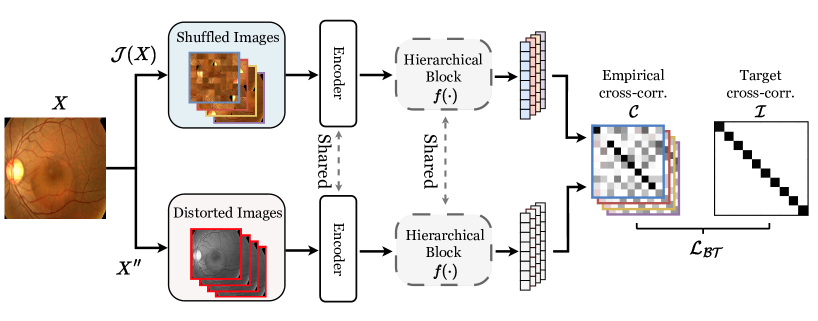
In this section, we present a method dubbed Fine-Grained Self-Supervised Learning (FG-SSL) for medical image recognition. The overall workflow of the proposed method is shown in Fig. 1. In Sec. 2.1., we first discuss the basis of this work regarding the single-level Jigsaw puzzle problem, and introduce our multi-level fine-grained self-supervised learning with a progressive Jigsaw puzzle. In Sec. 2.2., a regularization approach is also provided to improve the proposed fine-grained self-supervised method, and lastly, the step of fine-tuning employing the self-supervised pretrained model for classifying lesion classes of the medical data.
2.1 Self-supervision with Progressive Jigsaw puzzle
Single-level Jigsaw puzzle. The Jigsaw puzzle problem has been widely used in various self-supervised learning methodsLiu et al. (2021); Zhuang et al. (2019); Li et al. (2020). Since it can learn the unique form of the original image without labeling, self-supervised learning helps supervised learning as a pretext. Before applying the target task with supervised learning, a network is pretrained with the self-supervision of the single-level Jigsaw puzzle. The original image is compared to the Jigsaw puzzle that is shuffled in the single-level grid, as shown in Fig. 1. This single-level Jigsaw puzzle for self-supervised learning is formulated as:
| (1) |
where denotes features extracted from a Jigsaw patch , represents the shuffling order of Jigsaw patches, and stands for the number of patches.Noroozi and Favaro (2016) Although this single-level Jigsaw puzzle shows competitive performance, we argue that it is limited to learning fine-grained information about medical images, especially lesion classes.
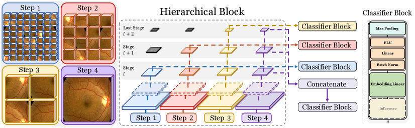
Progressive multi-level Jigsaw puzzle. Therefore, we introduce the multi-level Jigsaw puzzle to a progressive training strategy. We assume that at the stage level of a network, distinct self-supervisions should be learned hierarchically in order to understand the generic form of an object; early-stage layers should encode detailed low-level information, while late-stage layers should consider high-level abstracted features. For this purpose, we use the recently proposed PMG Du et al. (2020) for our multi-level self-supervised learning method. The PMG trains a network with different granularity levels of the Jigsaw puzzle. We apply the progressive Jigsaw puzzles of the PMG model in our self-supervised learning by shuffling the puzzles with different levels, as shown in Fig. 2. In the early stages of a network, the distorted images are compared to a Jigsaw puzzle shuffled in a grid size of , while subsequent stages employ more abstracted grid sizes such as , and . To learn the self-supervision from the multiple Jigsaw puzzles, we extract PMG features from each stage of a network as:
| (2) |
where denotes the index of a stage, represents the convolution layer to extract the patch feature , and is the concatenation function of multiple feature maps which generate the stage feature . We then train a network with self-supervised learning as
| (3) |
is the cross-entropy loss, and represent the ground truth and our empirical label of the Jigsaw puzzle ordering from each stage, denotes the classifier layer. We compute the final loss with the cross-entropy on each stage of a network progressively: .
Using this progressive learning process, our network can be pre-trained with finely shuffled Jigsaw puzzles. This fine-grained self-supervised learning has the advantage of learning various fine-grained hierarchical features in multi-stage Jigsaw puzzles.
2.2 Augmentation for Regularizing Self-supervision
Whereas the previous subsection introduced the SSL method of a target Jigsaw puzzle, this subsection provides the extensive fine-grained augmentation method of an original image directly for enhancing our self-supervision ability. We use the BarlowTwinsZbontar et al. (2021) method for fine-grained augmentation of original images, which uses the approach. BarlowTwins learn self-supervision by using random augmentation on the original image to minimize cross-correlation with the shuffled jigsaw images. Therefore, it computes the cross-correlation between the features of augmented original image with the Jigsaw puzzle as:
| (4) |
where represents the feature extractor, denotes the expectation function, and stand for the indices of training samples. Based on this definition of cross-correlation , we define loss function of BarlowTwins for comparing both augmented original and Jigsaw puzzles as
| (5) |
where the left term considers the invariance between the augmented original image and its Jigsaw puzzle, and the right term takes into account the redundancy reduction between the augmented original image and with Jigsaw puzzle from a different image. The positive constant , which is a user-input hyperparameter, is used to balance the importance of the first and second terms in the loss function. The invariance term, in particular, requires cross-correlation of the same images to resemble the identity matrix, resulting in Jigsaw puzzle invariance for fine-grained augmentations. Furthermore, by decreasing the cross-correlation between different images, the redundancy reduction term serves to push them away from the feature space. We then eventually acquire the pre-trained model by using this loss function of Eq. 5 under our fine-grained self-supervision. We dub this fine-grained self-supervised learning in the remaining sections as FG-SSL.
Fine-tuning. We fine-tune our networks from self-supervised learning for medical classification. With this fine-tuning, we also adopt the progressive Jigsaw puzzle of the PMG model. However, unlike the self-supervised learning as a pretext, we use the cross entropy(CE) with ground truth and estimated lesion labels as :
| (6) |
where and are the ground truth and estimated labels, and denote the stage of our hierarchical architecture.
3 Experiments
3.1 Dataset and Evaluation
We evaluate the proposed FG-SSL method with ISIC2018, APTOS2019, and ISIC2017 datasets. The ISIC2017 and ISIC2018 are available for download throu-gh the skin detection challengeCodella et al. (2018)111https://challenge.isic-archive.com/data/. The APTOS2019Karthik (2019) dataset can also be found in the public 222https://www.kaggle.com/competitions/aptos2019-blindness-detection/data. The ISIC2017 dataset has two classes with 2000 training and 600 test images. The ISIC2018 dataset contains seven classes and 10015 skin images, and APTOS2019 has five classes and a total of 3662 diabetic retinopathy images. In order to compare the performance fairly with existing methodsYang et al. (2022); Wu et al. (2020), we evaluate the proposed FG-SSL by randomly dividing the ISIC2018 and APTOS2019 datasets into train and test at a ratio of 7:3, in the same way as in the previous methods. Also, we perform experiments of ISIC2017 Task-3A, i.e., melanoma detection. We evaluate APTOS2019 and ISIC2018 using accuracy and F1-score, as in previous studies, and ISIC2017 using accuracy and area under the curve (AUC) as the comparative metrics. On the ISIC2018, APTOS2019, and ISIC2017 datasets, we compare existing methods, including state-of-the-art methods, in Table 1.
| Methods | APTOS2019 | ISIC2018 | Methods | ISIC2017 | ||||
| Acc | F1 | Acc | F1 | Acc | AUC | |||
| CE | 0.812 | 0.608 | 0.850 | 0.716 | GoogLeNet | 0.764 | 0.767 | |
| CE+resample | 0.802 | 0.583 | 0.861 | 0.735 | ResNet50 | 0.835 | 0.852 | |
| Focal Loss Lin et al. (2017) | 0.815 | 0.629 | 0.849 | 0.728 | ResNet101 | 0.839 | 0.861 | |
| OHEM | 0.813 | 0.631 | 0.818 | 0.660 | DenseNet101 | 0.842 | 0.861 | |
| LDAM Cao et al. (2019) | 0.813 | 0.620 | 0.857 | 0.734 | SDL Zhang et al. (2019) | 0.830 | 0.830 | |
| MTL | 0.813 | 0.632 | 0.811 | 0.667 | Galdran Galdran et al. (2017) | 0.480 | 0.765 | |
| DANIL Gong et al. (2020) | 0.825 | 0.660 | 0.825 | 0.674 | Vasconcelos Vasconcelos and Vasconcelos (2017) | 0.830 | 0.791 | |
| CL Marrakchi et al. (2021) | 0.825 | 0.652 | 0.865 | 0.739 | Yang Yang et al. (2017) | 0.830 | 0.830 | |
| CL + resample Marrakchi et al. (2021) | 0.816 | 0.608 | 0.868 | 0.751 | Diaz Díaz (2017) | 0.868 | 0.879 | |
| ProCo Yang et al. (2022) | 0.837 | 0.674 | 0.887 | 0.763 | ARDT-DenseNetWu et al. (2020) | 0.868 | 0.879 | |
| FG-SSL(Ours) | 0.858 | 0.720 | 0.897 | 0.816 | FG-SSL(Ours) | 0.913 | 0.927 | |
3.2 Implementation Details
The proposed FG-SSL uses ResNet50He et al. (2016) as the backbone in the same way as the existing methods. In the FG-SSL method for self-supervised learning, resize the input image and resize the input image to with a center crop. We apply the data augmentation method following previous studiesYang et al. (2022); Chen et al. (2020). During transfer learning, we resize the input image 256 and resize the input image to with a center crop. We set the hyperparameter to the one proposed by the existing authorZbontar et al. (2021). We utilize the features extracted from the 3rd to the last 5th stages in a total of 5 stages of the ResNet50 model for our progressive learning. We set a batch size of 256 and used SGD optimizer with a momentum of and weight decay of . The initial learning rate of 0.002 and continuously reduced it using a cosine annealing strategy. We set training epochs as 1000 and trained our model using the PyTorch version 1.11.0 with RTX A5000 GPUs.
| Method | Model | Single-level | Multi-level | ||
| Acc | F1 | Acc | F1 | ||
| SimSiam | Baseline | 0.860 | 0.732 | 0.865 | 0.743 |
| Rotation | 0.847 | 0.733 | 0.856 | 0.747 | |
| FG-SSL(Ours) | 0.856 | 0.759 | 0.873 | 0.764 | |
| SimSiam | PMG | 0.867 | 0.764 | 0.886 | 0.798 |
| MoCoV2 | 0.818 | 0.720 | 0.841 | 0.783 | |
| Rotation | 0.841 | 0.739 | 0.867 | 0.751 | |
| FG-SSL(Ours) | PMG | 0.882 | 0.794 | 0.897 | 0.816 |
| Granularity | FG-SSL | |||||||
| 1st | 2nd | 3rd | 4th | Acc | F1 | |||
| 8 | 8 | 8 | 8 | 0.864 | 0.763 | |||
| 8 | 8 | 4 | 2 | 0.878 | 0.784 | |||
| 8 | 4 | 4 | 4 | 0.867 | 0.775 | |||
| 8 | 4 | 2 | 2 | 0.885 | 0.809 | |||
| 8 | 4 | 2 | 1 | 0.897 | 0.816 | |||
| 16 | 8 | 4 | 1 | 0.884 | 0.797 | |||
| Method | Batch size | ||||
| 16 | 32 | 64 | 128 | 256 | |
| MoCoV2 | 0.785 | 0.793 | 0.812 | 0.834 | 0.841 |
| FG-SSL(Ours) | 0.874 | 0.878 | 0.882 | 0.891 | 0.897 |
| Jigsaw | PMG | Barlow | Acc | F1 |
| ✓ | 0.862 | 0.742 | ||
| ✓ | ✓ | 0.883 | 0.779 | |
| ✓ | ✓ | ✓ | 0.897 | 0.816 |
| Dimension | 16 | 32 | 128 | 256 | 512 |
| Accuracy | 0.826 | 0.835 | 0.859 | 0.878 | 0.897 |
| F1-score | 0.656 | 0.672 | 0.734 | 0.769 | 0.816 |
3.3 Comparison with Existing State-of-the-Art Methods
In this subsection, we compare the performance of the proposed FG-SSL with existing methods on the ISIC2018, APTOS2019, and ISIC2017 datasets. Table 1 shows the experimental results from all three datasets. In previous studies, contrastive learningYang et al. (2022) using negative sampling achieved the best performance on the APTOS2019 and ISIC2018 datasets. Negative sampling strategyHe et al. (2020) that relies on large batch sizes are unsuitable for medical datasets due to data scarcity. As a result, advances in performance utilizing such methods are frequently limited in medical tasks. Yet, because our FG-SSL method does not require a high batch size, it performs well compared to state-of-the-art methods in the small sample size of the medical image recognition task, as shown in Table 1.
3.4 Ablation Study
We conduct three ablation studies to demonstrate the efficacy of the proposed FG-SSL. Our models are trained in the same environment as in Sec. 3.2 and evaluated using the ISIC2018 Dataset in this ablation study.
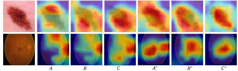
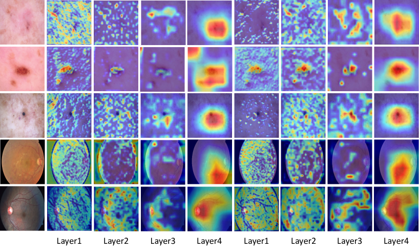
Quantitative analysis: We visualize the Grad-CAMSelvaraju et al. (2017) to evaluate the proposed method as shown in Fig. 3 qualitatively. In this visualization, we confirm that our progressive learning (A*, B, and C*) more focus on the subtle lesion region, and further, the BarlowTwins C* highlights it best. Further, we visualize the GradCam for each layer and found that FG-SSL more focus on subtle regions than the baseline method as show in Fig. 4.
Factor analysis: To show the effectiveness of the progressive Jigsaw puzzles, we compare ours with other self-supervised learning methods in Table 2(a). We confirm that our FG-SSL performs well compared to SimSiam and Rotation methods, while our multi-level setting also improves the performance. For evaluating our progressive learning strategy, we compare different settings as shown in Table 2(b). As the optimized hierarchical grid setting of PMG Du et al. (2020), we achieve the best result on among various settings. We also demonstrate that the proposed FG-SSL is insensitive to batch size, unlike the contrastive learning algorithm with the negative sampling (i.e., MoCoV2 Chen et al. (2020)), as shown in Table 2(c). Additionally, we validate the performance of our FG-SSL regarding different channel dimensionality as shown in Table 2(e). This result is significant in that our method will work well for a small sample size of medical datasets. To evaluate the effectiveness of the component of our self-supervised learning method, we carry out an ablation study in Table 2(d). When using Jigsaw puzzle and PMG, it shows competitive performance compared to ProCoYang et al. (2022). In addition, we validate that the proposed FG-SSL method combining Jigsaw puzzle, PMG, and BarlowTwin improves the performance significantly.
4 Conclusion
This paper introduces FG-SSL, a novel method for classifying subtle lesions from medical images. Our FG-SSL learns fine-grained self-supervision from progressive Jigsaw puzzles and fine-tunes it with the PMG method. Using the ISIC2018, APTOS2019, and ISIC2017 datasets, it is demonstrated that the proposed FG-SSL method performs well compared to the existing state-of-the-art methods. The efficiency of the proposed method is shown in the ablation study through extensive experiments. We validate the Jigsaw puzzle method on medical images in learning fine-grained features even without labels. We believe that our FG-SSL method will be useful in the future medical imaging community.
References
- Cao et al. (2019) Kaidi Cao, Colin Wei, Adrien Gaidon, Nikos Arechiga, and Tengyu Ma. Learning imbalanced datasets with label-distribution-aware margin loss. Advances in neural information processing systems, 32, 2019.
- Chen et al. (2023) Shaobin Chen, Zhenquan Wu, Mingzhu Li, Yun Zhu, Hai Xie, Peng Yang, Cheng Zhao, Yongtao Zhang, Shaochong Zhang, Xinyu Zhao, et al. Fit-net: Feature interaction transformer network for pathologic myopia diagnosis. IEEE Transactions on Medical Imaging, 2023.
- Chen and He (2021) Xinlei Chen and Kaiming He. Exploring simple siamese representation learning. In IEEE Conference on Computer Vision and Pattern Recognition, pages 15750–15758, 2021.
- Chen et al. (2020) Xinlei Chen, Haoqi Fan, Ross Girshick, and Kaiming He. Improved baselines with momentum contrastive learning. Arxiv, 2020.
- Codella et al. (2018) Noel C. F. Codella, David Gutman, M. Emre Celebi, Brian Helba, Michael A. Marchetti, Stephen W. Dusza, Aadi Kalloo, Konstantinos Liopyris, Nabin Mishra, Harald Kittler, and Allan Halpern. Skin lesion analysis toward melanoma detection: A challenge at the 2017 international symposium on biomedical imaging (isbi), hosted by the international skin imaging collaboration (isic). In 2018 IEEE 15th International Symposium on Biomedical Imaging (ISBI 2018), pages 168–172, 2018. doi: 10.1109/ISBI.2018.8363547.
- Díaz (2017) Iván González Díaz. Incorporating the knowledge of dermatologists to convolutional neural networks for the diagnosis of skin lesions. Arxiv, 2017.
- Du et al. (2020) Ruoyi Du, Dongliang Chang, Ayan Kumar Bhunia, Jiyang Xie, Zhanyu Ma, Yi-Zhe Song, and Jun Guo. Fine-grained visual classification via progressive multi-granularity training of jigsaw patches. In European Conference on Computer Vision, pages 153–168. Springer, 2020.
- Feng et al. (2019) Zeyu Feng, Chang Xu, and Dacheng Tao. Self-supervised representation learning by rotation feature decoupling. In IEEE Conference on Computer Vision and Pattern Recognition, pages 10364–10374, 2019.
- Galdran et al. (2017) Adrian Galdran, Aitor Alvarez-Gila, Maria Ines Meyer, Cristina L Saratxaga, Teresa Araújo, Estibaliz Garrote, Guilherme Aresta, Pedro Costa, Ana Maria Mendonça, and Aurélio Campilho. Data-driven color augmentation techniques for deep skin image analysis. Arxiv, 2017.
- Gong et al. (2020) Lijun Gong, Kai Ma, and Yefeng Zheng. Distractor-aware neuron intrinsic learning for generic 2d medical image classifications. In International Conference on Medical Image Computing and Computer-Assisted Intervention, pages 591–601. Springer, 2020.
- He et al. (2016) Kaiming He, Xiangyu Zhang, Shaoqing Ren, and Jian Sun. Deep residual learning for image recognition. In IEEE Conference on Computer Vision and Pattern Recognition, pages 770–778, 2016.
- He et al. (2020) Kaiming He, Haoqi Fan, Yuxin Wu, Saining Xie, and Ross Girshick. Momentum contrast for unsupervised visual representation learning. In IEEE Conference on Computer Vision and Pattern Recognition, pages 9729–9738, 2020.
- Jiao et al. (2023) Jing Jiao, Hongshuang Sun, Yi Huang, Menghua Xia, Mengyun Qiao, Yunyun Ren, Yuanyuan Wang, and Yi Guo. Gmrlnet: A graph-based manifold regularization learning framework for placental insufficiency diagnosis on incomplete multimodal ultrasound data. IEEE Transactions on Medical Imaging, 2023.
- Kaczmarzyk et al. (2023) Jakub R Kaczmarzyk, Rajarsi Gupta, Tahsin M Kurc, Shahira Abousamra, Joel H Saltz, and Peter K Koo. Champkit: a framework for rapid evaluation of deep neural networks for patch-based histopathology classification. Computer Methods and Programs in Biomedicine, page 107631, 2023.
- Karthik (2019) Sohier Dane Karthik, Maggie. Aptos 2019 blindness detection, 2019. URL https://kaggle.com/competitions/aptos2019-blindness-detection.
- Kim et al. (2018) Dahun Kim, Donghyeon Cho, Donggeun Yoo, and In So Kweon. Learning image representations by completing damaged jigsaw puzzles. In 2018 IEEE Winter Conference on Applications of Computer Vision (WACV), pages 793–802. IEEE, 2018.
- Li et al. (2020) Yuexiang Li, Jiawei Chen, Xinpeng Xie, Kai Ma, and Yefeng Zheng. Self-loop uncertainty: A novel pseudo-label for semi-supervised medical image segmentation. In International Conference on Medical Image Computing and Computer-Assisted Intervention, pages 614–623. Springer, 2020.
- Lin et al. (2017) Tsung-Yi Lin, Priya Goyal, Ross Girshick, Kaiming He, and Piotr Dollár. Focal loss for dense object detection. In IEEE International Conference on Computer Vision, pages 2980–2988, 2017.
- Liu et al. (2021) Quan Liu, Peter C Louis, Yuzhe Lu, Aadarsh Jha, Mengyang Zhao, Ruining Deng, Tianyuan Yao, Joseph T Roland, Haichun Yang, Shilin Zhao, et al. Simtriplet: Simple triplet representation learning with a single gpu. In International Conference on Medical Image Computing and Computer-Assisted Intervention, pages 102–112. Springer, 2021.
- Liu et al. (2022) Xiaofeng Liu, Fangxu Xing, Nadya Shusharina, Ruth Lim, C-C Jay Kuo, Georges El Fakhri, and Jonghye Woo. Act: Semi-supervised domain-adaptive medical image segmentation with asymmetric co-training. In International Conference on Medical Image Computing and Computer-Assisted Intervention, pages 66–76. Springer, 2022.
- Marrakchi et al. (2021) Yassine Marrakchi, Osama Makansi, and Thomas Brox. Fighting class imbalance with contrastive learning. In International Conference on Medical Image Computing and Computer-Assisted Intervention, pages 466–476. Springer, 2021.
- Noroozi and Favaro (2016) Mehdi Noroozi and Paolo Favaro. Unsupervised learning of visual representations by solving jigsaw puzzles. In European Conference on Computer Vision, pages 69–84. Springer, 2016.
- Selvaraju et al. (2017) Ramprasaath R Selvaraju, Michael Cogswell, Abhishek Das, Ramakrishna Vedantam, Devi Parikh, and Dhruv Batra. Grad-cam: Visual explanations from deep networks via gradient-based localization. In IEEE International Conference on Computer Vision, pages 618–626, 2017.
- Vasconcelos and Vasconcelos (2017) Cristina Nader Vasconcelos and B Nader Vasconcelos. Increasing deep learning melanoma classification by classical and expert knowledge based image transforms. CoRR, abs/1702.07025, 1, 2017.
- Wargnier-Dauchelle et al. (2023) Valentine Wargnier-Dauchelle, Thomas Grenier, Françoise Durand-Dubief, François Cotton, and Michaël Sdika. A weakly supervised gradient attribution constraint for interpretable classification and anomaly detection. IEEE Transactions on Medical Imaging, 2023.
- Wu et al. (2020) Jing Wu, Wei Hu, Yuan Wen, Wenli Tu, and Xiaoming Liu. Skin lesion classification using densely connected convolutional networks with attention residual learning. Sensors, 20(24):70–80, 2020.
- Yang et al. (2023) Qiushi Yang, Zhen Chen, and Yixuan Yuan. Hierarchical bias mitigation for semi-supervised medical image classification. IEEE Transactions on Medical Imaging, 2023.
- Yang et al. (2017) Xulei Yang, Zeng Zeng, Si Yong Yeo, Colin Tan, Hong Liang Tey, and Yi Su. A novel multi-task deep learning model for skin lesion segmentation and classification. Arxiv, 2017.
- Yang et al. (2022) Zhixiong Yang, Junwen Pan, Yanzhan Yang, Xiaozhou Shi, Hong-Yu Zhou, Zhicheng Zhang, and Cheng Bian. Proco: Prototype-aware contrastive learning for long-tailed medical image classification. In International Conference on Medical Image Computing and Computer-Assisted Intervention, pages 173–182. Springer, 2022.
- Yu et al. (2023) Jin-Gang Yu, Zihao Wu, Yu Ming, Shule Deng, Qihang Wu, Zhongtang Xiong, Tianyou Yu, Gui-Song Xia, Qingping Jiang, and Yuanqing Li. Bayesian collaborative learning for whole-slide image classification. IEEE Transactions on Medical Imaging, 2023.
- Zbontar et al. (2021) Jure Zbontar, Li Jing, Ishan Misra, Yann LeCun, and Stéphane Deny. Barlow twins: Self-supervised learning via redundancy reduction. In International Conference on Machine Learning, pages 12310–12320. PMLR, 2021.
- Zhang et al. (2019) Jianpeng Zhang, Yutong Xie, Qi Wu, and Yong Xia. Medical image classification using synergic deep learning. Medical image analysis, 54:10–19, 2019.
- Zhao et al. (2023) Yue Zhao, Hong Zeng, Haohao Zheng, Jing Wu, Wanzeng Kong, and Guojun Dai. A bidirectional interaction-based hybrid network architecture for eeg cognitive recognition. Computer Methods and Programs in Biomedicine, 238:107593, 2023.
- Zhuang et al. (2019) Xinrui Zhuang, Yuexiang Li, Yifan Hu, Kai Ma, Yujiu Yang, and Yefeng Zheng. Self-supervised feature learning for 3d medical images by playing a rubik’s cube. In International Conference on Medical Image Computing and Computer-Assisted Intervention, pages 420–428. Springer, 2019.