Henriques
Harnessing Artificial Intelligence
To Reduce Phototoxicity in Live Imaging
Abstract
Fluorescence microscopy, widely used in the study of living cells, tissues, and organisms, often faces the challenge of photodamage. This is primarily caused by the interaction between light and biochemical components during the imaging process, leading to compromised accuracy and reliability of biological results. Methods necessitating extended high-intensity illumination, such as super-resolution microscopy or thick sample imaging, are particularly susceptible to this issue. As part of the solution to these problems, advanced imaging approaches involving artificial intelligence (AI) have been developed. Here we underscore the necessity of establishing constraints to maintain light-induced damage at levels that permit cells to sustain their live behaviour. From this perspective, data-driven live-cell imaging bears significant potential in aiding the development of AI-enhanced photodamage-aware microscopy. These technologies could streamline precise observations of natural biological dynamics while minimising phototoxicity risks.
keywords:
photodamage | phototoxicity | live-microscopy | artificial intelligence | deep learning | data driven microscopy | fluorescence microscopy | live-cell super-resolution microscopyrjhenriques and igc.gulbenkian.pt
Introduction
The ability to comprehend biological dynamics is inherently linked to the capacity for non-invasive observation. Fluorescence microscopy has been instrumental in facilitating these analyses across a range of scales, with molecular specificity (1, 2, 3). Over the past two decades, technological advancements such as light sheet microscopy (4, 5, 6, 7), structured illumination microscopy (SIM) (8, 9), and single molecule localisation microscopy (SMLM) (10, 11, 12), have revolutionised fluorescence light microscopy, enabling us to characterise biological events from molecular interactions up to larger living organisms.
A key challenge associated with advanced microscopy analysis is its likely need to increase fluorescence excitation light levels, which results in phototoxicity or photodamage. These terms refer to the detrimental impacts of light, especially when employing photosensitising agents or high-intensity illumination (13, 14, 15). Despite certain distinctions like photodamage occurring also in non-living materials, both terms are here used interchangeably, for simplicity. Sample illumination may also result in photobleaching, a process characterised by an irreversible loss of a fluorescent signal attributed to the destruction of the fluorophore. This is one manifestation of light damage, among other possible effects. Phototoxicity severely influences the experimental outcomes by altering biological processes under observation, skewing findings and impeding consistency. Therefore, it’s crucial during live-cell microscopy to carefully consider these factors to prolong imaging durations and achieve dependable research outcomes.
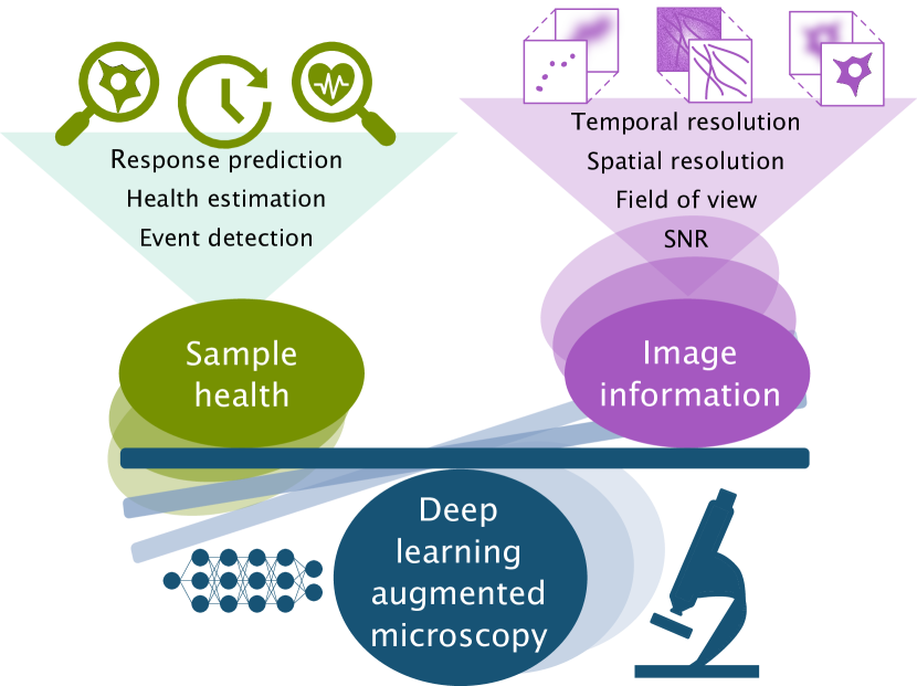
The biological validity of live-cell imaging experiments requires a precise balance between the data quality and the specimen’s health, as depicted in Fig. . Major advancements have been made in both hardware and software technologies aiming to reduce sample light damage. Hardware innovations such as Lattice Light Sheet Microscopy (LLSM) (16) and Airyscan Microscopy (17) are notable examples. Additionally, computational advancements like Fluctuation-based Super Resolution Microscopy offer promising solutions to these issues (18, 19). A recent study has shown that a two-colour illumination scheme combining near-infrared illumination with fluorescence excitation presents an interesting capacity to limit the phototoxicity caused by light-induced interactions with fluorescent proteins (20). These technological breakthroughs have the potential to optimise observation accuracy while mitigating photodamage.
In parallel, artificial intelligence (AI), specifically deep learning, can significantly improve imaging information in low illumination scenarios by considerably enhancing image quality and quantification (21, 22, 23). This has inspired the search for integrated solutions by the microscopy community (24, 25, 26). The fusion of advanced optical hardware with computational models and intelligent analytics heralds new breakthroughs in overcoming sample damage induced by traditional live fluorescence microscopy methodologies, marking the advent of AI-enhanced intelligent microscopy.

Understanding phototoxicity
Cellular environments undergo continuous adaptation to maintain homeostasis, with oxygen playing a key role in multiple chemical processes (27). Reactive Oxygen Species (ROS), resulting from oxidative processes involving oxygen radicals (Table 1), are integral to cell growth, differentiation, and other functions. However, uncontrolled ROS processes lead to oxidative stress and cellular damage, resulting in autophagy, inflammation, and potentially cell death (27). For these reasons, a balance between these states is critical for cellular physiology.
| Oxygen Radicals | Biomolecules Affected |
|---|---|
| Hydrogen Peroxide | Flavins |
| Singlet molecular oxygen | Porphyrins |
| Nitric Oxide | NAD(P)H |
| Superoxide anion radicals | Tyrosine |
| Hydroxyl radical | Catecholamines |
| Hydroxide ion | Cysteinyl thiols |
Light irradiation to excite fluorescence, in addition to inducing oxidative stress by producing high amounts of ROS, triggers multiscale alterations across biological samples that alter the homeostasis of oxidative processes. Moreover, higher doses of light irradiation increase this oxidative stress, further aggravating cellular homeostasis. Photochemical processes at the molecular level lead to intracellular component damage or toxic compound formation (33, 34). The literature extensively documents the impacts of fluorescence excitation light, especially UV light on DNA, such as DNA double-stranded breaks (35), thymidine dimerisations (35), UV-induced apoptosis (36), and tumour factor activation (36). These effects are even routinely employed in research settings under controlled conditions (37, 38, 39, 40, 41). Moreover, molecules naturally present in cells (Table ) undergo degradation via exposure to light-induced oxidative stress, canonically produced during the fluorescence excitation process. This degradation culminates in the generation of reactive oxygen compounds that directly impair cell health by oxidising DNA, proteins, unsaturated fatty acids, and enzyme cofactors (15, 42). Excessive ROS shifts the homeostasis of the cell cytoplasmic environment’ leading to organelle damage like mitochondrial fragmentation due to compromised membrane potential and integrity, among others (43, 35, 44, 45, 13).
While helpful and versatile, fluorescence microscopy illumination entails an additional source of ROS formation (46). When excited, fluorophores undergo autocatalysis through dioxygen, releasing hydrogen peroxide and consequently degrading - a process commonly known as photobleaching (47). Photobleaching produces ROS similar to other biomolecules, intensifying phototoxic effects (46). However, despite the interrelation between photobleaching and phototoxicity (20), these phenomena exhibit distinct features and can occur independently. Namely, identical oxygen radical compounds originate from photobleaching and direct fluorescence excitation light interactions with other cellular components (46, 47). Suggesting that a reduction of photobleaching does not necessarily imply a decrease in phototoxicity, and the other way around. The intricate mechanisms underlying these phenomena have been thoroughly researched, especially concerning various fluorophores’ photosensitising effects in live microscopy. Given the common necessity for oxygen in cell cultures, ROS formation during live-cell imaging is an unavoidable aspect of the imaging process.
At lower thresholds, the impact of light exposure may be minimal and reversible, allowing the cell to return to physiological conditions. This cell recovery will depend on several factors, namely how resilient the sample is and the degree of phototoxicity incurred by the experiment (13). In severe cases, the behaviour of cells will be permanently altered (15). Moreover, further alterations normally accumulate within a cell’s homeostatic environment until reaching an irreversible point causing pronounced impairment of cell health that leads to unwanted cell behaviour (cellular collapse) or death (apoptosis) (Figure a) (42, 15, 34, 35).
Although scientists have identified several indicators of photodamage, the correlation between fluorescent excitation light dose and light damage is not yet fully understood due to variability in experimental conditions. These variations include culture conditions, illumination modes (Figure b) and biological sample variability, hindering the understanding between fluorescent excitation light dose and light damage (15, 14). Despite challenges associated with varying specimen resistances to light damage, establishing universal quantitative benchmarks across diverse samples could harmonise these effects and enhance reproducibility in the imaging context.
Phototoxicity quantification
The literature describes several phototoxicity hallmarks with numerous known markers available to identify and characterise sample damage (33). However, using these markers presents additional challenges in live-cell experiments. Firstly, incorporating phototoxicity markers necessitates extra planning and may require allocating fluorescent channels usually reserved for observing conditions of interest (e.g., markers for DNA oxidative damage). Not only does this limit the number of fluorescent channels available for the experiment, but when paired with live cell experiments could enhance ROS formation, thereby escalating the phototoxicity risk on the specimen. Secondly, despite well-documented phototoxicity markers, quantification-based screenings are less commonly employed (34). The absence of live-cell imaging universal metrics relatable to light exposure and consequent damage across various biological systems hampers experimental reproducibility, undermines results’ robustness, and limits the obtainable biological readouts. Furthermore, without these universal metrics, it’s challenging to fully leverage the capacity to image biological systems. This results in the incorrect assessment of experimental conditions to achieve maximum spatial and temporal resolution while preserving cell viability.
Researchers primarily rely on observation and experience to assess cell health and viability during photodamage evaluation (Figure a) (13, 34, 15). While there are some attempts to provide quantifiable metrics for improving sample viability (14, 42, 13), they often simplify the impact of fluorescence excitation light to a binary classification of viable/healthy or non-viable/dead. These approaches may overlook subtle effects that could disturb a specimen’s normal physiology. A gradient model that considers the accumulation of such discrete minor effects would more accurately depict the spectrum of effects documented in the existing literature. Namely, these approaches could be even more flexible by considering both cell health decline and recovery.
Yet an important aspect to consider is the collective physiological and behavioural outcome, rather than individual effects. In controlled experimental conditions, specimens typically exhibit consistent behaviours such as cell division, motility, and membrane dynamics. By modelling and quantifying these behaviours, deviations can be correlated to the negative effects of fluorescent light exposure on the sample.
Phototoxicity is a known issue in live-cell imaging, and a plethora of strategies exist to mitigate its effects, ranging from reducing light irradiation by either reducing the acquisition points or the light dose (7) to using more sensitive light detectors (17). Other strategies focus on controlling oxidative stress effects in biological samples by reducing its effects by supplementing antioxidants (48, 49), or increasing the resistance of the sample itself (50). Although some strategies are simpler to integrate than others, they require using specific and costly equipment or altering the conditions of the specimen.
As advanced image analysis tools become increasingly available, a future strategy for fully reproducible live-cell imaging procedures could incorporate specific phototoxicity reporters within automated image acquisition and analysis workflows. This approach, paired with standardised experimental guidelines for identifying and quantifying phototoxic events, represents a promising solution. As imaging technologies evolve, adopting such methods should be prioritised by scientists aiming to create robust imaging strategies for visualising biological phenomena.
The relationship between fluorescence excitation light, image information and phototoxicity
Fluorescence live-cell imaging involves the excitation of fluorophores using light, resulting in photon emission. These photons can be captured by a camera or sensor, facilitating the visualisation of cellular targets. Various microscopy methods are available for sample illumination as depicted in Figure b. Some strategies, such as widefield, confocal microscopy, and Stimulated Emission Depletion (STED), illuminate the sample across the optical axis. In contrast, two-photon excitation primarily excites sample regions near the imaging focal plane while also subjecting the sample to less harmful infrared light across the optical axis. Total Internal Reflection Fluorescence (TIRF) imaging optically limits illumination to near the coverslip surface, substantially reducing in-depth illumination. However, it generally only illuminates the interface between the sample and the coverslip. Light-sheet microscopy, a more live-cell-friendly approach, illuminates only around the focal plane of the sample but often poses challenges in achieving high resolution compared to alternative methods. Hence, the choice of microscopy depends on sample characteristics; more sensitive specimens like embryos benefit from gentler modalities such as light-sheet microscopy. Inversely, more resilient samples can withstand intense irradiation experienced during Single-Molecule Localization Microscopy (SMLM) or STED microscopy (Figure b).
As introduced earlier, modern microscopy technologies seek to minimise the required illumination by becoming more specific towards the type of information that needs to be visualised. However, the acquired information’s quality may depend on factors such as the signal-to-noise ratio (SNR), image contrast, and temporal and spatial resolution of the image data (Figure c). Each microscopy equipment has inherent limitations that affect these properties’ optimisation, necessitating a trade-off, which is often expressed by the microscope ’pyramid of frustration’ (51), or as characterised explicitly for super-resolution microscopy by Jaquemet et al. (52). This trade-off can be optimised based on experimental needs by adjusting light exposure intensity and acquisition speed - common parameters used to achieve less harmful imaging configurations. As such, methods that enhance image quality from sample-friendly setups, like deep learning methods, are particularly valuable when pushing against intertwined image information and phototoxicity limitations (Figures and c). Specifically, deep learning’s growing ability to augment and refine image-based information is attracting considerable interest in the microscopy community. It is becoming one of the most popular strategies to enable imaging setups with reduced phototoxicity (53, 25). In succeeding sections, we explore different strategies conceived to enhance microscopy image data’s information contrast and quality, which could facilitate varied live-cell imaging setups with decreased fluorescent light illumination.
Deep learning for microscopy to the rescue
The exceptional ability of deep learning to automatically discern, uncover, and summarise complex patterns within images is undeniably one of its most transformative contributions to bioimaging. It has significantly influenced microscopy image analysis by ushering in a shift from traditional mathematical feature modelling to data-driven methods (57, 21, 58, 22, 23). We have observed the potential of deep learning to achieve remarkable accuracy and, in some cases, human-level performance in diverse computer vision tasks such as segmentation, denoising, detection, reconstruction, unmixing, and response prediction. Additionally, emerging imaging processing tasks like cross-modal style transfer (59, 60) and virtual labelling (61, 62, 63, 64) hold promising prospects due to the flexibility they introduce when designing imaging experiments (e.g., spectral unmixing or rapid low-resolution acquisitions that can be virtually super-resolved for subsequent quantification) (21). Indeed, microscopy imaging naturally allows for creating paired image datasets by alternating acquisition setups and combining different modalities (65, 54), simulating data (66, 67, 68, 69, 70), or, recently, developing correlative approaches such as CLEM (71). Cumulatively, all these advancements have laid a solid foundation for the growing field of deep learning-augmented microscopy (Figure ).
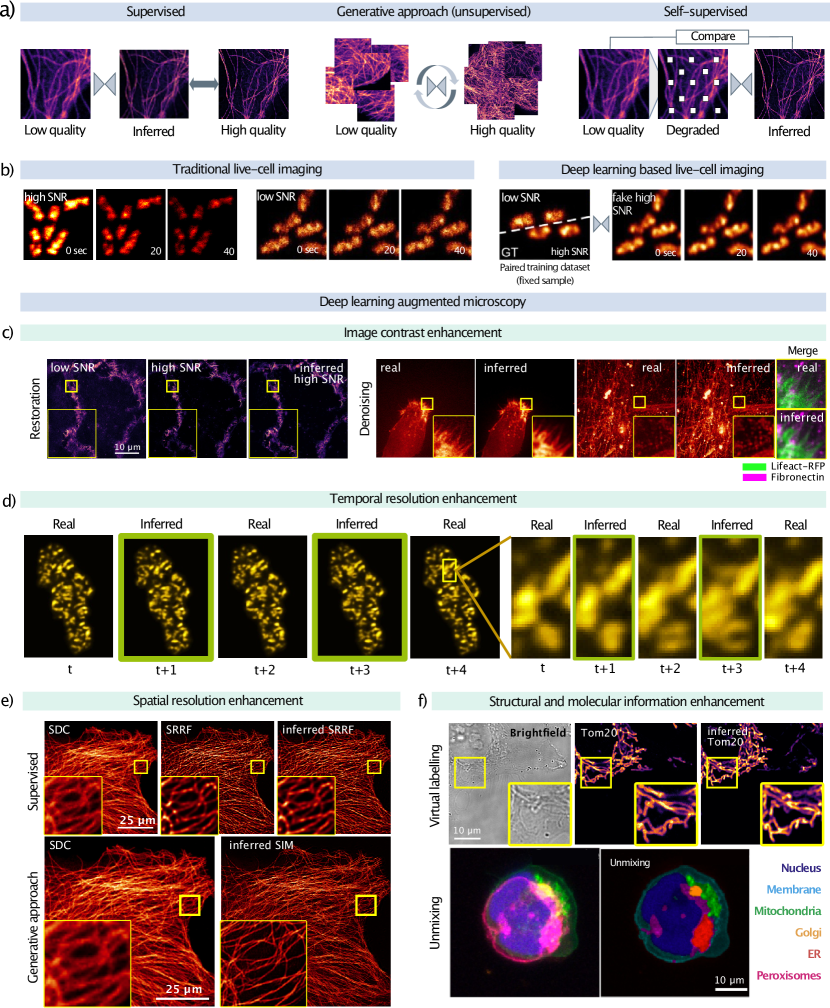
Regardless of the strategy, the key idea behind deep learning is to define models that learn to identify or enhance specific features directly from the data. Traditionally, the learning process, or training, has been classified as supervised, where the model is trained with paired input-output image datasets, or unsupervised, where the model is only exposed to input images during training (Figure a). Typically, supervised approaches have demonstrated superior accuracy and specificity to the task and data distribution, but their versatility relates to the availability of paired images, which could be a limitation in live microscopy. Some alternatives (51, 55, 72) propose training models using paired images of ex vivo samples—providing perfectly aligned images for training and assessment—to subsequently perform inference with in vivo images (Figure b). Importantly, collecting images from fixed images supports faster creation of more extensive and diverse datasets than live imaging. Depending upon the image features, simulated data may also be viable for training such models (67, 69, 66, 68). However, there remain scenarios where obtaining such paired datasets does not encapsulate the complexity of live experiments, is not experimentally feasible or cross-modality acquisition devices are inaccessible. Given this, alongside time-consuming data annotation processes, has propelled exploration into alternative approaches such as semi- or weakly-supervised (73), self-supervised (74, 75), or generative techniques (59, 63, 76) (Figure a).
Many possibilities exist to exploit deep learning-augmented microscopy and reduce phototoxicity. Drawing inspiration from the delineation provided in (22), we distinguish strategies that aim either to surmount the physical limitations intrinsic to live fluorescence microscopy imaging (i.e., acquisition speed or illumination) or to enhance the content in less qualitatively superior but more sample-friendly image data. The former includes techniques such as denoising, restoration, or temporal interpolation. The latter, referred to by the original authors as "augmentation of microscopy data contrast", includes techniques such as virtual super-resolution (59, 77, 78, 65, 79, 80).
Recent denoising (81, 74, 75) and restoration approaches (51, 82, 83, 84, 85, 86) have successfully virtualised noise removal, enhanced the SNR, improved fluorescence channel contrast with unparalleled accuracy, or provided isotropic reconstruction of volumetric information of images with low SNR (Figure c). That is, they support imaging setups with reduced fluorescence illumination that results in lower SNR or reduced optical D sectioning, which in turn, are gentler for the sample. Similarly, acquisition can be slowed down and use intelligent interpolators like CAFI (87) or DBlink (69) to recover temporal information (Figure d). As we discussed earlier, reducing the number of illumination time points can significantly decrease the cumulative phototoxicity while potentially enabling some degree of photodamage recovery.
Another innovative approach to enable reduced illumination imaging setups is exploiting cross-modal style transfer methodologies. In brief, these methods involve training a model to translate images between varied microscopy modalities (Figure e). For example, it was shown that SIM images could be inferred from input images acquired with wide-field illumination (65), reducing the photon dose by a factor of / in D/D. This capability extends to numerous types of fluorescence microscopy modalities such as confocal to STED (59, 26), SIM and SRRF (54), or wide-field to SMLM (88, 67, 89). The enhancement in spatial resolution via learning fine details is similar in objective to traditional deconvolution. It offers comparable benefits for mitigating phototoxicity, such as using less aggressive imaging modalities (widefield and/or confocal against SIM, STED, and SMLM). While the extent these methodologies can contribute towards scientific discovery is up for debate (21, 90), they undoubtedly elevate image data quality which directly impacts subsequent tasks like accurate tracking or segmentation (51, 87). As suggested in (51), less aggressive live imaging approaches may yield visually less appealing but easier-to-analyse data due to the reduction in artefacts induced by photodamage (e.g., apoptosis, stressed cells or specimen shrinkage during illumination) with the benefit of preserving close-to physiological conditions.
Exploiting the prowess of data-driven methods, artificial labelling approaches have emerged (Figure f). They span from inferring cell nuclei (e.g., Hoechst staining) from actin (e.g., Lifeact staining) (54), to estimating specific fluorescence information (e.g., nucleoli, cell membrane, nuclear envelope, mitochondria or neuron-specific tubulin) from brightfield input images (91, 61). It’s worth noting that the latter technique is also categorized as a cross-modality style transfer approach. Moreover, artificial labelling can also be employed for channel unmixing (92, 93, 94), which offers several key advantages, including illumination channel reduction, acceleration of image acquisition, or support for more straightforward or cost-effective imaging experiments (Figure f). Among these benefits, the former is pivotal in enabling more sample-friendly setups. Indeed, artificial labelling often works as an intermediary step for further quantification such as segmentation or tracking (60, 54).
Beyond the questions present also in the natural domain, novel inpainting and modality transfer techniques pose an additional challenge in bioimaging: they must accurately infer reliable and quantifiable physiological information. Thus, further validation, more assessment methodologies, and standard quantitative strategies ought to ascertain both the biological reliability of restored images and the integrity of recovered signal intensity (i.e., virtual images) are needed (21, 95, 96).
Biomedical data typically exhibit high variability due to factors such as the biological sample’s physiology, experimental protocol, imaging setup, and even the individual researcher conducting the experiments. Defining and establishing an accurate ground truth becomes crucial (95, 97), as deep learning model training highly depends on the data quality. Quality in terms of image processing ranges from the number of images available to train to how suitable the features of these images are for the task to train the model on. For instance, it remains to be seen how to strike an optimal balance between data acquisition and model accuracy (i.e., defining an ideal sampling frequency for precise cell tracking). A prevalent notion suggests that larger training datasets enhance model performance. Simultaneously, many publicly available annotated or paired databases are growing (98, 99, 97). However, strategies to combine these datasets effectively while preserving each experimental setup’s specificity still need to be clarified. Furthermore, pre-trained models that facilitate fine-tuning and transfer learning are readily accessible thanks to trained deep learning model repositories such as the Bioimage Model Zoo (100) and MONAI (101). Nonetheless, further enquiry is necessary to ascertain the best practices for assembling adequate training data and executing effective transfer learning while considering data quality, image features, and task-specific analytical requirements. Because live-cell image data is usually highly redundant, such optimisation could maximise information extraction from images while minimising photodamage during the acquisition.
Life scientists hold the top authority in deciding parameters such as sampling frequencies, resolution, or fields of view. While these decisions may sometimes be sub-optimal for subsequent quantification, they are our best reference for accurate performance. Incorporating users in the loop or their expertise as priors to guide model performance towards more specific and biologically relevant results is one of the pro missing directions to take into AI-enhanced microscopy. Recent technological advances such as the analytical representation of sparse and raw information to create priors - an approach already proposed for the segmentation of natural pictures in works such as the Segment Anything Model (SAM) (102) - is an important step towards it.
Future Outlook
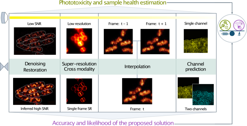
With the advance of fluorescence microscopy technology, the field is developing intelligent imaging techniques to minimise photodamage and enable accurate observations of biological dynamics. Similar to automatic cars or smart robots in industrial environments, microscopes can be empowered with AI components that enable real-time decisions by integrating the information extracted from the observed data into an intelligent feedback loop that balances the health of the sample and the image data quality (Figures c and ) (53). Note that we refer to image quality as the combination of features (e.g., resolution in time and space, SNR, the size of the field of view, or the number of fluorescent channels) that allow getting the most information relevant for the understanding of the observed process. In line with these, some have already conceptualised and proved such systems. In the first place, we find event-driven approaches, which automatically identify specific objects or incidents in the image that trigger the acquisition in real-time (103, 104, 105, 106, 107). While these adaptive approaches reduce the induced phototoxicity by only illuminating the sample when needed, in most cases, they are equipped with deep learning models trained to recognise predefined objects or elements in the images, which is not always the case in biology and may limit, even bias, the observation of novel physiological processes. Alternative approaches propose the integration of image resolution enhancement in the loop to get faster and gentler setups. In (24), a deep learning model is trained and validated in the acquisition loop to enhance the volumetric reconstruction and provide an adaptive light field microscopy (LFM) setup. In the context of super-resolution imaging, Bouchard et al. (26) propose evaluating the quality of virtually inferred STED images from confocal microscopy images to determine the uncertainty in the observed sample and determine whether a new STED image should be acquired or not. All these works pose new paradigms in the realm of smart microscopy.
Deep learning approaches, particularly the unsupervised ones, learn and match data distributions even in highly heterogeneous or complex scenarios without the need for human descriptions or annotations. As mentioned in (108), such methods could be exploited to identify the events that deviate from the general distribution, i.e., to discover new biological patterns. Thus, advancing generative models and unsupervised/self-supervised approaches that can effectively learn from unpaired data alone can provide flexibility when paired datasets are difficult to obtain experimentally or when investigating new dynamics, and contribute to unbiased observations.
Straightforwardly, one would realise that despite sample health preservation being a strong motivation and a major limitation in live-cell imaging, none of the existing solutions analyses or estimates it directly. Robust photodamage reporters that provide quantitative assessments of sample health without requiring additional fluorescence channels can directly contribute to more reproducible biological readouts. This could involve exploring modalities like transmitted light microscopy or label-free techniques. Namely, quantitative reporters would support the design of automated workflows that analyse sample health in real-time during image acquisition rather than only evaluating the image quality (Figure ). This would allow the detection of early signs of photodamage and the adaptive determination of optimal imaging conditions. In other words, it will open the door for data-driven sample-oriented live microscopy.
Pursuing such technical innovations while deepening our understanding of photodamage mechanisms will enable microscopists to unlock the full potential of intelligent imaging. With photodamage-aware AI and automated tools, the goal of observing undisturbed physiological processes can be realised. This will profoundly enhance fluorescence microscopy’s capacity to uncover ground truths in biology.
Discussion
Fluorescence microscopy has become an indispensable tool in cell biology, providing unparalleled insights into biomolecular dynamics. However, phototoxicity remains a major impediment, necessitating a deeper mechanistic understanding alongside the development of imaging techniques that mitigate this limitation. While the intercourse between microscopy hardware innovation and computational imaging has yielded promising solutions, standardised methodologies to comprehensively assess photodamage are lacking. Recent strides in deep learning provide optimism by enhancing information extraction from low-light or accelerated acquisitions. Nonetheless, robust validation strategies are still required to ensure biological fidelity.
A key opportunity lies in constructing universal photodamage metrics that account for subtle, cumulative deviations in sample physiology. Integrating such quantifications in intelligent automated analysis enables microscopes to optimise imaging conditions dynamically. This calls for a convergence of synergistic advancements spanning photodamage biology, microscopy hardware, computational imaging and model interpretation. An outstanding challenge is model training, which requires extensive paired datasets that sufficiently encapsulate biological variability. Alternatives like unsupervised learning provide flexibility but may compromise accuracy. Incorporating biological expertise through techniques like priors and prompts appears promising to guide models. Additionally, optimised strategies for effective model training and validation must be developed through empirical examination.
While deep learning has elevated imaging capabilities, over-reliance on its reconstruction prowess could promote complacency. Achieving gentler acquisition first requires re-evaluating illumination intensities and sampling frequencies. We claim that AI should extract the maximum information from the least invasive data rather than recover information already compromised by excessive phototoxicity. Keeping biological relevance at the crux while exploiting technology will lead to microscopes that truly observe life undisturbed.
E.G.M., M.DR., and R.H. conceptualised the majority of the manuscript. E.G.M. and M.DR. wrote the manuscript with input from J.W.P., G.J., and R.H. J.W.P. and G.J. contributed text, figure design, critical comments, and conceptual suggestions to improve the manuscript. J.W.P., G.J., and R.H. reviewed and edited the manuscript.
Acknowledgements.
M.DR., E.G.M. and R.H. acknowledge the support of the Gulbenkian Foundation (Fundação Calouste Gulbenkian), the European Research Council (ERC) under the European Union’s Horizon 2020 research and innovation programme (grant agreement No. 101001332), the European Commission through the Horizon Europe program (AI4LIFE project with grant agreement 101057970-AI4LIFE, and RT-SuperES project with grant agreement 101099654-RT-SuperES), the European Molecular Biology Organization (EMBO) Installation Grant (EMBO-2020-IG-4734) and the Chan Zuckerberg Initiative Visual Proteomics Grant (vpi-0000000044 with DOI:10.37921/743590vtudfp). E.G.M. was also supported by EMBO Postdoctoral Fellowship (EMBO ALTF 174-2022). This study was supported by the Academy of Finland (338537 to G.J.), the Sigrid Juselius Foundation (to G.J.), the Cancer Society of Finland (Syöpäjärjestöt; to G.J.), and the Solutions for Health strategic funding to Åbo Akademi University (to G.J.). This research was supported by InFLAMES Flagship Programme of the Academy of Finland (decision number: 337531).Bibliography
References
- Heimstädt (1911) Oskar Heimstädt. Das fluoreszenzmikroskop. Z Wiss Mikrosk, 28:330–337, 1911.
- Reichert (1911) K. Reichert. Das Fluoreszenzmikroskop. Physik Zeits, 12:1010, 1911.
- Lehmann (1913) H. Lehmann. Das Luminszenz-Mikroskop: seine Grundlagen und seine Anwendungen. 1913.
- Huisken et al. (2004) Jan Huisken, Jim Swoger, Filippo Del Bene, Joachim Wittbrodt, and Ernst H. K. Stelzer. Optical sectioning deep inside live embryos by selective plane illumination microscopy. Science, 305(5686):1007–1009, August 2004. ISSN 0036-8075, 1095-9203. 10.1126/science.1100035.
- Dodt et al. (2007) Hans-Ulrich Dodt, Ulrich Leischner, Anja Schierloh, Nina Jährling, Christoph Peter Mauch, Katrin Deininger, Jan Michael Deussing, Matthias Eder, Walter Zieglgänsberger, and Klaus Becker. Ultramicroscopy: three-dimensional visualization of neuronal networks in the whole mouse brain. Nature Methods, 4:331–336, 4 2007. ISSN 1548-7091. 10.1038/nmeth1036.
- Verveer et al. (2007) Peter J Verveer, Jim Swoger, Francesco Pampaloni, Klaus Greger, Marco Marcello, and Ernst H K Stelzer. High-resolution three-dimensional imaging of large specimens with light sheet–based microscopy. Nature Methods, 4(4):311–313, April 2007. ISSN 1548-7091, 1548-7105. 10.1038/nmeth1017.
- Reynaud et al. (2008) Emmanuel G. Reynaud, Uroš Kržič, Klaus Greger, and Ernst H. K. Stelzer. Light sheet-based fluorescence microscopy: More dimensions, more photons, and less photodamage. HFSP Journal, 2:266–275, 10 2008. ISSN 1955-2068. 10.2976/1.2974980.
- Heintzmann and Huser (2017) Rainer Heintzmann and Thomas Huser. Super-resolution structured illumination microscopy. Chemical Reviews, 117(23):13890–13908, December 2017. ISSN 0009-2665, 1520-6890. 10.1021/acs.chemrev.7b00218.
- Gustafsson (2000) M. G. L. Gustafsson. Surpassing the lateral resolution limit by a factor of two using structured illumination microscopy. Short communication. Journal of Microscopy, 198(2):82–87, May 2000. ISSN 0022-2720. 10.1046/j.1365-2818.2000.00710.x.
- Betzig et al. (2006) Eric Betzig, George H. Patterson, Rachid Sougrat, O. Wolf Lindwasser, Scott Olenych, Juan S. Bonifacino, Michael W. Davidson, Jennifer Lippincott-Schwartz, and Harald F. Hess. Imaging Intracellular Fluorescent Proteins at Nanometer Resolution. Science, 313(5793):1642–1645, September 2006. ISSN 0036-8075. 10.1126/science.1127344.
- Hess et al. (2006) Samuel T. Hess, Thanu P.K. Girirajan, and Michael D. Mason. Ultra-high resolution imaging by fluorescence photoactivation localization microscopy. Biophysical Journal, 91:4258–4272, 12 2006. ISSN 00063495. 10.1529/biophysj.106.091116.
- Lelek et al. (2021) Mickaël Lelek, Melina T. Gyparaki, Gerti Beliu, Florian Schueder, Juliette Griffié, Suliana Manley, Ralf Jungmann, Markus Sauer, Melike Lakadamyali, and Christophe Zimmer. Single-molecule localization microscopy. Nature Reviews Methods Primers, 1:39, 6 2021. ISSN 2662-8449. 10.1038/s43586-021-00038-x.
- Wäldchen et al. (2015) Sina Wäldchen, Julian Lehmann, Teresa Klein, Sebastian van de Linde, and Markus Sauer. Light-induced cell damage in live-cell super-resolution microscopy. Scientific Reports, 5(1):15348, October 2015. ISSN 2045-2322. 10.1038/srep15348. Number: 1 Publisher: Nature Publishing Group.
- Tinevez et al. (2012) Jean-Yves Tinevez, Joe Dragavon, Lamya Baba-Aissa, Pascal Roux, Emmanuelle Perret, Astrid Canivet, Vincent Galy, and Spencer Shorte. A quantitative method for measuring phototoxicity of a live cell imaging microscope. In Methods in Enzymology, volume 506, pages 291–309. Elsevier, 2012. ISBN 978-0-12-391856-7. 10.1016/B978-0-12-391856-7.00039-1.
- Laissue et al. (2017) P. Philippe Laissue, Rana A. Alghamdi, Pavel Tomancak, Emmanuel G. Reynaud, and Hari Shroff. Assessing phototoxicity in live fluorescence imaging. Nature Methods, 14(7):657–661, July 2017. ISSN 1548-7105. 10.1038/nmeth.4344. Number: 7 Publisher: Nature Publishing Group.
- Chen et al. (2014) Bi-Chang Chen, Wesley R. Legant, Kai Wang, Lin Shao, Daniel E. Milkie, Michael W. Davidson, Chris Janetopoulos, Xufeng S. Wu, John A. Hammer, Zhe Liu, Brian P. English, Yuko Mimori-Kiyosue, Daniel P. Romero, Alex T. Ritter, Jennifer Lippincott-Schwartz, Lillian Fritz-Laylin, R. Dyche Mullins, Diana M. Mitchell, Joshua N. Bembenek, Anne-Cecile Reymann, Ralph Böhme, Stephan W. Grill, Jennifer T. Wang, Geraldine Seydoux, U. Serdar Tulu, Daniel P. Kiehart, and Eric Betzig. Lattice light-sheet microscopy: Imaging molecules to embryos at high spatiotemporal resolution. Science, 346(6208):1257998, October 2014. ISSN 0036-8075, 1095-9203. 10.1126/science.1257998.
- Huff (2015) Joseph Huff. The Airyscan detector from Zeiss: confocal imaging with improved signal-to-noise ratio and super-resolution. Nature Methods, 12(12):i–ii, December 2015. ISSN 1548-7091, 1548-7105. 10.1038/nmeth.f.388.
- Dertinger et al. (2009) T. Dertinger, R. Colyer, G. Iyer, S. Weiss, and J. Enderlein. Fast, background-free, 3D super-resolution optical fluctuation imaging (SOFI). Proceedings of the National Academy of Sciences, 106(52):22287–22292, December 2009. ISSN 0027-8424, 1091-6490. 10.1073/pnas.0907866106.
- Gustafsson et al. (2016) Nils Gustafsson, Siân Culley, George Ashdown, Dylan M. Owen, Pedro Matos Pereira, and Ricardo Henriques. Fast live-cell conventional fluorophore nanoscopy with ImageJ through super-resolution radial fluctuations. Nature Communications, 7(1):12471, August 2016. ISSN 2041-1723. 10.1038/ncomms12471.
- Ludvikova et al. (2023) Lucie Ludvikova, Emma Simon, Mathieu Deygas, Thomas Panier, Marie-Aude Plamont, Jean Ollion, Alison Tebo, Matthieu Piel, Ludovic Jullien, Lydia Robert, Thomas Le Saux, and Agathe Espagne. Near-infrared co-illumination of fluorescent proteins reduces photobleaching and phototoxicity. Nature Biotechnology, pages 1–5, 8 2023. ISSN 1087-0156. 10.1038/s41587-023-01893-7.
- Belthangady and Royer (2019) Chinmay Belthangady and Loic A. Royer. Applications, promises, and pitfalls of deep learning for fluorescence image reconstruction. Nature Methods, 16(12):1215–1225, December 2019. ISSN 1548-7091. 10.1038/s41592-019-0458-z. Publisher: Nature Publishing Group.
- Tian et al. (2021) Lei Tian, Brady Hunt, Muyinatu A. Lediju Bell, Ji Yi, Jason T. Smith, Marien Ochoa, Xavier Intes, and Nicholas J. Durr. Deep Learning in Biomedical Optics. Lasers in Surgery and Medicine, 53(6):748–775, August 2021. ISSN 0196-8092. 10.1002/lsm.23414. Publisher: John Wiley and Sons Inc.
- Melanthota et al. (2022) Sindhoora Kaniyala Melanthota, Dharshini Gopal, Shweta Chakrabarti, Anirudh Ameya Kashyap, Raghu Radhakrishnan, and Nirmal Mazumder. Deep learning-based image processing in optical microscopy. Biophysical Reviews, 14:463–481, 4 2022. ISSN 18672469. 10.1007/S12551-022-00949-3/FIGURES/8.
- Wagner et al. (2021) Nils Wagner, Fynn Beuttenmueller, Nils Norlin, Jakob Gierten, Juan Carlos Boffi, Joachim Wittbrodt, Martin Weigert, Lars Hufnagel, Robert Prevedel, and Anna Kreshuk. Deep learning-enhanced light-field imaging with continuous validation. Nature Methods, 18:557–563, 5 2021. ISSN 1548-7091. 10.1038/s41592-021-01136-0.
- Ebrahimi et al. (2023) Vahid Ebrahimi, Till Stephan, Jiah Kim, Pablo Carravilla, Christian Eggeling, Stefan Jakobs, and Kyu Young Han. Deep learning enables fast, gentle STED microscopy. Communications Biology, 6:674, 6 2023. ISSN 2399-3642. 10.1038/s42003-023-05054-z.
- Bouchard et al. (2023) Catherine Bouchard, Theresa Wiesner, Andréanne Deschênes, Anthony Bilodeau, Benoît Turcotte, Christian Gagné, and Flavie Lavoie-Cardinal. Resolution enhancement with a task-assisted gan to guide optical nanoscopy image analysis and acquisition. Nature Machine Intelligence, pages 1–15, 7 2023. ISSN 2522-5839. 10.1038/s42256-023-00689-3.
- Sies and Jones (2020) Helmut Sies and Dean P. Jones. Reactive oxygen species (ROS) as pleiotropic physiological signalling agents. Nature Reviews Molecular Cell Biology, 21(7):363–383, July 2020. ISSN 1471-0072, 1471-0080. 10.1038/s41580-020-0230-3.
- Eichler et al. (2005) Maor Eichler, Ronit Lavi, Asher Shainberg, and Rachel Lubart. Flavins are source of visible-light-induced free radical formation in cells. Lasers in Surgery and Medicine, 37(4):314–319, October 2005. ISSN 0196-8092, 1096-9101. 10.1002/lsm.20239.
- Hockberger et al. (1999) Philip E. Hockberger, Timothy A. Skimina, Victoria E. Centonze, Colleen Lavin, Su Chu, Soheil Dadras, Janardan K. Reddy, and John G. White. Activation of flavin-containing oxidases underlies light-induced production of HO in mammalian cells. Proceedings of the National Academy of Sciences, 96(11):6255–6260, May 1999. ISSN 0027-8424, 1091-6490. 10.1073/pnas.96.11.6255.
- Fraikin et al. (1996) G.Ya. Fraikin, M.G. Strakhovskaya, and A.B. Rubin. The role of membrane-bound porphyrin-type compound as endogenous sensitizer in photodynamic damage to yeast plasma membranes. Journal of Photochemistry and Photobiology B: Biology, 34(2-3):129–135, July 1996. ISSN 10111344. 10.1016/1011-1344(96)07287-9.
- Ricchelli (1995) Fernanda Ricchelli. Photophysical properties of porphyrins in biological membranes. Journal of Photochemistry and Photobiology B: Biology, 29(2-3):109–118, August 1995. ISSN 10111344. 10.1016/1011-1344(95)07155-U.
- Cunningham et al. (1985) Michael L. Cunningham, Jennifer S. Johnson, Susan M. Giovanazzi, and Meyrick J. Peak. Photosensitized production of superoxide anion by monochromatic (290–405 nm) ultraviolet irradiation of NADH and NADPH coenzymes. Photochemistry and Photobiology, 42(2):125–128, August 1985. ISSN 0031-8655, 1751-1097. 10.1111/j.1751-1097.1985.tb01549.x.
- Laissue (2021) Philippe Laissue. Phototoxicity - the good, the bad and the quantified., May 2021.
- Tosheva et al. (2020) Kalina L. Tosheva, Yue Yuan, Pedro Matos Pereira, Siân Culley, and Ricardo Henriques. Between life and death: strategies to reduce phototoxicity in super-resolution microscopy. Journal of Physics D: Applied Physics, 53(16):163001, February 2020. ISSN 0022-3727. 10.1088/1361-6463/ab6b95. Publisher: IOP Publishing.
- Zhang et al. (2022a) Xinyi Zhang, Gabriel Dorlhiac, Markita P. Landry, and Aaron Streets. Phototoxic effects of nonlinear optical microscopy on cell cycle, oxidative states, and gene expression. Scientific Reports, 12(1):18796, November 2022a. ISSN 2045-2322. 10.1038/s41598-022-23054-7. Number: 1 Publisher: Nature Publishing Group.
- Mateos-Pujante et al. (2022) Alejandro Mateos-Pujante, María Consuelo Jiménez, and Inmaculada Andreu. Evaluation of phototoxicity induced by the anticancer drug rucaparib. Scientific Reports, 12(1):3434, March 2022. ISSN 2045-2322. 10.1038/s41598-022-07319-9. Number: 1 Publisher: Nature Publishing Group.
- Ikehata and Ono (2011) Hironobu Ikehata and Tetsuya Ono. The mechanisms of UV mutagenesis. Journal of Radiation Research, 52(2):115–125, 03 2011. ISSN 0449-3060. 10.1269/jrr.10175.
- Shibai et al. (2017) Atsushi Shibai, Yusuke Takahashi, Yuka Ishizawa, Daisuke Motooka, Shota Nakamura, Bei-Wen Ying, and Saburo Tsuru. Mutation accumulation under UV radiation in Escherichia coli. Scientific Reports, 7(1):14531, November 2017. ISSN 2045-2322. 10.1038/s41598-017-15008-1.
- Pfeifer et al. (2005) Gerd P. Pfeifer, Young-Hyun You, and Ahmad Besaratinia. Mutations induced by ultraviolet light. Mutation Research/Fundamental and Molecular Mechanisms of Mutagenesis, 571(1-2):19–31, April 2005. ISSN 00275107. 10.1016/j.mrfmmm.2004.06.057.
- Kumar (2015) Adepu K Kumar. UV mutagenesis treatment for improved production of endoglucanase and β-glucosidase from newly isolated thermotolerant actinomycetes, Streptomyces griseoaurantiacus. Bioresources and Bioprocessing, 2(1):22, December 2015. ISSN 2197-4365. 10.1186/s40643-015-0052-x.
- Tan et al. (2021) Melissa Tan, Yanis Caro, Alain Shum Cheong Sing, Héloïse Reiss, Jean-Marie Francois, and Thomas Petit. Selection by UV mutagenesis and physiological characterization of mutant strains of the yeast saprochaete suaveolens (former geotrichum fragrans) with higher capacity to produce flavor compounds. Journal of Fungi, 7(12):1031, November 2021. ISSN 2309-608X. 10.3390/jof7121031.
- Icha et al. (2017) Jaroslav Icha, Michael Weber, Jennifer C. Waters, and Caren Norden. Phototoxicity in live fluorescence microscopy, and how to avoid it. BioEssays: News and Reviews in Molecular, Cellular and Developmental Biology, 39(8), August 2017. ISSN 1521-1878. 10.1002/bies.201700003.
- Alam et al. (2022) Shagufta Rehman Alam, Horst Wallrabe, Kathryn G. Christopher, Karsten H. Siller, and Ammasi Periasamy. Characterization of mitochondrial dysfunction due to laser damage by 2-photon FLIM microscopy. Scientific Reports, 12(1):11938, July 2022. ISSN 2045-2322. 10.1038/s41598-022-15639-z. Number: 1 Publisher: Nature Publishing Group.
- Kim et al. (2015) Kyuri Kim, Hyeonji Park, and Kyung-Min Lim. Phototoxicity: Its mechanism and animal alternative test methods. Toxicological Research, 31(2):97–104, June 2015. ISSN 1976-8257. 10.5487/TR.2015.31.2.097.
- McDonald et al. (2012) Alison McDonald, John Harris, Debbi MacMillan, John Dempster, and Gail McConnell. Light-induced Ca transients observed in widefield epi-fluorescence microscopy of excitable cells. Biomedical Optics Express, 3(6):1266–1273, June 2012. ISSN 2156-7085. 10.1364/BOE.3.001266. Publisher: Optica Publishing Group.
- Demchenko (2020) Alexander P Demchenko. Photobleaching of organic fluorophores: quantitative characterization, mechanisms, protection. Methods and Applications in Fluorescence, 8(2):022001, February 2020. ISSN 2050-6120. 10.1088/2050-6120/ab7365.
- Nienhaus and Nienhaus (2016) Karin Nienhaus and G Ulrich Nienhaus. Chromophore photophysics and dynamics in fluorescent proteins of the GFP family. Journal of Physics: Condensed Matter, 28(44):443001, November 2016. ISSN 0953-8984, 1361-648X. 10.1088/0953-8984/28/44/443001.
- Harada et al. (2022) Tomoki Harada, Shoji Hata, Masamitsu Fukuyama, Takumi Chinen, and Daiju Kitagawa. An antioxidant screen identifies ascorbic acid for prevention of light-induced mitotic prolongation in live cell imaging. preprint, Cell Biology, June 2022.
- Kesari et al. (2020) Kavindra Kumar Kesari, Anupam Dhasmana, Shruti Shandilya, Neeraj Prabhakar, Ahmed Shaukat, Jinze Dou, Jessica M. Rosenholm, Tapani Vuorinen, and Janne Ruokolainen. Plant-derived natural biomolecule picein attenuates menadione induced oxidative stress on neuroblastoma cell mitochondria. Antioxidants, 9(6):552, June 2020. ISSN 2076-3921. 10.3390/antiox9060552.
- Kunkel et al. (2018) Marius Kunkel, Stefan Schildknecht, Klaus Boldt, Lukas Zeyffert, David Schleheck, Marcel Leist, and Sebastian Polarz. Increasing the resistance of living cells against oxidative stress by nonnatural surfactants as membrane guards. ACS Applied Materials & Interfaces, 10(28):23638–23646, July 2018. ISSN 1944-8244, 1944-8252. 10.1021/acsami.8b07032.
- Weigert et al. (2018) Martin Weigert, Uwe Schmidt, Tobias Boothe, Andreas Müller, Alexandr Dibrov, Akanksha Jain, Benjamin Wilhelm, Deborah Schmidt, Coleman Broaddus, Siân Culley, Mauricio Rocha-Martins, Fabián Segovia-Miranda, Caren Norden, Ricardo Henriques, Marino Zerial, Michele Solimena, Jochen Rink, Pavel Tomancak, Loic Royer, Florian Jug, and Eugene W. Myers. Content-aware image restoration: pushing the limits of fluorescence microscopy. Nature Methods, 15(12):1090–1097, December 2018. ISSN 1548-7105. 10.1038/s41592-018-0216-7. Number: 12 Publisher: Nature Publishing Group.
- Jacquemet et al. (2020) Guillaume Jacquemet, Alexandre F. Carisey, Hellyeh Hamidi, Ricardo Henriques, and Christophe Leterrier. The cell biologist’s guide to super-resolution microscopy. Journal of Cell Science, 133(11), June 2020. ISSN 14779137. 10.1242/JCS.240713. Publisher: Company of Biologists Ltd.
- Scherf and Huisken (2015) Nico Scherf and Jan Huisken. The smart and gentle microscope. Nature Biotechnology, 33:815–818, 8 2015. ISSN 1087-0156. 10.1038/nbt.3310.
- von Chamier et al. (2021) Lucas von Chamier, Romain F. Laine, Johanna Jukkala, Christoph Spahn, Daniel Krentzel, Elias Nehme, Martina Lerche, Sara Hernández-Pérez, Pieta K. Mattila, Eleni Karinou, Séamus Holden, Ahmet Can Solak, Alexander Krull, Tim-Oliver Buchholz, Martin L. Jones, Loïc A. Royer, Christophe Leterrier, Yoav Shechtman, Florian Jug, Mike Heilemann, Guillaume Jacquemet, and Ricardo Henriques. Democratising deep learning for microscopy with ZeroCostDL4Mic. Nature Communications, 12(1):2276, December 2021. ISSN 2041-1723. 10.1038/s41467-021-22518-0.
- Spahn et al. (2022) Christoph Spahn, Estibaliz Gómez-de Mariscal, Romain F. Laine, Pedro M. Pereira, Lucas von Chamier, Mia Conduit, Mariana G. Pinho, Guillaume Jacquemet, Séamus Holden, Mike Heilemann, and Ricardo Henriques. DeepBacs for multi-task bacterial image analysis using open-source deep learning approaches. Communications Biology, 5(1):688, July 2022. ISSN 2399-3642. 10.1038/s42003-022-03634-z. Publisher: Nature Publishing Group.
- McRae et al. (2019a) Tristan D. McRae, David Oleksyn, Jim Miller, and Yu-Rong Gao. Robust blind spectral unmixing for fluorescence microscopy using unsupervised learning. PLOS ONE, 14:e0225410, 12 2019a. ISSN 1932-6203. 10.1371/journal.pone.0225410.
- Moen et al. (2019) Erick Moen, Dylan Bannon, Takamasa Kudo, William Graf, Markus Covert, and David Van Valen. Deep learning for cellular image analysis. Nature Methods, 16:1233–1246, 12 2019. ISSN 1548-7091. 10.1038/s41592-019-0403-1.
- Meijering (2020) Erik Meijering. A bird’s-eye view of deep learning in bioimage analysis. Computational and Structural Biotechnology Journal, 18:2312–2325, 2020. ISSN 20010370. 10.1016/j.csbj.2020.08.003. Publisher: The Author(s).
- Wang et al. (2019) Hongda Wang, Yair Rivenson, Yiyin Jin, Zhensong Wei, Ronald Gao, Harun Günaydın, Laurent A. Bentolila, Comert Kural, and Aydogan Ozcan. Deep learning enables cross-modality super-resolution in fluorescence microscopy. Nature Methods, 16(1):103–110, January 2019. ISSN 1548-7091. 10.1038/s41592-018-0239-0.
- Hollandi et al. (2020) Reka Hollandi, Abel Szkalisity, Timea Toth, Ervin Tasnadi, Csaba Molnar, Botond Mathe, Istvan Grexa, Jozsef Molnar, Arpad Balind, Mate Gorbe, Maria Kovacs, Ede Migh, Allen Goodman, Tamas Balassa, Krisztian Koos, Wenyu Wang, Juan Carlos Caicedo, Norbert Bara, Ferenc Kovacs, Lassi Paavolainen, Tivadar Danka, Andras Kriston, Anne Elizabeth Carpenter, Kevin Smith, and Peter Horvath. nucleAIzer: A Parameter-free Deep Learning Framework for Nucleus Segmentation Using Image Style Transfer. Cell Systems, 10(5):453–458.e6, May 2020. ISSN 2405-4712. 10.1016/J.CELS.2020.04.003. Publisher: Cell Press.
- Christiansen et al. (2018) Eric M. Christiansen, Samuel J. Yang, D. Michael Ando, Ashkan Javaherian, Gaia Skibinski, Scott Lipnick, Elliot Mount, Alison O’Neil, Kevan Shah, Alicia K. Lee, Piyush Goyal, William Fedus, Ryan Poplin, Andre Esteva, Marc Berndl, Lee L. Rubin, Philip Nelson, and Steven Finkbeiner. In silico labeling: Predicting fluorescent labels in unlabeled images. Cell, 173(3):792–803.e19, April 2018. ISSN 00928674. 10.1016/j.cell.2018.03.040.
- Ounkomol et al. (2018a) Chawin Ounkomol, Sharmishtaa Seshamani, Mary M. Maleckar, Forrest Collman, and Gregory R. Johnson. Label-free prediction of three-dimensional fluorescence images from transmitted-light microscopy. Nature Methods, 15(11):917–920, November 2018a. ISSN 1548-7091. 10.1038/s41592-018-0111-2. Publisher: Nature Publishing Group.
- Xu et al. (2019) Zhaoyang Xu, Carlos Fernández Moro, Béla Bozóky, and Qianni Zhang. GAN-based Virtual Re-Staining: A Promising Solution for Whole Slide Image Analysis. arXiv, January 2019.
- Rivenson et al. (2019) Yair Rivenson, Hongda Wang, Zhensong Wei, Kevin de Haan, Yibo Zhang, Yichen Wu, Harun Günaydın, Jonathan E. Zuckerman, Thomas Chong, Anthony E. Sisk, Lindsey M. Westbrook, W. Dean Wallace, and Aydogan Ozcan. Virtual histological staining of unlabelled tissue-autofluorescence images via deep learning. Nature Biomedical Engineering, 3(6):466–477, June 2019. ISSN 2157-846X. 10.1038/s41551-019-0362-y.
- Qiao et al. (2021) Chang Qiao, Di Li, Yuting Guo, Chong Liu, Tao Jiang, Qionghai Dai, and Dong Li. Evaluation and development of deep neural networks for image super-resolution in optical microscopy. Nature Methods, 18(2):194–202, February 2021. ISSN 1548-7091. 10.1038/s41592-020-01048-5.
- Sage et al. (2019) Daniel Sage, Thanh-An Pham, Hazen Babcock, Tomas Lukes, Thomas Pengo, Jerry Chao, Ramraj Velmurugan, Alex Herbert, Anurag Agrawal, Silvia Colabrese, Ann Wheeler, Anna Archetti, Bernd Rieger, Raimund Ober, Guy M Hagen, Jean-Baptiste Sibarita, Jonas Ries, Ricardo Henriques, Michael Unser, and Seamus Holden. Super-resolution fight club: assessment of 2d and 3d single-molecule localization microscopy software. Nature methods, 16:387–395, 5 2019. ISSN 1548-7105. 10.1038/s41592-019-0364-4.
- Nehme et al. (2018) Elias Nehme, Lucien E. Weiss, Tomer Michaeli, and Yoav Shechtman. Deep-STORM: super-resolution single-molecule microscopy by deep learning. Optica, 5(4):458, April 2018. ISSN 2334-2536. 10.1364/OPTICA.5.000458. Publisher: arXiv.
- Fang et al. (2021) Linjing Fang, Fred Monroe, Sammy Weiser Novak, Lyndsey Kirk, Cara R. Schiavon, Seungyoon B. Yu, Tong Zhang, Melissa Wu, Kyle Kastner, Alaa Abdel Latif, Zijun Lin, Andrew Shaw, Yoshiyuki Kubota, John Mendenhall, Zhao Zhang, Gulcin Pekkurnaz, Kristen Harris, Jeremy Howard, and Uri Manor. Deep learning-based point-scanning super-resolution imaging. Nature Methods, 18(4):406–416, April 2021. ISSN 1548-7091. 10.1038/s41592-021-01080-z. Publisher: Nature Research.
- Saguy et al. (2023) Alon Saguy, Onit Alalouf, Nadav Opatovski, Soohyen Jang, Mike Heilemann, and Yoav Shechtman. DBlink: dynamic localization microscopy in super spatiotemporal resolution via deep learning. Nature Methods, pages 1–10, 7 2023. ISSN 1548-7091. 10.1038/s41592-023-01966-0.
- Oh and Jeong (2023) Hyun-Jic Oh and Won-Ki Jeong. DiffMix: Diffusion model-based data synthesis for nuclei segmentation and classification in imbalanced pathology image datasets. 6 2023.
- de Boer et al. (2015) Pascal de Boer, Jacob P Hoogenboom, and Ben N G Giepmans. Correlated light and electron microscopy: ultrastructure lights up! Nature Methods, 12:503–513, 6 2015. ISSN 1548-7091. 10.1038/nmeth.3400.
- Xu et al. (2022) Y. K. T. Xu, A. R. Graves, G. I. Coste, R. L. Huganir, D. E. Bergles, A. S. Charles, and J. Sulam. Cross-modality supervised image restoration enables nanoscale tracking of synaptic plasticity in living mice. bioRxiv, page 2022.01.27.478042, January 2022. 10.1101/2022.01.27.478042. Publisher: Cold Spring Harbor Laboratory.
- Bilodeau et al. (2022) Anthony Bilodeau, Constantin V. L. Delmas, Martin Parent, Paul De Koninck, Audrey Durand, and Flavie Lavoie-Cardinal. Microscopy analysis neural network to solve detection, enumeration and segmentation from image-level annotations. Nature Machine Intelligence, 4:455–466, 4 2022. ISSN 2522-5839. 10.1038/s42256-022-00472-w.
- Krull et al. (2019) Alexander Krull, Tim-Oliver Buchholz, and Florian Jug. Noise2Void-Learning Denoising from Single Noisy Images. In Proceedings of the IEEE/CVF Conference on Computer Vision and Pattern Recognition (CVPR), 2019.
- Krull et al. (2020) Alexander Krull, Tomáš Vičar, Mangal Prakash, Manan Lalit, and Florian Jug. Probabilistic Noise2Void: Unsupervised Content-Aware Denoising. Frontiers in Computer Science, 2, February 2020. ISSN 2624-9898. 10.3389/fcomp.2020.00005. Publisher: arXiv.
- Li et al. (2022a) Honghan Li, Daiki Matsunaga, Tsubasa S. Matsui, Hiroki Aosaki, Genki Kinoshita, Koki Inoue, Amin Doostmohammadi, and Shinji Deguchi. Wrinkle force microscopy: a machine learning based approach to predict cell mechanics from images. Communications Biology, 5:361, 4 2022a. ISSN 2399-3642. 10.1038/s42003-022-03288-x.
- Jin et al. (2020) Luhong Jin, Bei Liu, Fenqiang Zhao, Stephen Hahn, Bowei Dong, Ruiyan Song, Timothy C. Elston, Yingke Xu, and Klaus M. Hahn. Deep learning enables structured illumination microscopy with low light levels and enhanced speed. Nature Communications, 11:1934, 4 2020. ISSN 2041-1723. 10.1038/s41467-020-15784-x.
- Chen et al. (2021a) Jiji Chen, Hideki Sasaki, Hoyin Lai, Yijun Su, Jiamin Liu, Yicong Wu, Alexander Zhovmer, Christian A. Combs, Ivan Rey-Suarez, Hung-Yu Chang, Chi Chou Huang, Xuesong Li, Min Guo, Srineil Nizambad, Arpita Upadhyaya, Shih-Jong J. Lee, Luciano A. G. Lucas, and Hari Shroff. Three-dimensional residual channel attention networks denoise and sharpen fluorescence microscopy image volumes. Nature Methods, 18(6):678–687, June 2021a. ISSN 1548-7091. 10.1038/s41592-021-01155-x. ISBN: 4159202101155x Publisher: Nature Publishing Group.
- Zhang et al. (2022b) Qinnan Zhang, Jiawei Chen, Jiaosheng Li, En Bo, Heming Jiang, Xiaoxu Lu, Liyun Zhong, and Jindong Tian. Deep learning-based single-shot structured illumination microscopy. Optics and Lasers in Engineering, 155:107066, 8 2022b. ISSN 01438166. 10.1016/j.optlaseng.2022.107066.
- Qiao et al. (2022) Chang Qiao, Di Li, Yong Liu, Siwei Zhang, Kan Liu, Chong Liu, Yuting Guo, Tao Jiang, Chuyu Fang, Nan Li, Yunmin Zeng, Kangmin He, Xueliang Zhu, Jennifer Lippincott-Schwartz, Qionghai Dai, and Dong Li. Rationalized deep learning super-resolution microscopy for sustained live imaging of rapid subcellular processes. Nature Biotechnology 2022, pages 1–11, October 2022. ISSN 1546-1696. 10.1038/s41587-022-01471-3. Publisher: Nature Publishing Group.
- Chen et al. (2021b) Rong Chen, Xiao Tang, Zeyu Shen, Yusheng Shen, Tiantian Li, Ji Wang, Binbin Cui, Yusong Guo, Shengwang Du, and Shuhuai Yao. Deep-Learning Super-Resolution Microscopy Reveals Nanometer-Scale Intracellular Dynamics at the Millisecond Temporal Resolution. bioRxiv, page 2021.10.08.463746, October 2021b. 10.1101/2021.10.08.463746. Publisher: Cold Spring Harbor Laboratory.
- Guo et al. (2020) Min Guo, Yue Li, Yijun Su, Talley Lambert, Damian Dalle Nogare, Mark W. Moyle, Leighton H. Duncan, Richard Ikegami, Anthony Santella, Ivan Rey-Suarez, Daniel Green, Anastasia Beiriger, Jiji Chen, Harshad Vishwasrao, Sundar Ganesan, Victoria Prince, Jennifer C. Waters, Christina M. Annunziata, Markus Hafner, William A. Mohler, Ajay B. Chitnis, Arpita Upadhyaya, Ted B. Usdin, Zhirong Bao, Daniel Colón-Ramos, Patrick La Riviere, Huafeng Liu, Yicong Wu, and Hari Shroff. Rapid image deconvolution and multiview fusion for optical microscopy. Nature Biotechnology, 38:1337–1346, 11 2020. ISSN 1087-0156. 10.1038/s41587-020-0560-x.
- Chen et al. (2021c) Jiji Chen, Hideki Sasaki, Hoyin Lai, Yijun Su, Jiamin Liu, Yicong Wu, Alexander Zhovmer, Christian A. Combs, Ivan Rey-Suarez, Hung-Yu Chang, Chi Chou Huang, Xuesong Li, Min Guo, Srineil Nizambad, Arpita Upadhyaya, Shih-Jong J. Lee, Luciano A. G. Lucas, and Hari Shroff. Three-dimensional residual channel attention networks denoise and sharpen fluorescence microscopy image volumes. Nature Methods, 18:678–687, 6 2021c. ISSN 1548-7091. 10.1038/s41592-021-01155-x.
- Park et al. (2022) Hyoungjun Park, Myeongsu Na, Bumju Kim, Soohyun Park, Ki Hean Kim, Sunghoe Chang, and Jong Chul Ye. Deep learning enables reference-free isotropic super-resolution for volumetric fluorescence microscopy. Nature Communications 2022 13:1, 13:1–12, 6 2022. ISSN 2041-1723. 10.1038/s41467-022-30949-6.
- Li et al. (2022b) Yue Li, Yijun Su, Min Guo, Xiaofei Han, Jiamin Liu, Harshad D. Vishwasrao, Xuesong Li, Ryan Christensen, Titas Sengupta, Mark W. Moyle, Ivan Rey-Suarez, Jiji Chen, Arpita Upadhyaya, Ted B. Usdin, Daniel Alfonso Colón-Ramos, Huafeng Liu, Yicong Wu, and Hari Shroff. Incorporating the image formation process into deep learning improves network performance. Nature Methods, 19:1427–1437, 11 2022b. ISSN 1548-7091. 10.1038/s41592-022-01652-7.
- Li et al. (2023) Xuesong Li, Yicong Wu, Yijun Su, Ivan Rey-Suarez, Claudia Matthaeus, Taylor B. Updegrove, Zhuang Wei, Lixia Zhang, Hideki Sasaki, Yue Li, Min Guo, John P. Giannini, Harshad D. Vishwasrao, Jiji Chen, Shih-Jong J. Lee, Lin Shao, Huafeng Liu, Kumaran S. Ramamurthi, Justin W. Taraska, Arpita Upadhyaya, Patrick La Riviere, and Hari Shroff. Three-dimensional structured illumination microscopy with enhanced axial resolution. Nature Biotechnology, pages 1–13, 1 2023. ISSN 1087-0156. 10.1038/s41587-022-01651-1.
- Priessner et al. (2021) Martin Priessner, David C A Gaboriau, Arlo Sheridan, Tchern Lenn, Jonathan R Chubb, Uri Manor, Ramon Vilar, and Romain F Laine. Content-aware frame interpolation (CAFI): Deep learning-based temporal super-resolution for fast bioimaging. bioRxiv 2021, 2021. https://doi.org/10.1101/2021.11.02.466664.
- Ouyang et al. (2018) Wei Ouyang, Andrey Aristov, Mickaël Lelek, Xian Hao, and Christophe Zimmer. Deep learning massively accelerates super-resolution localization microscopy. Nature Biotechnology, 36(5):460–468, April 2018. ISSN 1087-0156. 10.1038/nbt.4106.
- Macke et al. (2021) Jakob H. Macke, Jonas Ries, Srinivas C. Turaga, Artur Speiser, Lucas-Raphael Müller, Philipp Hoess, Ulf Matti, Christopher J. Obara, Wesley R. Legant, and Anna Kreshuk. Deep learning enables fast and dense single-molecule localization with high accuracy. Nature Methods, 18, 2021. 10.1038/s41592-021-01236-x.
- Hutson (2018) Matthew Hutson. Artificial intelligence faces reproducibility crisis. Science, 359(6377):725–726, February 2018. ISSN 0036-8075. 10.1126/science.359.6377.725. Publisher: American Association for the Advancement of Science.
- Ounkomol et al. (2018b) Chawin Ounkomol, Sharmishtaa Seshamani, Mary M. Maleckar, Forrest Collman, and Gregory R. Johnson. Label-free prediction of three-dimensional fluorescence images from transmitted light microscopy. biorXiv preprint, 2018b. 10.1101/289504.
- McRae et al. (2019b) Tristan D. McRae, David Oleksyn, Jim Miller, and Yu Rong Gao. Robust blind spectral unmixing for fluorescence microscopy using unsupervised learning. PLOS ONE, 14(12):e0225410, December 2019b. ISSN 1932-6203. 10.1371/JOURNAL.PONE.0225410. ISBN: 1111111111 Publisher: Public Library of Science.
- Xue et al. (2022) Min-Qi Xue, Xi-Liang Zhu, Ge Wang, and Ying-Ying Xu. DULoc: quantitatively unmixing protein subcellular location patterns in immunofluorescence images based on deep learning features. Bioinformatics, 38:827–833, 1 2022. ISSN 1367-4803. 10.1093/bioinformatics/btab730.
- Jiang et al. (2023) Yuan Jiang, Hao Sha, Shuai Liu, Peiwu Qin, and Yongbing Zhang. AutoUnmix: an autoencoder-based spectral unmixing method for multi-color fluorescence microscopy imaging. bioRxiv, page 2023.05.30.542836, 5 2023. 10.1101/2023.05.30.542836.
- Laine et al. (2021) Romain F. Laine, Ignacio Arganda-Carreras, Ricardo Henriques, and Guillaume Jacquemet. Avoiding a replication crisis in deep-learning-based bioimage analysis. Nature Methods 2021 18:10, 18:1136–1144, 10 2021. ISSN 1548-7105. 10.1038/s41592-021-01284-3.
- Lambert and Waters (2023) Talley Lambert and Jennifer Waters. Towards effective adoption of novel image analysis methods. Nature Methods, 20:971–972, 7 2023. ISSN 1548-7091. 10.1038/s41592-023-01910-2.
- Maška et al. (2023) Martin Maška, Vladimír Ulman, Pablo Delgado-Rodriguez, Estibaliz Gómez de Mariscal, Tereza Nečasová, Fidel A. Guerrero Peña, Tsang Ing Ren, Elliot M. Meyerowitz, Tim Scherr, Katharina Löffler, Ralf Mikut, Tianqi Guo, Yin Wang, Jan P. Allebach, Rina Bao, Noor M. Al-Shakarji, Gani Rahmon, Imad Eddine Toubal, Kannappan Palaniappan, Filip Lux, Petr Matula, Ko Sugawara, Klas E. G. Magnusson, Layton Aho, Andrew R. Cohen, Assaf Arbelle, Tal Ben-Haim, Tammy Riklin Raviv, Fabian Isensee, Paul F. Jäger, Klaus H. Maier-Hein, Yanming Zhu, Cristina Ederra, Ainhoa Urbiola, Erik Meijering, Alexandre Cunha, Arrate Muñoz-Barrutia, Michal Kozubek, and Carlos Ortiz de Solórzano. The Cell Tracking Challenge: 10 years of objective benchmarking. Nature Methods, 20:1010–1020, 7 2023. ISSN 1548-7091. 10.1038/s41592-023-01879-y.
- Caicedo et al. (2019) Juan C. Caicedo, Allen Goodman, Kyle W. Karhohs, Beth A. Cimini, Jeanelle Ackerman, Marzieh Haghighi, CherKeng Heng, Tim Becker, Minh Doan, Claire McQuin, Mohammad Rohban, Shantanu Singh, and Anne E. Carpenter. Nucleus segmentation across imaging experiments: the 2018 Data Science Bowl. Nature Methods, 16(12):1247–1253, December 2019. ISSN 1548-7091. 10.1038/s41592-019-0612-7. Publisher: Springer US.
- Ouyang et al. (2019) Wei Ouyang, Casper F. Winsnes, Martin Hjelmare, Anthony J. Cesnik, Lovisa Åkesson, Hao Xu, Devin P. Sullivan, Shubin Dai, Jun Lan, Park Jinmo, Shaikat M. Galib, Christof Henkel, Kevin Hwang, Dmytro Poplavskiy, Bojan Tunguz, Russel D. Wolfinger, Yinzheng Gu, Chuanpeng Li, Jinbin Xie, Dmitry Buslov, Sergei Fironov, Alexander Kiselev, Dmytro Panchenko, Xuan Cao, Runmin Wei, Yuanhao Wu, Xun Zhu, Kuan-Lun Tseng, Zhifeng Gao, Cheng Ju, Xiaohan Yi, Hongdong Zheng, Constantin Kappel, and Emma Lundberg. Analysis of the human protein atlas image classification competition. Nature Methods, 16:1254–1261, 12 2019. ISSN 1548-7091. 10.1038/s41592-019-0658-6.
- Ouyang et al. (2022) Wei Ouyang, Fynn Beuttenmueller, Estibaliz Gómez-De-Mariscal, Constantin Pape, Tom Burke, Carlos Garcia-López-De-Haro, Craig Russell, Lucía Moya-Sans, Cristina De-La-Torre-Gutiérrez, Deborah Schmidt, Dominik Kutra, Maksim Novikov, Martin Weigert, Uwe Schmidt, Peter Bankhead, Guillaume Jacquemet, Daniel Sage, Ricardo Henriques, Arrate Muñoz-Barrutia, Emma Lundberg, Florian Jug, Anna Kreshuk, and Chan Zuckerberg Biohub. Bioimage Model Zoo: A community-driven resource for accessible deep learning in bioimage analysis. bioRxiv, page 2022.06.07.495102, 6 2022. 10.1101/2022.06.07.495102.
- Cardoso et al. (2022) M. Jorge Cardoso, Wenqi Li, Richard Brown, Nic Ma, Eric Kerfoot, Yiheng Wang, Benjamin Murrey, Andriy Myronenko, Can Zhao, Dong Yang, Vishwesh Nath, Yufan He, Ziyue Xu, Ali Hatamizadeh, Andriy Myronenko, Wentao Zhu, Yun Liu, Mingxin Zheng, Yucheng Tang, Isaac Yang, Michael Zephyr, Behrooz Hashemian, Sachidanand Alle, Mohammad Zalbagi Darestani, Charlie Budd, Marc Modat, Tom Vercauteren, Guotai Wang, Yiwen Li, Yipeng Hu, Yunguan Fu, Benjamin Gorman, Hans Johnson, Brad Genereaux, Barbaros S. Erdal, Vikash Gupta, Andres Diaz-Pinto, Andre Dourson, Lena Maier-Hein, Paul F. Jaeger, Michael Baumgartner, Jayashree Kalpathy-Cramer, Mona Flores, Justin Kirby, Lee A. D. Cooper, Holger R. Roth, Daguang Xu, David Bericat, Ralf Floca, S. Kevin Zhou, Haris Shuaib, Keyvan Farahani, Klaus H. Maier-Hein, Stephen Aylward, Prerna Dogra, Sebastien Ourselin, and Andrew Feng. MONAI: An open-source framework for deep learning in healthcare. 11 2022.
- Kirillov et al. (2023) Alexander Kirillov, Eric Mintun, Nikhila Ravi, Hanzi Mao, Chloe Rolland, Laura Gustafson, Tete Xiao, Spencer Whitehead, Alexander C. Berg, Wan-Yen Lo, Piotr Dollár, and Ross Girshick. Segment anything. 4 2023.
- Alvelid et al. (2022) Jonatan Alvelid, Martina Damenti, Chiara Sgattoni, and Ilaria Testa. Event-triggered STED imaging. Nature Methods, 19:1268–1275, 10 2022. ISSN 1548-7091. 10.1038/s41592-022-01588-y.
- Fox et al. (2022) Zachary R. Fox, Steven Fletcher, Achille Fraisse, Chetan Aditya, Sebastián Sosa-Carrillo, Julienne Petit, Sébastien Gilles, François Bertaux, Jakob Ruess, and Gregory Batt. Enabling reactive microscopy with micromator. Nature Communications, 13:2199, 4 2022. ISSN 2041-1723. 10.1038/s41467-022-29888-z.
- Mahecic et al. (2022) Dora Mahecic, Willi L. Stepp, Chen Zhang, Juliette Griffié, Martin Weigert, and Suliana Manley. Event-driven acquisition for content-enriched microscopy. Nature Methods, 19:1262–1267, 10 2022. ISSN 1548-7091. 10.1038/s41592-022-01589-x.
- Chiron et al. (2022) Lionel Chiron, Matthias Le Bec, Céline Cordier, Sylvain Pouzet, Dimitrije Milunov, Alvaro Banderas, Jean-Marc Di Meglio, Benoit Sorre, and Pascal Hersen. CyberSco.Py an open-source software for event-based, conditional microscopy. Scientific Reports, 12:11579, 7 2022. ISSN 2045-2322. 10.1038/s41598-022-15207-5.
- André et al. (2023) Oscar André, Johannes Kumra Ahnlide, Nils Norlin, Vinay Swaminathan, and Pontus Nordenfelt. Data-driven microscopy allows for automated context-specific acquisition of high-fidelity image data. Cell Reports Methods, 3:100419, 3 2023. ISSN 26672375. 10.1016/j.crmeth.2023.100419.
- Pinkard and Waller (2022) Henry Pinkard and Laura Waller. Microscopes are coming for your job. Nature Methods 2022 19:10, 19:1175–1176, 9 2022. ISSN 1548-7105. 10.1038/s41592-022-01566-4.