Cardiac MRI Orientation Recognition and Standardization using Deep Neural Networks
Abstract
Orientation recognition and standardization play a crucial role in the effectiveness of medical image processing tasks. Deep learning-based methods have proven highly advantageous in orientation recognition and prediction tasks. In this paper, we address the challenge of imaging orientation in cardiac MRI and present a method that employs deep neural networks to categorize and standardize the orientation. To cater to multiple sequences and modalities of MRI, we propose a transfer learning strategy, enabling adaptation of our model from a single modality to diverse modalities. We conducted comprehensive experiments on CMR images from various modalities, including bSSFP, T2, and LGE. The validation accuracies achieved were 100.0%, 100.0%, and 99.4%, confirming the robustness and effectiveness of our model. Our source code and network models are available at https://github.com/rxzhen/MSCMR-orient
1 Introduction
Cardiac Magnetic Resonance (CMR) images may exhibit variations in image orientations when recorded in DICOM format and stored in PACS systems. Recognizing and comprehending these differences are of crucial importance in deep neural network (DNN)-based image processing and computation, as DNN systems typically treat images merely as matrices or tensors, disregarding the imaging orientation and real-world coordinates. This study aims to investigate CMR image orientation, with a focus on referencing human anatomy and a standardized real-world coordinate system. The goal is to develop an efficient method for recognizing and standardizing the orientation of CMR images. By achieving this goal, we can ensure consistency and enhance the accuracy of DNN-based image analysis in the context of cardiac MRI.
For CMR images, standardization of their orientations is a prerequisite for subsequent computing tasks utilizing DNN-based methodologies, such as image segmentation [4] and myocardial pathology analysis [1]. Deep learning methods have found widespread use in orientation recognition and prediction tasks. For instance, Wolterink et al. introduced an algorithm that employs a Convolutional Neural Network (CNN) to extract coronary artery centerlines in cardiac CT angiography (CCTA) images [5]. Building upon CMR orientation recognition, our work focuses on developing a method for standardizing and adjusting the image orientations.
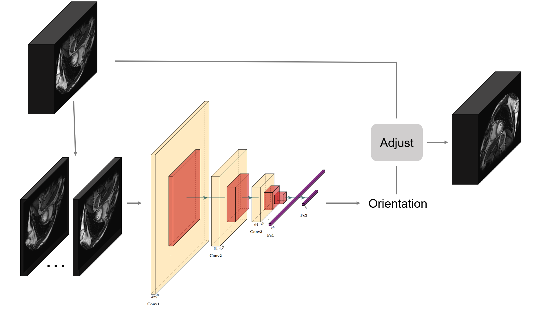
This study aims to design a DNN-based approach for achieving orientation recognition and standardization across multiple CMR modalities. Figure 1 illustrates the pipeline of our proposed method. The key contributions of this work are summarized as follows:
-
(1)
We propose a scheme to standardize the CMR image orientations and categorize them for classification purposes.
-
(2)
We present a DNN-based orientation recognition method tailored for CMR images and demonstrate its transferability to other modalities.
-
(3)
We develop a CMR image orientation adjustment tool embedded with a orientation recognition network. This tool greatly facilitates CMR image orientation recognition and standardization in clinical and medical image processing practices.
2 Method
In this section, we introduce our proposed method for orientation recognition and standardization. Our proposed framework is built on the categorization of CMR image orientations. We propose a DNN to recognize the orientation of CMR images and embed it into the CMR orientation adjust tool.
2.1 CMR Image Orientation Categorization
Due to different data sources and scanning habits, the orientation of different cardiac magnetic resonance images may be different, and the orientation vector corresponding to the image itself may not correspond correctly. This may cause problems in tasks such as image segmentation or registration. Taking a 2D image as an example, we set the orientation of an image as the initial image and set the four corners of the image to , then the orientation of the 2D MR image may have the following 8 variations, which is listed in Table 1. For each image-label pair , we can flip towards a picked orientation to get a new image-label pair. If we correctly recognize the orientation of an image, we can perform the reverse flip to standardize it.
| Label | Operation | Image | Correspondence of coordinates |
|---|---|---|---|
| 0 | Initial state | ||
| 1 | Horizontal flip | ||
| 2 | Vertical flip | ||
| 3 | Rotate clockwise | ||
| 4 | Flip along the main diagonal | ||
| 5 | Rotate clockwise | ||
| 6 | Rotate clockwise | ||
| 7 | Flip along the secondary diagonal |
2.2 Deep Neural Network
We employ a classical convolutional neural network for orientation recognition. It is a widely adopted approach in image classification tasks, adhering to the standard design pattern for CNNs. The neural network architecture comprises 3 convolutional blocks, each housing a convolutional layer, batch normalization, ReLU activation, and max pooling. These blocks effectively capture features from the input images. Additionally, an average pooling layer and 2 fully connected layers, with 8 units in the output layer, complete the network, enabling orientation prediction.
To train the model effectively, we utilize the cross-entropy loss, which efficiently measures the discrepancy between the predicted orientation and the ground truth orientation label.
2.3 Transfer Learning
When adapting the proposed orientation recognition network to new datasets of different modalities, we employ a transfer learning approach to obtain the transferred model. Initially, we freeze the weights of the convolutional layers and fine-tune the fully connected layers. We repeat this process for the subsequent fine-tuning steps until the model converges. Afterwards, we unfreeze the weights of all layers and proceed to fine-tune the entire model.
3 Experiment
3.1 Dataset
We experiment with the MyoPS dataset [1,2], which provides the three-sequence CMR (LGE, T2, and bSSFP) from the 45 patients. We divide the CMR data of 45 patients into training and validation sets at the ratio of 80% and 20%.
3.2 Data Pre-processing
For each CMR data, we initially apply 7 transformations according to Table 1 to ensure the dataset encompasses all 8 orientations. Subsequently, we slice the 3D CMR data into multiple 2D data instances.
Given an image-label pair , for each , we identify the maximum gray value, denoted as . Subsequently, three truncation operations are performed on using thresholds of and to generate and , respectively. In this operation, pixels with gray values higher than the threshold are mapped to the threshold gray value. The utilization of different thresholds allows us to capture image characteristics under various gray value window widths, mitigating the impact of extreme gray values. Additionally, grayscale histogram equalization is applied to and , resulting in and . Finally, we concatenate these three 2D images into a 3-channel image , which serves as the input to our proposed DNN.
We perform data augmentation by randomly rotating the image slightly and applying random crops and resizing. These approaches introduce variability in the orientation of the images, which aids in improving model generalization and enhances robustness to varying image sizes and aspect ratios.
3.3 Results
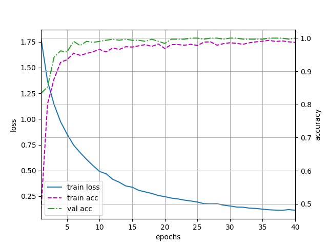
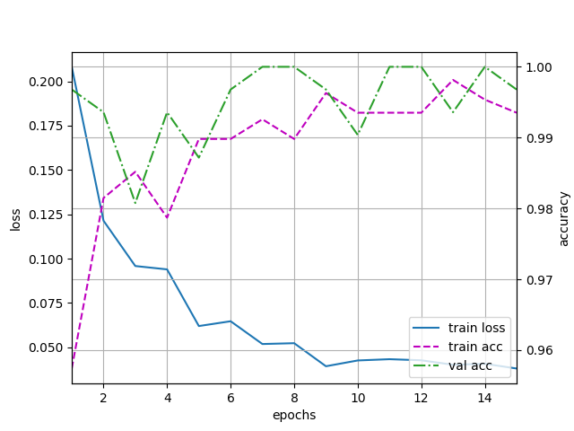
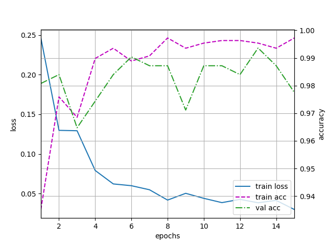
We initially train the model on the bSSFP modality and subsequently fine-tune it on the T2 and LGE modalities. The training process is depicted in Figure 2. The average accuracy on the dataset is presented in Table 2. The results highlight the model’s ability to transfer learning to other modalities, showcasing a remarkable level of accuracy.
| Modality | Train accuracy | Validation accuracy |
|---|---|---|
| bSSFP | 99.4% | 100.0% |
| LGE | 99.5% | 99.4% |
| T2 | 99.5% | 100.0% |
4 Conclusion
We have introduced a DNN-based method for multi-sequence MRI images. The experimental results validate the effectiveness of the orientation recognition network in accurately classifying the orientation of multi-sequence CMR images. Our future research will focus on expanding the categorization of the CMR image orientation and refining the classification network to further enhance the classification accuracy.
References
[1] Xiahai Zhuang: Multivariate mixture model for myocardial segmentation combining multi-source images. IEEE Transactions on Pattern Analysis and Machine Intelligence 41(12), 2933–2946, 2019
[2] Junyi Qiu, Lei Li, Sihan Wang, Ke Zhang, Yinyin Chen, Shan Yang, Xiahai Zhuang. MyoPS-Net: Myocardial Pathology Segmentation with Flexible Combination of Multi-Sequence CMR Images. Medical Image Analysis 84, 102694, 2023
[3] Ke Zhang and Xiahai Zhuang: Recognition and Standardization of Cardiac MRI Orientation via Multi-tasking Learning and Deep Neural Networks. MyoPS 2020, LNCS 12554, 167–176, Springer Nature, 2020 https://github.com/BWGZK/Orientation-Adjust-Tool
[4] Zhuang X, Li L, Payer C, et al. Evaluation of algorithms for multi-modality whole heart segmentation: an open-access grand challenge[J]. Medical image analysis, 2019, 58: 101537.
[5] Wolterink J M, van Hamersvelt R W, Viergever M A, et al. Coronary artery centerline extraction in cardiac CT angiography using a CNN-based orientation classifier[J]. Medical image analysis, 2019, 51: 46-60.