Ensemble Learning with Residual Transformer for Brain Tumor Segmentation
Abstract
Brain tumor segmentation is an active research area due to the difficulty in delineating highly complex shaped and textured tumors as well as the failure of the commonly used U-Net architectures. The combination of different neural architectures is among the mainstream research recently, particularly the combination of U-Net with Transformers because of their innate attention mechanism and pixel-wise labeling. Different from previous efforts, this paper proposes a novel network architecture that integrates Transformers into a self-adaptive U-Net to draw out 3D volumetric contexts with reasonable computational costs. We further add a residual connection to prevent degradation in information flow and explore ensemble methods, as the evaluated models have edges on different cases and sub-regions. On the BraTS 2021 dataset (3D), our model achieves 87.6% mean Dice score and outperforms the state-of-the-art methods, demonstrating the potential for combining multiple architectures to optimize brain tumor segmentation.
Index Terms— Transformer, U-Net, Brain tumor segmentation, ensemble learning
1 Introduction
Brain tumor segmentation has been a challenging task in medical imaging research and plays a crucial role in diagnosis, prognosis, and determining treatment strategies. Magnetic resonance imaging (MRI) is the imaging modality of choice for brain tumor clinics due to its advantages [1]: no radiation, better resolution of soft tissues compared with computed tomography (CT), and various imaging contrasts that provide rich diagnostic information via varying characteristics of the tissues highlighted by multi-modality MRI. For example, the spatial extent and volume of solid or active tumor regions, edema, and necrotic regions are important diagnostic and prognostic markers, and they can be measured by MRI. While tumor tissues may be readily detectable or visible at most times, accurate and reproducible auto-segmentation and characterization are still active research fields. This paper presents yet another deep learning-based segmentation method for brain tumor analysis using multi-modal MRIs with considerable advantages to the current state-of-the-art methods such as improving (1) the accuracy of segmentation results for complex shaped and textured tumors, (2) the robustness of the models and (3) the efficiency of segmentation systems (i.e., reducing the training time and computational burden).
The dataset used in this paper comes from Multi-modal Brain Tumor Segmentation Challenge (BraTS) 2021 [2, 3, 4], which includes four MRI modalities: T1-weighted (T1), T2-weighted (T2), fluid-attenuated inversion recovery (FLAIR), and contrast-enhanced T1-weighted (T1ce) MRI volumes. The segmentation task is defined as accurately generating contours of brain tumor sub-regions.
Previous works. The U-Net [5] architecture and its numerous modifications [6, 7, 8, 9] have achieved the state-of-the-art results in medical image segmentation. Skip connections between the encoder and decoder enable the network to capture details from low-level layers for accurate localization of object of interest. However, due to limited kernel size, long-range dependency is often lost through this process. This is rather important in biomedical segmentation tasks because it is difficult to capture more complex relations in some tumors with complex shapes and texture.
To address this issue, many efforts of introducing the self-attention mechanism have recently been made [10]. For instance, TransUNet [11] takes image patches from U-Net and provides evidence that Transformers have the potential in making encoders stronger. U-Net Transformer [12] and SwinUNet [13] both apply the transformer blocks during the low-level encoding-decoding process to model long-range dependencies. These models have shown the ability to extract global context; therefore, leading to a better performance on medical image segmentation. Yet, the transformer brings a heavy computational and memory burden.
Despite the benefits of Transformers, the complexity required by its mechanism can largely impede the feasibility of its application on 3D datasets. The aforementioned approaches are applied mainly on 2D medical images [14], which leaves room for the further explorations of Transformers. However, for 3D images, since the transformer requires quadratic complexity calculations, the one more dimension of the dataset would increase the computation so dramatically that it cannot be naively integrated into the network architecture. One potential solution is to introduce a linear transformer for segmentation, however, previous research also shows there is a performance drop when making the transformer linear [15].
To this end, we propose a novel architecture by integrating Transformers into the nnU-Net architecture solely in the bottleneck layer of feature extraction so as to alleviate the heavy computation issue on 3D datasets. Our models are expected to combine the benefits of the self-attention mechanism and nnU-Net, which are to learn long-range dependencies, to reserve the details from low-level layers in the decoding process, and to auto-generate network configurations. To avoid hierarchical complexity brought by additional layers [16], we also add residual connection on the Transformer block to further stabilize the model performance.
Our contributions can be summarized as:
-
•
We design an effective architecture combining the Transformer algorithm with a self-adaptive U-Net and include residual connections on the Transformer to avoid possible degradation in information flow.
-
•
Further, we manage to apply our algorithm in 3D volumetric space instead of the conventional slice-by-slice approach.
-
•
We demonstrate a great potential in ensemble learning of the proposed architectures for further enhancement in the segmentation performance.
-
•
The proposed design shows improved robustness against poor-quality images, and the efficacy for brain tumor segmentation on 3D datasets and outperforms the state-of-the-art approaches in this field.
2 Methodology
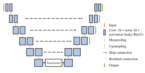
This study is based on nnU-Net, an extension of U-Net that automatically generates network configurations based on the dataset characteristics [6]. In our implementation, the parameters, including target spacing, patch size, batch size, downsampling strides, and convolutional kernel sizes, are self-adaptive and adjusted based on the specific dataset. To capture long-range contextual interactions, we introduce a self-attention mechanism by integrating a transformer block into the deepest layer of the encoder, where the dimensions of the input sequences are reduced to a size that can be easily handled by the transformer. We also include a residual connection to prevent information degradation. Figure 1 shows the architecture of our proposed model, illustrating the flow of information through the network, and highlighting the locations of the transformer block and residual connection.
2.1 U-Net architecture with transformer
Down-sampling. The input to our model is a 3D image with dimensions of 240 x 240 x 155 (height x width x depth), composed of four distinct channels derived from various MRI contrasts. Following pre-processing (i.e., inhomogeneity correction, denoising, and intensity standardization), the images are conveyed to the encoder for down-sampling. Each layer of the encoder comprises two consecutive blocks of stacked convolutional layers, incorporating 3D convolution, dropout, 3D instance normalization, and leaky ReLU activation in a sequential manner. The encoder extracts features from image patches that are subsequently utilized as input sequences for the Transformer block.
Transformer block. The Transformer block acts as the final component of the encoder, performing a sequence-to-sequence operation. It takes in non-overlapping encoded patches and transforms them into vectors. It repeats the self-attention layer times for multi-head attention. The self-attention [17] mechanism can be written as:
| (1) |
where the manipulation of queries (Q), keys (K), and values (V) requires O(N²) computational complexity. After being encoded down to the bottleneck layer, the sequence dimension could be reduced dramatically. Thus, on a 3D dataset, the transformer block placed in the last layer can alleviate the computational burden and can make it feasible to include self-attention in the overall network. The Transformer later outputs encoded image patches into the decoder, which reshapes them back to the original input size.
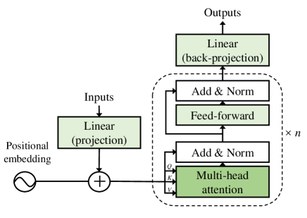
Residual connection. To avoid a fracture in information from previous layers and the difficulty in training complex models, we add a residual connection to the Transformer block. This allows the information to be propagated directly to the designated last layer. By doing so, we seek to ensure the integrity of the information flow while mitigating potential issues such as gradient explosion. We compare this model to one without a residual connection.
Up-sampling. During the up-sampling process, the decoder utilizes transposed convolutions to expand the image size and move up to low-level layers until the size matches the original input image. At each layer, the decoder concatenates input features with image features from the corresponding encoder level through a skip connection. It preserves crucial details in the final segmentation map. To further prevent information loss in the decoding process, we incorporate deep supervision. Overall, this up-sampling strategy enhances the accuracy and robustness of the proposed architecture.
2.2 Training and testing
We evaluate all our models on the BraTS2021 dataset. This dataset contains fully-annotated, multi-institutional, multi-parametric brain tumor MRI, covering diverse degrees of gliomas. There are 1,470 samples in the dataset, each of which has four MRI modalities: T1, T2, FLAIR, and T1ce. We split them into the training and validation set of 1,251 samples, and the test set of 219 samples. We use 5-fold cross-validation and optimization to train our models, where the loss function is defined as the sum of Dice loss and cross-entropy loss. Moreover, to assess the performance of our models, we conduct a blind-testing on the unseen validation dataset through an online evaluation provided by the BraTS challenge organizers.
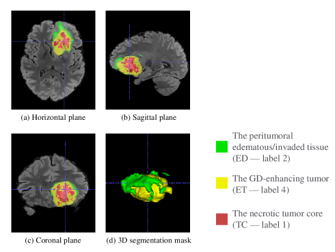
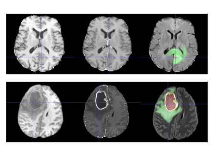
Figure 3 illustrates an example annotation of a single subject’s FLAIR image with four segmentation labels: peritumoral edema (ED), GD-enhancing tumor (ET), necrotic tumor core (TC), and background. A whole tumor (WT) refers to the complete extent of the disease, entailing TC and ED. When comparing T1ce to T1 images, ET shows hyper-intensity while TC shows hypo-intensity, as can be observed in Figure 4. Generally, all four labels are present in the expert manual annotation following a certain topological relationship, where ED is on the outside and TC is the innermost, but it varies from patient to patient. Some samples only have one or two labels (when there is no ET or TC to be found) due to different tumor development stages, which may affect the segmentation performance of certain models. This will be further discussed later.
3 Results
3.1 Single model performance
We evaluate the performance of four different models on the BraTS2021 dataset. The models include nnU-Net as the baseline, E1D3 [18] as a comparison to the state-of-the-art, and our proposed models transformer-nested nnU-Net (UNet+T) and its variation with residual connection (UNet+T+R). E1D3 is a variation of the U-Net that uses one encoder and three decoders for separate sub-regions of the brain tumor. This architecture may give E1D3 some advantages on certain samples that require examining sub-regions independently, therefore making it a good candidate for the later ensemble.
The evaluation metrics used in this study were the Dice coefficient score and Hausdorff Distance (HD). Table 1 provides the evaluation results on sub-region performance for all four models. The results show that UNet+T has a slightly better Dice score on ET and TC, and a better HD score on WT. UNet+T+R demonstrates significant improvement in the prediction of ET and better HD scores on ET and TC. As a result, our models demonstrate the best overall performance as evaluated by both mean Dice and mean HD.
Furthermore, nnU-Net is observed to be robust to poor image quality and displacement noise, as it gives the highest Dice score for WT. We also observe that E1D3 performs better on the ET region for many selected samples that contains less than or equal to two labels in the segmentation map, excluding the background. This observation inspires us to ensemble the results generated by different models to combine their advantages on sub-region segmentation.
3.2 Ensemble models
Table 2 presents the results of four different ensemble strategies. The first three methods involves performing pixel-wise operations on the segmentation maps from the four models, using mode, mean, and median, respectively. We should note that the weight of each model is not equal, as we double the weight of the UNet+T model based on its better overall performance and empirical evidence. For the threshold ensemble, we set a threshold of TC volume smaller than 60 and ET volume larger than 60 to automatically select subjects to be predicted by the E1D3 model, and the remaining subjects are predicted using the UNet+T model. This method is based on the observation that E1D3 performs better for subjects meeting these criteria, which may suggest fewer than four labels in the ground truth. Therefore, this method is only an ensemble of two models: E1D3 and UNet+T. The threshold method shows the best overall performance, both in terms of Dice score and HD, suggesting that this criterion helps improve our models.
Methods Dice (%) HD (mm) ET TC WT Mean ET TC WT Mean nnU-Net 80.5 87.5 92.7 86.9 24.66 7.62 3.60 11.96 E1D3 81.6 80.6 91.5 84.6 18.46 17.65 4.73 13.61 UNet+T 82.0 87.8 92.6 87.5 18.13 9.27 3.49 10.30 UNet+T+R 82.2 87.7 92.5 87.5 16.24 7.53 3.73 9.17
Methods Dice (%) HD (mm) ET TC WT Mean ET TC WT Mean Mode 81.6 87.6 92.6 87.3 19.72 9.26 3.61 10.86 Average 82.4 80.6 92.2 85.1 12.87 16.63 5.27 11.59 Median 82.0 87.7 92.6 87.4 18.00 9.20 3.61 10.27 Threshold 82.0 88.3 92.6 87.6 18.13 7.56 3.57 9.75
We also manually pick the best model to apply for each case, but the results are not compared in the table because it is not an automatic process. However, it has achieved the highest Dice (ET 84.0%, TC 88.3%, WT 92.8%) and lowest HD (ET 11.17mm, TC 7.51mm, WT 3.40mm) across all sub-regions, as well as the mean Dice (88.4%) and mean HD (6.82mm). It indicates that the four architectures evaluated in this paper have the potential to improve combined accuracy significantly if a more complex ensemble method were specified. All these ensemble methods have improved accuracy on selected sub-regions, and the threshold method gives the best overall Dice score and HD among automatic methods.
4 Discussion
We present a residual Transformer integrated nnU-Net architecture for brain tumor segmentation. Simple yet effective, both models with Transformer nested demonstrated impressive performance on 3D multi-modality datasets and have surpassed the state-of-the-art algorithms, including nnU-Net itself. We also explore ensemble models in an effort to combine their advantages on sub-regions and different types of subjects. Although the ensemble methods are not complex and the threshold is empirical, we have still observed notable improvements in segmentation performance.
We observe that the performance of the model is affected by various aspects of the subject, such as the number of sub-regions, the percentage of each class, the clarity of the boundary, and other unidentified factors. To test this hypothesis, we manually pick the best-performing model for each subject, and it achieves the best Dice scores among all models across metrics. This observation suggests that human-in-the-loop decision-making, as is common in current clinical practice, can further push the boundary of current segmentation performance.
In the future, we plan to investigate better ensemble methods and select appropriate models for combination. The potential demonstrated in our results is convincing and has the potential to be applied to other medical imaging tasks. Overall, our work offers a promising ensemble approach to brain tumor segmentation, opening up new possibilities for the integration of Transformers in medical image analysis.
References
- [1] Jessica M Winfield, Geoffrey S Payne, Alex Weller, and Nandita M deSouza, “Dce-mri, dw-mri, and mrs in cancer: challenges and advantages of implementing qualitative and quantitative multi-parametric imaging in the clinic,” Topics in Magnetic Resonance Imaging, vol. 25, no. 5, pp. 245, 2016.
- [2] Ujjwal Baid, Satyam Ghodasara, Suyash Mohan, Michel Bilello, Evan Calabrese, Errol Colak, Keyvan Farahani, Jayashree Kalpathy-Cramer, Felipe C Kitamura, Sarthak Pati, et al., “The rsna-asnr-miccai brats 2021 benchmark on brain tumor segmentation and radiogenomic classification,” arXiv preprint arXiv:2107.02314, 2021.
- [3] Bjoern H Menze, Andras Jakab, Stefan Bauer, Jayashree Kalpathy-Cramer, Keyvan Farahani, Justin Kirby, Yuliya Burren, Nicole Porz, Johannes Slotboom, Roland Wiest, et al., “The multimodal brain tumor image segmentation benchmark (brats),” IEEE transactions on medical imaging, vol. 34, no. 10, pp. 1993–2024, 2014.
- [4] Spyridon Bakas, Hamed Akbari, Aristeidis Sotiras, Michel Bilello, Martin Rozycki, Justin S Kirby, John B Freymann, Keyvan Farahani, and Christos Davatzikos, “Advancing the cancer genome atlas glioma mri collections with expert segmentation labels and radiomic features,” Scientific data, vol. 4, no. 1, pp. 1–13, 2017.
- [5] Olaf Ronneberger, Philipp Fischer, and Thomas Brox, “U-net: Convolutional networks for biomedical image segmentation,” in International Conference on Medical image computing and computer-assisted intervention. Springer, 2015, pp. 234–241.
- [6] Fabian Isensee, Paul F Jaeger, Simon AA Kohl, Jens Petersen, and Klaus H Maier-Hein, “nnu-net: a self-configuring method for deep learning-based biomedical image segmentation,” Nature methods, vol. 18, no. 2, pp. 203–211, 2021.
- [7] Wei Chen, Boqiang Liu, Suting Peng, Jiawei Sun, and Xu Qiao, “S3d-unet: separable 3d u-net for brain tumor segmentation,” in International MICCAI Brainlesion Workshop. Springer, 2018, pp. 358–368.
- [8] Ozan Oktay, Jo Schlemper, Loic Le Folgoc, Matthew Lee, Mattias Heinrich, Kazunari Misawa, Kensaku Mori, Steven McDonagh, Nils Y Hammerla, Bernhard Kainz, et al., “Attention u-net: Learning where to look for the pancreas,” arXiv preprint arXiv:1804.03999, 2018.
- [9] Huan Minh Luu and Sung-Hong Park, “Extending nn-unet for brain tumor segmentation,” in International MICCAI Brainlesion Workshop. Springer, 2022, pp. 173–186.
- [10] Jeya Maria Jose Valanarasu, Poojan Oza, Ilker Hacihaliloglu, and Vishal M Patel, “Medical transformer: Gated axial-attention for medical image segmentation,” in International Conference on Medical Image Computing and Computer-Assisted Intervention. Springer, 2021, pp. 36–46.
- [11] Jieneng Chen, Yongyi Lu, Qihang Yu, Xiangde Luo, Ehsan Adeli, Yan Wang, Le Lu, Alan L Yuille, and Yuyin Zhou, “Transunet: Transformers make strong encoders for medical image segmentation,” arXiv preprint arXiv:2102.04306, 2021.
- [12] Olivier Petit, Nicolas Thome, Clement Rambour, Loic Themyr, Toby Collins, and Luc Soler, “U-net transformer: Self and cross attention for medical image segmentation,” in International Workshop on Machine Learning in Medical Imaging. Springer, 2021, pp. 267–276.
- [13] Hu Cao, Yueyue Wang, Joy Chen, Dongsheng Jiang, Xiaopeng Zhang, Qi Tian, and Manning Wang, “Swin-unet: Unet-like pure transformer for medical image segmentation,” arXiv preprint arXiv:2105.05537, 2021.
- [14] Yao Zhang, Nanjun He, Jiawei Yang, Yuexiang Li, Dong Wei, Yawen Huang, Yang Zhang, Zhiqiang He, and Yefeng Zheng, “mmformer: Multimodal medical transformer for incomplete multimodal learning of brain tumor segmentation,” arXiv preprint arXiv:2206.02425, 2022.
- [15] Zheyuan Zhang and Ulas Bagci, “Dynamic linear transformer for 3d biomedical image segmentation,” arXiv preprint arXiv:2206.00771, 2022.
- [16] Kaiming He, Xiangyu Zhang, Shaoqing Ren, and Jian Sun, “Deep residual learning for image recognition,” in Proceedings of the IEEE conference on computer vision and pattern recognition, 2016, pp. 770–778.
- [17] Ashish Vaswani, Noam Shazeer, Niki Parmar, Jakob Uszkoreit, Llion Jones, Aidan N Gomez, Lukasz Kaiser, and Illia Polosukhin, “Attention is all you need,” Advances in neural information processing systems, vol. 30, 2017.
- [18] Syed Talha Bukhari and Hassan Mohy-ud Din, “Ed u-net for brain tumor segmentation: Submission to the rsna-asnr-miccai brats 2021 challenge,” in International MICCAI Brainlesion Workshop. Springer, 2022, pp. 276–288.