eXplainable Artificial Intelligence (XAI) in aging clock models
kalyakulina.alena@gmail.com
\And
yusipov.igor@gmail.com
\And
amoskalev@list.ru
\And
claudio.franceschi@unibo.it
\And
ivanchenko.mv@gmail.com
Abstract
eXplainable Artificial Intelligence (XAI) is a rapidly progressing field of machine learning, aiming to unravel the predictions of complex models. XAI is especially required in sensitive applications, e.g. in health care, when diagnosis, recommendations and treatment choices might rely on the decisions made by artificial intelligence systems. AI approaches have become widely used in aging research as well, in particular, in developing biological clock models and identifying biomarkers of aging and age-related diseases. However, the potential of XAI here awaits to be fully appreciated.
We discuss the application of XAI for developing the "aging clocks" and present a comprehensive analysis of the literature categorized by the focus on particular physiological systems.
Keywords explainable artificial intelligence aging longevity age-related diseases machine learning
1 Introduction
Machine learning (ML), and deep learning (DL) in particular, is currently one of the most common data analysis approaches in applications. Deep models handle large amounts of input data, training many layers, but in most cases, their functioning is not transparent. In this regard they are often called black boxes (Saleem et al., 2022). Decision-making process in such deep architectures is difficult to explain, raising concerns about the trustworthiness of such models and the security of their deployment. The problem of explainability of artificial intelligence (AI) models has received much attention (Baehrens et al., 2010; Lipton, 2018; Samek et al., 2017; Simonyan et al., 2014), and made eXplainable Artificial Intelligence (XAI) an important area of AI (Nauta et al., 2023). Major goals of XAI are to develop approaches capable of uncovering the grounds behind model decision-making, and, more profoundly, to develop interpretable and logically explainable models. XAI explanations must be understandable, reliable, whereas the explained models must retain predictive accuracy (Saleem et al., 2022). Commonly, one distinguishes global and local explainability. Global interpretation allows one to ‘open’ the black box of AI models by explaining the predictions of the model as a whole. Local interpretation explains the model’s decision for each particular sample. These two types of explainability represent two sides of the same coin: global explainability allows one to establish general patterns, while local explainability tracks these patterns at the individual level. The lack of explainability significantly limits the use and deployment of models, especially in sensitive applications, where human life and health may depend on ML decision.
In medical applications AI based clinical decision support systems aim to assist clinicians in diagnosing diseases and making treatment decisions (Amann et al., 2020). One of the most important requirements for such systems is clinical validation and the ability to verify model decisions. Biomedical data is subject to various errors (recording and processing errors, natural noise, and others), so AI systems will inevitably make errors as well. Another source of errors may be systemic problems caused, in particular, by limited train subset. In this case, AI models may make errors because an individual sample may significantly differ from the set on which the model was trained (Amann et al., 2020). In any case, explainability is extremely important; it allows one to track different types of errors, identify their causes, and adjust the behavior of the system. Explainability can also offer a personalized approach based on individual patient characteristics and risk factors. XAI approaches can give an explanation in natural language or visualize how different factors affected the final outcome (risk score, diagnosis, or proposed treatment), which can increase patient awareness with proper use (Politi et al., 2013; Stacey et al., 2017), and allow clinicians to make confident clinical decisions, adapting predictions and recommendations to individual circumstances if necessary (Beil et al., 2019).
A central problem of geroscience is identifying biomarkers of aging, which has benefited much from the growth of ML and DL research (Moskalev et al., 2023; Zhavoronkov et al., 2019). Considerable attention here is devoted to estimating individual biological age, which would characterize personal health status in various aspects, being, in general, different from chronological age. This biologically meaningful characteristic is associated with risk of mortality, disease, and general well-being, and even though each individual feature employed in one or another mathematical estimator of biological age may not be explicitly related to age, a combination of features are known to have predictive power (Zhavoronkov and Mamoshina, 2019). Various types of data may be used to estimate age, including laboratory tests, magnetic resonance (MRI) and X-ray images, electrocardiogram (ECG) and electroencephalogram (EEG) signals. Development and analysis of age estimators can help, in particular, in the study of age-related diseases (Zhavoronkov and Cantor, 2011), immunological aging, response to medications and vaccines (Zhavoronkov and Mamoshina, 2019) and in many other healthcare applications. The recent promises on the possibility of extending life expectancy (Partridge et al., 2018) demand reliable models that estimate biological age and the aging rate to validate life extension techniques (Rutledge et al., 2022). Age estimation models are usually called “biological” or “aging clocks”. One of the first fundamental works in this field are the epigenetic clocks from Hannum (Hannum et al., 2013) and Horvath (Horvath, 2013), which estimates age using a linear model based on DNA methylation data. By now a plethora of models have been proposed employing different input data and implementing AI techniques (Mamoshina and Zhavoronkov, 2019; Zhavoronkov and Mamoshina, 2019). There are several challenges in overcoming which XAI can be helpful. In particular, it is not always possible to separate the chronological and biological aging components in age estimation models (Bell et al., 2019). Here, the global aspect of XAI approaches can be used to identify hallmarks for particular age stages in healthy individuals that are part of the chronological aging component, as well as the effects of the environment, medical history, and many other factors that contribute to variations in the biological age component. The XAI can also help with identifying the set of biomarkers that are most representative of an individual’s health status, as well as discarding redundant ones.
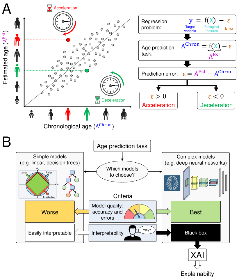
The considered points are summarized in Figure 1. Age estimation is commonly a regression problem that can take various biological features as an input. Models seek to predict a person’s chronological age with as small an error as possible. Commonly, one uses the model’s output as an estimate of the biological age (ideal estimation of person’s health status), and the mismatch between chronological age and biological age is usually treated as age acceleration (if biologically the person is older than chronologically) or age deceleration (if biologically the person is younger than chronologically), as shown in Figure 1A. It has to be kept in mind that the difference can be due to many reasons, like imperfection of the model, heterogeneity between individuals (not related to biological aging), actual difference between biological and chronological age and others. The problem of age estimation inevitably raises the question of choosing a suitable model. Among the many criteria that influence the final choice, there are two main ones: model performance in terms of predictive accuracy and interpretability (Figure 1B). Classical, simple models (e.g., linear, treelike ones) are usually easy to interpret, but they may not take into account the complex relationships between the input features, thereby showing worse results. More complex state-of-the-art models (like deep neural network (DNN) architectures) often give better results, but they are black boxes, which do not allow for explaining the principles by which models make decisions. This is where XAI approaches come in.
The primary aim of this review is to provide the current state-of-the art of XAI methods applied to human biological age estimation. It has the following structure. Section 2 describes the age estimation problem and XAI methods in the context of ML approaches, and gives an overview of XAI methods and their application to different types of data. Section 3 presents a comprehensive review of studies that use XAI methods to explain the results of age estimation models, grouped by the assessed body systems and the types of input data used. Section 4 concludes the work, summarizing the achievements of XAI in the field of age-associated changes in the human body.
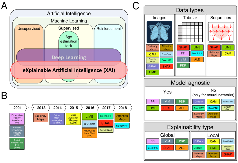
2 Artificial Intelligence, data types and methods
AI is an extremely broad field of research and development, finding its application in almost all spheres of modern life and helping to solve many complex problems. AI makes it possible to create data-driven decision-making systems (Ali et al., 2023). ML models are a broad class of AI approaches, combining both classical models and more complex DNNs. The main types of ML are supervised learning, unsupervised learning, and reinforcement learning. Depending on the type of the predicted value in supervised learning, there are classification tasks (class is predicted) and regression tasks (continuous value is predicted). Biological age estimation is usually done as a chronological age regression task (age - continuous value - is predicted) on different biomedical data, cf. Figure 2A. The formulation as an age range classification task is much less common. Biological age can be estimated using both simple methods and complex neural network architectures. At the same time, an increasing amount of data requires more and more advanced methods to be able to detect nonlinear relationships between input parameters and their impact on the final result of the biological age estimation. Most neural network architectures are ‘black boxes’, the models with unclear routes to decisions and unpredictable consequences to which changes in input data may lead. This is where XAI methods can help.
The timeline of the development of XAI methods mentioned in this review is shown in Figure 2B. It started with the permutation feature importance (PFI), variable importance measure (VIM) (Breiman, 2001), and partial dependence plot (PDP) (Friedman, 2001) proposed in 2001. PFI, one of the types of VIMs, is used for tabular data and represents a reduction in the model estimate when a single feature value is randomly shuffled. These metrics were originally proposed for tree models, but can also be used for other methods. PDPs are used to analyze and visualize the interaction between the set of interesting input features and the final model estimate. One of the first approaches developed for input image data is saliency maps (Simonyan et al., 2014), which are still widely used and have been adapted for sequence input data as well. Saliency maps show which regions of the input images were used by the AI model to make decisions and can provide a visual representation of how regions important to the model fit with human attitudes. Guided Backpropagation (Springenberg et al., 2015) builds on the ideas of Deconvolution (Zeiler and Fergus, 2013) and Saliency (Simonyan et al., 2014), solving the problem of negative gradient flux and minimizing the noise they cause. This approach is also used for input image data. Another well-known method for images and sequences is Class Activation Mapping (CAM) (Zhou et al., 2016). CAM usually uses a global average pooling layer after the convolutional layers and before the final fully connected layer. Later, a Grad-CAM (Gradient-weighted Class Activation Mapping) modification was proposed, which uses a gradient approach to generate CAM (Selvaraju et al., 2020). Accumulated local effects (ALE) (Apley and Zhu, 2020) are similar to the concept of PDPs in that they both aim to describe how functions on average affect model predictions. ALE eliminates the bias that occurs in PDPs when the feature of interest is highly correlated with other features. LIME (Local Interpretable Model-agnostic Explanations) provides locally accurate explanations in the neighborhood of the explained instance (Ribeiro et al., 2016). After obtaining a surrogate dataset, it weighs each row according to how close they are to the original sample and uses a feature selection method, such as Lasso, to obtain the most important features. As the name implies, this approach can be applied to any model and provides only local explainability. DeepLIFT (Deep Learning Important FeaTures) is a method of decomposing the output prediction of a neural network to a particular input by back propagating the contributions of all neurons in the network to each input feature, comparing the activation of each neuron to its ‘reference activation’ (Shrikumar et al., 2017). It is applied only to neural networks. One of the best known and most widely used XAI methods is SHAP (Shapley Additive exPlanations), a game-theoretic approach to explaining the results of any ML model. It relates optimal credit allocation to local explanations using classical Shapley values from game theory and related extensions (Lundberg and Lee, 2017). SHAP is applicable to almost any model, input data type, and is used for both global and local explainability. SmoothGrad (Smilkov et al., 2017) adds Gaussian noise to the input data and calculates the average of all samples to reduce the importance of less frequent features. This approach is also specific to neural networks and is typically used for image input data. One of the most recent approaches for explaining the results of convolutional neural networks (CNNs) with input image data is attention maps (Jetley et al., 2018). This approach generates intermediate representations of the input image at different stages of the CNN pipeline and outputs a two-dimensional matrix of scores for each map. As for saliency maps, CAM and Grad-CAM, this approach allows for comparing the representation of the neural network and the human in terms of important regions of the image highlighted for decision making. DeepPINK (Deep feature selection using Paired-Input Nonlinear Knockoffs) (Lu et al., 2018) offers a special pairwise-connected layer for the neural network to encourage competition between each original feature and its knockoff counterpart. DeepPINK offers an algorithm-independent measure of feature importance with more power than the naïve combination of the knockoffs idea with a vanilla multilayer perceptron.
ML models can be built on different types of data that they take as an input. Among the most common data types are images, tables, and sequences (like signals or texts). Images and sequences are examples of unstructured data, while for tabular data the features are already extracted. For different types of data, various types of methods dominate: for images and sequences, CNNs are the most common (Hershey et al., 2017; Sultana et al., 2018), for tabular data gradient-boosted decision trees (GBDTs) has long shown the best results, but now specialized neural network approaches that adapt techniques from other fields are also actively developing (Borisov et al., 2022; Grinsztajn et al., 2022; Shwartz-Ziv and Armon, 2022). XAI methods are also mostly specific to different types of data - some methods are applied to images and sequences, others are applied to tabular data (Figure 2C). But approaches like SHAP and LIME can be adapted to almost any type of input data and different models, and are very common in a wide range of applications. Different maps are usually used for images - saliency maps, CAM, attention maps and their modifications, as well as methods based on backpropagation. Classical approaches such as PFI, PDP and their modifications, as well as DeepPINK, are used for tabular data.
XAI methods are divided into model-agnostic, which can be applied to any type of ML models, and model-specific, which can be applied only to a certain class of models (usually to neural network architectures). Among model-agnostic methods there are such classical ones as PFI, VIM, PDP, ALE, as well as more modern SHAP and LIME. The other considered methods are specific for neural networks (usually convolutional ones). Another type of classification of XAI approaches includes global and local explainability. Global explainability attempts to interpret the behavior of the model as a whole, revealing general patterns. Local explainability attempts to obtain an interpretation for each individual sample and to identify the features that affect the decision in each case. The attribution of methods to different groups is shown in Figure 2C.
3 XAI in age prediction studies
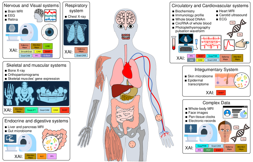
Among a wealth of age estimation models, some already implement XAI approaches to identify the most important features that contribute to the result. The models may address specific body systems or characterize an organism as a whole (Figure 3). Correspondingly, they take in system- or tissue-specific data, or aggregate information from multiple body systems.
3.1 Nervous and Visual Systems
3.1.1 Brain age estimation with MRI data
Brain age estimation is one of the most common tasks due to the close relationship between brain function, aging process, and neurodegenerative age-related diseases (Cole and Franke, 2017). MRI is a common noninvasive test that is very informative in assessing the functional state of the brain. Higher brain age compared to chronological age is evidence of accelerated aging and requires attention with regard to the potential progression of neurological, neurodegenerative, and psychiatric diseases (Boscolo Galazzo et al., 2022), while lower brain age compared to chronological age is interpreted as decelerated aging and corresponds to a good health status. To estimate brain age, various metrics and morphological characteristics of MRI scans or whole images can be used as input for ML models. The most advanced DL approaches demand that XAI techniques are employed in order to understand which MRI parameters affect the final model solutions and in what way, to increase the confidence in the models, required in clinical applications.
Brain aging clocks were proposed even before XAI methods became widespread. However, the need to determine the importance of certain features and to explain model predictions was articulated even at that time. In particular, (Cherubini et al., 2016) made one of the first attempts to visualize the most important MRI regions based on the analysis of individual voxels (single elements of a 3D brain image). Age estimation was performed by means of a simple linear model, where the weight coefficients for individual voxels allowed for a straightforward identification of the most important brain regions for age estimation on the 3D model.
The development of DL models led to architectures taking whole images as an input, like CNNs. Such models have also become widespread in the age estimation based on MRI images. However, the explanation here is significantly complicated by the large dimensionality and complexity of the model. Comparatively simple methods for explaining the importance of individual brain regions in age estimation are computationally costly, although they have been used quite successfully (e.g., PFI (Kolbeinsson et al., 2020)). More advanced XAI methods have also been applied, relying on various maps that highlight the most active regions in the image: attention maps, CAM, saliency maps (Feng et al., 2020; Hepp et al., 2021; Hu et al., 2021; Lam et al., 2020; Mouches et al., 2022; Ren et al., 2022; Wang et al., 2019; Yin et al., 2023), SmoothGrad (Levakov et al., 2020; Wilms et al., 2021). These approaches have also uncovered interesting relationships between aging, brain function, and neurodegenerative diseases. In particular, it allowed to identify brain regions associated with the development of dementia (Wang et al., 2019), as well as those associated with both age and Alzheimer’s disease (Lam et al., 2020). In (Levakov et al., 2020), the aggregation of explanation maps for many samples identified the association of cerebrospinal fluid (CSF) volume with age. Changes in the activation patterns of different brain regions with age were demonstrated (Feng et al., 2020). For younger participants, a smaller error was observed in (Hepp et al., 2021), and higher estimation accuracy in central brain regions was also shown. In (Hu et al., 2021) it was found that the most age-associated brain regions in children and adolescents are associated with movements, language, and processing of sensory information like vision and sense of touch (precentral gyrus, postcentral gyrus, inferior parietal lobule, middle temporal gyrus, medio ventral occipital cortex). In contrast, in the elderly, the amygdala, hippocampus, and thalamus, which are responsible for the limbic system, behavioral and emotional responses, and consolidating memories, were the most involved in age estimation (Ren et al., 2022). Sex-specific aspects of brain aging were revealed in (Yin et al., 2023).
U-Net architecture-based importance map extraction approach and related modifications (U-Noise) is less popular, but nevertheless noteworthy (Bintsi et al., 2021; Popescu et al., 2021), as well as the guided-backpropagation approach (Cho and Wallraven, 2022).
Another representation of MRI data is tabular, which characterizes morphological features: the sizes and volumes of the main structural brain regions measured from MRI images, as well as the ratios between them. As the input data in this case are in the tabular form (with numeric, ordinal, or categorical values), and all features have already been extracted, XAI approaches to explaining age estimation models based on such data are different, typically, SHAP and LIME (Ball et al., 2021; Ballester et al., 2023; Cumplido-Mayoral et al., 2023; Gómez-Ramírez et al., 2022; Han et al., 2022; Lombardi et al., 2021; Ran et al., 2022; Salih et al., 2021; Scheda and Diciotti, 2022). They also allow to make conclusions about the influence of morphological features on brain age estimation. Interestingly, a direct comparison of these methods for local explainability of brain age estimation showed little agreement between the methods: SHAP method emphasized the importance of metrics related to the precentral gyrus, inferior and lateral occipital cortex, and statistical descriptors of CSF volume, whereas LIME method valued more white matter volumes of opercular and triangular part of inferior frontal gyrus and inferior temporal gyrus (Lombardi et al., 2021). In (Ball et al., 2021) XAI was used for identifying typical changes during adulthood that appeared to reflect developmental remodeling in the cortex based on age estimation models for a cohort of children and adolescents. Similar observation was made in (Scheda and Diciotti, 2022): cortical thickness and brain complexity manifested maximum contribution to age estimation in both children and adults; like total intracranial volume and cortical thickness in (Han et al., 2022) for different models. Brain-to-intracranial-volume ratio appeared to be the most important in estimating brain age (Gómez-Ramírez et al., 2022). The brain age vector, stroked with Shapley values, has been shown to be a useful tool for the early screening of mild cognitive impairment and Parkinson’s disease (Ran et al., 2022). In (Ballester et al., 2023) the relationship between brain age and total gray matter volume in schizophrenia was revealed using SHAP. The use of different brain MRI metrics in (Cumplido-Mayoral et al., 2023) revealed the following most age-associated brain regions in both males and females: amygdala, nucleus accumbens, cerebellar white matter, lateral ventricles, and insula. Sex-specific traits were also found, for females: the thicknesses of the transverse temporal cortex, the pars triangularis, the inferior parietal cortex, and the left frontal pole, as well as the volume of the left entorhinal cortex; for males: the thicknesses of the left isthmus cingulate, the right cuneus, and the cortical volume of the superior frontal and right rostral middle regions. An unusual application of SHAP was proposed in (Sun et al., 2022): instead of morphological characteristics, voxel MRI parameters were used as input data, and SHAP highlighted important voxels, which were then used to build dynamic connectivity networks.
An original approach was proposed in (Monti et al., 2020): functional connectivity networks were constructed for different brain regions, which were then used to solve the age prediction problem. Self-explainable normalizing flows were proposed in (Wilms et al., 2021), demonstrating comparable results with CNN model and SmoothGrad XAI approach.
3.1.2 Brain age estimation with EEG data
Another common non-invasive test to evaluate brain function and activity is EEG. Changes in the brain caused by aging or age-associated diseases can affect the electrophysiological activity of the brain. Either raw EEG signals or their frequency, spectral, amplitude, and/or other characteristics serve the input for ML models.
XAI approaches in the age estimation based on EEG data are not commonly used; more often, built-in mechanisms for determining the feature importance are employed (Al Zoubi et al., 2018; Sun et al., 2019; Vandenbosch et al., 2019). Nevertheless, they are able to identify important EEG age-associated features. In particular, it was shown in (Vandenbosch et al., 2019) that low-frequency power decreases sharply from childhood to adolescence, with peak alpha frequency increasing with age and peak alpha power decreasing with age. A study of EEG during sleep (Sun et al., 2019) has shown that the duration of Non-REM (non-rapid eye movement) phases of sleep, as well as the total sleep time and the awakening time rate influence accelerated brain aging. In recent work (Khayretdinova et al., 2022), deep CNNs were used to estimate brain age, and attribution maps were used as an XAI method to identify the EEG signal elements whose activity most affects age estimation. Interestingly, the state of the eyes (open or closed), as well as the activity from the frontal electrodes (which may reflect eye movement activity) were found to be significant features.
3.1.3 Retinal age estimation
Health changes, symptoms of various diseases, age-associated conditions can be reflected in the eye and, in particular, the retina, so the retinal age estimation seems to be a promising and interesting direction. Age is a basic risk factor in the development of many eye diseases, leading to a significant decrease in visual acuity and even to vision loss. It is also worth noting that most tests that assess retinal age are non-invasive and can be conveniently used in clinical applications, for example, to track early signs of diseases (particularly cardiovascular diseases) or to estimate the rate of their development. The types of data used for the age estimation include retinal fundus photographs (Nusinovici et al., 2022; Poplin et al., 2018; Zhu et al., 2023), anterior segment morphological features (Ma et al., 2021), and macular optical coherence tomography (OCT) (Chueh et al., 2022; Shigueoka et al., 2021).
For image data in retinal age estimation, one employs CNNs and corresponding XAI methods: attention maps (Poplin et al., 2018; Zhu et al., 2023), Grad-CAM (Chueh et al., 2022; Shigueoka et al., 2021), and saliency maps (Nusinovici et al., 2022). In (Poplin et al., 2018) it was shown that vascular regions in the retina are not only associated with age, but can also indicate cardiovascular risk, while perivascular surroundings reflect changes in Haemoglobin A1c levels. Interestingly, sex differences in the ocular fundus were found to be concentrated in the optic disc, vessels and macula. Retinal vessels have also been associated with retinal age estimation in (Zhu et al., 2023). For age estimation by macular OCT, whole layers of retina were found to be the most important for all age groups from 20 to 80 years (Chueh et al., 2022; Shigueoka et al., 2021). Retinal fundus photographs were also used to estimate mortality, with macula, optic disc, and retinal vessels manifesting the highest contribution (Nusinovici et al., 2022).
Tabular data are also used to estimate retinal age, albeit not very often, and usually represent various morphological metrics measured from retinal fundus photographs. Such metrics can be measured manually, or modern medical image segmentation approaches can be used. In this case, one relies on the other XAI approaches, for example, PFI (Ma et al., 2021). In (Ma et al., 2021) the anterior chamber volume was identified as the most important metric for age estimation, negatively correlated with age, along with the absolute degree of anterior corneal astigmatism and corneal thickness parameters.
3.2 Circulatory and Cardiovascular Systems
3.2.1 Age estimation with blood data
Blood is an integral part of the cardiovascular system. It circulates within the human body, communicating with all systems, and it is an informative indicator of health status. The biochemical blood test is a common tool for assessing the human condition in clinical practice and is a rather sensitive indicator of many pathologies, including age-associated changes and diseases. Whole blood, its various products and indicators were among the first data types, which served as a basis for building models of human biological age estimation or the so-called ‘clocks’.
Blood biochemistry data stay in the focus of attention. These data always have a tabular format, representing a set of numerical values for each individual. In this regard, classical methods (Sagers et al., 2020), gradient ensemble approaches (Wood et al., 2019), and neural networks (Mamoshina et al., 2018a, 2019; Putin et al., 2016) are used to estimate age in this case, and XAI methods, such as PFI (Mamoshina et al., 2018a, 2019; Putin et al., 2016) and SHAP (Wood et al., 2019), are used to explain predictions. However, not all biochemical parameters and cell counts affect biological age equally. In (Putin et al., 2016), the levels of albumin, glucose, alkaline phosphatase, urea, and erythrocytes were found to be important markers of age in the constructed hematological clocks. When studying the ethnic specificities of age estimation, 5 markers, namely albumin, hemoglobin, urea, and glucose, were the most predictive for all the considered populations: Canadian, South Korean, and Eastern European (Mamoshina et al., 2018a). A higher rate of aging in smokers was shown by hematological clocks in (Mamoshina et al., 2019); it was also found that such cardiovascular risk indicators as high cholesterol ratio and fasting glucose significantly influenced age estimation in smokers. Glucose was also at the top by importance for both males and females (Wood et al., 2019), SHAP was also used to explain individual predictions, highlighting the contribution of levels of each individual feature to the resulting age estimation for each participant. In (Sagers et al., 2020), a built-in feature importance method for random forest showed that the ranking of features for age estimation was highly dependent on age range and highly correlated with sex and race/ethnicity.
Recently, there emerged a considerable interest in inflammatory profile data, also obtained from whole blood. It can characterize not only the status of the human immune system but also can be associated with the phenomenon of inflammaging - an increase in circulating inflammatory mediators with age. Immunological profile is characterized by tabular data (numerical values of cytokine levels) defining the choice of particular ML and XAI methods. The results on explaining inflammatory age models were published in (Kalyakulina et al., 2023). Here, by means of SHAP values, it was demonstrated that a crucial role in age estimation is played by levels of CXCL9, CD40LG, PDGFB in healthy participants and levels of CXCL9, IL6, CSF1 in patients with end-stage renal chronic disease.
Epigenetic clocks that rely on DNA methylation data have gained extreme attention over the last decade. Whereas DNA methylation of any human tissue and/or organ can be considered, the whole blood methylation is the most commonly used for age estimation due to its low invasiveness. A number of technologies allow for obtaining DNA methylation data with different resolutions. Low resolution data are more frequently used in forensic applications, and high resolution epigenome-wide data in research. Like the other whole blood derived data, they are tabular.
The celebrated works proposing epigenetic clocks for both research (Hannum et al., 2013; Horvath, 2013; Levine et al., 2018; Lu et al., 2019) and forensic applications (Park et al., 2016; Zbieć-Piekarska et al., 2015) used linear models, in particular ElasticNet, which are easily explainable. Beside, classical models, which have a built-in functionality for determining the importance of all input features, have been used to construct epigenetic clocks (Gao et al., 2020; Montesanto et al., 2020). However, more recent approaches to epigenetic age estimation, to name variational autoencoders (Levy et al., 2020), DNNs (de Lima Camillo et al., 2022), tabular data-handling architectures like TabNet (de Lima Camillo et al., 2022) required interpretations by XAI methods like SHAP (Levy et al., 2020; de Lima Camillo et al., 2022) and DeepPINK (de Lima Camillo et al., 2022). Such approaches allowed for identifying meaningful relationships between the methylation of different genome regions, aging, and age-associated diseases. In particular, (Levy et al., 2020) proposed MethylNet, one of the first models that beside accurate age estimation or disease classification, explained the obtained results using XAI. CpG sites significantly affecting age estimations differed significantly for participants younger than 44 years and older. This suggests a crossover in DNA methylation patterns change about a particular age, defining young and old age phenotypes. In (de Lima Camillo et al., 2022) a pan-tissue epigenetic clock called AltumAge was proposed, supplied with a mechanism to explain predictions based on SHAP and DeepPINK. Although methylation data from various organs and tissues were used, special attention was paid specifically to blood DNA methylation and comparison with existing models. The individual most age-related features with different types of dependence (linear and nonlinear) were identified. It also suggested features associated with various diseases affecting age-related acceleration in blood methylation (HIV, Down Syndrome, autism, atherosclerosis).
Toxicological blood tests giving levels of various metabolites were also investigated as a basis for age estimation models. In (Lassen et al., 2023), metabolic profiles of drivers suspected of being under drug influence were examined and used for age estimation by ML techniques. SHAP analysis demonstrated that high levels of such age-related, age-associated diseases and stress biomarkers as acylcarnitines, cortisol, and benzoic acid contribute to age acceleration. At the same time, high levels of tryptophan pathway metabolites, serotonin, and kynurenate contributed to age deceleration.
Another interesting type of data used for age estimation is circular RNA (circRNA) levels, which can also be obtained from human blood. It has a promising application in forensic science, especially in the case of fragmentary available data after the discovery of crime evidence, when it is necessary to determine the age of a victim or a criminal. Like all the previously discussed blood parameters, this one is numerical and the input data itself is tabular. Still, age estimation and model explanations based on circRNA data are not very common at the moment, with an exception of (Wang et al., 2022). VIM approach was taken there to determine the contribution of different circRNAs to age estimation in terms of the effect on model accuracy variation and standard deviations.
3.2.2 Heart and arterial age estimation with image data
Cardiovascular age can be assessed by MRI or ultrasound images. In this case, one typically makes use of specialized neural network architectures (CNNs) for age estimation, and, accordingly, specialized XAI methods like attention maps. In (Goallec et al., 2021c) frames from cardiac MRI videos and cardiac MRI images were employed to estimate heart age, and attention maps suggested that mitral and tricuspid valves, aorta, and interventricular septum were the most important regions. In (Goallec et al., 2021a) carotid ultrasound images were used to estimate arterial age, and attention maps showed that the carotid artery itself, as well as surrounding tissue and jugular veins made the highest contribution. Vascular images obtained by magnetic resonance angiography are also used in the assessment of cardiovascular age. It was shown in (Nam et al., 2020) that the vascular regions along the cerebral arteries are most contributing to the cerebral vascular aging estimation result.
3.2.3 Cardiovascular age estimation with signal data
Signals (sequential data) can also be used in age estimation. ECG is a noninvasive test, extremely common in diagnostic applications. So ECG signals are the straightforward and most popular choice for cardiovascular age. One elegant and fairly simple option is to reduce the data to tabular. To do this, numerical metrics (usually durations and amplitudes) of different waves and complexes of ECG signal are calculated and used as an input. Based on it, classical and linear models with a built-in functional for determining feature importance for heart age estimation were used (Attia et al., 2021; Lindow et al., 2022; Starc et al., 2012). In (Starc et al., 2012), the normalized RR-interval variability (informative of arrhythmia) turned out to be the most important feature. A coherent result was obtained in (Attia et al., 2021), where the mean RR-interval duration, as well as the maximum amplitudes of the peaks R and S of the QRS complex were among the most important features for age estimation. In this paper, the authors considered two sets of features: human-derived features and neural network-derived features. Although most of the neural network-derived features were correlated with human-derived features, the models based on neural network-derived features performed better in terms of coefficient of determination. The authors suggested that neural network-derived features may reflect signal components that are extremely difficult for humans to recognize for various reasons (need for very high medical skills, inability to describe them in any natural language) or may represent more complex features compared to traditional methodology. In (Lindow et al., 2022), the heart age was calculated separately for men and women, and it was shown that the sets of important features in the two sexes are very similar and include, in particular, length of P wave, QT interval, and heart rate.
More modern and advanced methods allow the signals themselves to be used as input data for prediction models. In particular, image-based approaches can be adapted to unidimensional data (signals), in which case the approaches previously considered for images, such as saliency maps (Lima et al., 2021), can also be used to explain predictions. Additionally, it was demonstrated that different physicians can perform much worse, making significantly different estimations of individual age after visual inspection of ECG recordings. At the same time, saliency maps suggested that the highest contribution to the prediction is made by the low-frequency components of the ECG, which include, in particular, P and T waves. Models capable of handling biomedical signals, like ECG12Net, were also developed, and XAI methods, such as CAM (Chang et al., 2022), were applied to them. In result, the association of relative irregular baseline, as well as aVL leads in general with accelerated cardiac age for patients with coronary artery disease has been shown (Chang et al., 2022).
Photoplethysmogram (PPG) that characterizes the pulse wave velocity and the filling of small vessels with blood is another type of signals widely used for a rapid and non-invasive assessment of cardiovascular health. As for ECG, one possibility is to convert a signal to tabular data. For this purpose, various temporal, amplitude, frequency metrics are calculated, which are used as input for ML models, in particular GBDTs (Shin, 2022). Although GBDT models have a built-in functionality for determining feature importance, it can benefit from a deeper analysis of global and local explainability by XAI approaches, like SHAP. In (Shin, 2022), the nasal PPG was analyzed to reveal the highest contribution to age estimation from the difference between incident wave peak and reflected wave peak amplitudes (that tends to decrease with age). As for ECG, modern methods like CNN, which take waveforms as input and use the Grad-Cam method of XAI to explain the predictions, were applied to PPG (Shin et al., 2022). In essence, this approach evaluates the shape of individual signal elements and features it as an integral part of the data, taking part in the age estimation. In particular, (Shin et al., 2022) demonstrated that the waveform near the systolic peak contributes most to the characterization of vascular aging and this result is consistent across age groups.
3.3 Respiratory System
3.3.1 Age estimation with chest X-ray data
Chest X-ray is a common medical procedure that is often performed to assess lung function and detect signs of various respiratory diseases. It can be used to assess not only the respiratory age of an individual but also the age of the structures adjacent to the lungs. As it was already pointed out, modern neural network architectures allow the direct use of X-ray images as an input for respiratory age estimation. XAI methods, such as saliency maps (Karargyris et al., 2019) and Grad-CAM (Ieki et al., 2022; Raghu et al., 2021), are used for identifying the parts of the X-ray images most contributing to age estimation. In (Karargyris et al., 2019), different projections of chest X-rays were investigated, and such regions as the neck, clavicles, mediastinum, ascending aortic arch and spine were highlighted as the most important for age estimation. Interestingly, the important regions appeared to be age-dependent: lungs and bone and joint regions (clavicles and spine) were more significant only in younger participants. In (Raghu et al., 2021), it was found that the mediastinum, heart silhouette, and aortic protrusion dilate and become tortuous with age. Age estimation was also affected by such parts of the images as the diaphragm silhouette, upper mediastinum and lower neck, associated with age-related degenerative changes in the lower cervical spine. Aortic tortuosity and calcification, also found in (Ieki et al., 2022), have been shown to be associated with aging and development of atherosclerotic diseases. Such changes in the lungs, such as fibrosis, detected on X-rays, significantly increased the estimated age.
3.4 Endocrine and Digestive Systems
3.4.1 Abdominal age estimation with MRI data
Age-related changes affect all systems and organs of the human body, including abdominal organs. Since these organs are deep inside the body, MRI has become a practical tool for assessing their condition. As it was already pointed out, MRI images have a potential in estimating biological age for different organs, and attention maps or other suitable XAI methods can be useful in identifying important image regions. Currently, the studies on age estimation from abdominal MRI scans are quite limited. In particular, liver and pancreas MRI images were used for constructing for respective age models (Le Goallec et al., 2022). Attention maps built separately on liver and pancreatic MRI images highlighted common important areas of the abdomen, including the liver, stomach, spleen, as well as muscle, bone, and fatty tissue. This may reflect age-associated changes in the liver associated with inflammation, decreased blood flow, and decreased liver volume. Age-related changes in the pancreas included fatty degeneration and lobularity. Interestingly, the areas of MRI indicative of decelerated and normal aging were concentrated in the liver, whereas those related to accelerated aging were found in the pancreas and stomach.
3.4.2 Age estimation with gut microbiome data
The gut microbiome is a huge community, a complex and constantly evolving system. It is responsible for many body functions, including digestive, immune, and metabolic ones, and can also be informative of an individual’s health status. The models for assessing age by the gut microbiome have been proposed, and in this case XAI methods have proved to be of particular importance. That is, they allow to estimate which microorganisms contribute most to age estimation and which composition of microbiota is associated with accelerated or decelerated aging. Gut microbiota data are quantitative estimates of microorganisms numbers, therefore, they are tabular. Accordingly, both built-in explainability methods for classical models (Gopu et al., 2020; Huang et al., 2020; Shen et al., 2022) and specialized XAI methods, like ALE (Galkin et al., 2020) and PFI (Chen et al., 2022), can suitably be applied.
To cite some interesting results, it was shown that the gut microbiome patterns that are important for age estimation can be region specific (Huang et al., 2020). In particular, Bifidobacterium level contributed to aging clocks in the Chinese cohort, while Lachnospiraceae, Ruminococcaceae, and Clostridiaceae levels were generically important. In (Gopu et al., 2020), it was found that Haemophilus, Turicibacter, and Romboutsia groups were the most ones negatively correlated with age (i.e., an increase in their number is associated with a decrease in the predicted age), while Streptococcus and Propionibacterium were the most positively correlated ones (an increase in their number is associated with an increase in the predicted age). In (Galkin et al., 2020), the list of important features contained both those that have a positive effect on intestinal function (Bifidobacterium spp., Akkermansia muciniphila, Bacteroides spp.) and those having a negative effect (Escherichia coli, Campylobacter jejuni). Finegoldia magna, Bifidobacterium dentium, and Clostridium clostridioforme, as were shown in (Chen et al., 2022), had an abundance with age. Interestingly, Cellulosilyticum was abundant in the long-lived group (Shen et al., 2022).
3.5 Skeletal and Muscular Systems
3.5.1 Bone age assessment
Estimation of bone age by hand X-rays is becoming widespread in clinical practice. They are most often used for two opposite age categories - in pediatrics and gerontology. During growth and development, particular parts of the child’s hand bones develop at specific times, so X-rays assessment and comparing to age norm allows to detect and monitor the progression of genetic, endocrinological and other diseases. On the other hand, disorders in the bone structure are associated with many age-related diseases, and bone fragility frequently develops in elderly. Moreover, the extension of methods for bone age assessment to all age ranges can also be practical. Since bone age assessment relies on images, XAI methods, such as attention maps (Lee et al., 2017; Wu et al., 2019), CAM (Bui et al., 2019; Zhao et al., 2018), and detection of regions of interest (ROIs) corresponding to the most active neurons (Spampinato et al., 2017), are used to identify the regions of X-rays most important for the outcome.
In (Spampinato et al., 2017) the authors proposed a CNN-based BoNet model developed specifically for processing X-rays images of hands. The most active neurons of this network highlighted radius and ulna, as well as tiny parts of carpal zones (at variance to the result based on the classical Tanner-Whitehouse method that highlights entire carpal zones). Interestingly, the regions highlighted as the most important for bone age estimation vary in different age groups (Lee et al., 2017). Attention maps for CNN showed that in prepuberty the model focuses on carpal bones and mid-distal phalanges, phalanges are most informative in early-mid and late-puberty, while in post puberty the wrist (where radius and ulna are close to each other) takes the lead. At the same time, no significant sex differences were found. However, a more detailed analysis in (Zhao et al., 2018) using CAM showed that the metacarpal bones are important for predicting bone age in males, while for females the method focuses on a large number of hand bones, including caudal phalanges, metacarpal bones and carpal bones. Carpals have been shown to be important for infants and toddlers, while metacarpals and phalanges for older ages (Wu et al., 2019). Instructively, it was shown that noise can introduce uncertainty into the outcome of attention maps and choose irrelevant (mostly background) regions of the image.
An interesting approach was proposed in (Bui et al., 2019). Instead of using the X-ray image of the whole hand for bone age estimation, they limited it to six parts, selected by Tanner-Whitehouse (TW3) methods: dp3 (distal phalanx of the third finger), mp3 (middle phalanx of the third finger), pp3 (proximal phalanx of the third finger), mc1 (first metacarpal), ulna, radius. Attention maps were used for a more detailed analysis of the regions important for age assessment. Differences between age groups were also quite pronounced in this case, indicating the size, shape and degree of skeletal maturity change.
An important application of bone age estimation models is forensic science. Estimation of age-at-death (exact value or range) is an important step in the study of human remains, and pelvic bones are most commonly used for this purpose. Bone characteristics such as surface estimates, texture, and structure of different parts of the pelvis are usually considered as an input for ML models. Correspondingly, the data have a tabular structure, and both classical methods and DNNs are used to estimate bone age. In (Kotěrová et al., 2018), the built-in XAI techniques for classical methods highlighted such regions of the pubic symphysis as posterior plate, ventral plate, dorsal lip. They allowed for distinguishing between the samples under 30 years old, 30-40 years old, and over 40 years old. Some less frequent rule-based XAI methods were also employed in explaining age-at-death estimation in forensic applications (Gámez-Granados et al., 2022). Here, the articular face, dorsal plateau, and ventral margin demonstrated importance in age assessment for all age ranges. Upper symphysial extremity, bony nodule and lower symphysial extremity played a major role in young samples, while irregular porosity and ventral bevel took it over in older samples.
3.5.2 Dental age estimation
Teeth are rather resistant to negative environmental factors, but at the same time, they explicitly reflect age changes. Age estimation using dental images is used in archaeology, anthropology, forensic science and other applications where it is necessary to verify age or provide evidence that a person is a child or an adult. Dental information can be assessed in different forms, among which orthopantomography, panoramic radiological imaging of teeth and surrounding bone structures, is the most common. Modern DL approaches can handle such images as input, and the already described XAI methods are used to identify the most important parts of the image taken by the model for decision: DeepLIFT (de Back et al., 2019), Grad-CAM (Atas et al., 2022; Guo et al., 2021; Kim et al., 2021; Sathyavathi and Baskaran, 2023; Vila-Blanco et al., 2020; Wallraff et al., 2021).
In (de Back et al., 2019) the orthopantomograms of participants 5-25 years old were considered. It is interesting that for the youngest participants, the maxillary sinus added to the molars as the most informative region. At older ages, the nasal septum was found to be highly informative, together with the molars. The mandibular molars made the highest contribution to age estimation in (Vila-Blanco et al., 2020) for samples younger than 25 years, in (Wallraff et al., 2021) for samples 11-20 years old, in (Sathyavathi and Baskaran, 2023) for samples 10-30 years old, and in (Hou et al., 2021) for samples 0-93 years old. Interestingly, in (Guo et al., 2021), the deep CNN for age estimation focused on low-density regions on X-ray imaging, like dental pulp cavity, periodontal membrane, area between adjacent teeth and area between deciduous and permanent teeth. In (Atas et al., 2022), the gingival tissue and bone of the maxilla were found to be important for age estimation, in addition to the teeth.
In Kim et al. (2021) authors proposed to focus on the images of the first molars only for each individual (two first molars for each jaw - 4 images in total). It should be noted that instead of age regression, they addressed the classification of age ranges. It was shown that even for a particular tooth, the details important for age estimation differ between age groups. The first molar pulp was the most important for all the considered decadal age groups under 50 years (0-9 years, 10-19 years, 20-29 years, 30-39 years, 40-49 years) and over 60 years. Age range of 0-9 years was also characterized by the eruption degree of the second molar; the age range of 10-19 years was better characterized by the condition of the alveolar bone and maxillary sinus. At the age range of 20-29 years, the periapical area of the first molar came to the focus, at the age of 30-39 years - the interdental space between the first molar and the second molar, and at the age of 40-49 and 50-59 years - the interdental space and level of alveolar bone between the first molar and the second molar. For patients over the age of 60 years, the occlusal levels of the teeth proved to be important.
An interesting approach was proposed in (Vila-Blanco et al., 2022). The authors presented a special architecture of the CNN, which first performed segmentation of the panoramic image and highlighted the regions corresponding to each individual tooth. Then, based on these regions, a separate network was constructed which generated estimated per-tooth age distributions for each subject and a final prediction based on certain aggregation policies. For young subjects, it has been shown that the distributions for canines and premolars are best centered on real age.
At the same time, like for the images of other body structures, the panoramic images of teeth can also be recast in the tabular form. In this case, various numeric, ordinal, and categorical features describing each individual image are taken as input for ML models. To determine the most important features, both built-in feature importance methods (Štepanovský et al., 2017) and special methods, such as SHAP (Lee et al., 2022; Patil et al., 2023), are employed.
The work (Štepanovský et al., 2017) used unusual input: for each tooth, the numerical value of each feature was the average age over some representative population, reflecting the same type and degree of dental development. Only samples from 3 to 20 years old were considered. It was found that for males, the most important teeth for age estimation are all mandibular (lower jaw) molars, as well as the 2nd premolars, 2nd and 3rd molars of the maxilla (upper jaw). For females, the 1st premolars, 2nd molars on the mandible and central incisors, canines, 1st premolars, 1st and 2nd molars on the maxilla head the importance list. The work (Lee et al., 2022) relied on more traditional metrics derived for panoramic radiographs, the sizes of certain teeth, interdental intervals, root and crown lengths. The most specific features for the young and elderly participants proved different. For the former, they were the distance between the mandibular canal and alveolar crest, tooth and pulp areas of the upper first molar, and pulp area of the lower first molar. For the latter, they were the number of teeth, the number of implants, the number of crown treatments, and the presence of periodontitis. In other words, the features for the younger group were mostly the characteristics of the first molars, and the features specific to the elderly refer more to the general condition of the teeth. In (Patil et al., 2023), where the authors focused on the images of the second and third molars, and even more specifically, on the values of the mesial and distal roots for each tooth. The length of the right side third molar mesial root proved to be the most important for the classification of all considered age ranges (12-25 years with division into 2, 3 and 5 equal groups).
3.5.3 Muscular age estimation with gene expression data
It is also possible to estimate an individual’s age using more complex data, in particular gene expression profiles. They are numerical values of the expression levels of multiple genes, thus representing typical tabular data. Obtaining such data is more costly and time-consuming, which limits the use in age estimation. Gene profiles can be obtained for different body systems, in particular, for skeletal muscles, as in (Mamoshina et al., 2018b). In this work, the authors considered classical methods with built-in functionality for determining the importance of individual features; they also used the Borda count algorithm to combine the rankings of the features for different models. Among the most important genes, authors found those known to be therapeutic targets for many drugs, as well as genes related to skeletal muscle relaxation. The authors suggested that this result may be important in the development of neuromuscular damage therapy.
3.6 Integumentary System
3.6.1 Age estimation with skin microbiome data
Skin has its own microbiome that mounts a complex system under contact with the environment. It changes with age and can therefore also be used to characterize the health status. The models estimate age by skin microbiome composition, and the respective XAI methods match the abundance of different groups of microorganisms with accelerated or decelerated aging. As for the gut microbiome the data are tabular, and, accordingly, both built-in explainability methods for classical models (Huang et al., 2020) and specialized XAI methods, like SHAP (Carrieri et al., 2021), are applicable.
In (Huang et al., 2020) it was found that the age-associated composition of the skin microbiome differs in males and females; the differences between the forehead and palm microbiomes were pointed out. Mycoplasma, Enterobacteriaceae, and Pasteurellaceae groups, involved in age-dependent changes in physiological skin characteristics, such as sebum production and dryness, were negatively correlated with age. In (Carrieri et al., 2021), it was suggested that different microbiome families manifest high importance in the age model for young and old age groups. In particular, it reported a decrease in the relative abundance of Propionibacterium with age, associated with the decrease in sebum secretion. Alloprevotella, Granulicatella, Gemella and Lactobacillus families were also found among the features specific for the younger age. Bacillus, Bacteroides, Pseudomonas, and Bergeyella families were identified as the most important features for age estimation of elderly people.
3.6.2 Epidermal age estimation with gene expression data
Gene expression profiles, as discussed earlier, are tabular data representing numerical values of gene expression levels. Transcriptome profiles for epidermis were investigated in (Holzscheck et al., 2021). The authors calculated pathway ranking based on the correlation of activations of intermediate neural network neurons with chronological age, which is an uncommon approach. The pathways responsible for p53 and TNFa/NFkB signaling, as well as responses to ultraviolet radiation and interferon gamma, were found to be the most significantly associated with age.
3.7 Complex Data
Not only biomarkers localized to a single body system can be used for age prediction. Aging is a complex process affecting the whole organism, so age-related changes can develop at different rates and intensities for different organism structures. In this case, more complex biomarkers affecting multiple systems of the human body can be considered. This potentially allows the identification of higher-level correlations between aging patterns between these systems.
3.7.1 Age estimation with whole-body MRI data
Whole-body MRIs survey the human body from the neck to the knees, and whole-body X-rays make it from head to feet, depicting internal organs as well as muscle and fat tissue distributions. Being less detailed than images of individual organs (e.g., the brain), they provide a more comprehensive assessment, and have a potential to yield integrative clocks that characterize the aging status of many body systems, possibly each having individual pace, and to infer interactions between them. MRI/X-ray images are most suitably addressed by CNNs for age estimation, whereas saliency maps (Langner et al., 2020) and Grad-RAM (Goallec et al., 2021b) are used to explain their predictions.
In (Langner et al., 2020), MRI image sets for each individual were combined into two types of images, based on the water and fat signals in two projections. The most active regions on the saliency maps included the knee joint, the aortic arch, the area covering the heart, the surrounding tissues, and part of the lungs. In younger participants, the knees, together with the contour of the tibia and the outer edge of the calf muscle, almost always appeared to be important, unlike the thoracic region. In (Goallec et al., 2021b), single projection X-ray images were used to construct the age predictor. The most important areas on the attention maps included neck, upper body, hips, and knees. Further on, the maps were also constructed specifically for selected body parts: they demonstrated importance of the lumbar region for X-ray images of the spine in the sagittal projection; the greater trochanter of the femur and the joint itself for the hip joint; and the thigh bone, the tibia, and the joint itself for the knee.
3.7.2 Face age estimation
Estimating age from facial images is frequently addressed in automatic facial analysis. Beside a chronological age, a visual age is influenced by the health status, environmental exposures, and many other factors. Aging signatures can be exposed on the face unevenly, get manifested at different levels and progress at different pace. In result, while obtaining data is simply taking a snapshot, facial age estimation is a difficult task. The models working specifically with images as input (mainly CNNs and their modifications) were first applied here.
In (Agustsson et al., 2017) a neural network architecture capable of analyzing facial images was proposed. It included pre-processing, aligning and predicting two types of age - real and apparent (perceived by human observers). Sensitivity maps for individual pixels in relation to the estimated age allowed the authors to determine which parts of the image the models rely on when making a decision. It appeared that sensitivity areas were changing with age: the forehead and the space between the eyes were important for young people, for middle-aged people the important areas were relatively uniformly distributed across the face, while for senior individuals the chin and the neck areas took the lead. In (Gao et al., 2018), activation (score) maps for different age groups (children 0-3 years old, adults 20-35 years old, seniors 65-100 years old) also highlighted different areas. The results for the first two groups agreed with (Agustsson et al., 2017): for infants, the eyes were the most important, for adults the eyes, nose, and mouth were decisive for age estimation. For seniors, the key areas included forehead, eyebrows, eyes, and nose. In (Abdolrashidi et al., 2020), edge patterns around facial parts as well as wrinkles proved central for predicting sex and age. (Letzgus et al., 2022) confirmed the importance of the eyes for age prediction in infants, the central part of the face in adults (mainly eyes, nose, and mouth), and the whole face in seniors (with a particular contribution of wrinkles).
3.7.3 Pan-tissue epigenetic and transcriptomic clocks
Age predictor models based on omics data, obtained from a specific tissue (most frequently, the whole blood) can be made more universal to accept data derived from different tissues. Such models are called pan-tissue clocks. The celebrated pan-tissue epigenetic clocks proposed by Horvath (Horvath, 2013) were based on the ElasticNet linear model and thus are easily interpreted. The more recent AltumAge model presented in (de Lima Camillo et al., 2022) employed a DNN architecture, and maked use of SHAP and DeepPINK approaches to explain predictions. In particular, it demonstrated age acceleration in brain samples from patients with autism and multiple sclerosis, in liver samples from patients with non-alcoholic fatty liver disease, and in pancreatic samples from patients with type 2 diabetes. XAI approaches also helped to identify the relationship between chromatin states and age predictions for a number of tissues.
In (Shokhirev and Johnson, 2021), the proposed age predictor based on transcriptomic data revealed the tissue-specificity and sex-specificity of feature rankings in ensemble models. The authors found age-associated genes shared by retina, brain, blood, heart and bones, constructing an age estimator based on transcriptomic data from the corresponding tissue.
The multimodal aging clock proposed in (Urban et al., 2023) incorporated both methylation and transcriptomic data in a tissue-agnostic fashion. SHAP was used to identify the most important genes that correspond to pathways associated with aging and age-related diseases: tRNA processing in mitochondrion, amino acid transport across plasma membrane, suppression of apoptosis, vasopressin-like receptors, highly sodium permeable postsynaptic acetylcholine nicotinic receptors, cytosolic sulfonation of small molecules. The authors also revealed the association of the obtained genes with drug targets for idiopathic pulmonary fibrosis, chronic obstructive pulmonary disease, Parkinson’s disease and heart failure.
3.7.4 Age estimation with medical records data
Medical records can include clinical history, anthropometric measures, laboratory tests (blood, urine, feces), physical examination results, and many other characteristics. They have a strong potential for a comprehensive assessment of a person’s health status and constructing informative clocks. These data are usually given in the tabular form with categorical, ordinal, and continuous values. Feature rating for classical models (Yang et al., 2022), PFI (Bae et al., 2021) and SHAP (Bernard et al., 2023) are used to determine the most important features among the input set and to explain model predictions.
In (Bae et al., 2021) different types of models for age estimation based on medical record data were considered: linear and polynomial models, ensemble models and DNNs. PFI scores showed that levels of creatinine and aspartate aminotransferase, as well as waist circumference were among the most important features for all models. Sex, body mass index, lean body mass, lactate dehydrogenase and blood urea nitrogen levels consistently manifested high importance as well. Indexes characterizing the body constitution were also at the top. In (Yang et al., 2022) the features ranking for an ensemble of classical models assigned the highest score to the values of diastolic and systolic blood pressure, height, sex, and platelet content. It was also shown that body shape index and waist-to-height ratio are associated with predicted age and serve health risk indicators. The physiological age model proposed in (Bernard et al., 2023) along with SHAP analysis suggested that the parameters related to metabolism, nitrogen (uric metabolites and creatinine), carbon (glycohemoglobin, triglycerides, and glucose), and liver function (albumin, ALT, and GGT) contribute the most to age estimation. The authors also found a threshold for glycohemoglobin levels that distinguishes between younger and older samples.
In addition, we refer to mortality risk prediction models that are based on the same or similar input variables. XAI approaches, most commonly SHAP, are applicable here too (Qiu et al., 2022a, b; Thorsen-Meyer et al., 2020). Along these lines Qiu et al. (2022a) demonstrated that red cell distribution width, serum albumin, arm circumference, platelet count, and serum chloride levels have the highest impact on 5-year mortality. Some of these parameters (red cell distribution width, serum albumin) had been known before as markers of mortality risk, while others (platelet count, serum chloride levels) were discovered for the first time. Further on, the importance of red cell distribution width was found to increase from 1-year to 10-year mortality, and the importance of serum albumin on the opposite. In (Qiu et al., 2022b) it was shown that cystatin C, smoking status, history for chronic and cancer diseases dominate the estimate of the all-cause mortality and neoplasm-cause mortality in women aged 65 years. An application of such mortality predictor models was described in (Thorsen-Meyer et al., 2020), which predicted 90-day mortality for patients in intensive care units. The most important parameters in this case were age at admission, heart rate, surgical intervention, blood pressure, blood oxygen saturation, Glasgow Coma Scale, temperature, length of stay in hospital before admission to the intensive care unit. Such models are particularly important for proper allocation of hospital workload and timely assessment of the risks of possible health deterioration.
4 Conclusion
XAI is a powerful tool for interpreting the results of complex models, which is crucial in sensitive applications, such as healthcare. Such models must not only demonstrate a good performance, but also have to allow experts to validate the results of their decisions. This allows for monitoring the behavior of the model, interpreting correct answers and sources of errors, making corrections to the model “on the fly”, and as a result, ensuring trustworthy AI. We would like to stress that interpretability is an aspect of reliability that becomes a golden standard for all modern AI systems, which are usually black boxes with a non-transparent decision-making process. The review suggests that the development of XAI approaches and their application to age prediction models drive in the top gear.
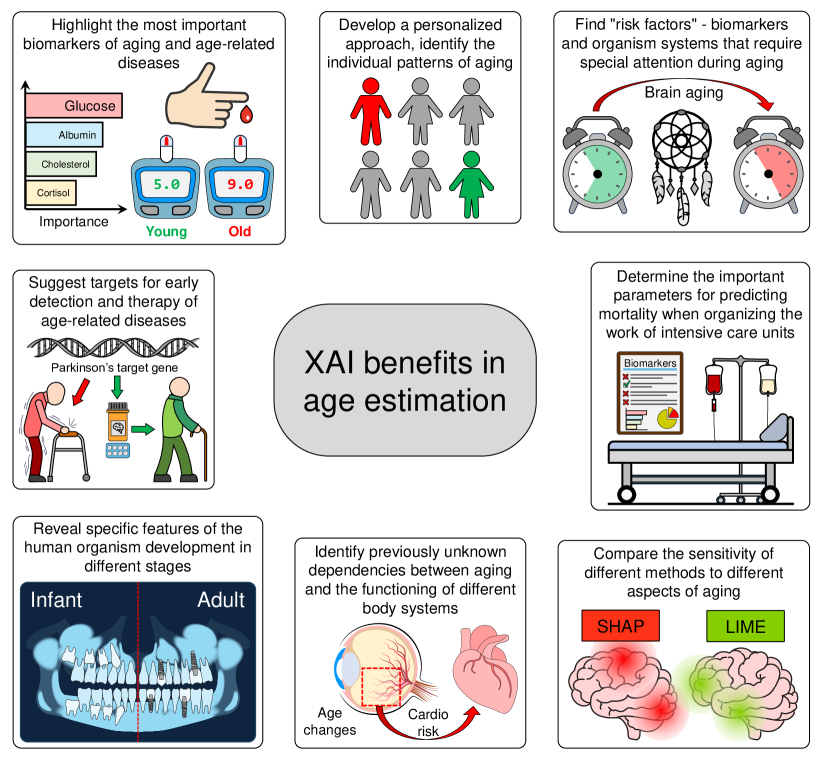
The application of XAI approaches to the biological age prediction task hits several targets, cf. Figure 4. One of the most significant and obvious achievements is identifying the most important biomarkers of aging and age-associated diseases, while keeping the benefits of the high performance of “black-box” AI models. In result, explainable clocks and age-associated parameters for a broad spectrum of body systems are becoming available. XAI is also helpful in deriving ‘risk factors’ from AI-based age estimation and mortality prediction models. Such early predictors can serve as ‘red flags’ before serious symptoms emerge and the model explainability can pave the way to recommending anti-age corrections. Next, the majority of XAI methods provide explanations for each specific sample, identifying the individual signatures that made the highest impact on the final prediction for that particular person. This opens the door for personalized preventive approaches and therapies. XAI methods have a clear potential in identifying the early signatures of age-related diseases and the targets for the early correction and treatment. Another important aspect of XAI for public health is related to support the organization of intensive care units. Determining the most important features in predicting mortality will allow the proper allocation of healthcare facility resources and provide the most efficient care. XAI also gives much promise in the fundamental research, suggesting the previously unknown dependencies between different body systems in the aging process, and the comparison of the sensitivity of different approaches to different aspects of aging.
In the horizon of several years, XAI approaches are highly likely to become ubiquitous in the aging research, development of various clocks, biomarkers of aging and age-related diseases, with the strong emphasis on determining individual aging status, personalized recommendations, corrections and therapies.
Abbreviations
AI - Artificial Intelligence; ALE - Accumulated Local Effects; ALT - ALanine Transaminase; CAM - Class Activation Mapping; CNN - Convolutional Neural Network; CSF - CerebroSpinal Fluid; DeepLIFT - Deep Learning Important FeaTures; DeepPINK - Deep feature selection using Paired-Input Nonlinear Knockoffs; DL - Deep Learning; DNA - DeoxyriboNucleic Acid; DNN - Deep Neural Network; ECG - Electrocardiogram; EEG - Electroencephalogram; GBDT - Gradient-Boosted Decision Tree; GGT - Gamma-Glutamyl Transferase; Grad-CAM - Gradient-weighted Class Activation Mapping; HIV - Human Immunodeficiency Virus; LIME - Local Interpretable Model-agnostic Explanations; ML - Machine Learning; MRI - Magnetic Resonance Imaging; OCT - Optical Coherence Tomography; PDP - Partial Dependence Plot; PFI - Permutation Feature Importance; PPG - PhotoPlethysmoGram; REM - Rapid Eye Movement; RNA - RiboNucleic Acid; ROI - Region Of Interest; SHAP - Shapley Additive exPlanations; VIM - Variable Importance Measure; XAI - eXplainable Artificial Intelligence.
References
- Abdolrashidi et al. [2020] Amirali Abdolrashidi, Mehdi Minaei, Elham Azimi, and Shervin Minaee. Age and gender prediction from face images using attentional convolutional network. arXiv, 2020. doi:10.48550/arXiv.2010.03791.
- Agustsson et al. [2017] Eirikur Agustsson, Radu Timofte, Sergio Escalera, Xavier Baro, Isabelle Guyon, and Rasmus Rothe. Apparent and real age estimation in still images with deep residual regressors on appa-real database. In 2017 12th IEEE International Conference on Automatic Face & Gesture Recognition (FG 2017), pages 87–94, 2017. doi:10.1109/FG.2017.20.
- Al Zoubi et al. [2018] Obada Al Zoubi, Chung Ki Wong, Rayus T. Kuplicki, Hung-wen Yeh, Ahmad Mayeli, Hazem Refai, Martin Paulus, and Jerzy Bodurka. Predicting age from brain EEG signals—a machine learning approach. Frontiers in Aging Neuroscience, 10, 2018. doi:10.3389/fnagi.2018.00184.
- Ali et al. [2023] Sajid Ali, Tamer Abuhmed, Shaker El-Sappagh, Khan Muhammad, Jose M. Alonso-Moral, Roberto Confalonieri, Riccardo Guidotti, Javier Del Ser, Natalia Díaz-Rodríguez, and Francisco Herrera. Explainable artificial intelligence (XAI): What we know and what is left to attain trustworthy artificial intelligence. Information Fusion, page 101805, 2023. doi:10.1016/j.inffus.2023.101805.
- Amann et al. [2020] Julia Amann, Alessandro Blasimme, Effy Vayena, Dietmar Frey, Vince I. Madai, and the Precise4Q consortium. Explainability for artificial intelligence in healthcare: a multidisciplinary perspective. BMC Medical Informatics and Decision Making, 20(1):310, 2020. doi:10.1186/s12911-020-01332-6.
- Apley and Zhu [2020] Daniel W. Apley and Jingyu Zhu. Visualizing the effects of predictor variables in black box supervised learning models. Journal of the Royal Statistical Society Series B: Statistical Methodology, 82(4):1059–1086, 2020. doi:10.1111/rssb.12377.
- Atas et al. [2022] Isa Atas, Cuneyt Ozdemir, Musa Atas, and Yahya Dogan. Forensic dental age estimation using modified deep learning neural network. arXiv, 2022. doi:10.48550/arXiv.2208.09799.
- Attia et al. [2021] Zachi I Attia, Gilad Lerman, and Paul A Friedman. Deep neural networks learn by using human-selected electrocardiogram features and novel features. European Heart Journal - Digital Health, 2(3):446–455, 2021. doi:10.1093/ehjdh/ztab060.
- Bae et al. [2021] Chul-Young Bae, Yoori Im, Jonghoon Lee, Choong-Shik Park, Miyoung Kim, Hojeong Kwon, Boseon Kim, Hye ri Park, Chun-Koo Lee, Inhee Kim, and JeongHoon Kim. Comparison of biological age prediction models using clinical biomarkers commonly measured in clinical practice settings: AI techniques vs. traditional statistical methods. Frontiers in Analytical Science, 1, 2021. doi:10.3389/frans.2021.709589.
- Baehrens et al. [2010] David Baehrens, Timon Schroeter, Stefan Harmeling, Motoaki Kawanabe, Katja Hansen, and Klaus-Robert Müller. How to explain individual classification decisions. The Journal of Machine Learning Research, 11:1803–1831, 2010.
- Ball et al. [2021] Gareth Ball, Claire E Kelly, Richard Beare, and Marc L Seal. Individual variation underlying brain age estimates in typical development. NeuroImage, 235:118036, 2021. doi:10.1016/j.neuroimage.2021.118036.
- Ballester et al. [2023] Pedro L. Ballester, Jee Su Suh, Natalie C. W. Ho, Liangbing Liang, Stefanie Hassel, Stephen C. Strother, Stephen R. Arnott, Luciano Minuzzi, Roberto B. Sassi, Raymond W. Lam, Roumen Milev, Daniel J. Müller, Valerie H. Taylor, Sidney H. Kennedy, James P. Reilly, Lena Palaniyappan, Katharine Dunlop, and Benicio N. Frey. Gray matter volume drives the brain age gap in schizophrenia: a SHAP study. Schizophrenia, 9(1):1–8, 2023. doi:10.1038/s41537-022-00330-z.
- Beil et al. [2019] Michael Beil, Ingo Proft, Daniel van Heerden, Sigal Sviri, and Peter Vernon van Heerden. Ethical considerations about artificial intelligence for prognostication in intensive care. Intensive Care Medicine Experimental, 7(1):70, 2019. doi:10.1186/s40635-019-0286-6.
- Bell et al. [2019] Christopher G. Bell, Robert Lowe, Peter D. Adams, Andrea A. Baccarelli, Stephan Beck, Jordana T. Bell, Brock C. Christensen, Vadim N. Gladyshev, Bastiaan T. Heijmans, Steve Horvath, Trey Ideker, Jean-Pierre J. Issa, Karl T. Kelsey, Riccardo E. Marioni, Wolf Reik, Caroline L. Relton, Leonard C. Schalkwyk, Andrew E. Teschendorff, Wolfgang Wagner, Kang Zhang, and Vardhman K. Rakyan. DNA methylation aging clocks: challenges and recommendations. Genome Biology, 20(1):249, 2019. doi:10.1186/s13059-019-1824-y.
- Bernard et al. [2023] David Bernard, Emmanuel Doumard, Isabelle Ader, Philippe Kemoun, Jean-Christophe Pagès, Anne Galinier, Sylvain Cussat-Blanc, Felix Furger, Luigi Ferrucci, Julien Aligon, Cyrille Delpierre, Luc Pénicaud, Paul Monsarrat, and Louis Casteilla. Explainable machine learning framework to predict personalized physiological aging. Aging Cell, page e13872, 2023. doi:10.1111/acel.13872.
- Bintsi et al. [2021] Kyriaki-Margarita Bintsi, Vasileios Baltatzis, Alexander Hammers, and Daniel Rueckert. Voxel-level importance maps for interpretable brain age estimation. In Mauricio Reyes, Pedro Henriques Abreu, Jaime Cardoso, Mustafa Hajij, Ghada Zamzmi, Paul Rahul, and Lokendra Thakur, editors, Interpretability of Machine Intelligence in Medical Image Computing, and Topological Data Analysis and Its Applications for Medical Data, Lecture Notes in Computer Science, pages 65–74. Springer International Publishing, 2021. doi:10.1007/978-3-030-87444-5_7.
- Borisov et al. [2022] Vadim Borisov, Tobias Leemann, Kathrin Seßler, Johannes Haug, Martin Pawelczyk, and Gjergji Kasneci. Deep neural networks and tabular data: A survey. IEEE Transactions on Neural Networks and Learning Systems, pages 1–21, 2022. doi:10.1109/TNNLS.2022.3229161.
- Boscolo Galazzo et al. [2022] Ilaria Boscolo Galazzo, Federica Cruciani, Lorenza Brusini, Ahmed Salih, Petia Radeva, Silvia Francesca Storti, and Gloria Menegaz. Explainable artificial intelligence for magnetic resonance imaging aging brainprints: Grounds and challenges. IEEE Signal Processing Magazine, 39(2):99–116, 2022. doi:10.1109/MSP.2021.3126573.
- Breiman [2001] Leo Breiman. Random forests. Machine Learning, 45(1):5–32, 2001. doi:10.1023/A:1010933404324.
- Bui et al. [2019] Toan Duc Bui, Jae-Joon Lee, and Jitae Shin. Incorporated region detection and classification using deep convolutional networks for bone age assessment. Artificial Intelligence in Medicine, 97:1–8, 2019. doi:10.1016/j.artmed.2019.04.005.
- Carrieri et al. [2021] Anna Paola Carrieri, Niina Haiminen, Sean Maudsley-Barton, Laura-Jayne Gardiner, Barry Murphy, Andrew E. Mayes, Sarah Paterson, Sally Grimshaw, Martyn Winn, Cameron Shand, Panagiotis Hadjidoukas, Will P. M. Rowe, Stacy Hawkins, Ashley MacGuire-Flanagan, Jane Tazzioli, John G. Kenny, Laxmi Parida, Michael Hoptroff, and Edward O. Pyzer-Knapp. Explainable AI reveals changes in skin microbiome composition linked to phenotypic differences. Scientific Reports, 11(1):4565, 2021. doi:10.1038/s41598-021-83922-6.
- Chang et al. [2022] Chiao-Hsiang Chang, Chin-Sheng Lin, Yu-Sheng Luo, Yung-Tsai Lee, and Chin Lin. Electrocardiogram-based heart age estimation by a deep learning model provides more information on the incidence of cardiovascular disorders. Frontiers in Cardiovascular Medicine, 9, 2022. doi:10.3389/fcvm.2022.754909.
- Chen et al. [2022] Yutao Chen, Hongchao Wang, Wenwei Lu, Tong Wu, Weiwei Yuan, Jinlin Zhu, Yuan Kun Lee, Jianxin Zhao, Hao Zhang, and Wei Chen. Human gut microbiome aging clocks based on taxonomic and functional signatures through multi-view learning. Gut Microbes, 14(1):2025016, 2022. doi:10.1080/19490976.2021.2025016.
- Cherubini et al. [2016] Andrea Cherubini, Maria Eugenia Caligiuri, Patrice Péran, Umberto Sabatini, Carlo Cosentino, and Francesco Amato. Importance of multimodal MRI in characterizing brain tissue and its potential application for individual age prediction. IEEE Journal of Biomedical and Health Informatics, 20(5):1232–1239, 2016. doi:10.1109/JBHI.2016.2559938.
- Cho and Wallraven [2022] Daehyun Cho and Christian Wallraven. Do pre-processing and augmentation help explainability? a multi-seed analysis for brain age estimation. In Mauricio Reyes, Pedro Henriques Abreu, and Jaime Cardoso, editors, Interpretability of Machine Intelligence in Medical Image Computing, Lecture Notes in Computer Science, pages 12–21. Springer Nature Switzerland, 2022. doi:10.1007/978-3-031-17976-1_2.
- Chueh et al. [2022] Kuan-Ming Chueh, Yi-Ting Hsieh, Homer H. Chen, I.-Hsin Ma, and Sheng-Lung Huang. Identification of sex and age from macular optical coherence tomography and feature analysis using deep learning. American Journal of Ophthalmology, 235:221–228, 2022. doi:10.1016/j.ajo.2021.09.015.
- Cole and Franke [2017] James H. Cole and Katja Franke. Predicting age using neuroimaging: Innovative brain ageing biomarkers. Trends in Neurosciences, 40(12):681–690, 2017. doi:10.1016/j.tins.2017.10.001.
- Cumplido-Mayoral et al. [2023] Irene Cumplido-Mayoral, Marina García-Prat, Grégory Operto, Carles Falcon, Mahnaz Shekari, Raffaele Cacciaglia, Marta Milà-Alomà, Luigi Lorenzini, Silvia Ingala, Alle Meije Wink, Henk JMM Mutsaerts, Carolina Minguillón, Karine Fauria, José Luis Molinuevo, Sven Haller, Gael Chetelat, Adam Waldman, Adam J Schwarz, Frederik Barkhof, Ivonne Suridjan, Gwendlyn Kollmorgen, Anna Bayfield, Henrik Zetterberg, Kaj Blennow, Marc Suárez-Calvet, Verónica Vilaplana, Juan Domingo Gispert, ALFA study, EPAD study, ADNI study, and OASIS study. Biological brain age prediction using machine learning on structural neuroimaging data: Multi-cohort validation against biomarkers of alzheimer’s disease and neurodegeneration stratified by sex. eLife, 12:e81067, 2023. doi:10.7554/eLife.81067.
- de Back et al. [2019] Walter de Back, Sebastian Seurig, Sebastian Wagner, Birgit Marré, Ingo Roeder, and Nico Scherf. Forensic age estimation with bayesian convolutional neural networks based on panoramic dental x-ray imaging. In Proceedings of Machine Learning Research, 2019.
- de Lima Camillo et al. [2022] Lucas Paulo de Lima Camillo, Louis R. Lapierre, and Ritambhara Singh. A pan-tissue DNA-methylation epigenetic clock based on deep learning. npj Aging, 8(1):1–15, 2022. doi:10.1038/s41514-022-00085-y.
- Feng et al. [2020] Xinyang Feng, Zachary C. Lipton, Jie Yang, Scott A. Small, and Frank A. Provenzano. Estimating brain age based on a uniform healthy population with deep learning and structural magnetic resonance imaging. Neurobiology of Aging, 91:15–25, 2020. doi:10.1016/j.neurobiolaging.2020.02.009.
- Friedman [2001] Jerome H. Friedman. Greedy function approximation: A gradient boosting machine. The Annals of Statistics, 29(5):1189–1232, 2001. doi:10.1214/aos/1013203451.
- Galkin et al. [2020] Fedor Galkin, Polina Mamoshina, Alex Aliper, Evgeny Putin, Vladimir Moskalev, Vadim N. Gladyshev, and Alex Zhavoronkov. Human gut microbiome aging clock based on taxonomic profiling and deep learning. iScience, 23(6), 2020. doi:10.1016/j.isci.2020.101199.
- Gao et al. [2018] Bin-Bin Gao, Hong-Yu Zhou, Jianxin Wu, and Xin Geng. Age estimation using expectation of label distribution learning. Proceedings of IJCAI 2018, pages 712–718, 2018. doi:10.24963/ijcai.2018/99.
- Gao et al. [2020] Xiaoqian Gao, Shuai Liu, Haoqiu Song, Xin Feng, Meiyu Duan, Lan Huang, and Fengfeng Zhou. AgeGuess, a methylomic prediction model for human ages. Frontiers in Bioengineering and Biotechnology, 8, 2020. doi:10.3389/fbioe.2020.00080.
- Gámez-Granados et al. [2022] Juan Carlos Gámez-Granados, Javier Irurita, Raúl Pérez, Antonio González, Sergio Damas, Inmaculada Alemán, and Oscar Cordón. Automating the decision making process of todd’s age estimation method from the pubic symphysis with explainable machine learning. Information Sciences, 612:514–535, 2022. doi:10.1016/j.ins.2022.08.110.
- Gómez-Ramírez et al. [2022] Jaime Gómez-Ramírez, Miguel A. Fernández-Blázquez, and Javier J. González-Rosa. Prediction of chronological age in healthy elderly subjects with machine learning from MRI brain segmentation and cortical parcellation. Brain Sciences, 12(5):579, 2022. doi:10.3390/brainsci12050579.
- Goallec et al. [2021a] Alan Le Goallec, Sasha Collin, Samuel Diai, Théo Vincent, and Chirag J. Patel. Predicting arterial age using carotid ultrasound images, pulse wave analysis records, cardiovascular biomarkers and deep learning. medRxiv, 2021a. doi:10.1101/2021.06.17.21259120.
- Goallec et al. [2021b] Alan Le Goallec, Samuel Diai, Sasha Collin, Théo Vincent, and Chirag J. Patel. Using deep learning to analyze the compositeness of musculoskeletal aging reveals that spine, hip and knee age at different rates, and are associated with different genetic and non-genetic factors. medRxiv, 2021b. doi:10.1101/2021.06.14.21258896.
- Goallec et al. [2021c] Alan Le Goallec, Jean-Baptiste Prost, Sasha Collin, Samuel Diai, Théo Vincent, and Chirag J. Patel. Dissecting heart age using cardiac magnetic resonance videos, electrocardiograms, biobanks, and deep learning, 2021c.
- Gopu et al. [2020] Vishakh Gopu, Ying Cai, Subha Krishnan, Sathyapriya Rajagopal, Francine R. Camacho, Ryan Toma, Pedro J. Torres, Momchilo Vuyisich, Ally Perlina, Guruduth Banavar, and Hal Tily. An accurate aging clock developed from the largest dataset of microbial and human gene expression reveals molecular mechanisms of aging. bioRxiv, 2020. doi:10.1101/2020.09.17.301887.
- Grinsztajn et al. [2022] Léo Grinsztajn, Edouard Oyallon, and Gaël Varoquaux. Why do tree-based models still outperform deep learning on tabular data? arXiv, 2022. doi:10.48550/arXiv.2207.08815.
- Guo et al. [2021] Yu-cheng Guo, Mengqi Han, Yuting Chi, Hong Long, Dong Zhang, Jing Yang, Yang Yang, Teng Chen, and Shaoyi Du. Accurate age classification using manual method and deep convolutional neural network based on orthopantomogram images. International Journal of Legal Medicine, 135(4):1589–1597, 2021. doi:10.1007/s00414-021-02542-x.
- Han et al. [2022] Juhyuk Han, Seo Yeong Kim, Junhyeok Lee, and Won Hee Lee. Brain age prediction: A comparison between machine learning models using brain morphometric data. Sensors, 22(20):8077, 2022. doi:10.3390/s22208077.
- Hannum et al. [2013] Gregory Hannum, Justin Guinney, Ling Zhao, Li Zhang, Guy Hughes, SriniVas Sadda, Brandy Klotzle, Marina Bibikova, Jian-Bing Fan, Yuan Gao, Rob Deconde, Menzies Chen, Indika Rajapakse, Stephen Friend, Trey Ideker, and Kang Zhang. Genome-wide methylation profiles reveal quantitative views of human aging rates. Molecular Cell, 49(2):359–367, 2013. doi:10.1016/j.molcel.2012.10.016.
- Hepp et al. [2021] Tobias Hepp, Dominik Blum, Karim Armanious, Bernhard Schölkopf, Darko Stern, Bin Yang, and Sergios Gatidis. Uncertainty estimation and explainability in deep learning-based age estimation of the human brain: Results from the german national cohort MRI study. Computerized Medical Imaging and Graphics, 92:101967, 2021. doi:10.1016/j.compmedimag.2021.101967.
- Hershey et al. [2017] Shawn Hershey, Sourish Chaudhuri, Daniel P. W. Ellis, Jort F. Gemmeke, Aren Jansen, R. Channing Moore, Manoj Plakal, Devin Platt, Rif A. Saurous, Bryan Seybold, Malcolm Slaney, Ron J. Weiss, and Kevin Wilson. CNN architectures for large-scale audio classification. arXiv, 2017. doi:10.48550/arXiv.1609.09430.
- Holzscheck et al. [2021] Nicholas Holzscheck, Cassandra Falckenhayn, Jörn Söhle, Boris Kristof, Ralf Siegner, André Werner, Janka Schössow, Clemens Jürgens, Henry Völzke, Horst Wenck, Marc Winnefeld, Elke Grönniger, and Lars Kaderali. Modeling transcriptomic age using knowledge-primed artificial neural networks. npj Aging and Mechanisms of Disease, 7(1):1–13, 2021. doi:10.1038/s41514-021-00068-5.
- Horvath [2013] Steve Horvath. DNA methylation age of human tissues and cell types. Genome Biology, 14(10):R115, 2013. doi:10.1186/gb-2013-14-10-r115.
- Hou et al. [2021] Wenxuan Hou, Longjun Liu, Jinxia Gao, Anguo Zhu, Keyang Pan, Hongbin Sun, and Nanning Zheng. Exploring effective DNN models for forensic age estimation based on panoramic radiograph images. In 2021 International Joint Conference on Neural Networks (IJCNN), pages 1–8, 2021. doi:10.1109/IJCNN52387.2021.9533672.
- Hu et al. [2021] Guozhen Hu, Qinjian Zhang, Zhi Yang, and Baobin Li. Accurate brain age prediction model for healthy children and adolescents using 3d-CNN and dimensional attention. In 2021 IEEE International Conference on Bioinformatics and Biomedicine (BIBM), pages 800–806, 2021. doi:10.1109/BIBM52615.2021.9669900.
- Huang et al. [2020] Shi Huang, Niina Haiminen, Anna-Paola Carrieri, Rebecca Hu, Lingjing Jiang, Laxmi Parida, Baylee Russell, Celeste Allaband, Amir Zarrinpar, Yoshiki Vázquez-Baeza, Pedro Belda-Ferre, Hongwei Zhou, Ho-Cheol Kim, Austin D. Swafford, Rob Knight, and Zhenjiang Zech Xu. Human skin, oral, and gut microbiomes predict chronological age. mSystems, 5(1):e00630–19, 2020. doi:10.1128/mSystems.00630-19.
- Ieki et al. [2022] Hirotaka Ieki, Kaoru Ito, Mike Saji, Rei Kawakami, Yuji Nagatomo, Kaori Takada, Toshiya Kariyasu, Haruhiko Machida, Satoshi Koyama, Hiroki Yoshida, Ryo Kurosawa, Hiroshi Matsunaga, Kazuo Miyazawa, Kouichi Ozaki, Yoshihiro Onouchi, Susumu Katsushika, Ryo Matsuoka, Hiroki Shinohara, Toshihiro Yamaguchi, Satoshi Kodera, Yasutomi Higashikuni, Katsuhito Fujiu, Hiroshi Akazawa, Nobuo Iguchi, Mitsuaki Isobe, Tsutomu Yoshikawa, and Issei Komuro. Deep learning-based age estimation from chest x-rays indicates cardiovascular prognosis. Communications Medicine, 2(1):1–12, 2022. doi:10.1038/s43856-022-00220-6.
- Jetley et al. [2018] Saumya Jetley, Nicholas A. Lord, Namhoon Lee, and Philip H. S. Torr. Learn to pay attention. arXiv, 2018. doi:10.48550/arXiv.1804.02391.
- Kalyakulina et al. [2023] ALena Kalyakulina, Igor Yusipov, Elena Kondakova, Maria Giulia Bacalini, Claudio Franceschi, Maria Vedunova, and Mikhail Ivanchenko. Small immunological clocks identified by deep learning and gradient boosting. Frontiers in Immunology, 14, 2023. doi:10.3389/fimmu.2023.1177611. URL https://doi.org/10.3389/fimmu.2023.1177611.
- Karargyris et al. [2019] A. Karargyris, S. Kashyap, J. T. Wu, A. Sharma, M. Moradi, and T. Syeda-Mahmood. Age prediction using a large chest x-ray dataset. In Medical Imaging 2019: Computer-Aided Diagnosis, volume 10950, pages 468–476. SPIE, 2019. doi:10.1117/12.2512922.
- Khayretdinova et al. [2022] Mariam Khayretdinova, Alexey Shovkun, Vladislav Degtyarev, Andrey Kiryasov, Polina Pshonkovskaya, and Ilya Zakharov. Predicting age from resting-state scalp EEG signals with deep convolutional neural networks on TD-brain dataset. Frontiers in Aging Neuroscience, 14, 2022. doi:10.3389/fnagi.2022.1019869.
- Kim et al. [2021] Seunghyeon Kim, Yeon-Hee Lee, Yung-Kyun Noh, Frank C. Park, and Q.-Schick Auh. Age-group determination of living individuals using first molar images based on artificial intelligence. Scientific Reports, 11(1):1073, 2021. doi:10.1038/s41598-020-80182-8.
- Kolbeinsson et al. [2020] Arinbjörn Kolbeinsson, Sarah Filippi, Yannis Panagakis, Paul M. Matthews, Paul Elliott, Abbas Dehghan, and Ioanna Tzoulaki. Accelerated MRI-predicted brain ageing and its associations with cardiometabolic and brain disorders. Scientific Reports, 10(1):19940, 2020. doi:10.1038/s41598-020-76518-z.
- Kotěrová et al. [2018] Anežka Kotěrová, David Navega, Michal Štepanovský, Zdeněk Buk, Jaroslav Brůžek, and Eugénia Cunha. Age estimation of adult human remains from hip bones using advanced methods. Forensic Science International, 287:163–175, 2018. doi:10.1016/j.forsciint.2018.03.047.
- Lam et al. [2020] Pradeep Lam, Alyssa H. Zhu, Iyad Ba Gari, Neda Jahanshad, and Paul M. Thompson. 3d grid-attention networks for interpretable age and alzheimer’s disease prediction from structural MRI. arXiv, 2020. doi:10.48550/arXiv.2011.09115.
- Langner et al. [2020] Taro Langner, Johan Wikström, Tomas Bjerner, Håkan Ahlström, and Joel Kullberg. Identifying morphological indicators of aging with neural networks on large-scale whole-body MRI. IEEE Transactions on Medical Imaging, 39(5):1430–1437, 2020. doi:10.1109/TMI.2019.2950092.
- Lassen et al. [2023] Johan K. Lassen, Tingting Wang, Kirstine L. Nielsen, Jørgen B. Hasselstrøm, Mogens Johannsen, and Palle Villesen. Large-scale metabolomics: Predicting biological age using 10,133 routine untargeted LC–MS measurements. Aging Cell, 22(5):e13813, 2023. doi:10.1111/acel.13813.
- Le Goallec et al. [2022] Alan Le Goallec, Samuel Diai, Sasha Collin, Jean-Baptiste Prost, Théo Vincent, and Chirag J. Patel. Using deep learning to predict abdominal age from liver and pancreas magnetic resonance images. Nature Communications, 13(1):1979, 2022. doi:10.1038/s41467-022-29525-9.
- Lee et al. [2017] Hyunkwang Lee, Shahein Tajmir, Jenny Lee, Maurice Zissen, Bethel Ayele Yeshiwas, Tarik K. Alkasab, Garry Choy, and Synho Do. Fully automated deep learning system for bone age assessment. Journal of Digital Imaging, 30(4):427–441, 2017. doi:10.1007/s10278-017-9955-8.
- Lee et al. [2022] Yeon-Hee Lee, Jong Hyun Won, Q.-Schick Auh, and Yung-Kyun Noh. Age group prediction with panoramic radiomorphometric parameters using machine learning algorithms. Scientific Reports, 12(1):11703, 2022. doi:10.1038/s41598-022-15691-9.
- Letzgus et al. [2022] Simon Letzgus, Patrick Wagner, Jonas Lederer, Wojciech Samek, Klaus-Robert Müller, and Grégoire Montavon. Toward explainable artificial intelligence for regression models: A methodological perspective. IEEE Signal Processing Magazine, 39(4):40–58, 2022. doi:10.1109/MSP.2022.3153277.
- Levakov et al. [2020] Gidon Levakov, Gideon Rosenthal, Ilan Shelef, Tammy Riklin Raviv, and Galia Avidan. From a deep learning model back to the brain—identifying regional predictors and their relation to aging. Human Brain Mapping, 41(12):3235–3252, 2020. doi:10.1002/hbm.25011.
- Levine et al. [2018] Morgan E. Levine, Ake T. Lu, Austin Quach, Brian H. Chen, Themistocles L. Assimes, Stefania Bandinelli, Lifang Hou, Andrea A. Baccarelli, James D. Stewart, Yun Li, Eric A. Whitsel, James G Wilson, Alex P Reiner, Abraham Aviv, Kurt Lohman, Yongmei Liu, Luigi Ferrucci, and Steve Horvath. An epigenetic biomarker of aging for lifespan and healthspan. Aging (Albany NY), 10(4):573–591, 2018. doi:10.18632/aging.101414.
- Levy et al. [2020] Joshua J. Levy, Alexander J. Titus, Curtis L. Petersen, Youdinghuan Chen, Lucas A. Salas, and Brock C. Christensen. MethylNet: an automated and modular deep learning approach for DNA methylation analysis. BMC Bioinformatics, 21(1):108, 2020. doi:10.1186/s12859-020-3443-8.
- Lima et al. [2021] Emilly M. Lima, Antônio H. Ribeiro, Gabriela M. M. Paixão, Manoel Horta Ribeiro, Marcelo M. Pinto-Filho, Paulo R. Gomes, Derick M. Oliveira, Ester C. Sabino, Bruce B. Duncan, Luana Giatti, Sandhi M. Barreto, Wagner Meira Jr, Thomas B. Schön, and Antonio Luiz P. Ribeiro. Deep neural network-estimated electrocardiographic age as a mortality predictor. Nature Communications, 12(1):5117, 2021. doi:10.1038/s41467-021-25351-7.
- Lindow et al. [2022] Thomas Lindow, Israel Palencia-Lamela, Todd T. Schlegel, and Martin Ugander. Heart age estimated using explainable advanced electrocardiography. Scientific Reports, 12(1):9840, 2022. doi:10.1038/s41598-022-13912-9.
- Lipton [2018] Zachary C. Lipton. The mythos of model interpretability: In machine learning, the concept of interpretability is both important and slippery. Queue, 16(3):31–57, 2018. doi:10.1145/3236386.3241340.
- Lombardi et al. [2021] Angela Lombardi, Domenico Diacono, Nicola Amoroso, Alfonso Monaco, João Manuel R. S. Tavares, Roberto Bellotti, and Sabina Tangaro. Explainable deep learning for personalized age prediction with brain morphology. Frontiers in Neuroscience, 15, 2021. doi:10.3389/fnins.2021.674055.
- Lu et al. [2019] Ake T. Lu, Austin Quach, James G. Wilson, Alex P. Reiner, Abraham Aviv, Kenneth Raj, Lifang Hou, Andrea A. Baccarelli, Yun Li, James D. Stewart, Eric A. Whitsel, Themistocles L. Assimes, Luigi Ferrucci, and Steve Horvath. DNA methylation GrimAge strongly predicts lifespan and healthspan. Aging (Albany NY), 11(2):303–327, 2019. doi:10.18632/aging.101684.
- Lu et al. [2018] Yang Young Lu, Yingying Fan, Jinchi Lv, and William Stafford Noble. DeepPINK: reproducible feature selection in deep neural networks. In Proceedings of the 32nd International Conference on Neural Information Processing Systems, NIPS’18, pages 8690–8700. Curran Associates Inc., 2018.
- Lundberg and Lee [2017] Scott M. Lundberg and Su-In Lee. A unified approach to interpreting model predictions. In Proceedings of the 31st International Conference on Neural Information Processing Systems, NIPS’17, pages 4768–4777. Curran Associates Inc., 2017. doi:10.5555/3295222.3295230.
- Ma et al. [2021] Jiaonan Ma, Xueli Xu, Mengdi Li, Yan Zhang, Lin Zhang, Ping Ma, Jie Hou, Yulin Lei, Jianguo Liu, Xiaojin Huangfu, Yang Yang, Xianglong Yi, George Cheng, Ji Bai, Xingwu Zhong, Ximing Xu, and Yan Wang. Predictive models of aging of the human eye based on ocular anterior segment morphology. Journal of Biomedical Informatics, 120:103855, 2021. doi:10.1016/j.jbi.2021.103855.
- Mamoshina and Zhavoronkov [2019] Polina Mamoshina and Alex Zhavoronkov. Deep integrated biomarkers of aging. In Alexey Moskalev, editor, Biomarkers of Human Aging, Healthy Ageing and Longevity, pages 281–291. Springer International Publishing, 2019. doi:10.1007/978-3-030-24970-0_18.
- Mamoshina et al. [2018a] Polina Mamoshina, Kirill Kochetov, Evgeny Putin, Franco Cortese, Alexander Aliper, Won-Suk Lee, Sung-Min Ahn, Lee Uhn, Neil Skjodt, Olga Kovalchuk, Morten Scheibye-Knudsen, and Alex Zhavoronkov. Population specific biomarkers of human aging: A big data study using south korean, canadian, and eastern european patient populations. The Journals of Gerontology: Series A, 73(11):1482–1490, 2018a. doi:10.1093/gerona/gly005.
- Mamoshina et al. [2018b] Polina Mamoshina, Marina Volosnikova, Ivan V. Ozerov, Evgeny Putin, Ekaterina Skibina, Franco Cortese, and Alex Zhavoronkov. Machine learning on human muscle transcriptomic data for biomarker discovery and tissue-specific drug target identification. Frontiers in Genetics, 9, 2018b. doi:10.3389/fgene.2018.00242.
- Mamoshina et al. [2019] Polina Mamoshina, Kirill Kochetov, Franco Cortese, Anna Kovalchuk, Alexander Aliper, Evgeny Putin, Morten Scheibye-Knudsen, Charles R. Cantor, Neil M. Skjodt, Olga Kovalchuk, and Alex Zhavoronkov. Blood biochemistry analysis to detect smoking status and quantify accelerated aging in smokers. Scientific Reports, 9(1):142, 2019. doi:10.1038/s41598-018-35704-w.
- Montesanto et al. [2020] Alberto Montesanto, Patrizia D’Aquila, Vincenzo Lagani, Ersilia Paparazzo, Silvana Geracitano, Laura Formentini, Robertina Giacconi, Maurizio Cardelli, Mauro Provinciali, Dina Bellizzi, and Giuseppe Passarino. A new robust epigenetic model for forensic age prediction. Journal of Forensic Sciences, 65(5):1424–1431, 2020. doi:10.1111/1556-4029.14460.
- Monti et al. [2020] Ricardo Pio Monti, Alex Gibberd, Sandipan Roy, Matthew Nunes, Romy Lorenz, Robert Leech, Takeshi Ogawa, Motoaki Kawanabe, and Aapo Hyvärinen. Interpretable brain age prediction using linear latent variable models of functional connectivity. PLOS ONE, 15(6):e0232296, 2020. doi:10.1371/journal.pone.0232296.
- Moskalev et al. [2023] Alexey Moskalev, Ilia Stambler, and Alex Zhavoronkov, editors. Artificial Intelligence for Healthy Longevity. Springer International Publishing, 2023. doi:10.1007/978-3-031-35176-1. URL https://doi.org/10.1007/978-3-031-35176-1.
- Mouches et al. [2022] Pauline Mouches, Matthias Wilms, Deepthi Rajashekar, Sönke Langner, and Nils D. Forkert. Multimodal biological brain age prediction using magnetic resonance imaging and angiography with the identification of predictive regions. Human Brain Mapping, 43(8):2554–2566, 2022. doi:10.1002/hbm.25805.
- Nam et al. [2020] Yoonho Nam, Jinhee Jang, Hea Yon Lee, Yangsean Choi, Na Young Shin, Kang-Hyun Ryu, Dong Hyun Kim, So-Lyung Jung, Kook-jin Ahn, and Bum-soo Kim. Estimating age-related changes in in vivo cerebral magnetic resonance angiography using convolutional neural network. Neurobiology of Aging, 87:125–131, 2020. doi:10.1016/j.neurobiolaging.2019.12.008.
- Nauta et al. [2023] Meike Nauta, Jan Trienes, Shreyasi Pathak, Elisa Nguyen, Michelle Peters, Yasmin Schmitt, Jörg Schlötterer, Maurice van Keulen, and Christin Seifert. From anecdotal evidence to quantitative evaluation methods: A systematic review on evaluating explainable AI. ACM Computing Surveys, 2023. doi:10.1145/3583558.
- Nusinovici et al. [2022] Simon Nusinovici, Tyler Hyungtaek Rim, Marco Yu, Geunyoung Lee, Yih-Chung Tham, Ning Cheung, Crystal Chun Yuen Chong, Zhi Da Soh, Sahil Thakur, Chan Joo Lee, Charumathi Sabanayagam, Byoung Kwon Lee, Sungha Park, Sung Soo Kim, Hyeon Chang Kim, Tien-Yin Wong, and Ching-Yu Cheng. Retinal photograph-based deep learning predicts biological age, and stratifies morbidity and mortality risk. Age and Ageing, 51(4):afac065, 2022. doi:10.1093/ageing/afac065.
- Park et al. [2016] Jong-Lyul Park, Jong Hwan Kim, Eunhye Seo, Dong Hyuck Bae, Seon-Young Kim, Han-Chul Lee, Kwang-Man Woo, and Yong Sung Kim. Identification and evaluation of age-correlated DNA methylation markers for forensic use. Forensic Science International: Genetics, 23:64–70, 2016. doi:10.1016/j.fsigen.2016.03.005.
- Partridge et al. [2018] Linda Partridge, Joris Deelen, and P. Eline Slagboom. Facing up to the global challenges of ageing. Nature, 561(7721):45–56, 2018. ISSN 1476-4687. doi:10.1038/s41586-018-0457-8.
- Patil et al. [2023] Vathsala Patil, Janhavi Saxena, Ravindranath Vineetha, Rahul Paul, Dasharathraj K. Shetty, Sonali Sharma, Komal Smriti, Deepak Kumar Singhal, and Nithesh Naik. Age assessment through root lengths of mandibular second and third permanent molars using machine learning and artificial neural networks. Journal of Imaging, 9(2):33, 2023. doi:10.3390/jimaging9020033.
- Politi et al. [2013] Mary C. Politi, Don S. Dizon, Dominick L. Frosch, Marie D. Kuzemchak, and Anne M. Stiggelbout. Importance of clarifying patients’ desired role in shared decision making to match their level of engagement with their preferences. BMJ (Clinical research ed.), 347:f7066, 2013. doi:10.1136/bmj.f7066.
- Popescu et al. [2021] Sebastian G. Popescu, Ben Glocker, David J. Sharp, and James H. Cole. Local brain-age: A u-net model. Frontiers in Aging Neuroscience, 13, 2021. doi:10.3389/fnagi.2021.761954.
- Poplin et al. [2018] Ryan Poplin, Avinash V. Varadarajan, Katy Blumer, Yun Liu, Michael V. McConnell, Greg S. Corrado, Lily Peng, and Dale R. Webster. Prediction of cardiovascular risk factors from retinal fundus photographs via deep learning. Nature Biomedical Engineering, 2(3):158–164, 2018. doi:10.1038/s41551-018-0195-0.
- Putin et al. [2016] Evgeny Putin, Polina Mamoshina, Alexander Aliper, Mikhail Korzinkin, Alexey Moskalev, Alexey Kolosov, Alexander Ostrovskiy, Charles Cantor, Jan Vijg, and Alex Zhavoronkov. Deep biomarkers of human aging: Application of deep neural networks to biomarker development. Aging, 8(5):1021–1030, 2016. doi:10.18632/aging.100968.
- Qiu et al. [2022a] Wei Qiu, Hugh Chen, Ayse Berceste Dincer, Scott Lundberg, Matt Kaeberlein, and Su-In Lee. Interpretable machine learning prediction of all-cause mortality. Communications Medicine, 2(1):1–15, 2022a. doi:10.1038/s43856-022-00180-x.
- Qiu et al. [2022b] Wei Qiu, Hugh Chen, Matt Kaeberlein, and Su-In Lee. An explainable AI framework for interpretable biological age. medRxiv, 2022b. doi:10.1101/2022.10.05.22280735.
- Raghu et al. [2021] Vineet K. Raghu, Jakob Weiss, Udo Hoffmann, Hugo J. W. L. Aerts, and Michael T. Lu. Deep learning to estimate biological age from chest radiographs. JACC: Cardiovascular Imaging, 14(11):2226–2236, 2021. doi:10.1016/j.jcmg.2021.01.008.
- Ran et al. [2022] Chen Ran, Yanwu Yang, Chenfei Ye, Haiyan Lv, and Ting Ma. Brain age vector: A measure of brain aging with enhanced neurodegenerative disorder specificity. Human Brain Mapping, 43(16):5017–5031, 2022. doi:10.1002/hbm.26066.
- Ren et al. [2022] Bingyu Ren, Yingtong Wu, Liumei Huang, Zhiguo Zhang, Bingsheng Huang, Huajie Zhang, Jinting Ma, Bing Li, Xukun Liu, Guangyao Wu, Jian Zhang, Liming Shen, Qiong Liu, and Jiazuan Ni. Deep transfer learning of structural magnetic resonance imaging fused with blood parameters improves brain age prediction. Human Brain Mapping, 43(5):1640–1656, 2022. doi:10.1002/hbm.25748.
- Ribeiro et al. [2016] Marco Tulio Ribeiro, Sameer Singh, and Carlos Guestrin. "why should i trust you?": Explaining the predictions of any classifier. In Proceedings of the 22nd ACM SIGKDD International Conference on Knowledge Discovery and Data Mining, KDD ’16, pages 1135–1144. Association for Computing Machinery, 2016. doi:10.1145/2939672.2939778.
- Rutledge et al. [2022] Jarod Rutledge, Hamilton Oh, and Tony Wyss-Coray. Measuring biological age using omics data. Nature Reviews Genetics, 23(12):715–727, 2022. doi:10.1038/s41576-022-00511-7.
- Sagers et al. [2020] Luke Sagers, Luke Melas-Kyriazi, Chirag J. Patel, and Arjun K. Manrai. Prediction of chronological and biological age from laboratory data. Aging, 12(9):7626–7638, 2020. doi:10.18632/aging.102900.
- Saleem et al. [2022] Rabia Saleem, Bo Yuan, Fatih Kurugollu, Ashiq Anjum, and Lu Liu. Explaining deep neural networks: A survey on the global interpretation methods. Neurocomputing, 513:165–180, 2022. doi:10.1016/j.neucom.2022.09.129.
- Salih et al. [2021] Ahmed Salih, Ilaria Boscolo Galazzo, Zahra Raisi-Estabragh, Steffen E. Petersen, Polyxeni Gkontra, Karim Lekadir, Gloria Menegaz, and Petia Radeva. A new scheme for the assessment of the robustness of explainable methods applied to brain age estimation. In 2021 IEEE 34th International Symposium on Computer-Based Medical Systems (CBMS), pages 492–497, 2021. doi:10.1109/CBMS52027.2021.00098.
- Samek et al. [2017] Wojciech Samek, Thomas Wiegand, and Klaus-Robert Müller. Explainable artificial intelligence: Understanding, visualizing and interpreting deep learning models. arXiv, 2017. doi:10.48550/arXiv.1708.08296.
- Sathyavathi and Baskaran [2023] S. Sathyavathi and K.R. Baskaran. Human age estimation using deep convolutional neural network based on dental images (orthopantomogram). IETE Journal of Research, 0(0):1–8, 2023. doi:10.1080/03772063.2023.2165177.
- Scheda and Diciotti [2022] Riccardo Scheda and Stefano Diciotti. Explanations of machine learning models in repeated nested cross-validation: An application in age prediction using brain complexity features. Applied Sciences, 12(13):6681, 2022. doi:10.3390/app12136681.
- Selvaraju et al. [2020] Ramprasaath R. Selvaraju, Michael Cogswell, Abhishek Das, Ramakrishna Vedantam, Devi Parikh, and Dhruv Batra. Grad-CAM: Visual explanations from deep networks via gradient-based localization. International Journal of Computer Vision, 128(2):336–359, 2020. doi:10.1007/s11263-019-01228-7.
- Shen et al. [2022] Jie Shen, Dake Zhang, and Boying liang. Prediction of host age and sex classification through gut microbes based on machine learning. Biochemical Engineering Journal, 178:108280, 2022. doi:10.1016/j.bej.2021.108280.
- Shigueoka et al. [2021] Leonardo S. Shigueoka, Eduardo B. Mariottoni, Atalie C. Thompson, Alessandro A. Jammal, Vital P. Costa, and Felipe A. Medeiros. Predicting age from optical coherence tomography scans with deep learning. Translational Vision Science & Technology, 10(1), 2021. doi:10.1167/tvst.10.1.12.
- Shin [2022] Hangsik Shin. XGBoost regression of the most significant photoplethysmogram features for assessing vascular aging. IEEE Journal of Biomedical and Health Informatics, 26(7):3354–3361, 2022. doi:10.1109/JBHI.2022.3151091.
- Shin et al. [2022] Hangsik Shin, Gyujeong Noh, and Byung-Moon Choi. Photoplethysmogram based vascular aging assessment using the deep convolutional neural network. Scientific Reports, 12(1):11377, 2022. doi:10.1038/s41598-022-15240-4.
- Shokhirev and Johnson [2021] Maxim N. Shokhirev and Adiv A. Johnson. Modeling the human aging transcriptome across tissues, health status, and sex. Aging Cell, 20(1):e13280, 2021. doi:10.1111/acel.13280.
- Shrikumar et al. [2017] Avanti Shrikumar, Peyton Greenside, and Anshul Kundaje. Learning important features through propagating activation differences. In Proceedings of the 34th International Conference on Machine Learning, pages 3145–3153. PMLR, 2017. doi:10.5555/3305890.3306006.
- Shwartz-Ziv and Armon [2022] Ravid Shwartz-Ziv and Amitai Armon. Tabular data: Deep learning is not all you need. Information Fusion, 81:84–90, 2022. doi:10.1016/j.inffus.2021.11.011.
- Simonyan et al. [2014] Karen Simonyan, Andrea Vedaldi, and Andrew Zisserman. Deep inside convolutional networks: Visualising image classification models and saliency maps. arXiv, 2014. doi:10.48550/arXiv.1312.6034.
- Smilkov et al. [2017] Daniel Smilkov, Nikhil Thorat, Been Kim, Fernanda Viégas, and Martin Wattenberg. SmoothGrad: removing noise by adding noise. arXiv, 2017. doi:10.48550/arXiv.1706.03825.
- Spampinato et al. [2017] C. Spampinato, S. Palazzo, D. Giordano, M. Aldinucci, and R. Leonardi. Deep learning for automated skeletal bone age assessment in x-ray images. Medical Image Analysis, 36:41–51, 2017. doi:10.1016/j.media.2016.10.010.
- Springenberg et al. [2015] Jost Tobias Springenberg, Alexey Dosovitskiy, Thomas Brox, and Martin Riedmiller. Striving for simplicity: The all convolutional net. arXiv, 2015. doi:10.48550/arXiv.1412.6806.
- Stacey et al. [2017] Dawn Stacey, France Légaré, Krystina Lewis, Michael J. Barry, Carol L. Bennett, Karen B. Eden, Margaret Holmes-Rovner, Hilary Llewellyn-Thomas, Anne Lyddiatt, Richard Thomson, and Lyndal Trevena. Decision aids for people facing health treatment or screening decisions. Cochrane Database of Systematic Reviews, 4(4), 2017. doi:10.1002/14651858.CD001431.pub5.
- Starc et al. [2012] Vito Starc, Manj A Leban, Petra Šinigoj, Milos Vrhovec, Nejka Potočnik, Eva Fernlund, Petru Liuba, and Todd T Schlegel. Can functional cardiac age be predicted from the ECG in a normal healthy population? In 2012 Computing in Cardiology, pages 101–104, 2012.
- Sultana et al. [2018] Farhana Sultana, A. Sufian, and Paramartha Dutta. Advancements in image classification using convolutional neural network. In 2018 Fourth International Conference on Research in Computational Intelligence and Communication Networks (ICRCICN), pages 122–129, 2018. doi:10.1109/ICRCICN.2018.8718718.
- Sun et al. [2019] Haoqi Sun, Luis Paixao, Jefferson T. Oliva, Balaji Goparaju, Diego Z. Carvalho, Kicky G. van Leeuwen, Oluwaseun Akeju, Robert J. Thomas, Sydney S. Cash, Matt T. Bianchi, and M. Brandon Westover. Brain age from the electroencephalogram of sleep. Neurobiology of Aging, 74:112–120, 2019. doi:10.1016/j.neurobiolaging.2018.10.016.
- Sun et al. [2022] Jiancheng Sun, Zongqing Tu, Deqi Meng, Yizhou Gong, Mengmeng Zhang, and Jinsong Xu. Interpretation for individual brain age prediction based on gray matter volume. Brain Sciences, 12(11):1517, 2022. doi:10.3390/brainsci12111517.
- Štepanovský et al. [2017] Michal Štepanovský, Alexandra Ibrová, Zdeněk Buk, and Jana Velemínská. Novel age estimation model based on development of permanent teeth compared with classical approach and other modern data mining methods. Forensic Science International, 279:72–82, 2017. doi:10.1016/j.forsciint.2017.08.005.
- Thorsen-Meyer et al. [2020] Hans-Christian Thorsen-Meyer, Annelaura B. Nielsen, Anna P. Nielsen, Benjamin Skov Kaas-Hansen, Palle Toft, Jens Schierbeck, Thomas Strøm, Piotr J. Chmura, Marc Heimann, Lars Dybdahl, Lasse Spangsege, Patrick Hulsen, Kirstine Belling, Søren Brunak, and Anders Perner. Dynamic and explainable machine learning prediction of mortality in patients in the intensive care unit: a retrospective study of high-frequency data in electronic patient records. The Lancet Digital Health, 2(4):e179–e191, 2020. doi:10.1016/S2589-7500(20)30018-2.
- Urban et al. [2023] Anatoly Urban, Denis Sidorenko, Diana Zagirova, Ekaterina Kozlova, Aleksandr Kalashnikov, Stefan Pushkov, Vladimir Naumov, Viktoria Sarkisova, Geoffrey Ho Duen Leung, Hoi Wing Leung, Frank W. Pun, Ivan V. Ozerov, Alex Aliper, Feng Ren, and Alex Zhavoronkov. Precious1gpt: multimodal transformer-based transfer learning for aging clock development and feature importance analysis for aging and age-related disease target discovery. Aging, 15(11):4649–4666, 2023. doi:10.18632/aging.204788.
- Vandenbosch et al. [2019] Marjolein M. L. J. Z. Vandenbosch, Dennis van ’t Ent, Dorret I. Boomsma, Andrey P. Anokhin, and Dirk J. A. Smit. EEG-based age-prediction models as stable and heritable indicators of brain maturational level in children and adolescents. Human Brain Mapping, 40(6):1919–1926, 2019. doi:10.1002/hbm.24501.
- Vila-Blanco et al. [2020] Nicolás Vila-Blanco, María J. Carreira, Paulina Varas-Quintana, Carlos Balsa-Castro, and Inmaculada Tomás. Deep neural networks for chronological age estimation from OPG images. IEEE Transactions on Medical Imaging, 39(7):2374–2384, 2020. doi:10.1109/TMI.2020.2968765.
- Vila-Blanco et al. [2022] Nicolás Vila-Blanco, Paulina Varas-Quintana, Ángela Aneiros-Ardao, Inmaculada Tomás, and María J. Carreira. XAS: Automatic yet eXplainable age and sex determination by combining imprecise per-tooth predictions. Computers in Biology and Medicine, 149:106072, 2022. doi:10.1016/j.compbiomed.2022.106072.
- Wallraff et al. [2021] Sarah Wallraff, Sulaiman Vesal, Christopher Syben, Rainer Lutz, and Andreas Maier. Age estimation on panoramic dental x-ray images using deep learning. In Christoph Palm, Thomas M. Deserno, Heinz Handels, Andreas Maier, Klaus Maier-Hein, and Thomas Tolxdorff, editors, Bildverarbeitung für die Medizin 2021, Informatik aktuell, pages 186–191. Springer Fachmedien, 2021. doi:10.1007/978-3-658-33198-6_46.
- Wang et al. [2019] Johnny Wang, Maria J. Knol, Aleksei Tiulpin, Florian Dubost, Marleen de Bruijne, Meike W. Vernooij, Hieab H. H. Adams, M. Arfan Ikram, Wiro J. Niessen, and Gennady V. Roshchupkin. Gray matter age prediction as a biomarker for risk of dementia. Proceedings of the National Academy of Sciences, 116(42):21213–21218, 2019. doi:10.1073/pnas.1902376116.
- Wang et al. [2022] Junyan Wang, Chunyan Wang, Yangyan Wei, Yanhao Zhao, Can Wang, Chaolong Lu, Jin Feng, Shujin Li, and Bin Cong. Circular RNA as a potential biomarker for forensic age prediction. Frontiers in Genetics, 13, 2022. doi:10.3389/fgene.2022.825443.
- Wilms et al. [2021] Matthias Wilms, Pauline Mouches, Jordan J. Bannister, Deepthi Rajashekar, Sönke Langner, and Nils D. Forkert. Towards self-explainable classifiers and regressors in neuroimaging with normalizing flows. In Ahmed Abdulkadir, Seyed Mostafa Kia, Mohamad Habes, Vinod Kumar, Jane Maryam Rondina, Chantal Tax, and Thomas Wolfers, editors, Machine Learning in Clinical Neuroimaging, Lecture Notes in Computer Science, pages 23–33. Springer International Publishing, 2021. doi:10.1007/978-3-030-87586-2_3.
- Wood et al. [2019] Thomas R. Wood, Christopher Kelly, Megan Roberts, and Bryan Walsh. An interpretable machine learning model of biological age. F1000Research, 8(17), 2019. doi:10.12688/f1000research.17555.1.
- Wu et al. [2019] Eric Wu, Bin Kong, Xin Wang, Junjie Bai, Yi Lu, Feng Gao, Shaoting Zhang, Kunlin Cao, Qi Song, Siwei Lyu, and Youbing Yin. Residual attention based network for hand bone age assessment. In 2019 IEEE 16th International Symposium on Biomedical Imaging (ISBI 2019), pages 1158–1161, 2019. doi:10.1109/ISBI.2019.8759332.
- Yang et al. [2022] Qing Yang, Sunan Gao, Junfen Lin, Ke Lyu, Zexu Wu, Yuhao Chen, Yinwei Qiu, Yanrong Zhao, Wei Wang, Tianxiang Lin, Huiyun Pan, and Ming Chen. A machine learning-based data mining in medical examination data: a biological features-based biological age prediction model. BMC Bioinformatics, 23(1):411, 2022. doi:10.1186/s12859-022-04966-7.
- Yin et al. [2023] Chenzhong Yin, Phoebe Imms, Mingxi Cheng, Anar Amgalan, Nahian F. Chowdhury, Roy J. Massett, Nikhil N. Chaudhari, Xinghe Chen, Paul M. Thompson, Paul Bogdan, Andrei Irimia, and the Alzheimer’s Disease Neuroimaging Initiative. Anatomically interpretable deep learning of brain age captures domain-specific cognitive impairment. Proceedings of the National Academy of Sciences, 120(2):e2214634120, 2023. doi:10.1073/pnas.2214634120.
- Zbieć-Piekarska et al. [2015] Renata Zbieć-Piekarska, Magdalena Spólnicka, Tomasz Kupiec, Agnieszka Parys-Proszek, Żanetta Makowska, Anna Pałeczka, Krzysztof Kucharczyk, Rafał Płoski, and Wojciech Branicki. Development of a forensically useful age prediction method based on DNA methylation analysis. Forensic Science International: Genetics, 17:173–179, 2015. doi:10.1016/j.fsigen.2015.05.001.
- Zeiler and Fergus [2013] Matthew D. Zeiler and Rob Fergus. Visualizing and understanding convolutional networks. arXiv, 2013. doi:10.48550/arXiv.1311.2901.
- Zhao et al. [2018] Chen Zhao, Jungang Han, Yang Jia, Lianghui Fan, and Fan Gou. Versatile framework for medical image processing and analysis with application to automatic bone age assessment. Journal of Electrical and Computer Engineering, 2018:e2187247, 2018. doi:10.1155/2018/2187247.
- Zhavoronkov and Cantor [2011] Alex Zhavoronkov and Charles R. Cantor. Methods for structuring scientific knowledge from many areas related to aging research. PLOS ONE, 6(7):e22597, 2011. doi:10.1371/journal.pone.0022597.
- Zhavoronkov and Mamoshina [2019] Alex Zhavoronkov and Polina Mamoshina. Deep aging clocks: The emergence of AI-based biomarkers of aging and longevity. Trends in Pharmacological Sciences, 40(8):546–549, 2019. doi:10.1016/j.tips.2019.05.004.
- Zhavoronkov et al. [2019] Alex Zhavoronkov, Polina Mamoshina, Quentin Vanhaelen, Morten Scheibye-Knudsen, Alexey Moskalev, and Alex Aliper. Artificial intelligence for aging and longevity research: Recent advances and perspectives. Ageing Research Reviews, 49:49–66, January 2019. doi:10.1016/j.arr.2018.11.003. URL https://doi.org/10.1016/j.arr.2018.11.003.
- Zhou et al. [2016] Bolei Zhou, Aditya Khosla, Agata Lapedriza, Aude Oliva, and Antonio Torralba. Learning deep features for discriminative localization. In 2016 IEEE Conference on Computer Vision and Pattern Recognition (CVPR). Conference Proceedings, pages 2921–2929. IEEE Computer Society, 2016. doi:10.1109/CVPR.2016.319.
- Zhu et al. [2023] Zhuoting Zhu, Danli Shi, Peng Guankai, Zachary Tan, Xianwen Shang, Wenyi Hu, Huan Liao, Xueli Zhang, Yu Huang, Honghua Yu, Wei Meng, Wei Wang, Zongyuan Ge, Xiaohong Yang, and Mingguang He. Retinal age gap as a predictive biomarker for mortality risk. British Journal of Ophthalmology, 107(4):547–554, 2023. doi:10.1136/bjophthalmol-2021-319807.