Treatment and Follow-up Guidelines for Multiple Brain Metastases: A Systematic Review
Abstract
Brain metastases are a complication of primary cancer, representing the most common type of brain tumor in adults. The management of multiple brain metastases represents a clinical challenge worldwide in finding the optimal treatment for patients considering various individual aspects. Managing multiple metastases with stereotactic radiosurgery (SRS) is being increasingly used because of quality of life and neurocognitive preservation, which do not present such good outcomes when dealt with whole brain radiation therapy (WBRT). After treatment, analyzing the progression of the disease still represents a clinical issue, since it is difficult to determine a standard schedule for image acquisition. A solution could be the applying artificial intelligence, namely predictive models to forecast the incidence of new metastases in post-treatment images. Although there aren’t many works on this subject, this could potentially bennefit medical professionals in early decision of the best treatment approaches.
Keywords Multiple Brain Metastases, Stereotactic Radiosurgery (SRS), Whole Brain Radiation Therapy (WBRT), Guidelines, Prediction of brain metastases.
1 Introduction
Brain metastases, are a complication of primary cancer, representing the most common type of brain tumor in adults [1, 2]. The improvement of imaging techniques has enabled earlier detection of smaller metastases therefore, the incidence of brain metastases has increased worldwide. A population-based study from Canada found that the annual average number of patients with BMs was over 3,200 at the time of diagnosis (data between the years of 2010 and 2017) [3]. An earlier population-based study conducted in Ontario estimated that the annual incidence of intracranial metastatic disease was over 3,500 (around 24.2 among a population of 100,000 people) between the years of 2010 and 2018 [4]. In general, the incidence of brain metastases is not certainly known, however, it is thought to be increasing [5, 6, 3]. Brain metastases are secondary tumors consisting of cancer cells that typically migrate through the bloodstream from the part of the body where they originally started, to the brain. The majority of patients who develop metastases to the brain have primary cancers originating in the lung, breast, skin (melanoma), kidney, esophagus, and colon/rectum [3, 4, 6, 7, 8].
Because they occur in a very prompt proportion of patients, the appropriate management of this disease is essential for improving outcomes and quality of life in patients with advanced cancer. Treatments for brain metastases range from several methods including whole-brain radiation therapy (WBRT), stereotactic radiosurgery (SRS), surgery resection, targeted therapies, and immunotherapies [9, 10]. The optimal treatment for dealing with multiple metastases is an issue that lacks consensus, although several data is constantly released. [11]. The success of managing multiple metastases interferes with different variables such as overall survival, quality of life, local tumor control, neurocognitive preservation, among several others. Depending on a patient's overall health, the stage and characteristics of the cancer, and other individual factors, the medical team decides which treatment is most suitable for metastatic cancer in the brain accounting for overall surviving rates as well as treatment and follow-up care guidelines [12, 13, 14, 15, 16, 17, 18, 19]. These guidelines are traced by several professional societies around the world who gather experts from different fields to collaborate and share knowledge, with the ultimate goal of improving cancer prevention, diagnosis, and treatment. This review intends to (i) analyze the management of brain metastases considering individual patient cases; (ii) provide an overview of the guidelines from different societies related to the treatment and follow-up care associated with multiple brain metastases; (iii) assess state-of-the-art relating to the application of artificial intelligence algorithms capable of predicting the incidence of brain metastases after radiotherapy treatment.
1.1 Research Questions
The major goal of this systematic review is to assess recent articles on management and follow-up recommendations for multiple brain metastases, published between 2005 and May 2023. Therefore, the purpose of this review is to respond to the following questions: 1) Are there specific patient characteristics or tumor features that influence the choice of treatment modality or affect treatment outcomes? 2) What are the recommended follow-up protocols after treatment for multiple brain metastases? 3) Are there artificial intelligence applications to predict recurrence of metastases during monitoring? 4) What gaps exist in the current literature and guidelines regarding the treatment and follow-up of patients with multiple brain metastases?
1.2 Search Strategy
The methodology used for elaborating the current systematic review and answer the previous questions was PRISMA. The search was performed in multiple databases including PubMed, ScienceDirect and BioMed Central with the search query ‘((“Management of multiple brain metastases” OR “Treatment of multiple brain metastases” OR “Multiple brain metastases” OR “Multiple BMs”) AND (“Follow-up care of multiple brain metastases” OR “Monitoring of multiple brain metastases” OR “Follow-up of multiple brain metastases after radiotherapy treatment”) AND (“Guidelines for treatment of multiple brain metastases” OR “Recommendations for treatment of multiple brain metastases” OR “EANO and ESMO guidelines for multiple brain metastases” OR “ASTRO and ASCO guidelines for multiple brain metastases” OR “JASTRO guidelines for multiple brain metastases” OR “DEGRO guidelines for multiple brain metastases” OR “KSNO guidelines for multiple brain metastases” OR “Guidelines for follow-up care of multiple brain metastases” OR “Guidelines for monitoring multiple brain metastases”) AND (“AI applications for predicting brain metastases” OR “AI to predict probability of brain metastases” OR “Artificial intelligence in forecasting recurrence of brain metastases” OR “Machine learning to predict incidence of brain metastases” OR “Predicting incidence of brain metastases”) AND multiple brain metastases AND multiple BMs)’ to find specific papers to access the several topics previously mentioned. The articles covered in this review were published after 2005 due to the ongoing upgrading and transformation on the domain of accessing multiple brain metastases. However, it should be noted that older papers are in minor number and were mentioned in this review to enhance the advancements in the field.
There were found a total of 138 records, of which 5 were removed from being prior to 2005. From the remaining 133 records, 34 were then excluded based on the titles and abstracts, that did not mention treatments associated with multiple brain metastases. The resulting 99 papers were assessed and from these, 6 surveys were excluded due to lack of substantial information relating to the research questions and other 4 papers did not present relevant results to be compared to others works. This resulted in a total of 89 core papers, from which 13 are other reviews regarding topics related to multiple BMs. The PRISMA [20] diagram in Figure 1 provides a summary overview of the screening.

1.3 Manuscript Outline
The current systematic review on the guidelines for treatment and follow-up care of multiple brain metastases, includes an approach of the general recommendations currently carried out by various societies around the world on the management of metastases according to multiple scenarios, as well as a general explanation of the treatments available and comparisons between them, evaluating which one is better according to a patient’s circumstances. Furthermore, this review also explores the usage of artificial intelligence in forecasting the incidence of multiple metastases which would be an advancement in the strategy of treatment, helping medical professionals to explore different case scenarios and improve the outcomes for patients. This section presents the manuscript outline, the search strategy, and the research questions. The rest of this review is organized as follows.
-
•
Section 2 covers the treatments for multiple brain metastases in great detail. A comparison of the treatments is made, and the best courses of action are determined based on the patient's condition.
-
•
Section 3 discusses current techniques used in monitoring the disease after main treatment, including several medical image modalities.
-
•
Section 4 provides insight into the current guidelines by different societies around the world including EANO, ESMO, ASTRO, ASCO, SNO and KSNO on their management of multiple metastases and follow-up care considering various factors such as number, size and location of the tumors, and the patient’s overall health among others.
-
•
Section 5 is the primary focus of this review since it provides a relatively fresh perspective on the potential application of artificial intelligence models to predict recurrence of brain metastases while the disease is being monitored, assisting healthcare professionals in early treatment planning.
-
•
Finally, Section 6, concludes on the topics explored in the previous sections, emphasizing research options that should be pursued. It should be made clear that this is an overview focusing majorly on the treatment of numerous metastases. The motivation for this decision is the desire to enhance the application of artificial intelligence for the management of brain metastases, a poorly established area with significant untapped potential.
1.4 Acronyms and abbreviations
The following Table 1 shows abbreviations that are used more than once throughout the review. Other abbreviations are defined directly before each table, when used only once.
| AHS | Adaptive Hybrid Surgery |
| AHSA | Adaptive Hybrid Surgery Analysis |
| ASCO | American Society of Clinical Oncology |
| ASTRO | American Society for Radiation Oncology |
| BMs | Brain Metastases |
| BTR | Brain Tumor Recurrence |
| CEST | Chemical Exchange Saturation Transfer |
| CITV | Cumulative Intracranial Tumor Volume |
| COVID-19 | Coronavirus Disease 2019 |
| CT | Computed Tomography |
| DVH | Dose Volume Histogram |
| DWI | Diffusion Weighted Imaging |
| EANO | European Association of Neuro-Oncology |
| EFS | Event-Free Survival |
| Elements MBM | Elements Multiple Brain Mets (BrainLab®) |
| ESMO | European Society for Medical Oncology |
| FSRS | Fractioned Stereotactic Radiosurgery |
| GI | Gradient Index |
| GTR | Gross Total Resection |
| GTV | Gross Tumor Volume |
| Gy | Gray |
| HA | Hyperarc |
| IMD | Intracranial Metastatic Disease |
| KPS | Karnofsky Performance Score |
| KSNO | Korean Society for Neuro-Oncology |
| LBRT | Local Brain Radiation Therapy |
| LGP | Leksell Gamma Plan |
| LMM | Leptomeningeal Metastases |
| LUAD | Lung Adenocarcinoma |
| MBM | Melanoma Brain Metastases |
| MRI | Magnetic Resonance Imaging |
| MRS | Magnetic Resonance Spectroscopy |
| MTR | Margin Total Resection |
| NSCLC | Non-Small Cell Lung Carcinoma |
| OS | Overall Survival |
| PET | Positron Emission Tomography |
| PI | Paddick Index |
| PTV | Planning Tumor Volume |
| PWI | Perfusion Weighted Imaging |
| RCTs | Randomized Controlled Trials |
| RD-WBRT | Reduced-dose Whole-brain Radiotherapy |
| SCLC | Small-cell Lung Cancer |
| SD-WBRT | Standard-dose Whole-brain Radiotherapy |
| SFRT | Stereotactic Fractioned Radiotherapy |
| SNO | Society for Neuro-Oncology |
| SRS | Stereotactic Radiosurgery |
| SRT | Stereotactic Radiotherapy |
| STR | Subtotal Resection |
| T1W | T1-Weighted |
| TTIP | Time To Intracranial Progression |
| VS | Vestibular Schwannoma |
| WBRT | Whole-Brain Radiation Therapy |
2 Treatment for Brain Metastases
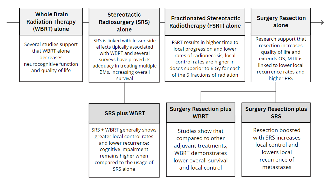
Treatments for brain metastases may ease symptoms, slow tumor growth, and increase the patient’s overall survival. Management options include systemic therapies, surgery, stereotactic radiotherapy (SRT), stereotactic radiosurgery (SRS), whole-brain radiation therapy (WBRT) or some combination of these. Nowadays treatments are adapted to individual cases and have the ultimate goal to reduce toxicity outcomes, while being the most effective, hence, promoting long term survival, as well as quality of life [10]. The medical team first gathers information including patient factors (such as age, sex, overall health, performance status), tumor factors (number and size of brain metastases, primary tumor type, extracranial disease activity), and available treatment options (such as access to neurosurgery or stereotactic radiosurgery) to accurately recommend the most suitable treatment [10, 13, 21]. The following subsections (which are condensed in Figure 2) will go over several treatments.
2.1 Whole Brain Radiation Therapy (WBRT) alone
Whole brain radiation therapy is a type of radiation treatment that delivers radiation to the entire brain, rather than targeting a specific area. Historically speaking, WBRT was considered the standard treatment for patients with brain metastases, as it showed efficacy on metastatic dissemination in the brain, provided good tumor control, and improved symptomatology [22, 23, 24]. WBRT is typically advised for patients with more than 3 brain metastases, with a dose of 30 Gy in 10 daily fractions. This because the treatment covers all brain tissue and has the potential of irradiating yet non-visible lesions [25, 10]. The use of WBRT, however, has decreased throughout the years owing to the progress in radiation technology that allows targeted radiation delivery, as well as the mounting apprehensions concerning the toxicity outcome effects linked with WBRT [22, 26, 27, 28]. Studies (Table 2) have concluded that management of brain metastases using WBRT alone can result in a decline of the patient’s neurocognitive capacity [29, 30]. Salzmann et al. [30] investigated a cohort of 8 patients who had suffered from melanoma brain metastases (MBM) and treated with WBRT to assess whether they had suffered cognitive decline after treatment and analyze the long-term neurocognitive effects of WBRT. Results showed that 5 patients experienced little restriction on day-to-day life and the remaining 3 experienced cognitive decline associated with the area where the tumor mass developed. The study was found to be limited for its small patient sample, although it proved to be relevant for the topic’s discussion. More recently, Yao et al. [29] aimed to evaluate the correlations between psychological distress, cognitive impairment, and quality of life in patients with brain metastases after WBRT. The study enrolled 71 patients with brain metastasis treated with WBRT, who were investigated with several scales before and after WBRT. Results showed that after receiving WBRT, the cognitive function and quality of life of patients decreased, while psychological distress increased.
The management techniques regarding multiple brain metastases are in constant trial to expand the barriers of treatment approaches aiming different scenarios (i.e., number and size of metastases, primary tumor characteristics, among others). In a 2017 study, Khan et al. [31] compared the effectiveness of WBRT alone, SRS alone, and their combination in the treatment of brain metastases based on randomized controlled trial studies. Five studies (n=763) were included in this analysis and the results showed that no significant survival benefit was observed for any treatment approach, but local control was poorly achieved when WBRT was applied alone, compared to its combination with SRS. On the other hand, no difference in radiation-related toxicities was found among the three approaches. Patil et al. [32] also discussed the efficacy of WBRT plus SRS versus WBRT alone in the treatment of brain metastases. By conducting a meta-analysis of two studies, with a total of 358 participants, no difference in overall survival (OS) between the two groups was found, similarly to the work previously stated. However, participants with one brain metastasis had significantly shorter median survival in the WBRT-alone group. Participants in the combined therapy group had better local control, performance status scores, and decreased steroid use compared to the WBRT-alone group, as in the study by Khan et al. In 2022, Gaebe et al. [33] also compared the efficacy of SRS and WBRT in the treatment of intracranial metastatic disease (IMD) in patients with small-cell lung cancer (SCLC). A systematic review and meta-analysis of 31 studies were conducted, and the primary outcome was overall survival. The results showed that SRS had longer overall survival compared to WBRT alone or with SRS boost, but not with WBRT plus SRS boost. Single-arm studies reported a pooled median overall survival of 8.99 months. The study suggests that survival outcomes are similar between SRS and WBRT in treating IMD in SCLC patients, and future studies should focus on differences in progression between the two treatments.
Whole brain radiation therapy is often used as adjunctive therapy or as monotherapy for metastases that are not removable by surgery or unresponsive to radiation. The treatment is often applied to patients with multiple metastases (3 metastases). Stereotactic radiosurgery (SRS), on the other hand, has become a more widely used treatment option for multiple brain metastases due to advancements in technology, but upfront WBRT remains the standard approach for such patients [10, 23, 28].
| Study | Year | Purpose | Arm | Clinical Outcomes |
|---|---|---|---|---|
| Khan et al. [31] | 2017 | Compare the effectiveness of WBRT alone, SRS alone, and their combination in the treatment of BMs | SRS alone WBRT alone SRS + WBRT | No significant survival benefit was observed for any treatment approach; local control was better in SRS+WBRT arm; no difference in radiation-related toxicities was found among the three approaches |
| Patil et al. [32] | 2017 | Compare the efficacy of WBRT + SRS versus WBRT alone in the treatment of BMs | WBRT alone SRS + WBRT | No difference in OS between the two groups; WBRT alone had shorter median survival in patients with 1 BM; WBRT+SRS had better local tumor control |
| Salzmann et al. [30] | 2022 | Assess long-term neurocognitive effects on patients diagnoses with MBM, treated with WBRT | WBRT | Three patients experienced cognitive decline associated with tumor location |
| Gaebe et al. [33] | 2022 | Compare the efficacy of SRS + WBRT in the treatment of IMD associated with SCLC | SRS alone WBRT alone SRS + WBRT | Survival outcomes are similar in the SRS and WBRT arms |
| Yao et al. [29] | 2023 | Evaluate psychological distress, cognitive impairment, and quality of life in patients with brain metastases after WBRT | WBRT | Cognitive function and quality of life, while psychological distress increased |
2.2 Stereotactic Radiosurgery (SRS) alone
Stereotactic radiosurgery (SRS) is a non-invasive technique for treating both intracranial and extracranial lesions. Throughout history, neurosurgeons and physicists developed various stereotactic devices, imaging techniques, and radiation energy sources, however, it would only be in 1951, that a Swedish neurosurgeon by the name of Lars Leksell would come to introduce the concept of SRS. Over the course of the past 70 years, technological advancements greatly improved treatment precision, patient comfort, and dose fractionation making SRS today a highly versatile and cost-effective therapy [34, 35].
Contrary to WBRT, SRS involves the precise focusing of radiation from multiple angles and provides a confined area of high-dose radiation. By using multiple, convergent beams of high energy, SRS decreases the dose of radiation reaching healthy tissue and allows avoidance of radiation sensitive tissue like the optic nerve [36]. SRS has become a popular alternative to WBRT due to its shorter and less invasive treatment course. It can be used alone for patients with a limited number of BMs or in combination with WBRT, systemic therapies, targeted therapies, and immunotherapies [37]. Earlier research already favored the usage of SRS alone to preserve neurocognitive function. Chang et al. [38] compared the effects of SRS alone to SRS plus WBRT on the learning and memory functions of patients with 1-3 newly diagnosed BMs. The cohort included a total of 58 patients (n=30 in the SRS alone group, n=28 in the SRS plus WBRT group) from which those who received SRS plus WBRT had significant chance of learning and memory function decline at 4 months, compared to those receiving SRS alone. Ultimately, the study showed that SRS alone entails lesser toxicity effects usually associated with WBRT, making it a far better choice as primary treatment for multiple metastases.
The improvement of radiation therapy and imaging technology, along with the acknowledgement of the adverse effects of WBRT have sparked the interest in using SRS as an alternative for patients with multiple BMs [39]. Several clinical evidence (Table 3) could support such statement [39, 40, 41, 42]. A study published in 2014 by Yamamoto et al. [42] aimed to determine whether SRS alone as the initial treatment for patients with 5-10 BMs was non-inferior to that for patients with 2-4 BMs in terms of overall survival (OS). A total of 1,194 eligible patients were enrolled, and standard SRS procedures were used in all patients. The study found that overall survival did not differ significantly between patients with 2-4 metastases and those with 5-10. Hence, considering the minimal invasiveness of SRS and the minimal outcome toxicity compared to WBRT, SRS alone proved to be a suitable alternative for patients with up to 10 brain metastases.
Following this investigation, Amaan Ali et al. [41] intended to understand whether the number of BMs alone had major impact on the choice between SRS or WBRT treatments. The investigation gathered a cohort of 5750 patients with BMs who underwent SRS. The researchers categorized the patients based on the number of BMs they had, with categories of 1, 2-4, 5-10, and 10 metastases. The median OS was compared between the categories, and a multivariate analysis was performed to account for other factors such as age, Karnofsky Performance Score (KPS), systemic disease status, tumor histology, and cumulative intracranial tumor volume (CITV). The results showed that patients with a single metastases (1 BM) had superior median OS compared to those with 2-4 BMs (7.1 months vs. 6.4 months, p=0.009); the OS in patients with 2-4 BMs was not significantly different from those with 5-10 BMs (6.4 months v. 6.3 months, p=0.170). Patients with 10 BMs had lower median OS than those with 2-10 BMs (5.5 months vs. 6.3 months, p=0.025). The multivariate analysis showed that when comparing patients with 1 BM to those with 2-10 BMs, or those with 10 BMs to those with 10 BMs, risk of death increased by 10%. In other words, patients with a higher number of BMs had a higher risk of death. On the other hand, when the number of BMs was modeled as a continuous variable, the analysis showed that there was a 5% increase in the risk of death for every increment of 5-6 BMs. This means that even small increases in the number of BMs can have an impact on survival. Accordingly, the research suggests that the number of BMs alone doesn’t have primary impact on OS and therefore should not be considered as a major factor when deciding between SRS and WBRT. Other factors, such as total tumor volume, prescribed dose, age, systemic disease status, among others, should also be considered when making treatment decisions.
Treating each BM separately with radiation therapy can be time-consuming and may limit the feasibility of SRS for multiple BMs. Following this, a retrospective survey by Limon et al. [39] explored how the single-isocenter, multitarget (SIMT) for SRS planning and delivery would impact the treatment of BMs in order to identify those patients who might benefit the most from this procedure. The cohort included 59 patients with a median follow-up of 15.2 months and a median OS of 5.8 months. The study suggested that the number of metastases did not represent a major impact on OS. On the other hand, better OS was registered on planning tumor volume (PTV) 10 cc versus 10 cc (7.1 vs 4.2 months, respectively; ), which indicates the treatment works more effectively in smaller volumes. Another factor influencing OS was the dose administered to the PTV. Results showed that PTVs treated with 19 Gy were associated with increased overall survival. Conversely, patients receiving a dose of 12 Gy to 10 cc of normal brain had worse survival. Although it presented a limited follow-up period and a small cohort of patients, this study suggested that using SRS for the treatment of multiple BMs is effective and that outcomes such as OS might be less affected by the number of lesions and more by the tumor volume of those.
Another study conducted by Mizuno et al. [40] suggests that SRS as an upfront treatment for 10-20 BMs could potentially delay the need for WBRT and therefore, its associated adverse events. This retrospective research intended to compare the efficacy and safety of SRS and WBRT in patients with advanced non‑small cell lung cancer (NSCLC) presenting 10‑20 BMs. The cohort included 44 patients with 10 to 20 brain metastases from NSCLC who had been treated with SRS or WBRT as an initial treatment. The findings indicated no significant difference in OS between the two groups, however, the median time to intracranial progression (TTIP) was significantly shorter in the SRS group than in the WBRT group (7.1 months vs. 19.1 months, P=0.009), meaning the disease progressed more rapidly in the SRS group. Neurological survival did not differ between the two groups, and the type of initial treatment (WBRT or SRS) was not found to be a significant prognostic factor in the univariate and multivariate analyses. However, other factors such as histology, performance status, subsequent molecular targeted drugs, subsequent chemotherapy, and salvage treatment were identified as independent prognostic factors. More recently, Shafie et al. [43] also compared the effectiveness of SRS alone compared to WBRT alone for treating multiple BMs. The research analyzed data from 128 patients over a 5-year period, assessing various outcomes. Results revealed that patients who received SRS had a median of 4 BMs, and the 1-year local control of individual BM after SRS was roughly 92%. Median OS was significantly longer in the SRS arm, over 15 months, compared to the 8 months in the WBRT subgroup. The distant intracranial progression-free survival (PFS) was shorter in the SRS subgroup (close to 9 months) compared to the WBRT subgroup (about 22 months), indicating that patients who receive WBRT might have lower hazard of distant intracranial progression. The survey also found that synchronous BM diagnosis, higher initial number of BMs and lung cancer histology, were detrimental factors negatively affecting survival after SRS. Ultimately, the study supported the use of SRS as a feasible and effective treatment option for patients with multiple BMs. SRS alone was associated with longer OS compared to WBRT.
More recent research by Ogawa et al. [44] assessed the effectiveness of fractionated stereotactic radiosurgery (FSRS) as a treatment for larger BMs ( 20 mm). 105 patients with a total of 116 BMs were examined with a median maximum tumor diameter of 25 mm, and median prescribed dose of 35 Gy in 3 fractions. The findings revealed that FSRS achieved good local control with a local failure rate of 12.5% and an intracranial failure rate within 1 year of nearly 57%. Because of multiple and local recurrences, 21 and 4 patients had to be submitted to WBRT and surgery after FSRS, respectively. The study concluded that BMs greater than 20 mm present worthy local control when treated with fractioned SRS. Moreover, the eligibility of the survey proved that FSRS is a potential alternative to surgery since few patients were administered to surgery after FSRS treatment.
Recent research by Chea et al. [45] compared the efficacy of Linac-based mono-isocentric SRS with BrainLab® Elements Multiple Brain Mets (MBM) SRS to the Gamma Knife for the treatment of patients with 3-9 BMs. The study involved 20 patients who were previously treated with Gamma Knife SRS. The patients had 3 to 9 BMs from different primary malignant tumors. Totally there were 95 metastases with a major axis diameter ranging from 0.3 to 4 cm and volume ranging from 0.02 to 9.61 cc. Various statistical methods were used to evaluate the dosimetric impact of target volume geometric characteristics, including the Paddick Index (PI), Gradient Index (GI), dose fall-off, volume of healthy brain receiving more than 12 Gy, and dose volume histogram (DVH). Results showed that both approaches have similar plan qualities, with MBM having somewhat better selectivity for smaller lesions but Leksell Gamma Plan (LGP) having slightly better healthy tissue sparing at the cost of a significantly longer irradiation period. For larger volumes, MBM strategies must take a higher healthy tissue exposure into account. Several research supports the usage of Elements MBM in generating high quality plans for treatment of multiple brain metastases. Raza et al. [46] concluded that MBM had the same capacity to generate high quality plans in patients presenting up to 25 BMs, as the Varian Hyperarc (HA) system. Additionally, Cui et al. [47] established that compared to knowledge-based planning (KBP), the MBM plans had a higher capacity to spare normal brain tissues in terms of total volumes receiving 5 Gy.
When managing multiple BMs, plenty of studies support the efficacy of treatment with SRS as a far better choice than WBRT, regarding the side effects. However, the number of brain metastases alone is not sufficient factor to determine the best treatment approach and medical professionals should account for several other aspects such as age, histology, performance status, tumor volume and prescribed dose.
| Study | Year | Purpose | Arm | Clinical Outcomes |
|---|---|---|---|---|
| Chang et al. [38] | 2009 | Compare the effects of SRS alone and SRS + WBRT on the cognitive functions of patients with 1-3 newly diagnosed BMs | SRS alone SRS+WBRT | SRS alone entailed lesser toxicity effects usually associated with WBRT |
| Yamamoto et al. [42] | 2014 | Compare OS in patients receiving SRS alone as the initial treatment of 5-10 BMs and 2-4 BMs | SRS alone | OS did not differ significantly between patients with 2-4 BMs and 5-10 BMs; SRS alone proved to be a suitable alternative for patients with up to 10 BMs |
| Amaan Ali et al. [41] | 2017 | Understand whether the number of BMs alone had a major impact on the choice between SRS or WBRT | SRS alone WBRT alone SRS + WBRT | Patients with 1 BM had superior OS compared to those with 2-4 BMs (7.1 months vs. 6.4 months); OS in patients with 2-4 BMs was not significantly different from those with 5-10 BMs (6.4 months vs. 6.3 months); patients with 10 BMs had lower median OS |
| Limon et al. [39] | 2017 | Understand how the single-isocenter, multitarget (SIMT) for SRS planning and delivery would impact the treatment of BMs | SIMT SRS | Number of metastases did not represent a major impact on OS; Better OS was registered on PTV 10 cc versus 10 cc (7.1 vs. 4.2 months, respectively); PTVs treated with 19 Gy were associated with increased OS |
| Mizuno et al. [40] | 2019 | Compare the efficacy and safety of SRS and WBRT in patients with advanced non‑small cell lung cancer (NSCLC) presenting 10‑20 BMs | SRS alone WBRT alone | Median TTIP was significantly shorter in the SRS arm compared to the WBRT arm (7.1 months vs. 19.1 months); The type of initial treatment was not found to be a significant prognostic factor |
| Shafie et al. [43] | 2020 | Compare the effectiveness of SRS alone vs WBRT alone for treating multiple BMs | SRS alone WBRT alone | Median OS was longer in the SRS arm, more than 15 months, compared to 8 months in the WBRT arm |
| Ogawa et al. [44] | 2022 | Assess the effectiveness of fractionated SRS as a treatment for larger BMs (20 mm) | FSRS (35 Gy in 3 fractions) | FSRS achieved good local control with a local failure rate of 12.5% and an intracranial failure rate within 1 year of nearly 57% |
2.3 Fractionated Stereotactic Radiotherapy (SFRT)
Fractionated stereotactic radiotherapy (FSRT) is a specialized technique in radiation therapy that combines the precision of stereotactic radiosurgery (SRS) with the delivery of radiation in multiple fractions. For patients with a small number of brain metastases, SRS is an enticing alternative. However, the efficacy and safety of SRS might be jeopardized when treating large lesions or those close to healthy tissue. In these situations, FSRT is frequently employed in an effort to raise the ratio between the desired therapeutic effects and the potential side effects caused by radiation [48, 49]. Several clinical trials (Table 4) have proven the efficacy of FSRT, namely five-fraction SRT, as an advantageous alternative for managing multiple lesions, especially regarding LC and radionecrosis rates.
A study by Putz et al. [50] compared the effectiveness and safety of FSRT and SRS for the treatment of BMs. The survey included a cohort of 120 patients (n=98 treated with FSRT and n=92 treated with SRS). Results showed that the biologically effective dose (BED) for metastases was related to local control, while the BED for normal brain tissue was associated with radionecrosis. The median time to local progression was nearly 23 months in the FSRT group compared to the 14.5 months in the SRS arm. There was a significant difference in overall rate of radionecrosis at 12 months with 3.4% vs. 14.8% for FSRT and SRS, respectively. The incidence of severe radionecrosis requiring resection was also significantly lower in the FSRT group. The study proved the efficacy of FSRT in treating both large and small BMs, highlighting its potential in local control and reduced hazard of radionecrosis.
A retrospective survey by Piras et al. [51] investigated the optimal dose schedule for five-fraction SRT in the treatment of BMs. The analysis was conducted on 41 patients treated with different five-fraction SRT dose schedules, administered over 5 consecutive days, with prescribed doses ranging from 30 to 40 Gy and covering at least 98% of the gross tumor volume (GTV). Magnetic resonance imaging (MRI) was performed at 3, 6, and 9 months to evaluate LC and clinical outcomes and data related to toxicity was also acquired. The evidence suggested that higher LC rates were found when dose schedules exceeded 6 Gy per fraction and that the toxicity rates were generally mild. Based on these findings, the study concludes that the management of BMs with five-fraction SRT is feasible. The analysis also suggests that a total dose of less than 30 Gy in 5 fractions should be avoided due to the expectation of lower LC. Another research by Layer et al. [52] also evaluated the safety of five-fraction SRT for the treatment of BMs as a definitive or adjuvant treatment. The study analyzed data from 36 patients who received FSRT (5 fractions of 7 Gy each) to BMs or cavities after resection. Findings showed that the overall RN rate was more than 14%, and the median time to RN occurrence was almost 13 months. Factors associated with RN occurrence were immunotherapy, young age (45 years), and larger planning target volume (PTV). The cumulative 1-year LC rate was over 83%, indicating effective tumor control. The estimated median LPF survival was roughly 19 months. Ultimately, the study concluded that FSRT is a feasible and safe treatment approach for brain metastases. It demonstrated acceptable local control rates and comparable rates of radiation necrosis in both adjuvant and definitive radiotherapy settings. Similar to the previous paper by Piras et al. this research also supports the efficiency of five-fraction SRT as a great treatment option for patients with BMs.
Following this study, Ding et al. [53] intended to evaluate the feasibility and potential benefits of online adaptive MR-guided FSRT for patients with BMs. This survey included 28 patients diagnosed with BMs, treated with FSRT (30 Gy in 5 fractions) using a 1.5 T MR-Linac. Daily MRI scans were used, and customized treatment plans based on the contours identified from the MR images were developed. For all lesions assessed, the results demonstrated a significant decrease in tumor volume during FSRT in comparison to the pre-treatment simulation. The inter-fractional alterations in lesions with perilesional edema, which accounted for more than 53% of the lesions, were substantially different from those in lesions without it. Compared to patients with a single metastasis, patients with multiple metastases experienced more inter-fractional tumor alterations, such as tumor volume reduction and anatomical shift. In terms of treatment planning, 19% of the fractions in the non-adaptive plans showed insufficient PTV coverage. The study concluded that significant inter-fractional tumor changes can occur during FSRT, and that the daily MR-guided re-optimization of treatment plans provided dosimetric benefits, particularly for patients with perilesional edema or multiple lesions. This highlights the potential advantages of online adaptive MR-guided FSRT in optimizing treatment delivery and maintaining target coverage during the course of treatment.
| Study | Year | Purpose | Arm | Clinical Outcomes |
|---|---|---|---|---|
| Putz et al. [50] | 2020 | Compare the efficacy of FSRT and SRS in the treatment of BMs | FSRT alone SRS alone | Median time to local progression was 22.9 months in the FSRT group and 14.5 months in the SRS arm; there was significant difference in overall rate of radionecrosis at 12 months (3.4% for FSRT vs. 14.8% for SRS); the incidence of severe radionecrosis was significantly lower in the FSRT group |
| Piras et al. [51] | 2022 | Investigate the optimal dose schedule for five-SRT in the treatment of BMs | FFSRT alone | Higher LC rates for dose schedules superior to 6 Gy per fraction; toxicity rates were not found to be higher than Grade 1 (mild) |
| Layer et al. [52] | 2023 | Determine the safety of five-fraction SRT for BMs, either as a unique or adjuvant treatment | FFSRT alone Resection + FFSRT | Overall RN rate was 14.3%; median time to RN was roughly 12.9 months; RN occurrence was associated with immunotherapy, young age (45 years), and large PTV; estimated cumulative 1-year LC rate was 83.1%; the median local PFS was 18.8 months; estimated median OS was 11 months |
| Ding et al. [53] | 2023 | Assess the potential benefits of online adaptive MR-guided FSRT in patients with BMs | FSRT alone | Significant tumor volume reduction was found during FSRT compared to initial fraction; patients with multiple lesions had more significant inter-fractional tumor changes than those with single lesion |
2.4 Surgery Resection
Surgical resection is one of the treatment options for brain metastases and involves the removal of the tumor from the brain by surgical procedure, without damaging regions responsible for critical brain functions, surrounding the lesions. The decision to perform surgery depends on several factors, including the number, size, location of the tumors, the patient's overall health, and the presence of symptoms such as neurological deficits [5, 23]. Although there aren’t many prospective studies confirming the effectiveness of surgery resection in the treatment of multiple BMs, there is growing evidence that it can improve functional outcomes and increase the possibility of receiving additional therapies that are important for overall cancer prognosis, such as SRS or systemic therapies. Although surgical resection is generally recommended for patients with a single BM, studies suggest that this procedure can also be effective when applied to multiple metastases. Moreover, surgical resection can improve clinical status and overall survival rates, improve quality of life and patient’s neurocognitive ability (Table 5) [23, 54, 55, 56, 57].
A retrospective study by Schödel et al. [55] investigated the relationship between surgical resection of BMs and subsequent treatment and clinical outcomes. The research concluded that surgical resection could improve clinical status and enhance the probability of receiving a second treatment, leading to an increased overall survival. The authors analyzed a large cohort of 750 patients who underwent resection of symptomatic BMs. Results showed that surgical resection significantly improved patients' functional status, with a median Karnofsky Performance Score of 80 before surgery, increasing to 90 after surgery. Hence, after resection patients presented a higher level of function and performance of daily activities independently. Also, patients who received postoperative local radiotherapy and systemic treatment had significantly longer survival. Another research carried out by Jünger et al. [56] also suggested that resection of symptomatic BMs could be indicated to patients with multiple BMs to ease their neurological symptoms and facilitate further consecutive treatments. The study gathered a cohort of 216 patients (n=129 were diagnosed with a single BM; n=64 had 2-3 BMs; n=23 had more than 3 BMs) who underwent surgical resection and collected demographic, clinical, and tumor-associated parameters. Similar to Schödel et al., they found that surgical resection of symptomatic BMs significantly improved the KPS and enabled adjuvant radiotherapy and systemic treatment for up to 90% and 55% of patients, respectively. Among other results, the number of BMs did not influence the local control rates and multiple prognostic factors such as age, preoperative and postoperative KPS, presence of extracranial metastases and consecutive radiation therapy and systemic treatment influenced overall survival. In a more recent study, Winther et al. [57] concluded that overall survival rates were improved with total resection of GTV compared to subtotal resection. The researchers reviewed 373 adult patients who underwent surgery for a single BM and found that gross total resection (complete removal of visible tumor) was associated with longer overall survival (13.0 months) compared to subtotal resection (8.0 months). In addition to the previous studies, these findings suggested that the extent of surgical resection also takes an important factor in the management of BMs allowing to improve overall survival.
Patients with BMs are becoming more prevalent as a result of improved treatment options, growing medical standards, and ongoing development. Although surgical resection of original BMs is well defined and supported by numerous research, the use of resection for recurring BMs is still poorly understood [54, 58]. Following the previous study by Winther et al., Gong et al. [59] further intended to investigate whether margin total resection (MTR) could reduce the local recurrence rate of BMs from lung adenocarcinoma compared to gross total resection (GTR). This retrospective research involved 48 patients diagnosed with BMs from lung adenocarcinoma (LUAD). The variable under study was the local tumor control from GTR and MTR. The MTR group consisted of patients who had undergone GTR and had the tumor periphery resected for more 5 mm. Results showed that the local recurrence rates were significantly lower in the MTR group, for 6 months after surgery the local recurrence rate was roughly 14% in the MTR group compared to around 42% in the GTR group. The median progression-free survival rate after surgery was twice as high in the MTR group compared to the GTR group, 14.0 months, and 7.0 months, respectively. In conclusion, although resection of the gross tumor volume might improve overall survival, expanded peripheral resection of 5 mm around the BMs can significantly reduce the local recurrence rate and prolong progression-free survival time.
A retrospective study performed by Heßler et al. [58] evaluated the effectiveness of surgical resection for recurrent symptomatic BMs. The research gathered a total of 107 patients with multiple occurrences of primary tumors (non‑small cell lung cancer (NSCLC), breast, melanoma, gastro‑intestinal, among others) who were also previously individually treated. The variables at study were intracranial event-free survival (EFS) and overall survival (OS), and results showed that the median postoperative EFS and OS were roughly 7 and 11 months, respectively. The median pre‑operative KPS was 70%, improving to 80% after surgery, meaning that resection increased patient's functional status and independence to perform activities of daily life. However, complication rate was greater than 26%. Furthermore, the patient’s clinical status proved to be the only factor to remain independent from survival analysis. The study suggested that although surgical resection of recurrent BMs might improve the clinical status and OS, it is associated with a high complication rate. Therefore, it is crucial to carefully select patients before deciding on surgical resection for recurrent BMs.
| Study | Year | Purpose | Arm | Clinical Outcomes |
|---|---|---|---|---|
| Schödel et al. [55] | 2020 | Understand the relationship between surgical resection of BMs and subsequent treatment, and clinical outcomes | Resection alone | After resection patients presented higher level of function; patients who received postoperative treatment had significantly longer survival |
| Jünger et al. [56] | 2021 | Evaluate the role of surgical resection of BMs in patients with NSCLC, regardless of the number of lesions | Resection alone | Significant improvement in KPS after resection; BM count did not significantly influence local control rates; the mean OS after surgery was 12.7 months |
| Winther et al. [57] | 2022 | Investigate the association between OS and residual tumor after surgery for single BM | Resection alone | Median OS was 8.0 months for patients with subtotal resection and 13.0 months for patients with gross total resection |
| Gong et al. [59] | 2022 | Investigate whether MTR can reduce the local recurrence rate of BM from lung adenocarcinoma compared with GTR | Marginal Tumor Resection alone Gross Tumor Resection alone | Local recurrence rates 6 months after surgery: GTR group was 42.3%, MTR group was 13.6%. Local recurrence rates 12 months after surgery: GTR group was 57.7%, MTR group was 22.7%. Median progression-free survival time after surgery: GTR group was 7.0 months, MTR group was 14.0 months |
| Heßler et al. [58] | 2022 | Evaluate the efficacy of surgery for pretreated, recurrent, and symptomatic BMs | Resection alone | Median KPS was 80% after surgery; median postoperative EFS and OS were 7.1 and 11.1 months, respectively; clinical status remained the only independent factor for survival in multivariate analysis |
2.5 Stereotactic Radiosurgery (SRS) plus WBRT
Treating brain metastases with WBRT alone has been the standard approach however, the toxicity outcomes have led to a wide appliance of SRS to manage multiple metastases [39, 40, 41, 42, 60]. Nonetheless, when WBRT is not used, there is a greater chance of brain tumor recurrence (BTR), which could potentially negatively impact the overall survival of certain patients [60]. Results from a study led by Stafinski et al. [61] showed that not only did combining SRS with WBRT improve OS in patients with a single BM, but also improved local tumor control and functional independence. The research intended to assess the effectiveness of SRS alone or in combination with WBRT, compared to surgery and/or WBRT in prolonging survival and improving quality of life and functional status of patients with BMs. Patients who presented multiple BMs, did not show any difference in OS when treated with WBRT plus SRS, compared to treatment with WBRT alone. However, in patients who had a single metastasis, the same combination therapy resulted in a significant improvement in survival rates. In terms of controlling the tumor locally over a 24-month period, the rates were significantly higher in the group of patients receiving WBRT plus SRS, regardless of the number of metastases. A study by Chang et al. [38] mentioned above, also compared the effectiveness of SRS plus WBRT compared to SRS alone. Although in terms of cognitive decline, SRS alone proved to have far better response, patients who had received SRS plus WBRT presented a higher rate of freedom from CNS recurrence at 1 year (73% of patients), compared to those who had received SRS treatment alone (27% of patients). This means that combining SRS with WBRT resulted in absence of cancer recurrence in the brain or spinal cord after treatment. When comparing SRS alone, WBRT alone and SRS plus WBRT, any of the treatments offer significant survival benefit. Nonetheless, SRS combined with WBRT has proven to be effective when it comes to control of local tumor volume and extended brain tumor recurrence (BTR) free time, period during which a patient does not experience the recurrence of brain tumors after being treated [61, 31, 62].
Another research conducted by Hasan et al. [63] determined the effectiveness of adding WBRT following SRS for treating brain metastases. The study analyzed three randomized controlled trials, involving 389 patients with 1 to 4 brain metastases, and five retrospective studies, reporting on outcomes such as survival, control, salvage therapy, and quality of life measures. The study found that adjuvant WBRT following SRS was more effective at tumor local control (1-year local control was roughly 90% for SRS + WBRT and little above 70% for SRS alone) and distant recurrence (mean crude distant recurrence rate for SRS + WBRT was near 40% and 54% for SRS alone) than SRS alone. However, the study also found that SRS plus adjuvant WBRT did not benefit in terms of overall survival (mean 1-year survival was little above 33% for SRS + WBRT and roughly 39% for SRS alone) or symptomology. The addition of WBRT was linked to a lower quality of life, and its known link to cognitive decline and neurotoxicity should be evaluated against the advantage of local control.
So far, the analysis of the three works reinforces the debated character of combining these treatments to manage brain metastases. The works suggest contrary findings in terms of the influence of SRS plus WBRT in terms of overall survival and preservation of neurocognitive ability depending on the number of metastases to be cured. Moreover, all the data indicated that in terms of recurrence and capacity of local control this combination seems to have greater effect than implementing SRS alone. In a later retrospective investigation by Brown et al. [64] concluded that for patients with 1 to 3 BMs, the use of SRS alone, compared with SRS combined with WBRT, resulted in less cognitive deterioration. The study also suggested that there was no difference in terms of overall survival between the two groups. The work’s primary quest was to answer the debating question of whether there could be less cognitive deterioration at SRS alone compared to SRS plus WBRT. The research involved 213 participants who randomly received either SRS alone or SRS plus WBRT. Findings indicated that there was less cognitive deterioration at 3 months after SRS alone (roughly 64% of patients) than when combined with WBRT (nearly 92% of patients) and quality of life was also higher at 3 months with SRS alone.
Many studies regarding treatment with SRS plus WBRT present inconclusive results regarding overall survival rates. Following this, Khan et al. [65] assessed whether the improved local control achieved with WBRT plus SRS leads to any survival benefit in patients with BMs and favorable prognostic factors. Five studies (n=2728) were identified, and the primary outcome was overall survival. It became apparent that WBRT plus SRS improved survival in brain metastatic cancer patients with better prognostic factors, particularly when compared to WBRT alone. Nonetheless, the survival advantage over SRS was only limited to non-small cell lung cancer primary tumor histology. This way, the research suggests that WBRT combined with SRS might indeed improve overall survival, considering selected patients with favorable prognostic factors. As seen previously, WBRT has an impact in tumor volume control, despite its toxicity side effects on cognitive function. A more recent investigation undertaken by Nakano et al. [60], intended to assess whether combining reduced-dose whole-brain radiotherapy (RD-WBRT) with SRS, could potentially minimize the risk of cognitive decline, without compromising the brain tumor control for patients with 1-4 BMs. The standard-dose WBRT (SD-WBRT) consists of 30 Gy in 10 fractions, whereas RD-WBRT consists of 25 Gy in 10 fractions). The study enrolled 40 patients from seven different institutions (28 patients with primary tumor in the lung; 20 patients with single BM). Results showed that the median OS time was 19 months; more than 75% of the patients didn’t experience the recurrence of brain tumors in distant locations at 6 months after treatment; 23% of patients experienced the recurrence of brain tumor at any location at 6 months after treatment, considering the possibility of death; and roughly 49% of patients experienced a decline in cognitive function that persisted over time. The work showed that RD-WBRT combined with SRS may reduce the risk of cognitive decline when compared to standard WBRT.
The issue with combining SRS with WBRT to treat brain metastases prevails, as incoherence whether this treatment option influences overall survival, neurocognitive decline, local control, and metastases recurrence persist. Moreover, results vary attending the number of BMs treated. Several studies (Table 6) refer WBRT combined with SRS, to have great effect on patients’ local tumor control over time and preventing metastases recurrence, however because of the different results concerning neurotoxicity and influence on survival, this topic remains unclear and in need of further research.
| Study | Year | Purpose | Arm | Clinical Outcomes |
|---|---|---|---|---|
| Stafinski et al. [61] | 2006 | Assess the effectiveness of SRS alone or SRS+WBRT, compared to surgery and/or WBRT in prolonging survival and improving quality of life and functional status of patients with BMs | SRS alone SRS+WBRT Resection alone Resection + WBRT | In patients with a single BM SRS+WBRT improved OS, local tumor control and functional independence |
| Chang et al. [38] | 2009 | Compare the effects of SRS alone and SRS + WBRT on the cognitive functions of patients with 1-3 newly diagnosed BMs | SRS alone SRS+WBRT | SRS alone entailed lesser toxicity effects usually associated with WBRT |
| Hasan et al. [63] | 2014 | Determine the effectiveness of SRS+WBRT for treating 1 to 4 BMs, reporting on outcomes such as survival, control, salvage therapy, and quality of life measures | SRS alone SRS+WBRT | At 1-year local control was roughly 90% for SRS + WBRT and little above 70% for SRS alone; mean crude distant recurrence rate for SRS + WBRT was near 40% and 54% for SRS alone; SRS+WBRT did not benefit in terms of OS or symptomology |
| Brown et al. [64] | 2016 | Answer the debating question of whether there could be less cognitive deterioration at SRS alone compared to SRS plus WBRT | SRS alone SRS+WBRT | There was less cognitive deterioration at 3 months after SRS alone (roughly 64% of patients) than when combined with WBRT (nearly 92% of patients); quality of life was higher at 3 months with SRS alone |
| Khan et al. [65] | 2019 | Assess whether improved local control achieved with WBRT plus SRS leads to any survival benefit in patients with BMs and favorable prognostic factors | SRS+WBRT | WBRT+SRS might improve OS, considering selected patients with favorable prognostic factors |
| Nakano et al. [60] | 2022 | Assess whether combining reduced-dose whole-brain radiotherapy (RD-WBRT) with SRS could potentially minimize the risk of cognitive decline, without compromising the brain tumor control for patients with 1–4 BMs | RD-WBRT (25 Gy in 10 fractions) + SRS | Median OS time was 19 months; more than 75% of the patients didn’t experience the recurrence of brain tumors in distant locations at 6 months after treatment; roughly 49% of patients experienced a decline in cognitive function that persisted over time |
2.6 Surgery Resection plus WBRT
SRS is frequently preferred to WBRT since neurocognition and quality of life are increasingly being used as key therapy objectives in patients with brain metastases, even though WBRT affords significant intracranial disease control by targeting the entire brain [23, 66, 67]. Moreover, while SRS might increase the hazard of distant brain failure it would seem to be as effective as WBRT when it comes to local tumor control and overall survival, withdrawing the associated risks within it [66].
Some early studies (Table 7) are indicative that surgery resection combined with WBRT might reduce intracranial relapses and neurologic deaths however it is not certain how it influences the duration of a patient’s functional independence since some studies present contrary results regarding this matter. In terms of overall survival, resection combined with WBRT does not reveal any advantages. A study by Hart et al. [68] intended to assess the clinical effectiveness of surgical resection combined with WBRT versus WBRT alone in the treatment of patients with a single BM. The research comprehended three randomized controlled trials (RCTs) involving a total of 195 patients. Results showed that although surgery plus WBRT did not significantly improve overall survival compared to WBRT alone, it did however increase the duration of functionally independent survival (FIS). Nevertheless, this study is contradicted by later research by Kocher et al. [69] who stated that though adjuvant WBRT decreases neurologic fatalities and intracranial relapses, it does not extend functional independence or increase overall survival. In this analysis the authors set out to determine whether adjuvant WBRT could lengthen the period of functional independence following BMs radiosurgery or resection. 359 patients (n=199 underwent SRS; n=160 underwent resection) with 1 to 3 BMs originated from solid tumors were randomly assigned to adjuvant WBRT or observation. According to the data, adjuvant WBRT reduced intracranial relapse rates (59% to 27% and 42% to 23%, at initial and new sites, respectively) and neurologic deaths regardless, it did not improve the duration of functional independence (approximately 10 months for both WBRT and observation groups) or overall survival (roughly 11 months for both WBRT and observation groups).
More recent studies have shown that the use of post-operative WBRT resulted in a shorter OS and did not provide as much of local tumor control as other methodologies of treatment such as SRS or FSRT. Moreover, WBRT reveals to accelerate the decline of cognitive function compared to other treatments. Research by Shafie et al. [70] compared the use of single-session SRS or fractionated SRS (FSRS) of the resection cavity versus WBRT in treating BMs after surgery. 101 patients participated in the study, 50 of which were treated with SRS/FSRS and 51 were treated with WBRT. Findings indicated that patients who received SRS/FSRT had longer OS and local control compared to those who received WBRT. Following this analysis, Koo et al. [71] retrospectively investigated which factors affected OS, local and distant control, and leptomeningeal metastases (LMM) in patients who had undergone resection of BMs. The study gathered 124 patients who had undergone resection of BM, and results showed that although both WBRT and localized radiotherapy (local brain radiotherapy or stereotactic radiosurgery (LBRT/SRS)) demonstrated great local control, LBRT/SRS would still be preferable to avoid WBRT effects on cognitive function and promote an extended survival of cancer patients. Ultimately, research suggests that after resection, alternative treatment options such as SRS may be better for managing brain metastases compared to adjuvant WBRT.
| Study | Year | Purpose | Arm | Clinical Outcomes |
|---|---|---|---|---|
| Hart et al. [68] | 2005 | Assess the clinical effectiveness of surgical resection combined with WBRT versus WBRT alone in the treatment of patients with a single BM | Resection plus WBRT WBRT alone | Surgery plus WBRT did not significantly improve OS compared to WBRT alone but increased the duration of functionally independent survival |
| Kocher et al. [69] | 2011 | Determine whether adjuvant WBRT could lengthen the period of functional independence following 1 to 3 BMs originated from solid tumors | Resection plus WBRT | Adjuvant WBRT reduced intracranial relapse rates and neurologic deaths; resection plus WBRT did not improve the duration of functional independence or OS |
| Shafie et al. [70] | 2020 | Compare the use of single-session SRS or FSRT of the resection cavity versus WBRT in treating BMs after surgery | Resection plus single-session SRS Resection plus FSRS Resection plus WBRT | Patients who received SRS/FSRS had longer OS and local control compared to those who received WBRT |
| Koo et al. [71] | 2021 | Investigate which factors affected OS, local and distant control, and leptomeningeal metastases (LMM) in patients who undergo resection of BMs | Resection plus SRS Resection plus WBRT | After resection, SRS may be better for managing brain metastases compared to adjuvant WBRT |
2.7 Surgery Resection plus SRS
Several published data suggests that resection combined with SRS is an effective treatment for patients with multiple metastases, tending towards longer survival outcomes [72]. Although WBRT might reduce the recurrence rate of BMs following resection, it does not increment overall survival neither quality of life, since WBRT is associated with outcome toxicities namely, neurocognitive decline. This way, surgical resection when combined with SRS is able to achieve great local control, while preventing cognitive defects [23, 73, 74]. An earlier study conducted by Mahajan et al. [74] suggests that SRS after BMs resection could potentially be more beneficial than WBRT, withdrawing the cognitive decline associated with it. The study intended to determine if SRS applied to the surgical cavity after resection could improve time to local recurrence compared with surgical resection alone. Patients were selected by presenting complete resection of 1-3 BMs (maximum diameter of the resection cavity 4 cm), among other characteristics. The findings indicated that resection boosted with SRS significantly lowers local recurrence compared to resection alone, with a 12-month freedom from local recurrence of 72% for the SRS group and 43% for the observation group. Comparing to WBRT, surgical resection followed by SRS could not only prevent local recurrence, but also provide longer quality of life to patients lowering the hazard of cognitive decline. Additionally, the previously stated study by Shafie et al. [70] supports the fact that using SRS or fractioned SRS after surgery resection promotes longer OS and local control. The enhancement of local tumor control by irradiating the surgical cavity was also recently proven by Huang et al. [75]. This retrospective study examined the effects of postoperative SRS on local tumor control and overall survival in 97 patients diagnosed with BMs who underwent Gamma Knife SRS. The study found that irradiating the surgical cavity after resection could enhance local tumor control rate by 75% and overall survival rate (12 months) by near 90%. Hence, combining surgical resection with SRS not only lowers local recurrence but also promotes tumor volume control.
Despite the advancements in surgical care of brain tumors, the complete resection of lesions located near vital neurovascular structures still poses a significant risk of morbidity. Because it is minimally invasive, SRS has low complication rates and excellent tumor control. However, when it comes to larger tumor volumes, SRS alone does not constitute the greatest treatment due to major radiation-induced problems. In recent years, there has been growing interest in Adaptive Hybrid Surgery (AHS) (BrainLab®, Munich, Germany) as a multimodal method to manage these large lesions. In AHS, a planned subtotal resection (STR) with adjuvant SRS for an anticipated residual tumor takes advantage of both strategies’ benefits [76, 77, 78]. Research by Sheppard et al. [78] compared the ideal radiosurgical target volumes defined by a manual method (surgeon) to those determined by Adaptive Hybrid Surgery (AHS) software in 7 patients with vestibular schwannoma (VS). The findings showed that the planned residual tumor volumes were smaller than the ideal radiosurgical target volumes defined by AHS (1.6 cm3 vs. 4.5 cm3, respectively) and the average difference between the ideal radiosurgical target volume defined by AHS and the planned residual tumor volume was 2.9 ± 1.7 cm3. Despite the fact that planned subtotal resection of VS by a surgeon differs from the ideal radiosurgical target defined by AHS, the study did not provide any favorable conclusion regarding the preferred method since both methods influence in clinical outcomes were not defined. Another survey by Cohen-Inbar et al. [76] favors Adaptive Hybrid Surgery (AHS) as a combined strategy for the treatment of large cerebral and skull base tumors. The study suggests that a planned STR followed by SRS to a preplanned residual tumor offers a desirable combination of high tumor control rates and favorable clinical outcomes, including preservation of neurological function and quality of life. Moreover, it recommends that patients with large tumors who cannot be treated with SRS lone due to neurotoxicity outcomes, should be managed with AHS. Recent research by Keinzler et al. [77] evaluated the feasibility and safety of the Adaptive Hybrid Surgery Analysis (AHSA) method in 5 patients with benign skull base tumors. Patients underwent planned partial tumor resection followed by SRS. The AHSA method was able to accurately assess the residual tumor volume during surgery and suggest safe hypo-fractionated radiation plans in all patients. No complications occurred after radiation treatment, and the authors attested the viability of this technology and its great potential in facilitating an optimal multidisciplinary approach and resection strategy, reducing surgical and radiosurgical risks.
Ultimately, surgical resection combined with SRS might be recommended for patients who have 1-3 brain metastases that are relatively small and located in areas of the brain that can be safely accessed through surgery (Table 8). The decision to use resection plus SRS will depend on a number of factors, including the size and location of the tumors, the patient's overall health and prognosis, and the potential risks and benefits of the treatment approach [23, 74, 75].
| Study | Year | Purpose | Arm | Clinical Outcomes |
|---|---|---|---|---|
| Mahajan et al. [74] | 2017 | Determine if SRS applied to the surgical cavity of 1 to 3 BMs after resection could improve time to local recurrence compared with surgical resection alone | Resection alone Resection plus SRS | Resection boosted with SRS lowered local recurrence compared to resection alone; 12-month freedom from local recurrence was 72% in the SRS group and 43% in the observation group |
| Huang et al. [75] | 2023 | Examine the effects of postoperative SRS on local tumor control and overall survival in patients diagnosed with BMs who underwent Gamma Knife SRS | Resection plus SRS | Irradiating the surgical cavity after resection could enhance local tumor control rate by 75% and OS rate (12 months) by near 90% |
3 Follow-up Care after Treatment of Multiple metastases
Even with successful treatment, BMs often recur, so medical professionals recommend close follow-up after treatment. Individuals who have BMs are at significant risk of experiencing recurrence of the tumor in the same area or the development of new lesions in other parts of the brain (distant recurrence). It is important to detect any potential recurrence of the disease, even asymptomatic, to receive prompt treatment. Follow-up care should be tailored to the individual patient and based on factors such as the patient's overall health, the extent of metastases, and the type of treatment initially received. Medical imaging in assistance to follow-up care is an essential tool for evaluating the effectiveness of treatment and distinguishing between any changes resulting from the treatment and those indicating regrowth of the tumor [79].
Monitoring patients after treatment include regular imaging to detect the progression of the disease and potential recurrence of new metastases. Based on each patient’s circumstances, the doctor will determine the frequency imaging should take place. An experiment executed by Jena et al. [80] concluded that imaging surveillance of the brain could detect asymptomatic metastases. The study revealed that the pattern of the lesion may influence the patients’ response to therapy and survival benefit, especially the asymptomatic patients with multiple metastases. The research included 175 patients with BMs derived from primary non-small cell lung cancer (NSCLC). Patients with asymptomatic BMs and those with symptomatic BMs, were parted into two separate groups. Results showed that more than 30% of patients had BMs, and roughly 47% of those were asymptomatic. Also, no difference was found in terms of number, size, site, nature, presence of perilesional edema, and intralesional hemorrhage of the metastasis. Results showed that the number of patients with and without neurological symptoms caused by BMs, was roughly the same. Medical imaging during follow-up of the brain, supports early detection of asymptomatic metastases though the prognosis for these patients doesn’t uniquely depend on the presence or absence of symptoms.
A more recent survey by Derks et al. [81] discussed the challenges posed by BMs and the role of imaging in clinical practice. The authors begin by outlining the rising prevalence of BMs from various primary tumors and draw attention to the difficulty in choosing the correct individuals for screening because not all cancer patients experience BM development. The paper then goes into the imaging methods that are employed to find BMs. A three-dimensional (3D) T1W MRI sequence is the gold standard for BM detection, but other anatomical, functional, and metabolic data from imaging methods like susceptibility weighted imaging, diffusion weighted imaging, perfusion MRI, MR spectroscopy, and positron emission tomography (PET) can help distinguish BMs from other intracranial conditions. The authors also discuss the function of imaging at various stages of BM treatment. For surgical resection, imaging is utilized to choose surgical candidates and provide intraoperative assistance using methods like fluorescence-guided surgery and ultrasound. MRI and CT are combined for SRS therapy planning. After both local and systemic therapy, conventional MRI is utilized for surveillance, but sophisticated imaging is increasingly used to identify real tumor progression.
Overall, effective follow-up care is an important part of the management of patients with multiple BMs. Regular imaging, symptom management, supportive care and patient education should be incorporated into an individualized follow-up care plan. Follow-up care after treatment for detecting new metastases, still represents a clinical challenge since predicting the incidence of new metastases would be a great assistance to medical professionals. This step forward in oncology would allow radiologists and physicians to elaborate earlier secondary treatment plans and study with more time alternatives to manage the newly potential metastases.
4 Guidelines from multiple societies
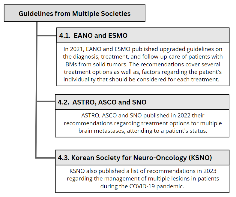
Brain metastases are a serious complication of primary cancer, with an estimated incidence of 10-30% among cancer patients. Although there are several treatment options available, the management of BMs is complex, requiring a multidisciplinary approach and their optimal management remains a topic of debate, with no clear consensus on the best approach [1, 82, 83]. Multiple societies have developed guidelines for the treatment and follow-up of multiple BMs, aiming to provide evidence-based recommendations for clinicians on the best course of action for managing patients with this condition, emphasizing the importance of individualized treatment, considering factors such as the patient’s age, overall health status, and the extent of their cancer. These guidelines typically cover a range of topics, including the most effective treatments for BMs and how to monitor patients for recurrence or new metastases. Because they are based on the available scientific literature, these recommendations are updated periodically to reflect new evidence or changes in clinical practice. Some of the societies that have developed guidelines for the treatment and monitoring of multiple BMs include the European Association of Neuro-Oncology (EANO) [84], the European Society for Medical Oncology (ESMO) [85], the American Society for Radiation Oncology (ASTRO) [86], the American Society of Clinical Oncology (ASCO) [87] and the Korean Society for Neuro-Oncology (KSNO) [88]. The following sections (condensed in Figure 3) will review the guidelines developed by each of these societies and highlight their recommendations for the treatment and follow-up of multiple BMs.
4.1 EANO and ESMO
The European Association of Neuro-Oncology (EANO) and the European Society for Medical Oncology (ESMO) are two prominent professional organizations that provide guidance on the management of BMs. EANO is a multidisciplinary organization that brings together professionals from various fields, to continuously launch new guidelines supporting the development of high-quality care of brain tumor patients across Europe [89]. The European Society for Medical Oncology (ESMO) is a leading organization for medical oncology professionals in Europe and around the world, with several missions including the improvement of cancer care from different sights, quality of prevention, diagnosis, treatment, supportive and palliative care, as well as the follow-up of patients with malignant diseases [90]. Based on an extensive examination of the scientific literature and professional opinion, EANO and ESMO have both developed guidelines for the management of BMs.
In a 2017 publication [91] EANO presented evidence-based guidelines to help manage patients with BMs from solid tumors. These guidelines were created by a multidisciplinary Task Force and covered various aspects of diagnosis, staging, prognostic factors, and different treatment options, including surgery, SRT, WBRT, chemotherapy, and targeted therapy. The study provides recommendations for the management of newly diagnosed BMs and specific attention to BMs from non-small cell lung cancer, melanoma, breast cancer, and renal cancer, as well as supportive care. A summary of these recommendations is presented on Table 9.
| Treatment Modality | Recommendations |
|---|---|
| Surgical Resection | Patients with 1 to 3 newly diagnosed BMs, particularly lesions 3 cm in diameter, necrotic or cystic appearance and edema/mass effect, lesions located in the posterior fossa with associated hydrocephalus, and lesions located in symptomatic eloquent areas |
| The systemic disease is absent/controlled and the KPS 60, as it can prolong survival | |
| The systemic disease is active but there are effective systemic treatment options or when the primary tumor presents resistance to radiation | |
| Stereotactic Fractionated Radiotherapy (SFRT) | Patients with metastases 3 cm in maximum diameter and a larger irradiation volume than 10 or 12 cm3 due to increased toxicity and radiation necrosis of normal brain tissue |
| Stereotactic Radiosurgery (SRS) | Patients with metastases less than 3 to 3.5 cm |
| WBRT or best supportive care | Patients with short life expectancy (low KPS score and/or progressive systemic disease) |
The recommendations also suggest that SRS and/or fractionated SRT should be considered in patients with metastases that are not resectable due to location or with other coexisting conditions. The use of adjuvant WBRT after complete surgical resection or SRS is not unequivocally recommended due to lack of survival advantage and risk of neurocognitive decline and a close monitoring with MRI (every 3-4 months) is recommended.
The decision regarding whether to employ surgical resection, SRS, fractionated SRT, WBRT, alone or in combination, for patients with multiple BMs comes down to clinical discretion, patient preference, and logistical considerations. The study also refers to recommendations regarding the treatment of recurrent BMs suggesting that resection must be considered in selected patients with favorable prognostic factors and accessible location, or when it is required a differential diagnosis between tumor regrowth and radionecrosis. SRS followed by adjuvant WBRT should be considered for local tumor control and OS and after an initial course of SRS, additional rounds of SRS may be used as an alternative to WBRT for new BMs.
In a more recent publication [17] both EANO and ESMO have stated upgraded recommendations on the diagnosis, treatment, and follow-up care of patients with BMs from solid tumors. Some of these guidelines recommend surgery resection when there is doubt on the neoplastic nature of a brain lesion (benign or malignant), when no primary tumor is known, when more than one tumour is known, or when there are acute symptoms of raised intracranial pressure. Both single BMs and multiple resectable BMs should be considered for resection and after surgery MRI should be carried out within 48 hours to determine the extent of resection. SRS is recommended for patients with 1 to 4 BMs and could be considered for patients with 5 to 10 BMs with tumor volume < 15mL. SRS to the resection cavity is recommended after complete or incomplete resection of BMs. Post-operative WBRT after neurosurgical resection or after SRS should be discouraged, and WBRT should only be considered to treat multiple BMs that are not eligible for SRS, depending on the symptomatology, size, number, and location of BMs, among others. Moreover, supportive care with omission of WBRT should be considered in patients with multiple BMs not eligible for SRS and poor preservation of neurological status.
Regarding follow-up care the same study [17] suggests that every 2-3 months, when radiological progression is suspected or neurological symptoms develop, a detailed neurological examination should be done. Moreover, during monitoring the neurocognitive function and the patient’s ability to consent should be regularly assessed.
4.2 ASTRO, ASCO and SNO
The American Society for Radiation Oncology (ASTRO), the American Society of Clinical Oncology (ASCO) and Society of Neuro Oncology (SNO) are three prominent medical societies that focus on advancing the field of oncology and improving cancer care for patients. ASTRO [92] is a professional organization of physicians, nurses, biologists, physicists, radiation therapists, dosimetrists and other health care professionals who specialize in treating patients with radiation therapies. They are dedicated in advancing the practice of radiation oncology by promoting excellence in patient care, providing opportunities for educational and professional development, promoting research, and disseminating research results and representing radiation oncology in a rapidly evolving health care environment. ASCO [93] is a professional organization of physicians and oncology professionals who specialize in treating people with cancer. ASCO is dedicated to advancing the field of oncology and ensuring that everyone with cancer receives high-quality, equitable care. SNO [94] is a multidisciplinary organization of neuro-oncologists, neurosurgeons, radiation oncologists, basic and translational scientists, neuropathologists, nurses, and other health care professionals, dedicated to promoting advances in neuro-oncology through research and education. In a paper published in 2022 [19] several guidelines for treatment of BMs were presented by these three societies, ASCO, ASTRO and SNO. The recommendations regarding several treatments are summarized in Table 10.
| Treatment Modality | Recommendations |
|---|---|
| Surgical Resection | Patients with suspected BMs without primary cancer diagnosis |
| Patients with large tumors with mass effect | |
| Patients with multiple BMs and/or uncontrolled systemic disease are less likely to benefit from surgery unless the remaining disease is controllable via other measures | |
| Stereotactic radiosurgery (SRS) | SRS alone (as opposed to WBRT or combination of WBRT and SRS) should be offered to patients with 1 to 4 unresected BMs, excluding small-cell carcinoma |
| Patients with 1 to 2 resected BMs if the surgical cavity can be safely treated and considering the extent of remaining intracranial disease |
The guidelines also propose that when resection is considered as treatment, no recommendation regarding the method of resection can be made. Regarding symptomatic BMs, local therapy (SRS and/or radiation therapy and/or surgery) should be the course of treatment and for asymptomatic metastases, local therapy should not be postponed unless it is specifically recommended. Concerning radiotherapy treatment, it should not be administered to patients with asymptomatic metastases who have KPS 50 or less, or KPS 70 and no systemic therapy options. For patients with more than 4 unresected or more than 2 resected metastases and improved performance status (KPS 70), SRS, WBRT, and the combination of the two are all viable treatments.
More recently, ASTRO [14] published a new survey updating clinical practice guidelines for treating BMs with radiation therapy. The paper recommendations suggest SRS for patients with limited BMs and Eastern Cooperative Oncology Group performance status 0 to 2, and suggests multidisciplinary discussion to consider surgical resections for all tumors causing mass effect and/or that are > 4 cm. It also recommends upfront local therapy for patients with symptomatic metastases and SRS for those with resected BMs to improve local control. The study also highlights the importance of multidisciplinary and patient-centered decision-making for asymptomatic patients eligible for central nervous system-active systemic therapy. Finally, the survey strongly recommends early palliative care for patients with poor prognosis. At the same time, ASCO released a paper [95] endorsing the above work by ASTRO. The ASCO Endorsement Panel found the recommendations to be clear and based on relevant scientific evidence. The guidelines recommend the use of SRS for patients with up to 4 intact BMs and with Eastern Cooperative Oncology Group performance status of 0-2. For patients with resected metastases, SRS, or WBRT is recommended to improve intracranial tumor control, with SRS recommended over WBRT if there are limited additional metastases. For patients with favorable prognosis and BMs not eligible for surgery and/or SRS, WBRT with hippocampal avoidance and memantine is recommended.
4.3 Korean Society for Neuro-Oncology (KSNO)
The Korean Society for Neuro-Oncology (KSNO) is a professional medical society in South Korea, dedicated to the study and treatment of brain and spinal cord tumors. This society gathers neurosurgeons, neuro-oncologists, radiation oncologists, and other healthcare professionals who specialize in the diagnosis and treatment of brain and spinal cord tumors aiming to improve patient’s outcomes through education, research, and clinical practice guidelines. KSNO has recently published a paper [96] describing treatment guidelines for patients with brain tumors during the COVID-19 pandemic. The society developed the guidelines using the Delphi method with a working group of 22 multidisciplinary experts on neuro-oncology in Korea. The guidelines prioritize surgery and radiotherapy based on appropriate time window-based criteria for management outcome. The highest priority for surgery is assigned to patients who are life-threatening or have a risk of significant impact on prognosis unless immediate intervention is given within 24 to 48 hours. Patients at risk of compromising their OS or neurological status within 4 to 6 weeks are assigned to the highest priority for radiotherapy. The study emphasizes the importance of maintaining and providing adequate medical care for brain tumor patients during times of crisis and provides a valuable resource for the delivery of treatment to brain tumor patients in future crises.
5 Predicting Brain Metastases Incidence after RT treatment
Historically, being diagnosed with intracranial metastases meant that life expectancy would be up to a month. With the introduction of WBRT, however, life expectancy rose to 4-6 months [26]. Although there has been an exponential development in treatments of BMs, predicting their recurrence following a primary tumour still represents a challenge in oncology. Discriminating radiation necrosis from local recurrence (LR) or tumor progression remains one of the most challenging tasks. Radiomics has demonstrated great promise in forecasting the likelihood of recurrence or progression. These endpoints can be measured by examination of radiomic features collected from pre-treatment imaging, such as MRI or CT scans [97, 98, 99, 100].
5.1 State-of-the-art technologies for predicting the incidence of brain metastases
Research on predicting the hazard of BMs after treatment is ongoing, and the state of the art is continually changing. Due to the novelty of this topic, there haven’t been many studies exploring this matter, however, research on the application of radiomics and machine learning algorithms regarding the management of BMs is ongoing [97, 98, 99, 100, 101, 102], and the approaches so far have significant potential for helping to treat metastases to the brain more successfully. The artificial intelligence discipline of radiomics enables the extraction of features from several modalities of medical images, followed by the creation of predictive models [99]. When performing radiomic studies, the associations between pixels or voxels in an image are quantified under the assumption that slight variations in pixel/voxel intensity, location, and density can act as prognostic and predictive biomarkers. The radiomics workflow follows five main steps beginning with image acquisition, followed by image segmentation, highlighting the region of interest, image preprocessing, feature extraction, and ultimately clinical outcome prediction and classification [100].
A retrospective survey by Wang et al. [103] investigated the potential of MRI-based radiomics features in predicting local recurrence of BMs after SRS. 18 patients who underwent SRS were included in the analysis and both pre-treatment and follow-up post-contrast T1-weighted MR images were used for radiomics analysis. Multiple radiomics features regarding image intensity and texture were extracted from the MRIs using PyRadiomics software. Logistic regression and receiver operating characteristic (ROC) analysis were performed to evaluate the association of these features with LR. The study also assessed the complementary value of post-SRS features for LR prognosis. The findings showed that tumor size was significantly associated with LR. Thirty pre-SRS radiomics features demonstrated significant associations with LR. According to the study's findings, MRI radiomics features before and after treatment can be used to predict the local recurrence of BMs treated with SRS. Compared to using only pre-SRS features, using post-SRS measures increased prediction accuracy. More recently, retrospective research by Mulford et al. [104] assessed the predictive capability of radiomic-based models compared to standard clinical features for local tumor control in patients who received adjuvant SRS for resected BMs. Radiomic features extracted from pre-radiosurgery MRI scans of the surgical cavities were used to train a gradient boosting model with K-fold cross validation. Results showed that the radiomic-based model provided more robust predictions of local control rates (AUC of 0.73) compared to models based solely on clinical features (AUC of 0.40). In the end, the study found that compared to clinical features alone, radiomics features offered more reliable prediction models of local control rates. Similar to the earlier study by Wang et al., the created model also showed improvements in performance over clinical characteristics alone for predicting local recurrence after adjuvant SRS to resected brain metastases. Radiomics analysis of MR images can potentially offer useful information for predicting LR in BMs after SRS, thus assisting in treatment planning and patient care. The clinical value of MRI-based radiomics in this situation needs to be established, nevertheless, and more study with larger patient cohorts is required.
Traditional prognostic models frequently rely on pathological and clinical variables. However, they might not adequately account for the complexity of the lesions. The use of machine learning (ML) and deep learning (DL) to improve the precision of recurrence prediction in this context has gained popularity in recent years. An early survey by Zhao et al. [101] intended to find brain metastasis-related microRNAs (miRNAs) in lung adenocarcinoma (LUAD) and create a predictive model using a random Forest supervised classification algorithm. The researchers screened miRNA expression profiles from 77 LUAD patients with and without brain metastasis. Results showed that the predictive model presented high accuracy in stratifying patients into two groups with different brain metastasis sub types (roughly 90% and 91% accuracy in the training and test sets, respectively). Moreover, the three miRNAs revealed to be an independent prediction factor closely connected with brain metastasis.
In order to predict local failure in patients with BMs receiving hypo-fractionated SRT, Jaberipour et al. [105] tested the ability of pre-treatment quantitative MRI, clinical characteristics, and ML algorithms. Predictive models were created using data from 100 patients, and they were then tested on a separate test set utilizing data from 20 patients. The contrast-enhanced T1w and T2-FLAIR images used for treatment planning were used to extract quantitative MRI radiomic characteristics and clinical features were used to predict therapy outcome. The developed quantitative MRI biomarker consisted of four features related to tumor heterogeneity and morphology. The predictive models with radiomic and clinical feature sets achieved AUCs of roughly 0.90 and 0.60, respectively, on the independent test set. Combining radiomic features with clinical features improved the model’s performance by up to 16%. The study’s results showed a significant difference in survival between patient cohorts identified at pre-treatment using the radiomics-based predictive model and at post-treatment using standard clinical criteria. Findings demonstrate the potential of quantitative MRI radiomic features in predicting local failure in BMs treated with SRT, contributing to the advancement of precision oncology in this field.
Kim et al. [102] has recently published a survey developing a ML algorithm capable of predicting the possibility of BMs in patients with renal cell carcinoma (RCC), this way avoiding detection delays. The cohort for the research gathered 1282 RCC patients who were submitted to comparison in terms of the performance of six machine learning algorithms. The adaptive boosting (AdaBoost) model outperformed the others with an area under the receiver operating characteristic (AUROC) curve of 0.716. The developed predictive model can predict the hazard of BMs incidence in RCC patients and potentially help avoid detection delays by performing CT scans on potentially asymptomatic patients.
Radiomics and ML models show great results in predicting the recurrence of BMs following treatment. After SRS, the forecasting of BMs can be achieved through MRI-based radiomics features, and accuracy is increased by including post-SRS data. Although traditional approaches are outperformed by ML models, this approach is yet to be further investigated. There are currently few works regarding the application of ML and DL models in forecasting the incidence of brain metastases after primary treatment, during monitoring, which indicates its relatively new application in this field of oncology.
| Study | Year | Purpose | Technology | Clinical Outcomes |
|---|---|---|---|---|
| Wang et al. [103] | 2018 | Investigate the potential of MRI-based radiomics features in predicting local recurrence of BMs after SRS | PyRadiomics Software | Tumor size was significantly associated with LR; 30 pre-SRS radiomics features showed significant association with LR; MRI radiomics features before and after treatment could be used to predict the local recurrence of BMs treated with SRS |
| Mulford et al. [104] | 2021 | Compare the predictive capability of radiomic-based models to standard clinical features for LC in patients who received adjuvant SRS for resected BMs | Radiomics + gradient boosting model | The radiomic-based model provided more robust predictions of LC rates compared to models based solely on clinical features (AUC of 0.73 vs 0.40, respectively) |
| Zhao et al. [101] | 2018 | Analyze miRNA expression patterns to find BMs-related miRNAs and create a predictive model in tumor tissues using microarray technology | Random Forest supervised classification algorithm | The predictive model demonstrated great performance in stratifying patients into two groups with different brain metastasis sub types with accuracy of with 90% and 91% in training and test sets, respectively |
| Jaberipour et al. [105] | 2021 | Predict local failure in patients with BMs receiving hypo-fractionated SRT | k-nearest neighbor (k-NN) | Predictive models with radiomic and clinical feature sets achieved AUCs of roughly 0.90 and 0.60, respectively. Combining radiomic features with clinical features improved the model’s performance by up to 16% |
| Kim et al. [102] | 2022 | Develop a ML algorithm capable of predicting the possibility of BMs in patients with RCC | ML (AdaBoost) | The AdaBoost model outperformed the others with AUROC curve of 0.716 |
5.2 Implications for clinical practice
Predicting the risk of BMs after treatment through models and algorithms brings numerous benefits, one of the most significant being the early detection, which allows doctors to monitor patients closely and intervene promptly to prevent or delay the development of BMs. Moreover, early detection would help doctors to have different approaches towards treatment planning and making the necessary alterations according to the progression of the disease. The employment of predictive models in this field of oncology, could also improve the patients’ quality of life. The developing of BMs can gradually impact a patient's day to day life, so the ability to predict the risk of developing these metastases could assist patients and their families to prepare for the upcoming changes and adjust their expectations accordingly. This can reduce anxiety and stress related to the uncertainty of the disease's progression and help patients make informed decisions about their care. Overall, the use of predictive models and algorithms has several advantages, mainly improving patient’s survival outcomes and quality of life. Further research could lead to better diagnosis, treatment, and patient care for those at risk of developing BMs.
6 Conclusion
The incidence of brain metastases is increasingly denoted around the world due to ongoing improvement in treatment technology. The optimal management of newly diagnosed metastases is often controversial especially regarding the use of WBRT as primary or adjuvant treatment, due to the neurocognitive toxicities and poor overall survival related to it. Recommendations from several societies commonly relate to management of a limited number of BMs with surgical resection, employing SRS as adjuvant treatment to the surgical cavity, improving control. Elements MBM also demonstrates efficacy comparable to Gamma Knife for treating multiple metastases, with better selectivity for smaller lesions and generating high-quality plans. Integrating artificial intelligence into Elements MBM would enhance radiotherapy treatment planning, making it a promising treatment option. Regarding monitoring of metastases, follow-up care also remains a clinical challenge as the suitable period for imaging is not universal. The employment of artificial intelligence models to assist medical professionals in the task of monitoring the incidence of new metastases or controlling the progression of the lesions, could anticipate several medical decisions and expand time of analysis by employing predictive models attending post treatment images. Despite conducting a comprehensive search across various databases, the literature review regarding this topic remains relatively scarce. The significance of brain metastases prediction in healthcare management cannot be understated. The paucity of research in this field may be attributed to the complex nature of the topic and the recent emergence of predictive technologies.
References
- [1] Mark J. Amsbaugh and Catherine S. Kim “Brain Metastasis” In StatPearls StatPearls Publishing, 2022 URL: https://www.ncbi.nlm.nih.gov/books/NBK470246/
- [2] Benjamin D. Fox et al. “Epidemiology of metastatic brain tumors” In Neurosurgery clinics of North America 22.1 Neurosurg Clin N Am, 2011, pp. 1–6 DOI: 10.1016/J.NEC.2010.08.007
- [3] Jiaqi L. Liu et al. “Brain Metastases among Cancer Patients Diagnosed from 2010-2017 in Canada: Incidence Proportion at Diagnosis and Estimated Lifetime Incidence” In Current oncology (Toronto, Ont.) 29.3 Curr Oncol, 2022, pp. 2091–2105 DOI: 10.3390/CURRONCOL29030169
- [4] Steven Habbous et al. “Incidence and real-world burden of brain metastases from solid tumors and hematologic malignancies in Ontario: a population-based study” In Neuro-oncology advances 3.1 Neurooncol Adv, 2020 DOI: 10.1093/NOAJNL/VDAA178
- [5] “Brain metastases: Literature review” URL: https://www.elsevier.es/en-revista-revista-medica-del-hospital-general-325-pdf-S0185106316300166
- [6] Wenqiang Che et al. “Recent Trends in Synchronous Brain Metastasis Incidence and Mortality in the United States: Ten-Year Multicenter Experience” In Current oncology (Toronto, Ont.) 29.11 Curr Oncol, 2022, pp. 8374–8389 DOI: 10.3390/CURRONCOL29110660
- [7] Raj Singh et al. “Epidemiology of synchronous brain metastases” In Neuro-oncology Advances 2.1 Oxford University Press, 2020, pp. 1–10 DOI: 10.1093/NOAJNL/VDAA041
- [8] Animesh Saha et al. “Demographic and clinical profile of patients with brain metastases: A retrospective study” In Asian Journal of Neurosurgery 8.3 Thieme Medical Publishers, 2013, pp. 157 DOI: 10.4103/1793-5482.121688
- [9] John H. Suh et al. “Current approaches to the management of brain metastases” In Nature Reviews Clinical Oncology 2020 17:5 17.5 Nature Publishing Group, 2020, pp. 279–299 DOI: 10.1038/s41571-019-0320-3
- [10] Xuling Lin and Lisa M. DeAngelis “Treatment of Brain Metastases” In Journal of Clinical Oncology 33.30 American Society of Clinical Oncology, 2015, pp. 3475 DOI: 10.1200/JCO.2015.60.9503
- [11] Sarah M.C. Sittenfeld et al. “Contemporary management of 1-4 brain metastases” In Frontiers in Oncology 8.SEP Frontiers Media S.A., 2018, pp. 385 DOI: 10.3389/FONC.2018.00385/BIBTEX
- [12] Adam Garsa et al. “Radiation Therapy for Brain Metastases: A Systematic Review” In Practical radiation oncology 11.5 Pract Radiat Oncol, 2021, pp. 354–365 DOI: 10.1016/J.PRRO.2021.04.002
- [13] May N. Tsao et al. “Radiotherapeutic and surgical management for newly diagnosed brain metastasis(es): An American Society for Radiation Oncology evidence-based guideline” In Practical Radiation Oncology 2.3 Elsevier, 2012, pp. 210–225 DOI: 10.1016/j.prro.2011.12.004
- [14] Vinai Gondi et al. “Radiation Therapy for Brain Metastases: An ASTRO Clinical Practice Guideline” In Practical radiation oncology 12.4 Pract Radiat Oncol, 2022, pp. 265–282 DOI: 10.1016/J.PRRO.2022.02.003
- [15] Minesh P. Mehta et al. “The American Society for Therapeutic Radiology and Oncology (ASTRO) evidence-based review of the role of radiosurgery for brain metastases” In International journal of radiation oncology, biology, physics 63.1 Int J Radiat Oncol Biol Phys, 2005, pp. 37–46 DOI: 10.1016/J.IJROBP.2005.05.023
- [16] S. Popat and L. Welsh “Brain metastases in solid tumours: new guidelines for a new era” In Annals of Oncology 32.11 Elsevier Ltd, 2021, pp. 1322–1324 DOI: 10.1016/j.annonc.2021.08.1992
- [17] E. Le Rhun et al. “EANO-ESMO Clinical Practice Guidelines for diagnosis, treatment and follow-up of patients with brain metastasis from solid tumours” In Annals of oncology : official journal of the European Society for Medical Oncology 32.11 Ann Oncol, 2021, pp. 1332–1347 DOI: 10.1016/J.ANNONC.2021.07.016
- [18] Arjun Sahgal et al. “A Cancer Care Ontario Organizational Guideline for the Delivery of Stereotactic Radiosurgery for Brain Metastasis in Ontario, Canada” In Practical radiation oncology 10.4 Pract Radiat Oncol, 2020, pp. 243–254 DOI: 10.1016/J.PRRO.2019.11.002
- [19] Michael A. Vogelbaum et al. “Treatment for Brain Metastases: ASCO-SNO-ASTRO Guideline” In Journal of Clinical Oncology 40.5 American Society of Clinical Oncology, 2022, pp. 492–516 DOI: 10.1200/JCO.21.02314
- [20] “PRISMA” URL: http://www.prisma-statement.org/PRISMAStatement/
- [21] Alessandra Fabi et al. “Brain metastases from solid tumors: Disease outcome according to type of treatment and therapeutic resources of the treating center” In Journal of Experimental and Clinical Cancer Research 30.1 BioMed Central, 2011, pp. 1–7 DOI: 10.1186/1756-9966-30-10/TABLES/5
- [22] Adam Lauko, Yasmeen Rauf and Manmeet S. Ahluwalia “Medical management of brain metastases” In Neuro-oncology Advances 2.1 Oxford University Press, 2020 DOI: 10.1093/NOAJNL/VDAA015
- [23] Mira A. Patel, Jacob Ruzevick and Michael Lim “Management of Cerebral Brain Metastasis” In Current Surgery Reports 2014 2:12 2.12 Springer, 2014, pp. 1–7 DOI: 10.1007/S40137-014-0074-X
- [24] Jen-Hung Chao, Ralph Phillips and James J Nickson “ROENTGEN-RAY THERAPY OF CEREBRAL METASTASES” DOI: 10.1002/1097-0142
- [25] Eva Elisabeth Van Grinsven, Steven H.J. Nagtegaal, Joost J.C. Verhoeff and Martine J.E. Van Zandvoort “The Impact of Stereotactic or Whole Brain Radiotherapy on Neurocognitive Functioning in Adult Patients with Brain Metastases: A Systematic Review and Meta-Analysis” In Oncology Research and Treatment 44.11 Karger Publishers, 2021, pp. 622–636 DOI: 10.1159/000518848
- [26] Qi Liu, Xuezhi Tong and Jiangfei Wang “Management of brain metastases: history and the present” In Chinese Neurosurgical Journal 5.1 BioMed Central, 2019 DOI: 10.1186/S41016-018-0149-0
- [27] Emory McTyre, Jacob Scott and Prakash Chinnaiyan “Whole brain radiotherapy for brain metastasis” In Surgical Neurology International 4.Suppl 4 Scientific Scholar, 2013, pp. S236 DOI: 10.4103/2152-7806.111301
- [28] Paul D. Brown et al. “Whole-Brain Radiotherapy for Brain Metastases: Evolution or Revolution?” In Journal of clinical oncology : official journal of the American Society of Clinical Oncology 36.5 J Clin Oncol, 2018, pp. 483–491 DOI: 10.1200/JCO.2017.75.9589
- [29] Senbang Yao et al. “The correlations between psychological distress, cognitive impairment and quality of life in patients with brain metastases after whole-brain radiotherapy” In Clinical and Translational Oncology 25.1 Springer ScienceBusiness Media Deutschland GmbH, 2023, pp. 207–217 DOI: 10.1007/S12094-022-02927-3/FIGURES/6
- [30] Martin Salzmann et al. “Long-term neurocognitive function after whole-brain radiotherapy in patients with melanoma brain metastases in the era of immunotherapy” In Strahlentherapie und Onkologie 198.10 Springer ScienceBusiness Media Deutschland GmbH, 2022, pp. 884–891 DOI: 10.1007/S00066-022-01950-1/TABLES/2
- [31] Muhammad Khan et al. “Comparison of WBRT alone, SRS alone, and their combination in the treatment of one or more brain metastases: Review and meta-analysis”, 2017 DOI: 10.1177/1010428317702903
- [32] Chirag G. Patil et al. “Whole brain radiation therapy (WBRT) alone versus WBRT and radiosurgery for the treatment of brain metastases” In The Cochrane database of systematic reviews 9.9 Cochrane Database Syst Rev, 2017 DOI: 10.1002/14651858.CD006121.PUB4
- [33] Karolina Gaebe et al. “Stereotactic radiosurgery versus whole brain radiotherapy in patients with intracranial metastatic disease and small-cell lung cancer: a systematic review and meta-analysis” In The Lancet. Oncology 23.7 Lancet Oncol, 2022, pp. 931–939 DOI: 10.1016/S1470-2045(22)00271-6
- [34] Joseph C T Chen and Michael R Girvigian “Stereotactic Radiosurgery: Instrumentation and Theoretical Aspects—Part 1” In The Permanente Journal 9.4 Kaiser Permanente, 2005, pp. 23 DOI: 10.7812/TPP/04-075
- [35] Marina Kushnirsky, Vaibhav Patil and Michael Schulder “The history of stereotactic radiosurgery” In Principles and Practice of Stereotactic Radiosurgery Springer New York, 2015, pp. 3–10 DOI: 10.1007/978-1-4614-8363-2–“˙˝1/COVER
- [36] Lauren Harris and Joe M Das “Stereotactic Radiosurgery” In StatPearls StatPearls Publishing, 2022 URL: https://www.ncbi.nlm.nih.gov/books/NBK542166/%20http://www.pubmedcentral.nih.gov/articlerender.fcgi?artid=PMC2556333
- [37] Shahed N. Badiyan, William F. Regine and Minesh Mehta “Stereotactic radiosurgery for treatment of brain metastases” In Journal of Oncology Practice 12.8 American Society of Clinical Oncology, 2016, pp. 703–712 DOI: 10.1200/JOP.2016.012922
- [38] Eric L. Chang et al. “Neurocognition in patients with brain metastases treated with radiosurgery or radiosurgery plus whole-brain irradiation: a randomised controlled trial” In The Lancet Oncology 10.11 Elsevier, 2009, pp. 1037–1044 DOI: 10.1016/S1470-2045(09)70263-3
- [39] Dror Limon et al. “Single fraction stereotactic radiosurgery for multiple brain metastases” In Advances in Radiation Oncology 2.4 Elsevier, 2017, pp. 555–563 DOI: 10.1016/J.ADRO.2017.09.002
- [40] Takaaki Mizuno et al. “Comparison between stereotactic radiosurgery and whole-brain radiotherapy for 10-20 brain metastases from non-small cell lung cancer” In Molecular and clinical oncology 10.5 Mol Clin Oncol, 2019 DOI: 10.3892/MCO.2019.1830
- [41] Mir Amaan Ali et al. “Survival patterns of 5750 SRS-treated brain metastasis patients as a function of the number of lesions” DOI: 10.1016/j.wneu.2017.07.062
- [42] Masaaki Yamamoto et al. “Stereotactic radiosurgery for patients with multiple brain metastases (JLGK0901): a multi-institutional prospective observational study” In The Lancet Oncology 15.4 Elsevier, 2014, pp. 387–395 DOI: 10.1016/S1470-2045(14)70061-0
- [43] Rami A. El Shafie et al. “A matched-pair analysis comparing stereotactic radiosurgery with whole-brain radiotherapy for patients with multiple brain metastases” In Journal of neuro-oncology 147.3 J Neurooncol, 2020, pp. 607–618 DOI: 10.1007/S11060-020-03447-2
- [44] Hiroaki Ogawa, Kei Ito and Katsuyuki Karasawa “Clinical Outcomes and Prognostic Factors of Fractionated Stereotactic Radiosurgery for Brain Metastases ¿=20 mm as a Potential Alternative to Surgery” In World neurosurgery 162 World Neurosurg, 2022, pp. e141–e146 DOI: 10.1016/J.WNEU.2022.02.098
- [45] Michel Chea et al. “Dosimetric study between a single isocenter dynamic conformal arc therapy technique and Gamma Knife radiosurgery for multiple brain metastases treatment: impact of target volume geometrical characteristics” In Radiation Oncology (London, England) 16.1 BioMed Central, 2021, pp. 45 DOI: 10.1186/S13014-021-01766-W
- [46] Giorgio Hamid Raza et al. “Single-isocenter multiple-target stereotactic radiosurgery for multiple brain metastases: dosimetric evaluation of two automated treatment planning systems” In Radiation oncology (London, England) 17.1 Radiat Oncol, 2022 DOI: 10.1186/S13014-022-02086-3
- [47] Guoqiang Cui et al. “Evaluation of two automated treatment planning techniques for multiple brain metastases using a single isocenter” In Journal of Radiosurgery and SBRT 8.1 Old City Publishing, 2022, pp. 47 DOI: 10.1016/j.ijrobp.2020.07.682
- [48] Sujith Baliga et al. “Fractionated stereotactic radiation therapy for brain metastases: a systematic review with tumour control probability modelling” In The British journal of radiology 90.1070 Br J Radiol, 2017 DOI: 10.1259/BJR.20160666
- [49] Marco Lupattelli et al. “Stereotactic radiotherapy for brain oligometastases” In Reports of Practical Oncology and Radiotherapy 27.1 Via Medica sp. z o.o. sp. k., 2022, pp. 15 DOI: 10.5603/RPOR.A2021.0133
- [50] Florian Putz et al. “FSRT vs. SRS in Brain Metastases—Differences in Local Control and Radiation Necrosis—A Volumetric Study” In Frontiers in Oncology 10 Frontiers Media S.A., 2020, pp. 559193 DOI: 10.3389/FONC.2020.559193/BIBTEX
- [51] Antonio Piras et al. “Five-Fraction Stereotactic Radiotherapy for Brain Metastases: A Single-Institution Experience on Different Dose Schedules” In Oncology research and treatment 45.7-8 Oncol Res Treat, 2022, pp. 408–414 DOI: 10.1159/000522645
- [52] Julian P. Layer et al. “Five-Fraction Stereotactic Radiotherapy for Brain Metastases-A Retrospective Analysis” In Current oncology (Toronto, Ont.) 30.2 Curr Oncol, 2023, pp. 1300–1313 DOI: 10.3390/CURRONCOL30020101
- [53] Shouliang Ding et al. “An exploratory analysis of MR-guided fractionated stereotactic radiotherapy in patients with brain metastases” In Clinical and translational radiation oncology 40 Clin Transl Radiat Oncol, 2023 DOI: 10.1016/J.CTRO.2023.100602
- [54] Chibawanye I. Ene and Sherise D. Ferguson “Surgical Management of Brain Metastasis: Challenges and Nuances” In Frontiers in oncology 12 Front Oncol, 2022 DOI: 10.3389/FONC.2022.847110
- [55] Petra Schödel et al. “Surgical resection of symptomatic brain metastases improves the clinical status and facilitates further treatment” In Cancer medicine 9.20 Cancer Med, 2020, pp. 7503–7510 DOI: 10.1002/CAM4.3402
- [56] Stephanie T. Jünger et al. “Resection of symptomatic non-small cell lung cancer brain metastasis in the setting of multiple brain metastases” In Journal of neurosurgery 136.6 J Neurosurg, 2021, pp. 1576–1582 DOI: 10.3171/2021.7.JNS211172
- [57] Rebecca Rootwelt Winther et al. “Surgery for brain metastases-impact of the extent of resection” In Acta neurochirurgica 164.10 Acta Neurochir (Wien), 2022, pp. 2773–2780 DOI: 10.1007/S00701-021-05104-7
- [58] Nadine Heßler et al. “Recurrent brain metastases: the role of resection of in a comprehensive multidisciplinary treatment setting” In BMC Cancer 22.1 BioMed Central, 2022, pp. 275 DOI: 10.1186/S12885-022-09317-6
- [59] Weizhao Gong, Taipeng Jiang and Dahui Zuo “Recurrence benefit from supramarginal resection in brain metastases of lung adenocarcinoma” In Heliyon 8.8 Elsevier, 2022, pp. e10109 DOI: 10.1016/J.HELIYON.2022.E10109
- [60] Toshimichi Nakano et al. “Reduced-dose WBRT combined with SRS for 1-4 brain metastases aiming at minimizing neurocognitive function deterioration without compromising brain tumor control” In Clinical and translational radiation oncology 37 Clin Transl Radiat Oncol, 2022, pp. 116–129 DOI: 10.1016/J.CTRO.2022.09.005
- [61] Tania Stafinski, Gian S. Jhangri, Elizabeth Yan and Devidas Menon “Effectiveness of stereotactic radiosurgery alone or in combination with whole brain radiotherapy compared to conventional surgery and/or whole brain radiotherapy for the treatment of one or more brain metastases: A systematic review and meta-analysis” In Cancer Treatment Reviews 32.3 W.B. Saunders, 2006, pp. 203–213 DOI: 10.1016/J.CTRV.2005.12.009
- [62] Shuai Qie et al. “Stereotactic radiosurgery (SRS) alone versus whole brain radiotherapy plus SRS in patients with 1 to 4 brain metastases from non-small cell lung cancer stratified by the graded prognostic assessment: A meta-analysis (PRISMA) of randomized control trials” In Medicine 97.33 Medicine (Baltimore), 2018 DOI: 10.1097/MD.0000000000011777
- [63] Shaakir Hasan et al. “The role of whole-brain radiation therapy after stereotactic radiation surgery for brain metastases” In Practical radiation oncology 4.5 Pract Radiat Oncol, 2014, pp. 306–315 DOI: 10.1016/J.PRRO.2013.09.006
- [64] Paul D. Brown et al. “Effect of Radiosurgery Alone vs Radiosurgery With Whole Brain Radiation Therapy on Cognitive Function in Patients With 1 to 3 Brain Metastases: A Randomized Clinical Trial” In JAMA 316.4 NIH Public Access, 2016, pp. 401 DOI: 10.1001/JAMA.2016.9839
- [65] Muhammad Khan et al. “Whole Brain Radiation Therapy Plus Stereotactic Radiosurgery in the Treatment of Brain Metastases Leading to Improved Survival in Patients With Favorable Prognostic Factors” In Frontiers in Oncology 9.MAR Frontiers Media SA, 2019, pp. 205 DOI: 10.3389/FONC.2019.00205
- [66] Nikolaos Vlachos et al. “Stereotactic radiosurgery versus whole-brain radiotherapy after resection of solitary brain metastasis: A systematic review and meta-analysis” In World Neurosurgery: X 18 Elsevier, 2023, pp. 100170 DOI: 10.1016/J.WNSX.2023.100170
- [67] Carsten Nieder, Nicolaus H. Andratschke and Anca L. Grosu “Brain Metastases: Is There Still a Role for Whole-Brain Radiation Therapy?” In Seminars in Radiation Oncology W.B. Saunders, 2023 DOI: 10.1016/J.SEMRADONC.2023.01.005
- [68] Michael G. Hart, Mark Walker, Heather O. Dickinson and Robin Grant “Surgical resection and whole brain radiation therapy versus whole brain radiation therapy alone for single brain metastases” In The Cochrane Database of Systematic Reviews 2005.1 John WileySons, Inc.the Cochrane Library, 2005 DOI: 10.1002/14651858.CD003292.PUB2
- [69] Martin Kocher et al. “Adjuvant Whole-Brain Radiotherapy Versus Observation After Radiosurgery or Surgical Resection of One to Three Cerebral Metastases: Results of the EORTC 22952-26001 Study” In Journal of Clinical Oncology 29.2 American Society of Clinical Oncology, 2011, pp. 134 DOI: 10.1200/JCO.2010.30.1655
- [70] Rami A. El Shafie et al. “Stereotactic Cavity Irradiation or Whole-Brain Radiotherapy Following Brain Metastases Resection—Outcome, Prognostic Factors, and Recurrence Patterns” In Frontiers in Oncology 10 Frontiers Media SA, 2020, pp. 693 DOI: 10.3389/FONC.2020.00693
- [71] Jaho Koo et al. “Whole-Brain Radiotherapy vs. Localized Radiotherapy after Resection of Brain Metastases in the Era of Targeted Therapy: A Retrospective Study” In Cancers 13.18 Cancers (Basel), 2021 DOI: 10.3390/CANCERS13184711
- [72] Uma V. Mahajan et al. “Stereotactic radiosurgery and resection for treatment of multiple brain metastases: a systematic review and analysis” In Neurosurgical focus 53.5 Neurosurg Focus, 2022 DOI: 10.3171/2022.8.FOCUS22369
- [73] Matthew R. Quigley et al. “Single session stereotactic radiosurgery boost to the post-operative site in lieu of whole brain radiation in metastatic brain disease” In Journal of neuro-oncology 87.3 J Neurooncol, 2008, pp. 327–332 DOI: 10.1007/S11060-007-9515-Z
- [74] Anita Mahajan et al. “Post-operative stereotactic radiosurgery versus observation for completely resected brain metastases: a single-centre, randomised, controlled, phase 3 trial” In The Lancet Oncology 18.8 Elsevier, 2017, pp. 1040–1048 DOI: 10.1016/S1470-2045(17)30414-X
- [75] Yi Han Huang et al. “Gamma Knife Radiosurgery Irradiation of Surgical Cavity of Brain Metastases: Factor Analysis and Gene Mutations” In Life (Basel, Switzerland) 13.1 Life (Basel), 2023 DOI: 10.3390/LIFE13010236
- [76] Or Cohen-Inbar and Gil E. Sviri “Adaptive Hybrid Surgery: Paradigm Shift for Patient-centered Neurosurgery” In Rambam Maimonides medical journal 9.3 Rambam Maimonides Med J, 2018, pp. e0025 DOI: 10.5041/RMMJ.10346
- [77] Jenny Christine Kienzler and Javier Fandino “Adaptive Hybrid Surgery Experiences in Benign Skull Base Tumors” In Brain sciences 12.10 Brain Sci, 2022 DOI: 10.3390/BRAINSCI12101326
- [78] John P. Sheppard et al. “Planned Subtotal Resection of Vestibular Schwannoma Differs from the Ideal Radiosurgical Target Defined by Adaptive Hybrid Surgery” In World neurosurgery 114 World Neurosurg, 2018, pp. e441–e446 DOI: 10.1016/J.WNEU.2018.03.005
- [79] National Guideline Alliance (UK) “Follow-up for brain metastases” National Institute for HealthCare Excellence (NICE), 2018 URL: https://www.ncbi.nlm.nih.gov/books/NBK570047/
- [80] Amarnath Jena, Sangeeta Taneja, Vineet Talwar and Jai Bhagwan Sharma “Magnetic resonance (MR) patterns of brain metastasis in lung cancer patients: Correlation of imaging findings with symptom” In Journal of Thoracic Oncology 3.2 International Association for the Study of Lung Cancer, 2008, pp. 140–144 DOI: 10.1097/JTO.0b013e318161d775
- [81] Sophie H.A.E. Derks, Astrid A.M. Veldt and Marion Smits “Brain metastases: the role of clinical imaging” In The British journal of radiology 95.1130 Br J Radiol, 2022 DOI: 10.1259/BJR.20210944
- [82] “How Common Are Brain Metastases? - Brainlab.org” URL: https://www.brainlab.org/get-educated/brain-metastasis/investigate-brain-metastasis/how-common-are-brain-metastases/
- [83] “Epidemiology, clinical manifestations, and diagnosis of brain metastases - UpToDate” URL: https://www.uptodate.com/contents/epidemiology-clinical-manifestations-and-diagnosis-of-brain-metastases
- [84] “EANO - European Association of Neuro-Oncology” URL: https://www.eano.eu/#
- [85] “European Society for Medical Oncology” URL: https://www.esmo.org/
- [86] “Home - American Society for Radiation Oncology (ASTRO)” URL: https://www.astro.org/
- [87] “ASCO Hub – American Society of Clinical Oncology” URL: https://www.asco.org/
- [88] “::: THE KOREAN SOCIETY FOR RADIATION ONCOLOGY (KOSRO) :::” URL: http://eng.kosro.or.kr/about/
- [89] “Guidelines Committee” URL: https://www.eano.eu/about/about-us/committees/guidelines-committee/
- [90] “ESMO Mission” URL: https://www.esmo.org/about-esmo/esmo-mission
- [91] Riccardo Soffietti et al. “Diagnosis and treatment of brain metastases from solid tumors: guidelines from the European Association of Neuro-Oncology (EANO)” In Neuro-oncology 19.2 Neuro Oncol, 2017, pp. 162–174 DOI: 10.1093/NEUONC/NOW241
- [92] “About ASTRO - American Society for Radiation Oncology (ASTRO) - American Society for Radiation Oncology (ASTRO)” URL: https://www.astro.org/About-ASTRO.aspx
- [93] “About ASCO — ASCO” URL: https://old-prod.asco.org/about-asco
- [94] “About” URL: https://www.soc-neuro-onc.org/WEB/About/WEB/About.aspx?hkey=4a508251-f3f5-4336-8612-7c1fef6877a2
- [95] David Schiff et al. “Radiation Therapy for Brain Metastases: ASCO Guideline Endorsement of ASTRO Guideline” In Journal of clinical oncology : official journal of the American Society of Clinical Oncology 40.20 J Clin Oncol, 2022 DOI: 10.1200/JCO.22.00333
- [96] Min-Sung Kim et al. “The Korean Society for Neuro-Oncology (KSNO) Guideline for the Management of Brain Tumor Patients During the Crisis Period: A Consensus Survey About Specific Clinical Scenarios (Version 2023.1)” In Brain tumor research and treatment 11.2 Brain Tumor Res Treat, 2023, pp. 133 DOI: 10.14791/BTRT.2023.0010
- [97] Ahmed Abdel Khalek Abdel Razek et al. “Clinical applications of artificial intelligence and radiomics in neuro-oncology imaging” In Insights into Imaging 2021 12:1 12.1 SpringerOpen, 2021, pp. 1–17 DOI: 10.1186/S13244-021-01102-6
- [98] Maurizio Cè et al. “Artificial Intelligence in Brain Tumor Imaging: A Step toward Personalized Medicine” In Current oncology (Toronto, Ont.) 30.3 Curr Oncol, 2023, pp. 2673–2701 DOI: 10.3390/CURRONCOL30030203
- [99] Viola Salvestrini et al. “The role of feature-based radiomics for predicting response and radiation injury after stereotactic radiation therapy for brain metastases: A critical review by the Young Group of the Italian Association of Radiotherapy and Clinical Oncology (yAIRO)” In Translational Oncology 15.1 Elsevier, 2022, pp. 101275 DOI: 10.1016/J.TRANON.2021.101275
- [100] Alexander Nowakowski et al. “Radiomics as an emerging tool in the management of brain metastases” In Neuro-Oncology Advances 4.1 Oxford Academic, 2022, pp. 1–16 DOI: 10.1093/NOAJNL/VDAC141
- [101] Shuangtao Zhao, Jiangyong Yu and Luhua Wang “Machine Learning Based Prediction of Brain Metastasis of Patients with IIIA-N2 Lung Adenocarcinoma by a Three-miRNA Signature” In Translational Oncology 11, 2018, pp. 157–167 DOI: 10.1016/j.tranon.2017.12.002
- [102] Hyung Min Kim et al. “A Machine Learning Approach to Predict the Probability of Brain Metastasis in Renal Cell Carcinoma Patients” In Applied Sciences 2022, Vol. 12, Page 6174 12.12 Multidisciplinary Digital Publishing Institute, 2022, pp. 6174 DOI: 10.3390/APP12126174
- [103] H. Wang et al. “Predicting Local Recurrence of Stereotactic Radiosurgery Brain Metastases Using MRI Radiomics Features” In International Journal of Radiation Oncology*Biology*Physics 102.3 Elsevier BV, 2018, pp. e563–e564 DOI: 10.1016/j.ijrobp.2018.07.1564
- [104] Kellen Mulford et al. “A radiomics-based model for predicting local control of resected brain metastases receiving adjuvant SRS” In Clinical and Translational Radiation Oncology 29 Elsevier, 2021, pp. 27–32 DOI: 10.1016/J.CTRO.2021.05.001
- [105] Majid Jaberipour, Hany Soliman, Arjun Sahgal and Ali Sadeghi-Naini “A priori prediction of local failure in brain metastasis after hypo-fractionated stereotactic radiotherapy using quantitative MRI and machine learning” In Scientific Reports 2021 11:1 11.1 Nature Publishing Group, 2021, pp. 1–10 DOI: 10.1038/s41598-021-01024-9