Opportunities and challenges for deep learning in cell dynamics research
Abstract.
ABSTRACT
With the growth of artificial intelligence (AI), there has been an increase in the adoption of computer vision and deep learning (DL) techniques for the evaluation of microscopy images and movies. This adoption has not only addressed hurdles in quantitative analysis of dynamic cell biological processes, but it has also started supporting advances in drug development, precision medicine and genome-phenome mapping. Here we survey existing AI-based techniques and tools, and open-source datasets, with a specific focus on the computational tasks of segmentation, classification, and tracking of cellular and subcellular structures and dynamics. We summarise long-standing challenges in microscopy video analysis from the computational perspective and review emerging research frontiers and innovative applications for deep learning-guided automation for cell dynamics research.
1. Introduction
Advances in microscopy have influenced a range of cell biology and biomedical research areas. Microscopy advances supported by automated or semi-automated image analysis are being transformed by deep learning (DL) approaches. DL approaches for the analysis and restoration of microscopy image datasets have been reviewed recently (xu2022deep, ; moen2019deep, ; krentzel2023deep, ; choi2021emerging, ; hallou2021deep, ; hollandi2022nucleus, ), but there is no comprehensive survey of the status of artificial intelligence (AI) methods for tracking or predicting trajectories of dynamic structures in microscopy movies. Time-lapse movies of dynamic cell biological processes are particularly a unique case because of the temporal discontinuity in image acquisition which is being offset through high-speed and volumetric imaging (efstathiou2021electrically, ; liu2023characterization, ; mimori2021imaging, ). Machine learning and deep learning (ML/DL) methodologies that demonstrate superior performance in most image analysis tasks need to be adapted for movie analysis tasks.
Implementing DL approaches involves data annotation, denoising, selection and training of a chosen neural network, evaluating and optimising the DL model, and assessment of outcomes - all dependent on specific imaging and analysis tasks. For a practical guide on how to build DL models for image analysis, we direct readers to reviews focussing on bioimage analysis workflows (gomez2022building, ; janowczyk2016deep, ; lv2022deep, ).
In this review, we present an in-depth survey of current AI-based microscopy image and movie analysis considering three key computational tasks: object segmentation, classification and tracking. We contrast conventional image analysis approaches against DL techniques (neural network architectures) that have been successfully used in cell biology. To benefit future DL tool development, we collate a list of existing open-sourced datasets. Throughout we discuss accurate and efficient ways of data preparation for use in DL applications. Finally, we highlight critical challenges and limitations with current deep learning applications in analysing dynamic cell biology movies, along with possible opportunities for future research.
2. AI-guided advances in image analysis
We open with a list of successes in microscopy image analysis brought in through machine learning or deep learning (ML/DL) methods and list how these can set new trends in cell biology:
-
•
Analysing large image datasets in a context-free and efficient way: Ideal for large time-lapse videos or genome-wide imaging screens.
-
•
Automation of computational tasks: Image segmentation, classification, tracking and transformation support high-fidelity spatiotemporal studies of cellular processes.
-
•
Learn/recognise complex structures: Recovering hidden patterns amidst known morphological features for hypothesis-building and better data interpretation.
-
•
Managing noise and variation: Handling morphological and intensity variations, can bolster data reproducibility and reduce the chances of human biases or errors.
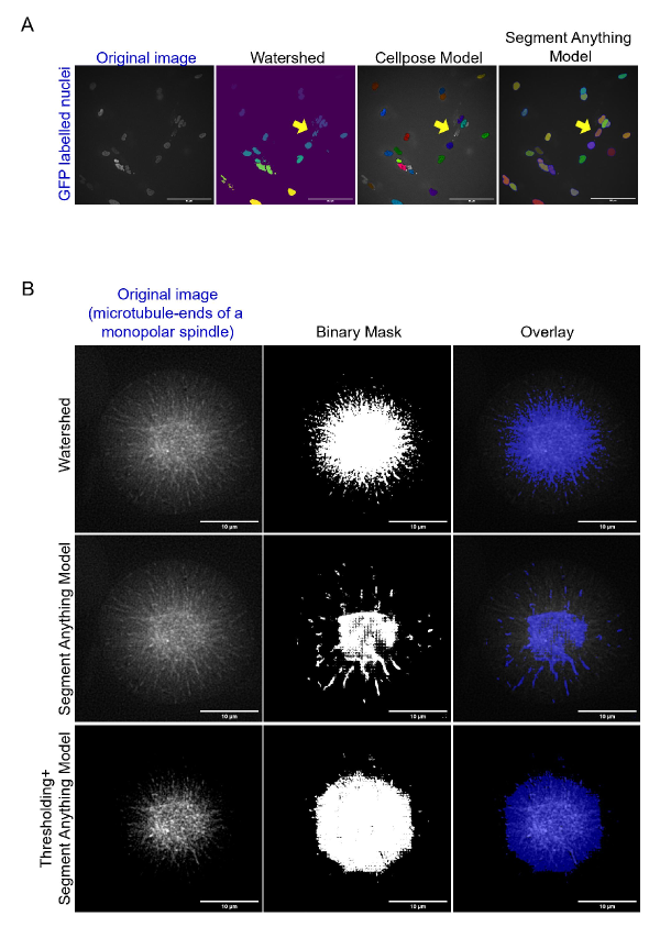
Table 1 displays the most widely used DL techniques for microscopy image analysis. Apart from these well-established techniques, a reusable and adaptable image segmentation architecture, the Segment Anything Model (SAM), has been proposed by Meta AI which is a zero-shot transfer learning approach (kirillov2023segment, ). Its performance appears to be competitive with or even superior to prior fully supervised trained models and is applied in medical imaging (he2023accuracy, ; mazurowski2023segment, ) and digital pathology (deng2023segment, ). SAM is unexplored for cellular or subcellular segmentation tasks. We tested SAM’s initial features on our data. Fig 1 (panel A) showcases SAM’s capability to segment complex nuclear morphologies without additional training. Some under/over-segmentations occur, but they are comparable to watershed segmentation (conventional) and CellPose (DL), making SAM a tool that could save considerable researcher time. However, it encounters challenges with intricate subcellular structures (wang2023empirical, ). For example in the case of microtubule-end segmentation, while the conventional watershed method can partially segment microtubule-ends found in a monopolar spindle, SAM segments the whole monopolar spindle instead (Fig 1.B). Evidently, SAM holds the capability to simplify segmentation, but it has not yet been tested in densely packed microscopy images. For instance, electron microscopy (EM) images displaying crowded organelles may pose challenges to achieving accurate segmentation without trained datasets of individual organelles.
3. AI-guided methods outperform conventional image analysis tools
DL neural networks are more effective than traditional computer vision techniques. They learn from large-scale datasets and have the capacity to extract high-level features without heavy reliance on domain knowledge for feature extraction (o2020deep, ). While many DL tools have focused on segmenting nuclei and whole cells with fluorescently-labelled markers, some specialised DL tools have been developed to segment distinct organelles such as Golgi apparatus, mitochondria and endoplasmic reticulum from Electron Microscopy (EM) data (Table 2). However, DL tools that can both segment and track dynamic subcellular structures in time-lapse fluorescent movies are currently limited. Mitochondria (lefebvre2021automated, ), microtubule-ends and mitotic spindles (dang2023deep, ) are among the few dynamically changing structures for which automated analysis tools are available, but deep learning has only been used in the last case. The most popular DL-based tools include U-net (ronneberger2015u, ; cciccek20163d, ; falk2019u, ), StarDist (schmidt2018cell, ; weigert2020star, ) and Cellpose (stringer2021cellpose, ; pachitariu2022cellpose, ; schiff2022integrating, ). Most DL-based solutions are data-driven and so there are no standards to inform biologists which model is the most suitable one for their own dataset for specific computational tasks. As a result, most people veer towards integrated platforms such as Fiji (through plugins) (schindelin2012fiji, ; gomez2021deepimagej, ), CellProfiler (mcquin2018cellprofiler, ), QuPath (bankhead2017qupath, ), ZEISS arivis Cloud (formerly APEER) (apeer2023, ; david2021apeer, ) and ZeroCostDL4Mic (von2021democratising, ; zerocostdl4micimplementation, ). Below we discuss the application of deep learning in cellular image and movie analysis: segmentation, classification, and tracking, and contrast it against conventional non-DL methods.
3.1. Segmentation
Two types of image segmentation, semantic and instance, serve different purposes. Semantic segmentation aims to classify individual pixels within an image into specific classes. It groups instances of a class together, lacking the ability to differentiate individual instances like overlapping nuclei. However, it effectively separates membrane outlines from intra or extracellular space. Instance segmentation differentiates instances of the same class (In Fig 1.A, Cellpose (stringer2021cellpose, ; pachitariu2022cellpose, ) and Segment Anything Model (SAM) (kirillov2023segment, ) separate overlapping nuclear objects could be separated as distinct instances). Recently, panoptic segmentation has been introduced which combines instance and semantic segmentation where each instance of an object in the image is segregated and the identification of each object is predicted (liu2020unsupervised, ).
Conventional segmentation methods include thresholding, edge-based algorithms and region-based segmentation (minaee2021image, ). Edge-based segmentation like Canny or Sobel edge-detectors followed by contour filling (minaee2021image, )performs better than thresholding, but can produce imperfect contours. Region-based segmentation, particularly watershed segmentation is widely used in cell biology (minaee2021image, ). Conventional segmentation methods are often used for automated annotation of large datasets, followed by manual correction to save annotation time (dang2023deep, ).
| Tool | Subcellular Structures | DL Architecture | Dynamics Tracking | Strengths in User Experience | Source |
|---|---|---|---|---|---|
| U-Net (ronneberger2015u, ; cciccek20163d, ; falk2019u, ) | Fluorescent and label-free cell membrane, fluorescent nuclei and EM neurites | CNN | No | Documentation (application), tutorials (Jupyter notebook). | https://lmb.informatik.uni-freiburg.de/people/ronneber/u-net/ |
| Cellpose (stringer2021cellpose, ; pachitariu2022cellpose, ) | Fluorescent cell membrane and nuclei | U-net | No | Documentation (installation and application), tutorials (Jupyter notebook), integrated through the ZEISS arivis Cloud (formerly APEER platform) (apeer2023, ; david2021apeer, ). | https://github.com/mouseland/cellpose |
| Stardist (3D) (schmidt2018cell, ; weigert2020star, ) | Fluorescent and H&E stained nuclei | U-net | No | Documentation (installation and application), tutorials (Jupyter notebook), integrated plugin (ImageJ/Fiji (schindelin2012fiji, ), Napari (napari, ), QuPath (bankhead2017qupath, )). | https://github.com/stardist/stardist |
| ASEM (gallusser2022deep, ) | EM Golgi apparatus, mitochondria, nuclear pore complexes, caveolae, endoplasmic reticulum, clathrin-coated pits, vesicles | 3D U-net | No | Documentation (installation and application). | https://github.com/kirchhausenlab/incasem |
| MitoSegNet (fischer2020mitosegnet, ) | Fluorescent mitochondria | U-net | No | Documentation (application), GUI (multiple operation systems). | https://github.com/MitoSegNet/MitoS-segmentation-tool |
| SpinX (dang2023deep, ) | Fluorescent mitotic spindle, label-free cell cortex | Mask R-CNN | Yes | Documentation (application), integrated through the ZEISS arivis Cloud (formerly APEER platform) (apeer2023, ; david2021apeer, ). | https://github.com/Draviam-lab/spinx_local |
| Multicut (beier2017multicut, ) | EM neural membrane | U-net | No | Documentation (installation), tutorials (Jupyter notebook). | https://github.com/ilastik/nature_methods_multicut_pipeline |
| nucleAIzer (hollandi2020nucleaizer, ) | Fluorescent and H&E stained nuclei | Mask R-CNN+U-net | No | Documentation (installation and application), tutorials (shell scripts). | https://github.com/spreka/biomagdsb |
| DenoiSeg (buchholz2021denoiseg, ) | Fluorescent cell membrane and nuclei | U-net | No | Documentation (installation), tutorials (Jupyter notebook). | https://github.com/juglab/DenoiSeg |
| InstantDL (waibel2021instantdl, ) | Fluorescent, H&E stained and label-free nuclei | U-net/Mask R-CNN | No | Documentation (installation and applications), dockerised. | https://github.com/marrlab/InstantDL |
| DeepCell (greenwald2022whole, ; van2016deep, ) | Fluorescent nuclei and cell membrane | Resnet50 | Yes | Documentation (application), tutorials (script), dockerised. | https://github.com/vanvalenlab/deepcell-applications |
| SplineDist (mandal2021splinedist, ) | Fluorescent and H&E stained nuclei | StarDist | No | Tutorials (Jupyter notebook). | https://github.com/uhlmanngroup/splinedist |
| CDeep3M (haberl2018cdeep3m, ) | XRM, ET, fluorescent and SBEM nuclei; SBEM synaptic vesicles, mitochondria and membranes | DeepEM3D-Net (dense CNN) | No | Documentation (installation), dockerised, implemented through Amazon Web Services. | https://github.com/CRBS/cdeep3m |
| CellSeg (lee2022cellseg, ) | Fluorescent nuclei and cell membrane | Mask R-CNN | No | Tutorials (Jupyter notebook). | https://github.com/michaellee1/CellSeg |
| EmbedSeg (lalit2022embedseg, ) | Fluorescent cell membrane and nuclei | Branched ERF-Net (3D) | No | Documentation (installation), datasets provided for reproducibility. | https://github.com/juglab/EmbedSeg |
DL methods not only surpass conventional techniques in the segmentation of subcellular structures in microscopy images, but also exhibit a remarkable generalisation capacity, accommodating diverse imaging conditions, fluorescent markers or proteins, and cell types (caicedo2019evaluation, ; fischer2020mitosegnet, ; stringer2021cellpose, ; dang2023deep, ; hollandi2022nucleus, ). This has led to the creation of several freely available tools providing pre-trained models for biologists to segment and subsequently analyse microscopy dataset quantitatively (Table 2).
3.2. Classification
Classification refers to assigning text labels to images and is frequently used in cell biology and digital pathology. Instance classification focuses on recognising and categorising individual objects within an image, rather than classifying the image as a whole. DL techniques are applied to identify and classify individual cells and nuclei and to provide quantitative information on cell populations and their distribution (graham2019hover, ; gamper2019pannuke, ). Cell type and subcellular structure identification are other applications of instance classification, which has allowed robust quantitative studies of cell function (simm2018repurposing, ), cell interaction (nitta2018intelligent, ), phenotype (’yes’ or ’no’ prediction) (godinez2017multi, ) and spatial patterns and protein localisation in fluorescence images (sullivan2018deep, ; kraus2016classifying, ; kraus2017automated, ). Classification has also been employed for large-scale phenotypic profiling of small molecules by analysing cellular responses to drug treatments at the single-cell level (scheeder2018machine, ) to evaluate drug efficacy, mechanism of action, and potential side effects.
Manual annotations by cell biology experts are robust but time-consuming and expensive. To offset this cost, active learning (monarch2021human, ) has been proposed. Active learning is a powerful human-in-the-loop process in deep learning. It involves annotating manually a subset of (not all) relevant objects in images, training with this subset, and generating initial segmentation and classification masks for all instances including unannotated ones (van2021biological, ). Then, the auto-generated initial segmentation and classification can be reviewed and manually corrected, serving as annotations of the next training iteration, making the human-in-the-loop process a cost-efficient approach (stringer2021cellpose, ; pachitariu2022cellpose, ; dang2023deep, ).
Unlike deep learning methods used for image classification, traditional machine learning (ML) based classifiers are humanly interpretable, which is important for failure analysis and model improvement (wang2021annotation, ). While the DL framework has higher recognition accuracy on large sample data sets, the traditional ML approach (eg., Support Vector Machine, SVM) is thought to be a better solution for small data sets (narotamo2021machine, ; wang2021comparative, ). So hybrid approaches that combine ML and DL techniques are being used for high accuracy and precision for cell-type classification problems (wahid2019performance, ; rani2022machine, ), as a step towards explainable AI.
3.3. Tracking
Tracking is the process of identifying and linking the movement of specific objects over time in a series of time-lapse images or a movie. Tracking methods in cell biology are primarily DL-independent, unlike real-world scenarios such as autonomous driving where DL-based tracking is being widely used (chen2017online, ; ciaparrone2020deep, ; marvasti2021deep, ; jiao2021deep, ; pal2021deep, ). From a computational perspective, the task of tracking consists of detection-based tracking (DBT), and detection-free tracking (DFT) (luo2021multiple, ). DBT, also commonly referred to as tracking-by-detection, usually consists of two main steps: detection of the objects of interest and linking their positions and properties across consecutive frames. On the other hand, DFT requires the manual initialisation of a fixed number of objects in the first frame and then localising these objects in the subsequent frames. DBT is widely used compared to DFT since objects can be newly discovered or transiently lost through time in most scenarios and DFT cannot deal with the case (luo2021multiple, ).
In many tracking studies, deep learning is used in the detection step, including techniques such as the R-CNN series (he2017mask, ; girshick2015fast, ; ren2015faster, ), YOLO (redmon2016you, ; redmon2018yolov3, ; jiang2022review, ) and SSD (liu2016ssd, ). Deep learning can also be used for trajectory or motion prediction to support tracking. Most of the DL-based trajectory predictions are through LSTM technique (sherstinsky2020fundamentals, ) which has extensively progressed by predicting the coordinators of selected objects in the upcoming time frame (chandra2019traphic, ; chandra2019robusttp, ; leon2021review, ; wang2019exploring, ). Some studies have taken advantage of convolutional feature extraction (chen2017online, ; he2017cell, ) for the prediction of trajectory. Currently (ciaparrone2020deep, ; marvasti2021deep, ; jiao2021deep, ; pal2021deep, ) the top application scenarios of DL-based tracking are pedestrian detection and autonomous vehicles, augmented reality (AR) and virtual reality (VR), and these could be brought to cell biology to advance multiscale system studies where subcellular, cellular and tissue levels changes are simultaneously modulated and measured (floerchinger2021optimizing, ; venkatesan2021virtual, ; razavian2019augmented, ).
Typical examples in cell biology applications include single-cell tracking (blockhuys2020single, ), multi-cell tracking during collective cell migration (song2023machine, ) or particle or organelle tracking within cells (jaqaman2008robust, ; tinevez2017trackmate, ). Tracking is challenging from both computational and biological perspectives for many reasons. First, objects can move from area to area, so each instance should be identified on a single-frame basis and these detections should be linked over time to avoid misconnections. Second, objects that are to be tracked can merge (mitochondria) or split (cell division), and this presents a discontinuity challenge in their morphology, leading to misrecognition. Third, there is a limitation in terms of the frame rate in time-lapse movies (nicovich2014acquisition, ; danuser2014reply, ), and this makes tracking in general and in 3-dimensions, in particular, challenging due to time discontinuity. Misconnection and misrecognition challenges could be at least in part overcome using DL methods for trajectory prediction.
Tracking subcellular structures and their changes through 3-Dimensional space is a challenging but rewarding application, as it can provide valuable insight into cell dynamics (zulkipli2018spindle, ; pennycook2021palbociclib, ) and support systems-level modelling efforts to explore complex signalling and regulatory pathways (corrigan2015modeling, ; min2019spontaneously, ). For example, analysing the patterns of cell movements following distinct molecular perturbations has helped dissect molecular principles that govern cellular migration (van2016deep, ; tsai2019usiigaci, ; he2017cell, ; mavska2023cell, ). Whole-cell tracking to monitor cell or nuclear size changes and the timing and duration of cell cycle phases (dang2023deep, ) or intracellular tracking to analyse the movement of intracellular organelles, vesicles, or proteins, within a cell, (newby2018convolutional, ; tinevez2017trackmate, ; ritter2021deep, ; spilger2021deep, ; ritter2021deep, ), have taken advantage of apriori knowledge of distinct features (structural or dynamic) which have been uniquely used to solve each individual tracking problem.
4. The challenges and opportunities
4.1. Challenges of AI-guided methods in cell dynamics studies
4.1.1. Lack of well-annotated datasets
Deep learning-based approaches require large amounts of labelled (annotated) data. Ideally, high-quality cell biology data need to be annotated by experts which is time-consuming. Although crowdsourcing can offer cost-effective solutions, annotation inconsistencies can require correction by experts (spiers2021deep, ). Furthermore, variations in subcellular morphologies, staining protocols, and imaging quality can make the annotation challenging for non-experts. Many solutions are being developed to tackle this challenge, including active learning (vununu2021classification, ; moen2019deep, ), transfer learning (vununu2021classification, ; moen2019deep, ; kensert2019transfer, ; minoofam2021trcla, ) and data augmentation techniques (moen2019deep, ; naghizadeh2021semantic, ; majurski2019cell, ; yu2021generative, ; chlap2021review, ). Augmentation strategies where an image is altered in scale or intensity provide additional samples without necessarily increasing the number of manually annotated samples (dang2023deep, ). Karabag et al investigate the impact of the amount of training data and shape variability on the U-net-based segmentation (karabaug2023impact, ). They suggest that data augmentation methodologies may not improve training if the acquired cell pairs are not representative of other cells. Therefore, a thorough investigation of various augmentations is recommended. Despite mentioned solutions, the shortage of high-quality labelled data remains a critical limitation for AI-guided images and time-lapse movie analysis. Only a limited number of open-sourced datasets are available, as listed chronologically in Table 3.
| Dataset | Description | Source |
|---|---|---|
| Broad Bioimage Benchmark Collection (ljosa2012annotated, ) | Over 11 million images from 52 datasets for segmentation, phenotype classification, and image-based profiling tasks. | https://bbbc.broadinstitute.org/image_sets |
| ISBI cell tracking challenge dataset collection (mavska2014benchmark, ) | Ten 2D image datasets and ten 3D time-lapse movie datasets of fluorescent counterstained nuclei or cells for segmentation and tracking tasks. | http://www.celltrackingchallenge.net/ |
| DeepCell dataset (van2016deep, ) | 75,000 single-cell annotations including live-cell movies of fluorescent nuclei (10,000 single-cell movie trajectories over 30 frames) and static images of whole cells for segmentation tasks. | https://github.com/vanvalenlab/deepcell-tf |
| Image data resource (IDR) (williams2017image, ) | Over 13 million images from 118 published studies. | https://idr.openmicroscopy.org/ |
| Human Protein Atlas (thul2017subcellular, ) | Over 80,000 high-resolution confocal Immunofluorescence images showing localisation patterns of thousands of proteins for a variety of human cell lines for segmentation tasks. | http://www.proteinatlas.org |
| The Cell Image Library (bray2017dataset, ) | 919,874 five-channel fields of morphologies of U20S cells and populations representing 30,616 tested compounds. | https://github.com/gigascience/paper-bray2017 |
| Salmonella-infected HeLa cells (antoniou2019high, ) | 93,300 multi-channel confocal fluorescence images. | https://dataverse.harvard.edu/dataset.xhtml?persistentId=doi:10.7910/DVN/FYGHFO |
| JUMP cell painting datasets (chandrasekaran2022three, ) | Images of osteosarcoma cells perturbed with CRISPR-mediated knockdowns and overexpression reagents and 120,000 compounds. | https://jump-cellpainting.broadinstitute.org/ |
| NYSCF automated deep phenotyping dataset (ADPD) (schiff2022integrating, ) | Cell painting dataset of 1.2 million images (48 TB). | https://nyscf.org/open-source/nyscf-adpd/ |
| Poisson-Gaussian Fluorescence Microscopy Denoising Dataset (zhang2019poisson, ) | Over 12,000 fluorescence microscopy images using confocal, two-photon and widefield microscopes. | https://drive.google.com/drive/folders/1aygMzSDdoq63IqSk-ly8cMq0_owup8UM |
4.1.2. The quality of image datasets
DL models rely on extracting patterns from the dataset, making the quality of annotated data crucial. Inconsistent ground truth yields incorrect analytical results, while biased data (highlighting some but not all phenotypes) leads to incorrect patterns or inaccurate predictions. Noise intrinsic to microscopy can also increase the complexity of the model required to accurately capture the underlying features. This may lead to overfitting, where the model becomes too complex and fails to generalise to new and unseen data. Noisy data can also cause adversarial attacks on deep learning models, leading to misclassification of cell types, incorrect tracking of cell movements, and under- or over- segmentation of cells (hirano2021universal, ; wang2019security, ). Meiniel et al. present a comprehensive review of microscopy data quality control and denoising using computational techniques (meiniel2018denoising, ). To manage the problem of high-quality image availability, the image data resource has been set up to allow easy image data access, storage and dissemination (williams2017image, ). Overall, it is essential to ensure that datasets used for deep learning are of high quality, with solid ground truth, minimal noise and free from bias (cimini2023twenty, ).
4.1.3. Model interpretability
The challenge of interpretability for deep learning models arises from the complex and black-box nature of these models (castelvecchi2016can, ; ekanayake2022novel, ). DL models can automatically extract complex features and patterns from large amounts of data through multiple layers of neurons (lecun2015deep, ). While this makes such models powerful, in tasks such as image segmentation or classification, it also presents a challenge in understanding how the models arrived at their predictions or decisions. One approach to addressing this challenge is to visualise and examine the activations of individual neurons or groups of neurons within the model (montavon2018methods, ). This technique provides insights into the patterns that the model has used to form its decision. However, these visualisations may be difficult to interpret without a deep understanding of the model architecture and data domain (see review (mohamed2022review, ) for more information).
4.1.4. High cost in real-world scenarios
Deep learning-based methods are often expensive due to two main factors. First, effective training of DL models requires a large amount of data which can be expensive to generate. Second, the training process can be computationally intensive requiring high-performance computing resources, such as hardware of graphics processing units (GPUs) and tensor processing units (TPUs). This infrastructure cost can dissuade the planning of imaging studies needed to build the DL model (nogare2023bioimage, ). DL model-building efforts supported by agencies/consortiums beyond individual researchers can help meet upfront costs, and maintain standards to make sure models are reusable (munappy2022data, ).
4.1.5. The generalisability issue
Generalisability denotes the extent to which a DL model trained on a specific dataset might perform well on new data, especially when the new data has different features or patterns compared to the training data. To showcase generalisability, DL models are deployed on data acquired from a different cell type or microscope (graham2019hover, ; gamper2019pannuke, ). Efforts to reuse or generalise workflow are ongoing (mavska2023cell, ; stringer2021cellpose, ). Generalisability issues arising due to sample variability or differences in image acquisition are being addressed through data augmentation, multi-task learning, swarm learning or collaboration with domain specialists (ali2022assessing, ; wang2019pathology, ; wang2019deep, ).
4.2. Opportunities of AI-guided methods in cell dynamics studies
With the advent of new AI-guided methods to identify, track and analyse objects in time-lapse movie datasets (Table 3), we expect new opportunities for large-scale cell biology applications in drug discovery, drug repositioning and phenome-genome interaction map-building efforts.
4.2.1. Drug discovery and repositioning
AI approaches in microscopy-based drug development or drug target identification primarily use still image datasets which are snapshots of dynamic processes (krentzel2023deep, ; tran2023artificial, ; karacosta2021imaging, ). Such still image-based drug screening efforts do not yet fully benefit from cellular and subcellular dynamics that can be visualised using high-speed live-imaging microscopes (wagner2021deep, ; efstathiou2021electrically, ; barazia2022imaging, ; yamashita2020digital, ). Incorporating dynamic changes through time can address challenges posed by cellular heterogeneity, cell cycle stages, cell fate dissimilarities, variations in protein expression variations, cellular or subcellular dimensions, inter/intra-cellular signalling (padovani2022segmentation, ). In addition to taking advantage of cell dynamics principles, AI-guided methods for movie datasets can accelerate several steps of drug discovery including cell toxicity assays (pulfer2022ades, ), cell cycle profiling and morphology analysis (padovani2022segmentation, ; ren2021deep, ). Increasing single-cell movie datasets along with the development of DL model standards can integrate image-omics with other -omics datasets that capture dynamic information and have accelerated drug repositioning studies (iorio2015semi, ; mertens2023drug, ). Investing in collaborative efforts to compile microscopy datasets can fuel the development of robust AI-guided methods. This, in turn, will unlock research and engineering opportunities, facilitating a cyclical learning process to uncover unexplored cellular transition states in frontier biology and drug discovery studies.
4.2.2. Genome-phenome mapping
Genetic interaction maps built using cell biology approaches are transforming our understanding of several biological processes (chessel2019observing, ), but their influence is limited to the specific model system or experimental set-up. We are just beginning to reliably link datasets from different cell types, fluorescent markers or imaging systems (roberts2017systematic, ; allenCell2017, ). AI-guided image analysis methods are well positioned to extract information across image and video datasets, across different databases, in an unbiased form as they can be trained to search for patterns (for example, nuclear atypia such as multinucleated, misshapen and binucleated structures (hart2021multinucleation, )) could be gathered across 100s of cell lines or drug treatments). Currently, high-throughput genome-phenome mapping image datasets of various cell types and models are deposited in a disconnected fashion because there is not much incentive to unify them. AI-guided methods may offer the possibility and the value in developing universal standards for collating data, in addition to existing global efforts to name and store large movie datasets (linkert2010metadata, ; moore2021ome, ; moore2023ome, ).
4.2.3. Precision medicine
Genetic variant interpretation and classification using high-throughput cell biology methods is still a nascent field. Germline variant guidelines are well established (richards2015standards, ) and somatic variant guidelines are being established (parikh2020identification, ). In both cases we expect single-cell imaging, the associated image dataset, and image analysis methods to play a crucial role in stratifying variant pathogenicity. To build stratification methods that are scalable, generalisable and interrogatable (crosscheck), DL models could be trained to detect and classify phenotype changes and hidden patterns. Swarm-learning has been proposed for decentralised and confidential X-ray image analysis (warnat2021swarm, ) and digital pathology (saldanha2022swarm, ) which could be extended to cell biology images and live-cell movies. As AI methods become incorporated within the clinical prognosis framework (oren2020artificial, ; cui2021artificial, ), we predict there will be a growing demand for robust models for the clinical actionability of molecular targets in cancer therapies, genetic rare diseases and infectious diseases.
5. Conclusion
The impact of deep learning methods in large-scale and complex microscopy data analysis has been significant. Deep learning techniques have already revolutionised still image analysis and are now beginning to transform time-lapse movie analysis through state-of-the-art performance in a wide range of applications, such as object detection and tracking, segmentation, unsupervised clustering and classification. Deep learning methods employed to segment and classify cells are beginning to detect novel anomalies in 3D structures (balkenhol2019deep, ) or time series data (ji2021novel, ), identify distinctive transient cellular transitions (ren2021deep, ), and reveal complex behaviours and movement patterns (molina2022acme, ; dang2023deep, ) which were previously unrecognised.
Automated and data-driven workflows and cloud-based large-scale solutions have significantly improved the speed, efficiency and accuracy of DL-guided image analysis tasks, while also increasing the ease with which biologists can implement AI tools. Overall, the use of deep learning methods in microscopy has enabled researchers to extract valuable information, some that are not obvious to the eye, from huge volumes of image data and opened up new opportunities in medical diagnosis and clinical translation.
It is important to recognise that deep learning methods rely on abundant, robustly-annotated data and careful parameter-tuning. Assessing their reliability and interpretability can be challenging (cimini2023twenty, ), which can restrict their applications in some domains. The establishment of universally accepted standards and frameworks to store and share human-annotated image datasets, DL models, and post-processing pipelines are complex challenges (nogare2023bioimage, ) that necessitate attention through international collaborations and consortia.
Acknowledgement
We thank Janeth Catalina Manjarrez-Gonzalez (Draviam group) for unpublished training images; Dr Sreenivasan Bhattiprolu (Zeiss, USA), Dr Peter Thorpe (QMUL) and Dr Nikola Ojkic (QMUL) for comments on the manuscript content and Draviam group members for useful discussions. We would like to acknowledge funding support from the Biotechnology and Biological Sciences Research Council (BBSRC) to V.M. Draviam (BB/R01003X/1, BB/T017716/1, BB/W002698/1 and BB/X511067/1), B. Chai (BB/X511067/1 and InnovateUK SBCY1K9R), and C. Efstathiou (LIDo-iCASE studentship [BB/T008709/1]). B Chai is a Knowledge Transfer Partnership associate collaborating with Zeiss UK.
Author contributions
Glossary
Data annotation
the process of adding attributes to training data and labelling them so that a DL model can learn what predictions it is expected to make.
Edge-based segmentation
a conventional segmentation approach that aims to first detect the contours of the specific object and then fill in the contours for segmentation.
Instance classification
usually consists of object detection, localising their position within the image and classifying them into predefined categories.
Long Short-Term Memory (LSTM)
a type of Recurrent Neural Network (RNN) architecture that was designed to overcome the problem of vanishing and exploding gradients faced by standard RNNs. LSTM is suited to tasks involving sequences with long-term dependencies, such as time series prediction, natural language processing, and speech recognition.
Neural network
a densely interconnected group of nodes. Each node connects to several nodes in the layer beneath it, from which it receives data (eg., training data in the last layer), and several nodes in the layer above it, to which it outputs data. Incoming connections are assigned weights. Active nodes multiply their respective weights and pass them forward if it exceeds a threshold. Training involves adjusting weights and thresholds are adjusted to produce similar outputs for data with the same labels.
Segmentation
the process of dividing an image into multiple regions or segments with each corresponding to a specific object or area of interest.
Single Shot Detector (SSD)
an object detection method that uses a single neural network for the entire image, similar to YOLO. Directly based on different image regions, it predicts bounding boxes and class probabilities directly. Unlike YOLO, SSD operates on multiple feature maps with different resolutions to handle objects of various sizes.
Thresholding segmentation
a conventional segmentation method by choosing a threshold based on the intensity histogram for segmenting an object.
You Only Look Once (YOLO)
an object detection method with the key idea of applying a single neural network to the full image, which divides the image into regions and predicts bounding boxes and probabilities for each region.
Zero-shot learning
a remarkable ML/DL method which refers to recognising new unseen objects, and so it can be applied to new image distributions and tasks.
References
- [1] Junde Xu, Donghao Zhou, Danruo Deng, Jingpeng Li, Cheng Chen, Xiangyun Liao, Guangyong Chen, and Pheng Ann Heng. Deep learning in cell image analysis. Intelligent Computing, 2022, 2022.
- [2] Erick Moen, Dylan Bannon, Takamasa Kudo, William Graf, Markus Covert, and David Van Valen. Deep learning for cellular image analysis. Nature methods, 16(12):1233–1246, 2019.
- [3] Daniel Krentzel, Spencer L Shorte, and Christophe Zimmer. Deep learning in image-based phenotypic drug discovery. Trends in Cell Biology, 2023.
- [4] Hee June Choi, Chuangqi Wang, Xiang Pan, Junbong Jang, Mengzhi Cao, Joseph A Brazzo, Yongho Bae, and Kwonmoo Lee. Emerging machine learning approaches to phenotyping cellular motility and morphodynamics. Physical biology, 18(4):041001, 2021.
- [5] Adrien Hallou, Hannah G Yevick, Bianca Dumitrascu, and Virginie Uhlmann. Deep learning for bioimage analysis in developmental biology. Development, 148(18):dev199616, 2021.
- [6] Reka Hollandi, Nikita Moshkov, Lassi Paavolainen, Ervin Tasnadi, Filippo Piccinini, and Peter Horvath. Nucleus segmentation: towards automated solutions. Trends in Cell Biology, 2022.
- [7] Christoforos Efstathiou and Viji M Draviam. Electrically tunable lenses–eliminating mechanical axial movements during high-speed 3d live imaging. Journal of Cell Science, 134(16):jcs258650, 2021.
- [8] Gaoxiang Liu, Xiongtao Ruan, Daniel E Milkie, Frederik Görlitz, Matthew Mueller, Wilmene Hercule, Alison Killilea, Eric Betzig, and Srigokul Upadhyayula. Characterization, comparison, and optimization of lattice light sheets. Science Advances, 9(13):eade6623, 2023.
- [9] Yuko Mimori-Kiyosue. Imaging mitotic processes in three dimensions with lattice light-sheet microscopy. Chromosome Research, 29(1):37–50, 2021.
- [10] Estibaliz Gómez-de Mariscal, Daniel Franco-Barranco, Arrate Muñoz-Barrutia, and Ignacio Arganda-Carreras. Building a bioimage analysis workflow using deep learning. In Bioimage Data Analysis Workflows–Advanced Components and Methods, pages 59–88. Springer International Publishing Cham, 2022.
- [11] Andrew Janowczyk and Anant Madabhushi. Deep learning for digital pathology image analysis: A comprehensive tutorial with selected use cases. Journal of pathology informatics, 7(1):29, 2016.
- [12] Qing Lv, Suzhen Zhang, and Yuechun Wang. Deep learning model of image classification using machine learning. Advances in Multimedia, 2022, 2022.
- [13] Yann LeCun, Yoshua Bengio, and Geoffrey Hinton. Deep learning. nature, 521(7553):436–444, 2015.
- [14] Alex Krizhevsky, Ilya Sutskever, and Geoffrey E Hinton. Imagenet classification with deep convolutional neural networks. Communications of the ACM, 60(6):84–90, 2017.
- [15] Shan E Ahmed Raza, Linda Cheung, Muhammad Shaban, Simon Graham, David Epstein, Stella Pelengaris, Michael Khan, and Nasir M Rajpoot. Micro-net: A unified model for segmentation of various objects in microscopy images. Medical image analysis, 52:160–173, 2019.
- [16] SanaUllah Khan, Naveed Islam, Zahoor Jan, Ikram Ud Din, and Joel JP C Rodrigues. A novel deep learning based framework for the detection and classification of breast cancer using transfer learning. Pattern Recognition Letters, 125:1–6, 2019.
- [17] Nicolas Coudray, Paolo Santiago Ocampo, Theodore Sakellaropoulos, Navneet Narula, Matija Snuderl, David Fenyö, Andre L Moreira, Narges Razavian, and Aristotelis Tsirigos. Classification and mutation prediction from non–small cell lung cancer histopathology images using deep learning. Nature medicine, 24(10):1559–1567, 2018.
- [18] Laith Alzubaidi, Mohammed A Fadhel, Omran Al-Shamma, Jinglan Zhang, and Ye Duan. Deep learning models for classification of red blood cells in microscopy images to aid in sickle cell anemia diagnosis. Electronics, 9(3):427, 2020.
- [19] Ahmed Ismail Shahin, Yanhui Guo, Khalid Mohamed Amin, and Amr A Sharawi. White blood cells identification system based on convolutional deep neural learning networks. Computer methods and programs in biomedicine, 168:69–80, 2019.
- [20] Krishna Kumar Jha and Himadri Sekhar Dutta. Mutual information based hybrid model and deep learning for acute lymphocytic leukemia detection in single cell blood smear images. Computer methods and programs in biomedicine, 179:104987, 2019.
- [21] Jay M Newby, Alison M Schaefer, Phoebe T Lee, M Gregory Forest, and Samuel K Lai. Convolutional neural networks automate detection for tracking of submicron-scale particles in 2d and 3d. Proceedings of the National Academy of Sciences, 115(36):9026–9031, 2018.
- [22] Jean-Baptiste Lugagne, Haonan Lin, and Mary J Dunlop. Delta: Automated cell segmentation, tracking, and lineage reconstruction using deep learning. PLoS computational biology, 16(4):e1007673, 2020.
- [23] Alex Sherstinsky. Fundamentals of recurrent neural network (rnn) and long short-term memory (lstm) network. Physica D: Nonlinear Phenomena, 404:132306, 2020.
- [24] Thomas Wollmann, Manuel Gunkel, Inn Chung, Holger Erfle, Karsten Rippe, and Karl Rohr. Gruu-net: Integrated convolutional and gated recurrent neural network for cell segmentation. Medical image analysis, 56:68–79, 2019.
- [25] Abin Jose, Rijo Roy, Daniel Moreno-Andrés, and Johannes Stegmaier. Automatic detection of cell-cycle stages using recurrent neural networks. bioRxiv, pages 2023–02, 2023.
- [26] Jacob C Kimmel, Andrew S Brack, and Wallace F Marshall. Deep convolutional and recurrent neural networks for cell motility discrimination and prediction. IEEE/ACM transactions on Computational Biology and Bioinformatics, 18(2):562–574, 2019.
- [27] Olaf Ronneberger, Philipp Fischer, and Thomas Brox. U-net: Convolutional networks for biomedical image segmentation. In Medical Image Computing and Computer-Assisted Intervention–MICCAI 2015: 18th International Conference, Munich, Germany, October 5-9, 2015, Proceedings, Part III 18, pages 234–241. Springer, 2015.
- [28] Cefa Karabağ, Mauricio Alberto Ortega-Ruíz, and Constantino Carlos Reyes-Aldasoro. Impact of training data, ground truth and shape variability in the deep learning-based semantic segmentation of hela cells observed with electron microscopy. Journal of Imaging, 9(3):59, 2023.
- [29] Naomi Joseph, Chaitanya Kolluru, Beth AM Benetz, Harry J Menegay, Jonathan H Lass, and David L Wilson. Quantitative and qualitative evaluation of deep learning automatic segmentations of corneal endothelial cell images of reduced image quality obtained following cornea transplant. Journal of Medical Imaging, 7(1):014503–014503, 2020.
- [30] Estibaliz Gómez-de Mariscal, Martin Maška, Anna Kotrbová, Vendula Pospíchalová, Pavel Matula, and Arrate Munoz-Barrutia. Deep-learning-based segmentation of small extracellular vesicles in transmission electron microscopy images. Scientific reports, 9(1):13211, 2019.
- [31] Özgün Çiçek, Ahmed Abdulkadir, Soeren S Lienkamp, Thomas Brox, and Olaf Ronneberger. 3d u-net: learning dense volumetric segmentation from sparse annotation. In Medical Image Computing and Computer-Assisted Intervention–MICCAI 2016: 19th International Conference, Athens, Greece, October 17-21, 2016, Proceedings, Part II 19, pages 424–432. Springer, 2016.
- [32] Thorsten Falk, Dominic Mai, Robert Bensch, Özgün Çiçek, Ahmed Abdulkadir, Yassine Marrakchi, Anton Böhm, Jan Deubner, Zoe Jäckel, Katharina Seiwald, et al. U-net: deep learning for cell counting, detection, and morphometry. Nature methods, 16(1):67–70, 2019.
- [33] Aya Saleh Ahmed, Wessam H El-Behaidy, and Aliaa AA Youssif. Medical image denoising system based on stacked convolutional autoencoder for enhancing 2-dimensional gel electrophoresis noise reduction. Biomedical Signal Processing and Control, 69:102842, 2021.
- [34] Karren Dai Yang, Anastasiya Belyaeva, Saradha Venkatachalapathy, Karthik Damodaran, Abigail Katcoff, Adityanarayanan Radhakrishnan, GV Shivashankar, and Caroline Uhler. Multi-domain translation between single-cell imaging and sequencing data using autoencoders. Nature communications, 12(1):31, 2021.
- [35] Ian Goodfellow, Jean Pouget-Abadie, Mehdi Mirza, Bing Xu, David Warde-Farley, Sherjil Ozair, Aaron Courville, and Yoshua Bengio. Generative adversarial networks. Communications of the ACM, 63(11):139–144, 2020.
- [36] Felix Fuentes-Hurtado, Tom Delaire, Florian Levet, Jean-Baptiste Sibarita, and Virgile Viasnoff. Mid3a: Microscopy image denoising meets differentiable data augmentation. In 2022 International Joint Conference on Neural Networks (IJCNN), pages 1–9. IEEE, 2022.
- [37] Liqun Zhong, Guole Liu, and Ge Yang. Blind denoising of fluorescence microscopy images using gan-based global noise modeling. In 2021 IEEE 18th International Symposium on Biomedical Imaging (ISBI), pages 863–867. IEEE, 2021.
- [38] Jiji Chen, Hideki Sasaki, Hoyin Lai, Yijun Su, Jiamin Liu, Yicong Wu, Alexander Zhovmer, Christian A Combs, Ivan Rey-Suarez, Hung-Yu Chang, et al. Three-dimensional residual channel attention networks denoise and sharpen fluorescence microscopy image volumes. Nature methods, 18(6):678–687, 2021.
- [39] Alireza Naghizadeh, Hongye Xu, Mohab Mohamed, Dimitris N Metaxas, and Dongfang Liu. Semantic aware data augmentation for cell nuclei microscopical images with artificial neural networks. In Proceedings of the IEEE/CVF international conference on computer vision, pages 3952–3961, 2021.
- [40] Michael Majurski, Petru Manescu, Sarala Padi, Nicholas Schaub, Nathan Hotaling, Carl Simon Jr, and Peter Bajcsy. Cell image segmentation using generative adversarial networks, transfer learning, and augmentations. In Proceedings of the IEEE/CVF conference on computer vision and pattern recognition workshops, pages 0–0, 2019.
- [41] Suxiang Yu, Shuai Zhang, Bin Wang, Hua Dun, Long Xu, Xin Huang, Ermin Shi, and Xinxing Feng. Generative adversarial network based data augmentation to improve cervical cell classification model. Math. Biosci. Eng, 18:1740–1752, 2021.
- [42] Phillip Chlap, Hang Min, Nym Vandenberg, Jason Dowling, Lois Holloway, and Annette Haworth. A review of medical image data augmentation techniques for deep learning applications. Journal of Medical Imaging and Radiation Oncology, 65(5):545–563, 2021.
- [43] Bijie Bai, Xilin Yang, Yuzhu Li, Yijie Zhang, Nir Pillar, and Aydogan Ozcan. Deep learning-enabled virtual histological staining of biological samples. Light: Science & Applications, 12(1):57, 2023.
- [44] Zonghan Wu, Shirui Pan, Fengwen Chen, Guodong Long, Chengqi Zhang, and S Yu Philip. A comprehensive survey on graph neural networks. IEEE transactions on neural networks and learning systems, 32(1):4–24, 2020.
- [45] Alexander Kirillov, Eric Mintun, Nikhila Ravi, Hanzi Mao, Chloe Rolland, Laura Gustafson, Tete Xiao, Spencer Whitehead, Alexander C Berg, Wan-Yen Lo, et al. Segment anything. arXiv preprint arXiv:2304.02643, 2023.
- [46] Sheng He, Rina Bao, Jingpeng Li, P Ellen Grant, and Yangming Ou. Accuracy of segment-anything model (sam) in medical image segmentation tasks. arXiv preprint arXiv:2304.09324, 2023.
- [47] Maciej A Mazurowski, Haoyu Dong, Hanxue Gu, Jichen Yang, Nicholas Konz, and Yixin Zhang. Segment anything model for medical image analysis: an experimental study. arXiv preprint arXiv:2304.10517, 2023.
- [48] Ruining Deng, Can Cui, Quan Liu, Tianyuan Yao, Lucas W Remedios, Shunxing Bao, Bennett A Landman, Lee E Wheless, Lori A Coburn, Keith T Wilson, et al. Segment anything model (sam) for digital pathology: Assess zero-shot segmentation on whole slide imaging. arXiv preprint arXiv:2304.04155, 2023.
- [49] Yuqing Wang, Yun Zhao, and Linda Petzold. An empirical study on the robustness of the segment anything model (sam). arXiv preprint arXiv:2305.06422, 2023.
- [50] Niall O’Mahony, Sean Campbell, Anderson Carvalho, Suman Harapanahalli, Gustavo Velasco Hernandez, Lenka Krpalkova, Daniel Riordan, and Joseph Walsh. Deep learning vs. traditional computer vision. In Advances in Computer Vision: Proceedings of the 2019 Computer Vision Conference (CVC), Volume 1 1, pages 128–144. Springer, 2020.
- [51] Austin EYT Lefebvre, Dennis Ma, Kai Kessenbrock, Devon A Lawson, and Michelle A Digman. Automated segmentation and tracking of mitochondria in live-cell time-lapse images. Nature Methods, 18(9):1091–1102, 2021.
- [52] David Dang, Christoforos Efstathiou, Dijue Sun, Haoran Yue, Nishanth R Sastry, and Viji M Draviam. Deep learning techniques and mathematical modeling allow 3d analysis of mitotic spindle dynamics. Journal of Cell Biology, 222(5):e202111094, 2023.
- [53] Uwe Schmidt, Martin Weigert, Coleman Broaddus, and Gene Myers. Cell detection with star-convex polygons. In Medical Image Computing and Computer Assisted Intervention–MICCAI 2018: 21st International Conference, Granada, Spain, September 16-20, 2018, Proceedings, Part II 11, pages 265–273. Springer, 2018.
- [54] Martin Weigert, Uwe Schmidt, Robert Haase, Ko Sugawara, and Gene Myers. Star-convex polyhedra for 3d object detection and segmentation in microscopy. In Proceedings of the IEEE/CVF winter conference on applications of computer vision, pages 3666–3673, 2020.
- [55] Carsen Stringer, Tim Wang, Michalis Michaelos, and Marius Pachitariu. Cellpose: a generalist algorithm for cellular segmentation. Nature methods, 18(1):100–106, 2021.
- [56] Marius Pachitariu and Carsen Stringer. Cellpose 2.0: how to train your own model. Nature Methods, pages 1–8, 2022.
- [57] Lauren Schiff, Bianca Migliori, Ye Chen, Deidre Carter, Caitlyn Bonilla, Jenna Hall, Minjie Fan, Edmund Tam, Sara Ahadi, Brodie Fischbacher, et al. Integrating deep learning and unbiased automated high-content screening to identify complex disease signatures in human fibroblasts. Nature Communications, 13(1):1590, 2022.
- [58] Johannes Schindelin, Ignacio Arganda-Carreras, Erwin Frise, Verena Kaynig, Mark Longair, Tobias Pietzsch, Stephan Preibisch, Curtis Rueden, Stephan Saalfeld, Benjamin Schmid, et al. Fiji: an open-source platform for biological-image analysis. Nature methods, 9(7):676–682, 2012.
- [59] Estibaliz Gómez-de Mariscal, Carlos García-López-de Haro, Wei Ouyang, Laurène Donati, Emma Lundberg, Michael Unser, Arrate Muñoz-Barrutia, and Daniel Sage. Deepimagej: A user-friendly environment to run deep learning models in imagej. Nature Methods, 18(10):1192–1195, 2021.
- [60] Claire McQuin, Allen Goodman, Vasiliy Chernyshev, Lee Kamentsky, Beth A Cimini, Kyle W Karhohs, Minh Doan, Liya Ding, Susanne M Rafelski, Derek Thirstrup, et al. Cellprofiler 3.0: Next-generation image processing for biology. PLoS biology, 16(7):e2005970, 2018.
- [61] Peter Bankhead, Maurice B Loughrey, José A Fernández, Yvonne Dombrowski, Darragh G McArt, Philip D Dunne, Stephen McQuaid, Ronan T Gray, Liam J Murray, Helen G Coleman, et al. Qupath: Open source software for digital pathology image analysis. Scientific reports, 7(1):1–7, 2017.
- [62] Carl Zeiss Microscopy GmbH. APEER [computer software]. www.apeer.com, 2023. Accessed: 2023-04-21.
- [63] David Dang, Mu Le, Thomas Irmer, Oguzhan Angay, Bernhard Fichtl, and Bernhard Schwarz. APEER: An interactive cloud platform for microscopists to easily deploy deep learning. 2021.
- [64] Lucas von Chamier, Romain F Laine, Johanna Jukkala, Christoph Spahn, Daniel Krentzel, Elias Nehme, Martina Lerche, Sara Hernández-Pérez, Pieta K Mattila, Eleni Karinou, et al. Democratising deep learning for microscopy with zerocostdl4mic. Nature communications, 12(1):2276, 2021.
- [65] ZeroCostDL4Mic implementation. https://github.com/HenriquesLab/ZeroCostDL4Mic/wiki. Accessed: 2023-04-21.
- [66] Dongnan Liu, Donghao Zhang, Yang Song, Fan Zhang, Lauren O’Donnell, Heng Huang, Mei Chen, and Weidong Cai. Unsupervised instance segmentation in microscopy images via panoptic domain adaptation and task re-weighting. In Proceedings of the IEEE/CVF conference on computer vision and pattern recognition, pages 4243–4252, 2020.
- [67] Shervin Minaee, Yuri Boykov, Fatih Porikli, Antonio Plaza, Nasser Kehtarnavaz, and Demetri Terzopoulos. Image segmentation using deep learning: A survey. IEEE transactions on pattern analysis and machine intelligence, 44(7):3523–3542, 2021.
- [68] Napari implementation. https://napari.org/stable/. Accessed: 2023-06-12.
- [69] Benjamin Gallusser, Giorgio Maltese, Giuseppe Di Caprio, Tegy John Vadakkan, Anwesha Sanyal, Elliott Somerville, Mihir Sahasrabudhe, Justin O’Connor, Martin Weigert, and Tom Kirchhausen. Deep neural network automated segmentation of cellular structures in volume electron microscopy. Journal of Cell Biology, 222(2):e202208005, 2022.
- [70] Christian A Fischer, Laura Besora-Casals, Stéphane G Rolland, Simon Haeussler, Kritarth Singh, Michael Duchen, Barbara Conradt, and Carsten Marr. Mitosegnet: easy-to-use deep learning segmentation for analyzing mitochondrial morphology. Iscience, 23(10):101601, 2020.
- [71] Thorsten Beier, Constantin Pape, Nasim Rahaman, Timo Prange, Stuart Berg, Davi D Bock, Albert Cardona, Graham W Knott, Stephen M Plaza, Louis K Scheffer, et al. Multicut brings automated neurite segmentation closer to human performance. Nature methods, 14(2):101–102, 2017.
- [72] Reka Hollandi, Abel Szkalisity, Timea Toth, Ervin Tasnadi, Csaba Molnar, Botond Mathe, Istvan Grexa, Jozsef Molnar, Arpad Balind, Mate Gorbe, et al. nucleaizer: a parameter-free deep learning framework for nucleus segmentation using image style transfer. Cell Systems, 10(5):453–458, 2020.
- [73] Tim-Oliver Buchholz, Mangal Prakash, Deborah Schmidt, Alexander Krull, and Florian Jug. Denoiseg: joint denoising and segmentation. In Computer Vision–ECCV 2020 Workshops: Glasgow, UK, August 23–28, 2020, Proceedings, Part I, pages 324–337. Springer, 2021.
- [74] Dominik Jens Elias Waibel, Sayedali Shetab Boushehri, and Carsten Marr. Instantdl: an easy-to-use deep learning pipeline for image segmentation and classification. BMC bioinformatics, 22:1–15, 2021.
- [75] Noah F Greenwald, Geneva Miller, Erick Moen, Alex Kong, Adam Kagel, Thomas Dougherty, Christine Camacho Fullaway, Brianna J McIntosh, Ke Xuan Leow, Morgan Sarah Schwartz, et al. Whole-cell segmentation of tissue images with human-level performance using large-scale data annotation and deep learning. Nature biotechnology, 40(4):555–565, 2022.
- [76] David A Van Valen, Takamasa Kudo, Keara M Lane, Derek N Macklin, Nicolas T Quach, Mialy M DeFelice, Inbal Maayan, Yu Tanouchi, Euan A Ashley, and Markus W Covert. Deep learning automates the quantitative analysis of individual cells in live-cell imaging experiments. PLoS computational biology, 12(11):e1005177, 2016.
- [77] Soham Mandal and Virginie Uhlmann. Splinedist: Automated cell segmentation with spline curves. In 2021 IEEE 18th International Symposium on Biomedical Imaging (ISBI), pages 1082–1086. IEEE, 2021.
- [78] Matthias G Haberl, Christopher Churas, Lucas Tindall, Daniela Boassa, Sébastien Phan, Eric A Bushong, Matthew Madany, Raffi Akay, Thomas J Deerinck, Steven T Peltier, et al. Cdeep3m—plug-and-play cloud-based deep learning for image segmentation. Nature methods, 15(9):677–680, 2018.
- [79] Michael Y Lee, Jacob S Bedia, Salil S Bhate, Graham L Barlow, Darci Phillips, Wendy J Fantl, Garry P Nolan, and Christian M Schürch. Cellseg: a robust, pre-trained nucleus segmentation and pixel quantification software for highly multiplexed fluorescence images. BMC bioinformatics, 23(1):46, 2022.
- [80] Manan Lalit, Pavel Tomancak, and Florian Jug. Embedseg: Embedding-based instance segmentation for biomedical microscopy data. Medical Image Analysis, 81:102523, 2022.
- [81] Juan C Caicedo, Jonathan Roth, Allen Goodman, Tim Becker, Kyle W Karhohs, Matthieu Broisin, Csaba Molnar, Claire McQuin, Shantanu Singh, Fabian J Theis, et al. Evaluation of deep learning strategies for nucleus segmentation in fluorescence images. Cytometry Part A, 95(9):952–965, 2019.
- [82] Simon Graham, Quoc Dang Vu, Shan E Ahmed Raza, Ayesha Azam, Yee Wah Tsang, Jin Tae Kwak, and Nasir Rajpoot. Hover-net: Simultaneous segmentation and classification of nuclei in multi-tissue histology images. Medical Image Analysis, 58:101563, 2019.
- [83] Jevgenij Gamper, Navid Alemi Koohbanani, Ksenija Benet, Ali Khuram, and Nasir Rajpoot. Pannuke: an open pan-cancer histology dataset for nuclei instance segmentation and classification. In Digital Pathology: 15th European Congress, ECDP 2019, Warwick, UK, April 10–13, 2019, Proceedings 15, pages 11–19. Springer, 2019.
- [84] Jaak Simm, Günter Klambauer, Adam Arany, Marvin Steijaert, Jörg Kurt Wegner, Emmanuel Gustin, Vladimir Chupakhin, Yolanda T Chong, Jorge Vialard, Peter Buijnsters, et al. Repurposing high-throughput image assays enables biological activity prediction for drug discovery. Cell chemical biology, 25(5):611–618, 2018.
- [85] Nao Nitta, Takeaki Sugimura, Akihiro Isozaki, Hideharu Mikami, Kei Hiraki, Shinya Sakuma, Takanori Iino, Fumihito Arai, Taichiro Endo, Yasuhiro Fujiwaki, et al. Intelligent image-activated cell sorting. Cell, 175(1):266–276, 2018.
- [86] William J Godinez, Imtiaz Hossain, Stanley E Lazic, John W Davies, and Xian Zhang. A multi-scale convolutional neural network for phenotyping high-content cellular images. Bioinformatics, 33(13):2010–2019, 2017.
- [87] Devin P Sullivan, Casper F Winsnes, Lovisa Åkesson, Martin Hjelmare, Mikaela Wiking, Rutger Schutten, Linzi Campbell, Hjalti Leifsson, Scott Rhodes, Andie Nordgren, et al. Deep learning is combined with massive-scale citizen science to improve large-scale image classification. Nature biotechnology, 36(9):820–828, 2018.
- [88] Oren Z Kraus, Jimmy Lei Ba, and Brendan J Frey. Classifying and segmenting microscopy images with deep multiple instance learning. Bioinformatics, 32(12):i52–i59, 2016.
- [89] Oren Z Kraus, Ben T Grys, Jimmy Ba, Yolanda Chong, Brendan J Frey, Charles Boone, and Brenda J Andrews. Automated analysis of high-content microscopy data with deep learning. Molecular systems biology, 13(4):924, 2017.
- [90] Christian Scheeder, Florian Heigwer, and Michael Boutros. Machine learning and image-based profiling in drug discovery. Current opinion in systems biology, 10:43–52, 2018.
- [91] Robert Munro Monarch. Human-in-the-Loop Machine Learning: Active learning and annotation for human-centered AI. Simon and Schuster, 2021.
- [92] Douwe van der Wal, Iny Jhun, Israa Laklouk, Jeff Nirschl, Lara Richer, Rebecca Rojansky, Talent Theparee, Joshua Wheeler, Jörg Sander, Felix Feng, et al. Biological data annotation via a human-augmenting ai-based labeling system. NPJ Digital Medicine, 4(1):145, 2021.
- [93] Zuhui Wang and Zhaozheng Yin. Annotation-efficient cell counting. In Medical Image Computing and Computer Assisted Intervention–MICCAI 2021: 24th International Conference, Strasbourg, France, September 27–October 1, 2021, Proceedings, Part VIII 24, pages 405–414. Springer, 2021.
- [94] Hemaxi Narotamo, Maria Sofia Fernandes, Ana Margarida Moreira, Soraia Melo, Raquel Seruca, Margarida Silveira, and João Miguel Sanches. A machine learning approach for single cell interphase cell cycle staging. Scientific Reports, 11(1):19278, 2021.
- [95] Pin Wang, En Fan, and Peng Wang. Comparative analysis of image classification algorithms based on traditional machine learning and deep learning. Pattern Recognition Letters, 141:61–67, 2021.
- [96] Md Ferdous Wahid, Md Jahid Hasan, Md Shahin Alom, and Shamim Mahbub. Performance analysis of machine learning techniques for microscopic bacteria image classification. In 2019 10th International Conference on Computing, Communication and Networking Technologies (ICCCNT), pages 1–4. IEEE, 2019.
- [97] Priya Rani, Shallu Kotwal, Jatinder Manhas, Vinod Sharma, and Sparsh Sharma. Machine learning and deep learning based computational approaches in automatic microorganisms image recognition: methodologies, challenges, and developments. Archives of Computational Methods in Engineering, 29(3):1801–1837, 2022.
- [98] Long Chen, Haizhou Ai, Chong Shang, Zijie Zhuang, and Bo Bai. Online multi-object tracking with convolutional neural networks. In 2017 IEEE international conference on image processing (ICIP), pages 645–649. IEEE, 2017.
- [99] Gioele Ciaparrone, Francisco Luque Sánchez, Siham Tabik, Luigi Troiano, Roberto Tagliaferri, and Francisco Herrera. Deep learning in video multi-object tracking: A survey. Neurocomputing, 381:61–88, 2020.
- [100] Seyed Mojtaba Marvasti-Zadeh, Li Cheng, Hossein Ghanei-Yakhdan, and Shohreh Kasaei. Deep learning for visual tracking: A comprehensive survey. IEEE Transactions on Intelligent Transportation Systems, 2021.
- [101] Licheng Jiao, Dan Wang, Yidong Bai, Puhua Chen, and Fang Liu. Deep learning in visual tracking: A review. IEEE Transactions on Neural Networks and Learning Systems, 2021.
- [102] Sankar K Pal, Anima Pramanik, Jhareswar Maiti, and Pabitra Mitra. Deep learning in multi-object detection and tracking: state of the art. Applied Intelligence, 51:6400–6429, 2021.
- [103] Wenhan Luo, Junliang Xing, Anton Milan, Xiaoqin Zhang, Wei Liu, and Tae-Kyun Kim. Multiple object tracking: A literature review. Artificial intelligence, 293:103448, 2021.
- [104] Kaiming He, Georgia Gkioxari, Piotr Dollár, and Ross Girshick. Mask r-cnn. In Proceedings of the IEEE international conference on computer vision, pages 2961–2969, 2017.
- [105] Ross Girshick. Fast r-cnn. In Proceedings of the IEEE international conference on computer vision, pages 1440–1448, 2015.
- [106] Shaoqing Ren, Kaiming He, Ross Girshick, and Jian Sun. Faster r-cnn: Towards real-time object detection with region proposal networks. Advances in neural information processing systems, 28, 2015.
- [107] Joseph Redmon, Santosh Divvala, Ross Girshick, and Ali Farhadi. You only look once: Unified, real-time object detection. In Proceedings of the IEEE conference on computer vision and pattern recognition, pages 779–788, 2016.
- [108] Joseph Redmon and Ali Farhadi. Yolov3: An incremental improvement. arXiv preprint arXiv:1804.02767, 2018.
- [109] Peiyuan Jiang, Daji Ergu, Fangyao Liu, Ying Cai, and Bo Ma. A review of yolo algorithm developments. Procedia Computer Science, 199:1066–1073, 2022.
- [110] Wei Liu, Dragomir Anguelov, Dumitru Erhan, Christian Szegedy, Scott Reed, Cheng-Yang Fu, and Alexander C Berg. Ssd: Single shot multibox detector. In Computer Vision–ECCV 2016: 14th European Conference, Amsterdam, The Netherlands, October 11–14, 2016, Proceedings, Part I 14, pages 21–37. Springer, 2016.
- [111] Rohan Chandra, Uttaran Bhattacharya, Aniket Bera, and Dinesh Manocha. Traphic: Trajectory prediction in dense and heterogeneous traffic using weighted interactions. In Proceedings of the IEEE/CVF Conference on Computer Vision and Pattern Recognition, pages 8483–8492, 2019.
- [112] Rohan Chandra, Uttaran Bhattacharya, Christian Roncal, Aniket Bera, and Dinesh Manocha. Robusttp: End-to-end trajectory prediction for heterogeneous road-agents in dense traffic with noisy sensor inputs. In Proceedings of the 3rd ACM Computer Science in Cars Symposium, pages 1–9, 2019.
- [113] Florin Leon and Marius Gavrilescu. A review of tracking and trajectory prediction methods for autonomous driving. Mathematics, 9(6):660, 2021.
- [114] Chujie Wang, Lin Ma, Rongpeng Li, Tariq S Durrani, and Honggang Zhang. Exploring trajectory prediction through machine learning methods. IEEE Access, 7:101441–101452, 2019.
- [115] Tao He, Hua Mao, Jixiang Guo, and Zhang Yi. Cell tracking using deep neural networks with multi-task learning. Image and Vision Computing, 60:142–153, 2017.
- [116] Alessia Floerchinger, Kendelle J Murphy, Sharissa L Latham, Sean C Warren, Andrew T McCulloch, Young-Kyung Lee, Janett Stoehr, Pauline Melenec, Cris S Guaman, Xanthe L Metcalf, et al. Optimizing metastatic-cascade-dependent rac1 targeting in breast cancer: Guidance using optical window intravital fret imaging. Cell Reports, 36(11):109689, 2021.
- [117] Mythreye Venkatesan, Harini Mohan, Justin R Ryan, Christian M Schürch, Garry P Nolan, David H Frakes, and Ahmet F Coskun. Virtual and augmented reality for biomedical applications. Cell reports medicine, 2(7), 2021.
- [118] Narges Razavian. Augmented reality microscopes for cancer histopathology. Nature medicine, 25(9):1334–1336, 2019.
- [119] Stéphanie Blockhuys, Xiaolu Zhang, and Pernilla Wittung-Stafshede. Single-cell tracking demonstrates copper chaperone atox1 to be required for breast cancer cell migration. Proceedings of the National Academy of Sciences, 117(4):2014–2019, 2020.
- [120] Taegeun Song, Yongjun Choi, Jae-Hyung Jeon, and Yoon-Kyoung Cho. A machine learning approach to discover migration modes and transition dynamics of heterogeneous dendritic cells. Frontiers in Immunology, 14:1321, 2023.
- [121] Khuloud Jaqaman, Dinah Loerke, Marcel Mettlen, Hirotaka Kuwata, Sergio Grinstein, Sandra L Schmid, and Gaudenz Danuser. Robust single-particle tracking in live-cell time-lapse sequences. Nature methods, 5(8):695–702, 2008.
- [122] Jean-Yves Tinevez, Nick Perry, Johannes Schindelin, Genevieve M Hoopes, Gregory D Reynolds, Emmanuel Laplantine, Sebastian Y Bednarek, Spencer L Shorte, and Kevin W Eliceiri. Trackmate: An open and extensible platform for single-particle tracking. Methods, 115:80–90, 2017.
- [123] Philip R Nicovich and Feng-Quan Zhou. Acquisition frame rate affects microtubule plus-end tracking analysis. nature methods, 11(3):219–220, 2014.
- [124] Gaudenz Danuser. Reply to” acquisition frame rate affects microtubule plus-end tracking analysis”. Nature methods, 11(3):220–220, 2014.
- [125] Ihsan Zulkipli, Joanna Clark, Madeleine Hart, Roshan L Shrestha, Parveen Gul, David Dang, Tami Kasichiwin, Izabela Kujawiak, Nishanth Sastry, and Viji M Draviam. Spindle rotation in human cells is reliant on a mark2-mediated equatorial spindle-centering mechanism. Journal of Cell Biology, 217(9):3057–3070, 2018.
- [126] Betheney R Pennycook and Alexis R Barr. Palbociclib-mediated cell cycle arrest can occur in the absence of the cdk inhibitors p21 and p27. Open Biology, 11(11):210125, 2021.
- [127] Adam M Corrigan, Roshan Shrestha, Viji M Draviam, and Athene M Donald. Modeling of noisy spindle dynamics reveals separable contributions to achieving correct orientation. Biophysical journal, 109(7):1398–1409, 2015.
- [128] Mingwei Min and Sabrina L Spencer. Spontaneously slow-cycling subpopulations of human cells originate from activation of stress-response pathways. PLoS Biology, 17(3):e3000178, 2019.
- [129] Hsieh-Fu Tsai, Joanna Gajda, Tyler FW Sloan, Andrei Rares, and Amy Q Shen. Usiigaci: Instance-aware cell tracking in stain-free phase contrast microscopy enabled by machine learning. SoftwareX, 9:230–237, 2019.
- [130] Martin Maška, Vladimír Ulman, Pablo Delgado-Rodriguez, Estibaliz Gómez-de Mariscal, Tereza Nečasová, Fidel A Guerrero Peña, Tsang Ing Ren, Elliot M Meyerowitz, Tim Scherr, Katharina Löffler, et al. The cell tracking challenge: 10 years of objective benchmarking. Nature Methods, pages 1–11, 2023.
- [131] Christian Ritter, Roman Spilger, J-Y Lee, Ralf Bartenschlager, and Karl Rohr. Deep learning for particle detection and tracking in fluorescence microscopy images. In 2021 IEEE 18th International Symposium on Biomedical Imaging (ISBI), pages 873–876. IEEE, 2021.
- [132] Roman Spilger, Ji-Young Lee, Vadim O Chagin, Lothar Schermelleh, M Cristina Cardoso, Ralf Bartenschlager, and Karl Rohr. Deep probabilistic tracking of particles in fluorescence microscopy images. Medical image analysis, 72:102128, 2021.
- [133] Helen Spiers, Harry Songhurst, Luke Nightingale, Joost De Folter, Zooniverse Volunteer Community, Roger Hutchings, Christopher J Peddie, Anne Weston, Amy Strange, Steve Hindmarsh, et al. Deep learning for automatic segmentation of the nuclear envelope in electron microscopy data, trained with volunteer segmentations. Traffic, 22(7):240–253, 2021.
- [134] Caleb Vununu, Suk-Hwan Lee, and Ki-Ryong Kwon. A classification method for the cellular images based on active learning and cross-modal transfer learning. Sensors, 21(4):1469, 2021.
- [135] Alexander Kensert, Philip J Harrison, and Ola Spjuth. Transfer learning with deep convolutional neural networks for classifying cellular morphological changes. SLAS Discovery: Advancing Life Sciences R&D, 24(4):466–475, 2019.
- [136] Seyyed Amir Hadi Minoofam, Azam Bastanfard, and Mohammad Reza Keyvanpour. Trcla: a transfer learning approach to reduce negative transfer for cellular learning automata. IEEE Transactions on Neural Networks and Learning Systems, 2021.
- [137] Vebjorn Ljosa, Katherine L Sokolnicki, and Anne E Carpenter. Annotated high-throughput microscopy image sets for validation. Nature methods, 9(7):637–637, 2012.
- [138] Martin Maška, Vladimír Ulman, David Svoboda, Pavel Matula, Petr Matula, Cristina Ederra, Ainhoa Urbiola, Tomás España, Subramanian Venkatesan, Deepak MW Balak, et al. A benchmark for comparison of cell tracking algorithms. Bioinformatics, 30(11):1609–1617, 2014.
- [139] Eleanor Williams, Josh Moore, Simon W Li, Gabriella Rustici, Aleksandra Tarkowska, Anatole Chessel, Simone Leo, Bálint Antal, Richard K Ferguson, Ugis Sarkans, et al. Image data resource: a bioimage data integration and publication platform. Nature methods, 14(8):775–781, 2017.
- [140] Peter J Thul, Lovisa Åkesson, Mikaela Wiking, Diana Mahdessian, Aikaterini Geladaki, Hammou Ait Blal, Tove Alm, Anna Asplund, Lars Björk, Lisa M Breckels, et al. A subcellular map of the human proteome. Science, 356(6340):eaal3321, 2017.
- [141] Mark-Anthony Bray, Sigrun M Gustafsdottir, Mohammad H Rohban, Shantanu Singh, Vebjorn Ljosa, Katherine L Sokolnicki, Joshua A Bittker, Nicole E Bodycombe, Vlado Dančík, Thomas P Hasaka, et al. A dataset of images and morphological profiles of 30 000 small-molecule treatments using the cell painting assay. Gigascience, 6(12):giw014, 2017.
- [142] Antony N Antoniou, Simon J Powis, and Janos Kriston-Vizi. High-content screening image dataset and quantitative image analysis of salmonella infected human cells. BMC Research Notes, 12:1–4, 2019.
- [143] Srinivas Niranj Chandrasekaran, Beth A Cimini, Amy Goodale, Lisa Miller, Maria Kost-Alimova, Nasim Jamali, John Doench, Briana Fritchman, Adam Skepner, Michelle Melanson, et al. Three million images and morphological profiles of cells treated with matched chemical and genetic perturbations. bioRxiv, pages 2022–01, 2022.
- [144] Yide Zhang, Yinhao Zhu, Evan Nichols, Qingfei Wang, Siyuan Zhang, Cody Smith, and Scott Howard. A poisson-gaussian denoising dataset with real fluorescence microscopy images. In Proceedings of the IEEE/CVF Conference on Computer Vision and Pattern Recognition, pages 11710–11718, 2019.
- [145] Hokuto Hirano, Akinori Minagi, and Kazuhiro Takemoto. Universal adversarial attacks on deep neural networks for medical image classification. BMC medical imaging, 21:1–13, 2021.
- [146] Xianmin Wang, Jing Li, Xiaohui Kuang, Yu-an Tan, and Jin Li. The security of machine learning in an adversarial setting: A survey. Journal of Parallel and Distributed Computing, 130:12–23, 2019.
- [147] William Meiniel, Jean-Christophe Olivo-Marin, and Elsa D Angelini. Denoising of microscopy images: a review of the state-of-the-art, and a new sparsity-based method. IEEE Transactions on Image Processing, 27(8):3842–3856, 2018.
- [148] Beth Cimini and Kevin Eliceiri. The twenty questions of bioimage object analysis. Nature Methods, 20:976–978, 2023.
- [149] Davide Castelvecchi. Can we open the black box of ai? Nature News, 538(7623):20, 2016.
- [150] IU Ekanayake, DPP Meddage, and Upaka Rathnayake. A novel approach to explain the black-box nature of machine learning in compressive strength predictions of concrete using shapley additive explanations (shap). Case Studies in Construction Materials, 16:e01059, 2022.
- [151] Grégoire Montavon, Wojciech Samek, and Klaus-Robert Müller. Methods for interpreting and understanding deep neural networks. Digital signal processing, 73:1–15, 2018.
- [152] Elhassan Mohamed, Konstantinos Sirlantzis, and Gareth Howells. A review of visualisation-as-explanation techniques for convolutional neural networks and their evaluation. Displays, page 102239, 2022.
- [153] Damian Dalle Nogare, Matthew Hartley, Joran Deschamps, Jan Ellenberg, and Florian Jug. Using ai in bioimage analysis to elevate the rate of scientific discovery as a community. Nature Methods, 20:973–975, 2023.
- [154] Aiswarya Raj Munappy, Jan Bosch, Helena Holmström Olsson, Anders Arpteg, and Björn Brinne. Data management for production quality deep learning models: Challenges and solutions. Journal of Systems and Software, 191:111359, 2022.
- [155] Sharib Ali, Noha Ghatwary, Debesh Jha, Ece Isik-Polat, Gorkem Polat, Chen Yang, Wuyang Li, Adrian Galdran, Miguel-Ángel González Ballester, Vajira Thambawita, et al. Assessing generalisability of deep learning-based polyp detection and segmentation methods through a computer vision challenge. arXiv preprint arXiv:2202.12031, 2022.
- [156] Shidan Wang, Donghan M Yang, Ruichen Rong, Xiaowei Zhan, and Guanghua Xiao. Pathology image analysis using segmentation deep learning algorithms. The American journal of pathology, 189(9):1686–1698, 2019.
- [157] Fei Wang, Lawrence Peter Casalino, and Dhruv Khullar. Deep learning in medicine—promise, progress, and challenges. JAMA internal medicine, 179(3):293–294, 2019.
- [158] Thi Tuyet Van Tran, Agung Surya Wibowo, Hilal Tayara, and Kil To Chong. Artificial intelligence in drug toxicity prediction: Recent advances, challenges, and future perspectives. Journal of Chemical Information and Modeling, 2023.
- [159] Loukia G Karacosta. From imaging a single cell to implementing precision medicine: an exciting new era. Emerging topics in life sciences, 5(6):837–847, 2021.
- [160] Nils Wagner, Fynn Beuttenmueller, Nils Norlin, Jakob Gierten, Juan Carlos Boffi, Joachim Wittbrodt, Martin Weigert, Lars Hufnagel, Robert Prevedel, and Anna Kreshuk. Deep learning-enhanced light-field imaging with continuous validation. Nature Methods, 18(5):557–563, 2021.
- [161] Andrew Barazia and Colin Monks. Imaging mitosis with lattice lightsheet. Mitosis: Methods and Protocols, pages 245–252, 2022.
- [162] Norio Yamashita, Masahiko Morita, Hideo Yokota, and Yuko Mimori-Kiyosue. Digital spindle: a new way to explore mitotic functions by whole cell data collection and a computational approach. Cells, 9(5):1255, 2020.
- [163] Francesco Padovani, Benedikt Mairhörmann, Pascal Falter-Braun, Jette Lengefeld, and Kurt M Schmoller. Segmentation, tracking and cell cycle analysis of live-cell imaging data with cell-acdc. BMC biology, 20(1):174, 2022.
- [164] Alain Pulfer, Diego Ulisse Pizzagalli, Paolo Armando Gagliardi, Lucien Hinderling, Paul Lopez, Romaniya Zayats, Pau Carrillo Barbera, Paola Antonello, Miguel Palomino Segura, Alessandro Giusti, et al. Ades: a deep learning based apoptosis detection system for live cell imaging. bioRxiv, pages 2022–11, 2022.
- [165] Edward Ren, Sungmin Kim, Saad Mohamad, Samuel F Huguet, Yulin Shi, Andrew R Cohen, Eugenia Piddini, and Rafael Carazo Salas. Deep learning-enhanced morphological profiling predicts cell fate dynamics in real-time in hpscs. bioRxiv, pages 2021–07, 2021.
- [166] Francesco Iorio, Roshan L Shrestha, Nicolas Levin, Viviane Boilot, Mathew J Garnett, Julio Saez-Rodriguez, and Viji M Draviam. A semi-supervised approach for refining transcriptional signatures of drug response and repositioning predictions. PloS one, 10(10):e0139446, 2015.
- [167] Sander Mertens, Maarten A Huismans, Carla S Verissimo, Bas Ponsioen, Rene Overmeer, Natalie Proost, Olaf van Tellingen, Marieke van de Ven, Harry Begthel, Sylvia F Boj, et al. Drug-repurposing screen on patient-derived organoids identifies therapy-induced vulnerability in kras-mutant colon cancer. Cell Reports, 42(4), 2023.
- [168] Anatole Chessel and Rafael E Carazo Salas. From observing to predicting single-cell structure and function with high-throughput/high-content microscopy. Essays in Biochemistry, 63(2):197–208, 2019.
- [169] Brock Roberts, Amanda Haupt, Andrew Tucker, Tanya Grancharova, Joy Arakaki, Margaret A Fuqua, Angelique Nelson, Caroline Hookway, Susan A Ludmann, Irina A Mueller, et al. Systematic gene tagging using crispr/cas9 in human stem cells to illuminate cell organization. Molecular biology of the cell, 28(21):2854–2874, 2017.
- [170] Allen Institute for Cell Science. Allen cell explorer web portal. https://imsc.allencell.org/, 2017. Accessed: 2023-05-19.
- [171] Madeleine Hart, Sophie D Adams, and Viji M Draviam. Multinucleation associated dna damage blocks proliferation in p53-compromised cells. Communications biology, 4(1):451, 2021.
- [172] Melissa Linkert, Curtis T Rueden, Chris Allan, Jean-Marie Burel, Will Moore, Andrew Patterson, Brian Loranger, Josh Moore, Carlos Neves, Donald MacDonald, et al. Metadata matters: access to image data in the real world. Journal of Cell Biology, 189(5):777–782, 2010.
- [173] Josh Moore, Chris Allan, Sébastien Besson, Jean-Marie Burel, Erin Diel, David Gault, Kevin Kozlowski, Dominik Lindner, Melissa Linkert, Trevor Manz, et al. Ome-ngff: a next-generation file format for expanding bioimaging data-access strategies. Nature methods, 18(12):1496–1498, 2021.
- [174] Josh Moore, Daniela Basurto-Lozada, Sébastien Besson, John Bogovic, Jordão Bragantini, Eva M Brown, Jean-Marie Burel, Xavier Casas Moreno, Gustavo de Medeiros, Erin E Diel, et al. Ome-zarr: a cloud-optimized bioimaging file format with international community support. bioRxiv, pages 2023–02, 2023.
- [175] Sue Richards, Nazneen Aziz, Sherri Bale, David Bick, Soma Das, Julie Gastier-Foster, Wayne W Grody, Madhuri Hegde, Elaine Lyon, Elaine Spector, et al. Standards and guidelines for the interpretation of sequence variants: a joint consensus recommendation of the american college of medical genetics and genomics and the association for molecular pathology. Genetics in medicine, 17(5):405–423, 2015.
- [176] Bijal A Parikh, Latisha Love-Gregory, Eric J Duncavage, and Jonathan W Heusel. Identification of challenges and a framework for implementation of the amp/asco/cap classification guidelines for reporting somatic variants. Practical Laboratory Medicine, 21:e00170, 2020.
- [177] Stefanie Warnat-Herresthal, Hartmut Schultze, Krishnaprasad Lingadahalli Shastry, Sathyanarayanan Manamohan, Saikat Mukherjee, Vishesh Garg, Ravi Sarveswara, Kristian Händler, Peter Pickkers, N Ahmad Aziz, et al. Swarm learning for decentralized and confidential clinical machine learning. Nature, 594(7862):265–270, 2021.
- [178] Oliver Lester Saldanha, Philip Quirke, Nicholas P West, Jacqueline A James, Maurice B Loughrey, Heike I Grabsch, Manuel Salto-Tellez, Elizabeth Alwers, Didem Cifci, Narmin Ghaffari Laleh, et al. Swarm learning for decentralized artificial intelligence in cancer histopathology. Nature Medicine, 28(6):1232–1239, 2022.
- [179] Ohad Oren, Bernard J Gersh, and Deepak L Bhatt. Artificial intelligence in medical imaging: switching from radiographic pathological data to clinically meaningful endpoints. The Lancet Digital Health, 2(9):e486–e488, 2020.
- [180] Miao Cui and David Y Zhang. Artificial intelligence and computational pathology. Laboratory Investigation, 101(4):412–422, 2021.
- [181] Maschenka CA Balkenhol, David Tellez, Willem Vreuls, Pieter C Clahsen, Hans Pinckaers, Francesco Ciompi, Peter Bult, and Jeroen AWM van der Laak. Deep learning assisted mitotic counting for breast cancer. Laboratory investigation, 99(11):1596–1606, 2019.
- [182] Zhiwei Ji, Jiaheng Gong, and Jiarui Feng. A novel deep learning approach for anomaly detection of time series data. Scientific Programming, 2021, 2021.
- [183] Miguel Molina-Moreno, Ivan Gonzalez-Diaz, Jon Sicilia, Georgiana Crainiciuc, Miguel Palomino-Segura, Andrés Hidalgo, and Fernando Diaz-de Maria. Acme: Automatic feature extraction for cell migration examination through intravital microscopy imaging. Medical Image Analysis, 77:102358, 2022.